high yield neuroanatomy 4th.edition
TRANSCRIPT


High-YieldNeuroanatomyF O U R T H E D I T I O N
TM
LWBK110-3895G-FM[i-xviii].qxd 8/14/08 5:57 AM Page i Aptara Inc.

LWBK110-3895G-FM[i-xviii].qxd 8/14/08 5:57 AM Page ii Aptara Inc.
High-YieldNeuroanatomyF O U R T H E D I T I O N
TM
James D. Fix, PhDProfessor Emeritus of Anatomy
Marshall University School of Medicine
Huntington, West Virginia
With Contributions by
Jennifer K. Brueckner, PhDAssociate Professor
Assistant Dean for Student Affairs
Department of Anatomy and Neurobiology
University of Kentucky College of Medicine
Lexington, Kentucky
LWBK110-3895G-FM[i-xviii].qxd 8/14/08 5:57 AM Page iii Aptara Inc.

Acquisitions Editor: Crystal TaylorManaging Editor: Kelley SquazzoMarketing Manager: Emilie MoyerDesigner: Terry Mallon Compositor: Aptara
Fourth Edition
Copyright © 2009, 2005, 2000, 1995 Lippincott Williams & Wilkins, a Wolters Kluwer business.
351 West Camden Street 530 Walnut Street
Baltimore, MD 21201 Philadelphia, PA 19106
Printed in the United States of America.
All rights reserved. This book is protected by copyright. No part of this book may be reproduced or transmitted in
any form or by any means, including as photocopies or scanned-in or other electronic copies, or utilized by any
information storage and retrieval system without written permission from the copyright owner, except for brief
quotations embodied in critical articles and reviews. Materials appearing in this book prepared by individuals as
part of their official duties as U.S. government employees are not covered by the above-mentioned copyright. To
request permission, please contact Lippincott Williams & Wilkins at 530 Walnut Street, Philadelphia, PA 19106,
via email at [email protected], or via website at http://www.lww.com (products and services).
9 8 7 6 5 4 3 2 1
Library of Congress Cataloging-in-Publication Data
Fix, James D.
High yield neuroanatomy / James D. Fix, Jennifer K. Brueckner. — 4th ed.
p. ; cm.
Includes bibliographical references and index.
ISBN 978-0-7817-7946-3
1. Neuroanatomy—Outlines, syllabi, etc. 2. Neuroanatomy—Examinations, questions, etc. I. Brueckner,
Jennifer K., 1970- II. Title.
[DNLM: 1. Nervous System—anatomy & histology—Examination Questions. 2. Nervous System—
anatomy & histology—Outlines. 3. Nervous System Diseases—Examination Questions. 4. Nervous System
Diseases—Outlines. WL 18.2 F566h 2009]
QM451.F588 2009
611'.8076—dc22
2008024078
DISCLAIMER
Care has been taken to confirm the accuracy of the information present and to describe generally accepted
practices. However, the authors, editors, and publisher are not responsible for errors or omissions or for any
consequences from application of the information in this book and make no warranty, expressed or implied,
with respect to the currency, completeness, or accuracy of the contents of the publication. Application of this
information in a particular situation remains the professional responsibility of the practitioner; the clinical
treatments described and recommended may not be considered absolute and universal recommendations.
The authors, editors, and publisher have exerted every effort to ensure that drug selection and dosage set
forth in this text are in accordance with the current recommendations and practice at the time of publication.
However, in view of ongoing research, changes in government regulations, and the constant flow of informa-
tion relating to drug therapy and drug reactions, the reader is urged to check the package insert for each drug
for any change in indications and dosage and for added warnings and precautions. This is particularly impor-
tant when the recommended agent is a new or infrequently employed drug.
Some drugs and medical devices presented in this publication have Food and Drug Administration (FDA)
clearance for limited use in restricted research settings. It is the responsibility of the health care provider to
ascertain the FDA status of each drug or device planned for use in their clinical practice.
To purchase additional copies of this book, call our customer service department at (800) 638-3030 or fax
orders to (301) 223-2320. International customers should call (301) 223-2300.
Visit Lippincott Williams & Wilkins on the Internet: http://www.lww.com. Lippincott Williams & Wilkins cus-
tomer service representatives are available from 8:30 am to 6:00 pm EST.
LWBK110-3895G-FM[i-xviii].qxd 8/14/08 5:57 AM Page iv Aptara Inc.

v
Preface
Based on your feedback on previous editions of this text, the fourth edition has been reorganizedand updated significantly. New features include chapter reorganization, terminology updates con-sistent with Terminologica Anatomica, addition of a table of common neurologic disease states, andan online ancillary of board-style review questions. To make the most effective use of this book,study the computed tomography scans and magnetic resonance images carefully and read the leg-ends. Test your knowledge of each topic area with board-style questions provided online. Finally,remember these tips as you scan the chapters:
Chapter 1: What is the difference between Lewy and Hirano bodies? Nerve cells contain Nissl sub-stance in their perikarya and dendrites but not in their axons. Remember that Nissl substance(rough endoplasmic reticulum) plays a role in protein synthesis. Study Figure 1-3 on the localiza-tion and prevalence of common brain and spinal cord tumors. Remember that in adults, glioblas-toma multiforme is the most common brain tumor, followed by astrocytoma and meningioma. Inchildren, astrocytoma is the most common brain tumor, followed by medulloblastoma and ependy-moma. In the spinal cord, ependymoma is the most common tumor.
Chapter 2: The neural crest and its derivatives, the dual origin of the pituitary gland, and the dif-ference between spina bifida and the Arnold-Chiari malformation are presented. Study the figuresthat illustrate the Arnold-Chiari and Dandy-Walker malformations.
Chapter 3: The mini-atlas provides you with the essential examination structures labeled on com-puted tomography scans and magnetic resonance images.
Chapter 4: Cerebrospinal fluid pathways are well demonstrated in Figure 4-1. Cerebrospinal fluid isproduced by the choroid plexus and absorbed by the arachnoid villi that jut into the venous sinuses.
Chapter 5: The essential arteries and the functional areas that they irrigate are shown. Study thecarotid and vertebral angiograms and the epidural and subdural hematomas in computed tomog-raphy scans and magnetic resonance images.
Chapter 6: The adult spinal cord terminates (conus terminalis) at the lower border of the first lum-bar vertebra. The newborn’s spinal cord extends to the third lumbar vertebra. In adults, the caudaequina extends from vertebral levels L-2 to Co.
Chapter 7: The important anatomy of the autonomic nervous system is clearly seen in Figures 7-1and 7-2.
Chapter 8: The tracts of the spinal cord are reduced to four: corticospinal (pyramidal), dorsalcolumns, pain and temperature, and Horner’s. Know them cold.
Chapter 9: Study the eight classic national board lesions of the spinal cord. Four heavy hitters arethe Brown-Sequard syndrome, B12 avitaminosis (subacute combined degeneration), syringomyelia,and amyotrophic lateral sclerosis (Lou Gehrig’s disease).
Chapter 10: Study the transverse sections of the brain stem and localize the cranial nerve nuclei.Study the ventral surface of the brain stem and identify the exiting and entering cranial nerves.On the dorsal surface of the brain stem, identify the only exiting cranial nerve, the trochlearnerve.
LWBK110-3895G-FM[i-xviii].qxd 8/14/08 5:57 AM Page v Aptara Inc.

Chapter 11: This chapter on the cranial nerves is pivotal. It spawns more neuroanatomy examina-tion questions than any other chapter. Carefully study all of the figures and legends. The seventhcranial nerve deserves special consideration (see Figures 11-5 and 11-6). Understand the differ-ence between an upper motor neuron and a lower motor neuron (Bell’s palsy).
Chapter 12: Cranial nerve (CN) V-1 is the afferent limb of the corneal reflex. CN V-1, V-2, III, IV,and VI and the postganglionic sympathetic fibers are all found in the cavernous sinus.
Chapter 13: Figure 13-1 shows the auditory pathway. What are the causes of conduction and sen-sorineural deafness? Describe the Weber and Rinne tuning fork tests. Remember that the auditorynerve and the organ of Corti are derived from the otic placode.
Chapter 14: This chapter describes the two types of vestibular nystagmus: postrotational and caloric(COWS acronym). Vestibuloocular reflexes in the unconscious patient are also discussed (see Fig-ure 14-3).
Chapter 15: Know the lesions of the visual system. How are quadrantanopias created? There aretwo major lesions of the optic chiasm. Know them! What is Meyer’s loop?
Chapter 16: The three most important lesions of the brain stem are occlusion of the anterior spinalartery (Figure 16-1), occlusion of the posterior inferior cerebellar artery (Figure 16-1), and mediallongitudinal fasciculus syndrome (Figure 16-2). Weber’s syndrome is the most common midbrainlesion (Figure 16-3).
Chapter 17: Figure 17-1 shows everything you need to know about what goes in and what comesout of the thalamus. Know the anatomy of the internal capsule; it will be on the examination. Whatis the blood supply of the internal capsule (stroke)?
Chapter 18: Figures 18-1 and 18-2 show that the paraventricular and supraoptic nuclei synthesizeand release antidiuretic hormone and oxytocin. The suprachiasmatic nucleus receives direct inputfrom the retina and plays a role in the regulation of circadian rhythms.
Chapter 19: Bilateral lesions of the amygdala result in Klüver-Bucy syndrome. Recall the triad hyper-phagia, hypersexuality, and psychic blindness. Memory loss is associated with bilateral lesions ofthe hippocampus. Wernicke’s encephalopathy results from a deficiency of thiamine (vitamin B1).Lesions are found in the mamillary bodies, thalamus, and midbrain tegmentum (Figure 19-3).Know the Papez circuit, a common board question.
Chapter 20: Figure 20-1 shows the most important cerebellar circuit. The inhibitory �-aminobu-tyric acid (GABA)-ergic Purkinje cells give rise to the cerebello-dentatothalamic tract. What aremossy and climbing fibers?
Chapter 21: Figure 21-6 shows the circuitry of the basal ganglia and their associated neurotrans-mitters. Parkinson’s disease is associated with a depopulation of neurons in the substantia nigra.Huntington’s disease results in a loss of nerve cells in the caudate nucleus and putamen. Hemibal-lism results from infarction of the contralateral subthalamic nucleus.
Chapter 22: This chapter describes the cortical localization of functional areas of the brain. Howdoes the dominant hemisphere differ from the nondominant hemisphere? Figure 22-5 shows theeffects of various major hemispheric lesions. What symptoms result from a lesion of the right infe-rior parietal lobe? What is Gerstmann’s syndrome?
Chapter 23: In this chapter, the pathways of the major neurotransmitters are shown in separatebrain maps. Glutamate is the major excitatory transmitter of the brain; GABA is the majorinhibitory transmitter. Purkinje cells of the cerebellum are GABA-ergic. In Alzheimer’s disease,there is a loss of acetylcholinergic neurons in the basal nucleus of Meynert. In Parkinson’s disease,there is a loss of dopaminergic neurons in the substantia nigra.
vi PREFACE
LWBK110-3895G-FM[i-xviii].qxd 8/14/08 5:57 AM Page vi Aptara Inc.
viiPREFACE
Chapter 24: This chapter describes apraxia, aphasia, and dysprosody. Be able to differentiate Broca’saphasia from Wernicke’s aphasia. What is conduction aphasia? This is board-relevant material.
While we have worked hard to ensure accuracy, we appreciate that some errors and omissions mayhave escaped our attention. We would welcome your comments and suggestions to improve thisbook in subsequent editions.
We wish you good luck.
James D. FixJennifer K. Brueckner
LWBK110-3895G-FM[i-xviii].qxd 8/14/08 5:57 AM Page vii Aptara Inc.

LWBK110-3895G-FM[i-xviii].qxd 8/14/08 5:57 AM Page viii Aptara Inc.

ix
Acknowledgments
The authors applaud all of the individuals at Lippincott Williams & Wilkins involved in this revision,including Crystal Taylor, Kelley Squazzo, Jennifer Verbiar, and Wendy Druck, Aptara Project Man-ager. Without their hard work, dedication, cooperation, and understanding, our vision for this newedition would not have been realized.
LWBK110-3895G-FM[i-xviii].qxd 8/14/08 5:57 AM Page ix Aptara Inc.

LWBK110-3895G-FM[i-xviii].qxd 8/14/08 5:57 AM Page x Aptara Inc.

Contents
Preface . . . . . . . . . . . . . . . . . . . . . . . . . . . . . . . . . . . . . . . . . . . . . . . . . . . . . . . . . . . . . . . vAcknowledgements . . . . . . . . . . . . . . . . . . . . . . . . . . . . . . . . . . . . . . . . . . . . . . . . . . . . . ix
Neurohistology . . . . . . . . . . . . . . . . . . . . . . . . . . . . . . . . . . . . . . . . . . . . . . . . . . . . . . . . 1
I. Neurons . . . . . . . . . . . . . . . . . . . . . . . . . . . . . . . . . . . . . . . . . . . . . . . . . . . . . . . . . . 1II. Nissl substance . . . . . . . . . . . . . . . . . . . . . . . . . . . . . . . . . . . . . . . . . . . . . . . . . . . . 1
III. Axonal transport . . . . . . . . . . . . . . . . . . . . . . . . . . . . . . . . . . . . . . . . . . . . . . . . . . . 1IV. Wallerian degeneration . . . . . . . . . . . . . . . . . . . . . . . . . . . . . . . . . . . . . . . . . . . . . . 2V. Chromatolysis . . . . . . . . . . . . . . . . . . . . . . . . . . . . . . . . . . . . . . . . . . . . . . . . . . . . . 3
VI. Regeneration of nerve cells . . . . . . . . . . . . . . . . . . . . . . . . . . . . . . . . . . . . . . . . . . . . 3VII. Glial cells . . . . . . . . . . . . . . . . . . . . . . . . . . . . . . . . . . . . . . . . . . . . . . . . . . . . . . . . . 3VIII. The blood–brain barrier . . . . . . . . . . . . . . . . . . . . . . . . . . . . . . . . . . . . . . . . . . . . . . 5
IX. The blood–CSF barrier . . . . . . . . . . . . . . . . . . . . . . . . . . . . . . . . . . . . . . . . . . . . . . . 5X. Pigments and inclusions . . . . . . . . . . . . . . . . . . . . . . . . . . . . . . . . . . . . . . . . . . . . . . 5
XI. The classification of nerve fibers . . . . . . . . . . . . . . . . . . . . . . . . . . . . . . . . . . . . . . . 5XII. Tumors of the CNS and PNS . . . . . . . . . . . . . . . . . . . . . . . . . . . . . . . . . . . . . . . . . . 5XIII. Cutaneous receptors . . . . . . . . . . . . . . . . . . . . . . . . . . . . . . . . . . . . . . . . . . . . . . . . . 7
Development of the Nervous System . . . . . . . . . . . . . . . . . . . . . . . . . . . . . . . . . . . . . . . 9
I. The neural tube . . . . . . . . . . . . . . . . . . . . . . . . . . . . . . . . . . . . . . . . . . . . . . . . . . . . 9II. The neural crest . . . . . . . . . . . . . . . . . . . . . . . . . . . . . . . . . . . . . . . . . . . . . . . . . . . . 9
III. The anterior neuropore . . . . . . . . . . . . . . . . . . . . . . . . . . . . . . . . . . . . . . . . . . . . . 11IV. The posterior neuropore . . . . . . . . . . . . . . . . . . . . . . . . . . . . . . . . . . . . . . . . . . . . . 11V. Microglia . . . . . . . . . . . . . . . . . . . . . . . . . . . . . . . . . . . . . . . . . . . . . . . . . . . . . . . . 11
VI. Myelination . . . . . . . . . . . . . . . . . . . . . . . . . . . . . . . . . . . . . . . . . . . . . . . . . . . . . . 11VII. Positional changes of the spinal cord . . . . . . . . . . . . . . . . . . . . . . . . . . . . . . . . . . . 13VIII. The optic nerve and chiasm . . . . . . . . . . . . . . . . . . . . . . . . . . . . . . . . . . . . . . . . . . 13
IX. The hypophysis . . . . . . . . . . . . . . . . . . . . . . . . . . . . . . . . . . . . . . . . . . . . . . . . . . . 13X. Congenital malformations of the CNS . . . . . . . . . . . . . . . . . . . . . . . . . . . . . . . . . . 13
Cross-Sectional Anatomy of the Brain . . . . . . . . . . . . . . . . . . . . . . . . . . . . . . . . . . . . . 17
I. Introduction . . . . . . . . . . . . . . . . . . . . . . . . . . . . . . . . . . . . . . . . . . . . . . . . . . . . . . 17II. Midsagittal section . . . . . . . . . . . . . . . . . . . . . . . . . . . . . . . . . . . . . . . . . . . . . . . . . 17
III. Coronal section through the optic chiasm . . . . . . . . . . . . . . . . . . . . . . . . . . . . . . . 17IV. Coronal section through the mamillary bodies . . . . . . . . . . . . . . . . . . . . . . . . . . . . 17V. Axial image through the thalamus and internal capsule . . . . . . . . . . . . . . . . . . . . . 17
VI. Axial image through the midbrain, mamillary bodies, and optic tract . . . . . . . . . . . 17VII. Atlas of the brain and brain stem . . . . . . . . . . . . . . . . . . . . . . . . . . . . . . . . . . . . . . 17
xi
1
2
3
LWBK110-3895G-FM[i-xviii].qxd 8/14/08 5:57 AM Page xi Aptara Inc.
xii CONTENTS
Meninges, Ventricles, and Cerebrospinal Fluid . . . . . . . . . . . . . . . . . . . . . . . . . . . . . . 30
I. Meninges . . . . . . . . . . . . . . . . . . . . . . . . . . . . . . . . . . . . . . . . . . . . . . . . . . . . . . . . 30II. Ventricular system . . . . . . . . . . . . . . . . . . . . . . . . . . . . . . . . . . . . . . . . . . . . . . . . . 32
III. Cerebrospinal fluid . . . . . . . . . . . . . . . . . . . . . . . . . . . . . . . . . . . . . . . . . . . . . . . . . 33IV. Herniation . . . . . . . . . . . . . . . . . . . . . . . . . . . . . . . . . . . . . . . . . . . . . . . . . . . . . . . 34
Blood Supply . . . . . . . . . . . . . . . . . . . . . . . . . . . . . . . . . . . . . . . . . . . . . . . . . . . . . . . . . 38
I. The spinal cord and lower brain stem . . . . . . . . . . . . . . . . . . . . . . . . . . . . . . . . . . . 38II. The internal carotid system . . . . . . . . . . . . . . . . . . . . . . . . . . . . . . . . . . . . . . . . . . 38
III. The vertebrobasilar system . . . . . . . . . . . . . . . . . . . . . . . . . . . . . . . . . . . . . . . . . . . 40IV. The blood supply of the internal capsule . . . . . . . . . . . . . . . . . . . . . . . . . . . . . . . . 41V. Veins of the brain . . . . . . . . . . . . . . . . . . . . . . . . . . . . . . . . . . . . . . . . . . . . . . . . . . 41
VI. Venous dural sinuses . . . . . . . . . . . . . . . . . . . . . . . . . . . . . . . . . . . . . . . . . . . . . . . 41VII. Angiography . . . . . . . . . . . . . . . . . . . . . . . . . . . . . . . . . . . . . . . . . . . . . . . . . . . . . 41VIII. The middle meningeal artery . . . . . . . . . . . . . . . . . . . . . . . . . . . . . . . . . . . . . . . . . 43
Spinal Cord . . . . . . . . . . . . . . . . . . . . . . . . . . . . . . . . . . . . . . . . . . . . . . . . . . . . . . . . . . 49
I. Gray and white rami communicans . . . . . . . . . . . . . . . . . . . . . . . . . . . . . . . . . . . . 49II. Termination of the conus medullaris . . . . . . . . . . . . . . . . . . . . . . . . . . . . . . . . . . . 49
III. Location of the major motor and sensory nuclei of the spinal cord . . . . . . . . . . . . . 49IV. The cauda equina . . . . . . . . . . . . . . . . . . . . . . . . . . . . . . . . . . . . . . . . . . . . . . . . . . 50V. The myotatic reflex . . . . . . . . . . . . . . . . . . . . . . . . . . . . . . . . . . . . . . . . . . . . . . . . 50
Autonomic Nervous System . . . . . . . . . . . . . . . . . . . . . . . . . . . . . . . . . . . . . . . . . . . . . . 53
I. Introduction . . . . . . . . . . . . . . . . . . . . . . . . . . . . . . . . . . . . . . . . . . . . . . . . . . . . . . 53II. Cranial nerves (CN) with parasympathetic components . . . . . . . . . . . . . . . . . . . . . 53
III. Communicating rami . . . . . . . . . . . . . . . . . . . . . . . . . . . . . . . . . . . . . . . . . . . . . . . 53IV. Neurotransmitters . . . . . . . . . . . . . . . . . . . . . . . . . . . . . . . . . . . . . . . . . . . . . . . . . 56V. Clinical correlation . . . . . . . . . . . . . . . . . . . . . . . . . . . . . . . . . . . . . . . . . . . . . . . . . 57
Tracts of the Spinal Cord . . . . . . . . . . . . . . . . . . . . . . . . . . . . . . . . . . . . . . . . . . . . . . . 58
I. Introduction . . . . . . . . . . . . . . . . . . . . . . . . . . . . . . . . . . . . . . . . . . . . . . . . . . . . . . 58II. Posterior (dorsal) column–medial lemniscus pathway . . . . . . . . . . . . . . . . . . . . . . 58
III. Lateral spinothalamic tract . . . . . . . . . . . . . . . . . . . . . . . . . . . . . . . . . . . . . . . . . . . 59IV. Lateral corticospinal tract . . . . . . . . . . . . . . . . . . . . . . . . . . . . . . . . . . . . . . . . . . . . 63V. Hypothalamospinal tract . . . . . . . . . . . . . . . . . . . . . . . . . . . . . . . . . . . . . . . . . . . . 63
Lesions of the Spinal Cord . . . . . . . . . . . . . . . . . . . . . . . . . . . . . . . . . . . . . . . . . . . . . . 65
I. Diseases of the motor neurons and corticospinal tracts . . . . . . . . . . . . . . . . . . . . . . 65II. Sensory pathway lesions . . . . . . . . . . . . . . . . . . . . . . . . . . . . . . . . . . . . . . . . . . . . . 65
III. Combined motor and sensory lesions . . . . . . . . . . . . . . . . . . . . . . . . . . . . . . . . . . . 65IV. Peripheral nervous system (PNS) lesions . . . . . . . . . . . . . . . . . . . . . . . . . . . . . . . . 68V. Intervertebral disk herniation . . . . . . . . . . . . . . . . . . . . . . . . . . . . . . . . . . . . . . . . . 68
VI. Cauda equina syndrome (spinal roots L3 to C0) . . . . . . . . . . . . . . . . . . . . . . . . . . 68VII. Conus medullaris syndrome (cord segments S3 to C0) . . . . . . . . . . . . . . . . . . . . . . 69
4
5
6
7
8
9
LWBK110-3895G-FM[i-xviii].qxd 8/14/08 5:57 AM Page xii Aptara Inc.

xiiiCONTENTS
Brain Stem . . . . . . . . . . . . . . . . . . . . . . . . . . . . . . . . . . . . . . . . . . . . . . . . . . . . . . . . . . . 70
I. Overview . . . . . . . . . . . . . . . . . . . . . . . . . . . . . . . . . . . . . . . . . . . . . . . . . . . . . . . . 70II. Cross section through the medulla . . . . . . . . . . . . . . . . . . . . . . . . . . . . . . . . . . . . . 70
III. Cross section through the pons . . . . . . . . . . . . . . . . . . . . . . . . . . . . . . . . . . . . . . . 70IV. Cross section through the rostral midbrain . . . . . . . . . . . . . . . . . . . . . . . . . . . . . . . 72V. Corticonuclear fibers . . . . . . . . . . . . . . . . . . . . . . . . . . . . . . . . . . . . . . . . . . . . . . . 73
Cranial Nerves . . . . . . . . . . . . . . . . . . . . . . . . . . . . . . . . . . . . . . . . . . . . . . . . . . . . . . . . 74
I. The olfactory nerve (CN I) . . . . . . . . . . . . . . . . . . . . . . . . . . . . . . . . . . . . . . . . . . . 74II. The optic nerve (CN II) . . . . . . . . . . . . . . . . . . . . . . . . . . . . . . . . . . . . . . . . . . . . . 74
III. The oculomotor nerve (CN III) . . . . . . . . . . . . . . . . . . . . . . . . . . . . . . . . . . . . . . . 75IV. The trochlear nerve (CN IV) . . . . . . . . . . . . . . . . . . . . . . . . . . . . . . . . . . . . . . . . . 76V. The trigeminal nerve (CN V) . . . . . . . . . . . . . . . . . . . . . . . . . . . . . . . . . . . . . . . . . 76
VI. The abducent nerve (CN VI) . . . . . . . . . . . . . . . . . . . . . . . . . . . . . . . . . . . . . . . . . 79VII. The facial nerve (CN VII) . . . . . . . . . . . . . . . . . . . . . . . . . . . . . . . . . . . . . . . . . . . . 79VIII. The vestibulocochlear nerve (CN VIII) . . . . . . . . . . . . . . . . . . . . . . . . . . . . . . . . . . 82
IX. The glossopharyngeal nerve (CN IX) . . . . . . . . . . . . . . . . . . . . . . . . . . . . . . . . . . . 82X. The vagal nerve (CN X) . . . . . . . . . . . . . . . . . . . . . . . . . . . . . . . . . . . . . . . . . . . . . 84
XI. The accessory nerve (CN XI) . . . . . . . . . . . . . . . . . . . . . . . . . . . . . . . . . . . . . . . . . 85XII. The hypoglossal nerve (CN XII) . . . . . . . . . . . . . . . . . . . . . . . . . . . . . . . . . . . . . . . 87
Trigeminal System . . . . . . . . . . . . . . . . . . . . . . . . . . . . . . . . . . . . . . . . . . . . . . . . . . . . 88
I. Overview . . . . . . . . . . . . . . . . . . . . . . . . . . . . . . . . . . . . . . . . . . . . . . . . . . . . . . . . 88II. The trigeminal ganglion . . . . . . . . . . . . . . . . . . . . . . . . . . . . . . . . . . . . . . . . . . . . . 88
III. Trigeminothalamic pathways . . . . . . . . . . . . . . . . . . . . . . . . . . . . . . . . . . . . . . . . . 88IV. Trigeminal reflexes . . . . . . . . . . . . . . . . . . . . . . . . . . . . . . . . . . . . . . . . . . . . . . . . . 90V. The cavernous sinus . . . . . . . . . . . . . . . . . . . . . . . . . . . . . . . . . . . . . . . . . . . . . . . . 92
Auditory System . . . . . . . . . . . . . . . . . . . . . . . . . . . . . . . . . . . . . . . . . . . . . . . . . . . . . . 93
I. Overview . . . . . . . . . . . . . . . . . . . . . . . . . . . . . . . . . . . . . . . . . . . . . . . . . . . . . . . . 93II. The auditory pathway . . . . . . . . . . . . . . . . . . . . . . . . . . . . . . . . . . . . . . . . . . . . . . 93
III. Hearing defects . . . . . . . . . . . . . . . . . . . . . . . . . . . . . . . . . . . . . . . . . . . . . . . . . . . 95IV. Auditory tests . . . . . . . . . . . . . . . . . . . . . . . . . . . . . . . . . . . . . . . . . . . . . . . . . . . . . 95
Vestibular System . . . . . . . . . . . . . . . . . . . . . . . . . . . . . . . . . . . . . . . . . . . . . . . . . . . . . 97
I. Overview . . . . . . . . . . . . . . . . . . . . . . . . . . . . . . . . . . . . . . . . . . . . . . . . . . . . . . . . 97II. The labyrinth . . . . . . . . . . . . . . . . . . . . . . . . . . . . . . . . . . . . . . . . . . . . . . . . . . . . . 97
III. The vestibular pathways . . . . . . . . . . . . . . . . . . . . . . . . . . . . . . . . . . . . . . . . . . . . . 97IV. Vestibuloocular reflexes . . . . . . . . . . . . . . . . . . . . . . . . . . . . . . . . . . . . . . . . . . . . . 99
Visual System . . . . . . . . . . . . . . . . . . . . . . . . . . . . . . . . . . . . . . . . . . . . . . . . . . . . . . . . 101
I. Introduction . . . . . . . . . . . . . . . . . . . . . . . . . . . . . . . . . . . . . . . . . . . . . . . . . . . . . 101II. The visual pathway . . . . . . . . . . . . . . . . . . . . . . . . . . . . . . . . . . . . . . . . . . . . . . . 101
III. The pupillary light reflex pathway . . . . . . . . . . . . . . . . . . . . . . . . . . . . . . . . . . . . 105IV. The pupillary dilation pathway . . . . . . . . . . . . . . . . . . . . . . . . . . . . . . . . . . . . . . . 105
10
11
12
13
14
15
LWBK110-3895G-FM[i-xviii].qxd 8/14/08 5:57 AM Page xiii Aptara Inc.
xiv CONTENTS
V. The near reflex and accommodation pathway . . . . . . . . . . . . . . . . . . . . . . . . . . . . 106VI. Cortical and subcortical centers for ocular motility . . . . . . . . . . . . . . . . . . . . . . . 106
VII. Clinical correlation . . . . . . . . . . . . . . . . . . . . . . . . . . . . . . . . . . . . . . . . . . . . . . . . 107
Lesions of the Brain Stem . . . . . . . . . . . . . . . . . . . . . . . . . . . . . . . . . . . . . . . . . . . . . 109
I. Lesions of the medulla . . . . . . . . . . . . . . . . . . . . . . . . . . . . . . . . . . . . . . . . . . . . . 109II. Lesions of the pons . . . . . . . . . . . . . . . . . . . . . . . . . . . . . . . . . . . . . . . . . . . . . . . 110
III. Lesions of the midbrain . . . . . . . . . . . . . . . . . . . . . . . . . . . . . . . . . . . . . . . . . . . . 111IV. Acoustic neuroma (schwannoma) . . . . . . . . . . . . . . . . . . . . . . . . . . . . . . . . . . . . 112V. Jugular foramen syndrome . . . . . . . . . . . . . . . . . . . . . . . . . . . . . . . . . . . . . . . . . . 113
VI. “Locked-in” syndrome . . . . . . . . . . . . . . . . . . . . . . . . . . . . . . . . . . . . . . . . . . . . . 114VII. Central pontine myelinolysis . . . . . . . . . . . . . . . . . . . . . . . . . . . . . . . . . . . . . . . . 114VIII. “Top of the basilar” syndrome . . . . . . . . . . . . . . . . . . . . . . . . . . . . . . . . . . . . . . . 115
IX. Subclavian steal syndrome . . . . . . . . . . . . . . . . . . . . . . . . . . . . . . . . . . . . . . . . . . 115X. The cerebellopontine angle . . . . . . . . . . . . . . . . . . . . . . . . . . . . . . . . . . . . . . . . . . 115
Thalamus . . . . . . . . . . . . . . . . . . . . . . . . . . . . . . . . . . . . . . . . . . . . . . . . . . . . . . . . . . . 116
I. Introduction . . . . . . . . . . . . . . . . . . . . . . . . . . . . . . . . . . . . . . . . . . . . . . . . . . . . . 116II. Major thalamic nuclei and their connections . . . . . . . . . . . . . . . . . . . . . . . . . . . . 116
III. Blood supply . . . . . . . . . . . . . . . . . . . . . . . . . . . . . . . . . . . . . . . . . . . . . . . . . . . . 118IV. The internal capsule . . . . . . . . . . . . . . . . . . . . . . . . . . . . . . . . . . . . . . . . . . . . . . . 118
Hypothalamus . . . . . . . . . . . . . . . . . . . . . . . . . . . . . . . . . . . . . . . . . . . . . . . . . . . . . . . 120
I. Introduction . . . . . . . . . . . . . . . . . . . . . . . . . . . . . . . . . . . . . . . . . . . . . . . . . . . . . 120II. Functions . . . . . . . . . . . . . . . . . . . . . . . . . . . . . . . . . . . . . . . . . . . . . . . . . . . . . . . 122
III. Clinical correlation . . . . . . . . . . . . . . . . . . . . . . . . . . . . . . . . . . . . . . . . . . . . . . . . 124
Limbic System . . . . . . . . . . . . . . . . . . . . . . . . . . . . . . . . . . . . . . . . . . . . . . . . . . . . . . . 125
I. Introduction . . . . . . . . . . . . . . . . . . . . . . . . . . . . . . . . . . . . . . . . . . . . . . . . . . . . . 125II. Major components and connections . . . . . . . . . . . . . . . . . . . . . . . . . . . . . . . . . . . 125
III. The Papez circuit . . . . . . . . . . . . . . . . . . . . . . . . . . . . . . . . . . . . . . . . . . . . . . . . . 127IV. Clinical correlation . . . . . . . . . . . . . . . . . . . . . . . . . . . . . . . . . . . . . . . . . . . . . . . . 127
Cerebellum . . . . . . . . . . . . . . . . . . . . . . . . . . . . . . . . . . . . . . . . . . . . . . . . . . . . . . . . . 130
I. Function . . . . . . . . . . . . . . . . . . . . . . . . . . . . . . . . . . . . . . . . . . . . . . . . . . . . . . . 130II. Anatomy . . . . . . . . . . . . . . . . . . . . . . . . . . . . . . . . . . . . . . . . . . . . . . . . . . . . . . . 130
III. The major cerebellar pathway . . . . . . . . . . . . . . . . . . . . . . . . . . . . . . . . . . . . . . . . 131IV. Cerebellar dysfunction . . . . . . . . . . . . . . . . . . . . . . . . . . . . . . . . . . . . . . . . . . . . . 132V. Cerebellar syndromes and tumors . . . . . . . . . . . . . . . . . . . . . . . . . . . . . . . . . . . . 132
Basal Nuclei (Ganglia) and Striatal Motor System . . . . . . . . . . . . . . . . . . . . . . . . . . 135
I. Basal nuclei (ganglia) . . . . . . . . . . . . . . . . . . . . . . . . . . . . . . . . . . . . . . . . . . . . . . 135II. The striatal (extrapyramidal) motor system . . . . . . . . . . . . . . . . . . . . . . . . . . . . . 135
III. Clinical correlation . . . . . . . . . . . . . . . . . . . . . . . . . . . . . . . . . . . . . . . . . . . . . . . 136
16
17
18
19
20
21
LWBK110-3895G-FM[i-xviii].qxd 8/14/08 5:57 AM Page xiv Aptara Inc.

xvCONTENTS
Cerebral Cortex . . . . . . . . . . . . . . . . . . . . . . . . . . . . . . . . . . . . . . . . . . . . . . . . . . . . . . 142
I. Introduction . . . . . . . . . . . . . . . . . . . . . . . . . . . . . . . . . . . . . . . . . . . . . . . . . . . . . 142II. The six-layered neocortex . . . . . . . . . . . . . . . . . . . . . . . . . . . . . . . . . . . . . . . . . . 142
III. Functional areas . . . . . . . . . . . . . . . . . . . . . . . . . . . . . . . . . . . . . . . . . . . . . . . . . . 142IV. Focal destructive hemispheric lesions and symptoms . . . . . . . . . . . . . . . . . . . . . . 148V. Cerebral dominance . . . . . . . . . . . . . . . . . . . . . . . . . . . . . . . . . . . . . . . . . . . . . . . 148
VI. Split-brain syndrome . . . . . . . . . . . . . . . . . . . . . . . . . . . . . . . . . . . . . . . . . . . . . . 148VII. Other lesions of the corpus callosum . . . . . . . . . . . . . . . . . . . . . . . . . . . . . . . . . . 149VIII. Brain and spinal cord tumors . . . . . . . . . . . . . . . . . . . . . . . . . . . . . . . . . . . . . . . . 149
Neurotransmitters . . . . . . . . . . . . . . . . . . . . . . . . . . . . . . . . . . . . . . . . . . . . . . . . . . . . 151
I. Important transmitters and their pathways . . . . . . . . . . . . . . . . . . . . . . . . . . . . . . 151II. Functional and clinical considerations . . . . . . . . . . . . . . . . . . . . . . . . . . . . . . . . . 156
Apraxia, Aphasia, and Dysprosody . . . . . . . . . . . . . . . . . . . . . . . . . . . . . . . . . . . . . . . 158
I. Apraxia . . . . . . . . . . . . . . . . . . . . . . . . . . . . . . . . . . . . . . . . . . . . . . . . . . . . . . . . 158II. Aphasia . . . . . . . . . . . . . . . . . . . . . . . . . . . . . . . . . . . . . . . . . . . . . . . . . . . . . . . . 158
III. Dysprosody . . . . . . . . . . . . . . . . . . . . . . . . . . . . . . . . . . . . . . . . . . . . . . . . . . . . . 160
Appendix I. Table of Cranial Nerves . . . . . . . . . . . . . . . . . . . . . . . . . . . . . . . . . . . . . . . 162Appendix II. Table of Common Neurologic Disease States . . . . . . . . . . . . . . . . . . . . . . 165Glossary . . . . . . . . . . . . . . . . . . . . . . . . . . . . . . . . . . . . . . . . . . . . . . . . . . . . . . . . . . . . 169Index . . . . . . . . . . . . . . . . . . . . . . . . . . . . . . . . . . . . . . . . . . . . . . . . . . . . . . . . . . . . . . 181
22
23
24
LWBK110-3895G-FM[i-xviii].qxd 8/14/08 8:07 PM Page xv KSC

LWBK110-3895G-FM[i-xviii].qxd 8/14/08 5:57 AM Page xvi Aptara Inc.

High-YieldNeuroanatomyF O U R T H E D I T I O N
TM
LWBK110-3895G-FM[i-xviii].qxd 8/14/08 5:57 AM Page xvii Aptara Inc.

LWBK110-3895G-FM[i-xviii].qxd 8/14/08 5:57 AM Page xviii Aptara Inc.

1
Chapter 1
Neurohistology
NEURONS are classified by the number of processes (Figure 1-1).
A. PSEUDOUNIPOLAR NEURONS are located in the spinal (dorsal root) ganglia and sen-sory ganglia of cranial nerves (CN) V, VII, IX, and X.
B. BIPOLAR NEURONS are found in the cochlear and vestibular ganglia of CN VIII, inthe olfactory nerve (CN I), and in the retina.
C. MULTIPOLAR NEURONS are the largest population of nerve cells in the nervous sys-tem. This group includes motor neurons, neurons of the autonomic nervous system,interneurons, pyramidal cells of the cerebral cortex, and Purkinje cells of the cerebel-lar cortex.
D. There are approximately 1011 neurons in the brain and approximately 1010 neurons inthe neocortex.
NISSL SUBSTANCE is characteristic of neurons. It consists of rosettes of polysomes andrough endoplasmic reticulum; therefore, it has a role in protein synthesis. Nissl substanceis found in the nerve cell body (perikaryon) and dendrites, not in the axon hillock or axon.
AXONAL TRANSPORT mediates the intracellular distribution of secretory proteins,organelles, and cytoskeletal elements. It is inhibited by colchicine, which depolymerizesmicrotubules.
III
II
I
Key Concepts1) What is the difference between Lewy and Hirano bodies? 2) Nerve cells contain Nissl substance in their perikarya and dendrites but not in their
axons. Remember that Nissl substance (rough endoplasmic reticulum) plays a role inprotein synthesis.
3) Study Figures 1-3 and 1-4 on the localization and prevalence of common brain andspinal cord tumors. Remember that, in adults, glioblastoma multiforme is the mostcommon brain tumor, followed by astrocytoma and meningioma. In children, astrocy-toma is the most common brain tumor, followed by medulloblastoma and ependymo-ma. In the spinal cord, ependymoma is the most common tumor.
✔
LWBK110-3895G-C01[1-8].qxd 7/10/08 7:15 AM Page 1 Aptara Inc.

A. FAST ANTEROGRADE AXONAL TRANSPORT is responsible for transporting allnewly synthesized membranous organelles (vesicles) and precursors of neurotransmit-ters. This process occurs at the rate of 200 to 400 mm/day. It is mediated by neuro-tubules and kinesin. (Fast transport is neurotubule-dependent.)
B. SLOW ANTEROGRADE TRANSPORT is responsible for transporting fibrillarcytoskeletal and protoplasmic elements. This process occurs at the rate of 1 to 5 mm/day.
C. FAST RETROGRADE TRANSPORT returns used materials from the axon terminalto the cell body for degradation and recycling at a rate of 100 to 200 mm/day. Ittransports nerve growth factor, neurotropic viruses, and toxins, such as herpessimplex, rabies, poliovirus, and tetanus toxin. It is mediated by neurotubules anddynein.
WALLERIAN DEGENERATION is anterograde degeneration characterized by the disap-pearance of axons and myelin sheaths and the secondary proliferation of Schwann cells. Itoccurs in the central nervous system (CNS) and the peripheral nervous system (PNS).
IV
2 CHAPTER 1
● Figure 1-1 Types of nerve cells. Olfactory neurons are bipolar and unmyelinated. Auditory neurons are bipolar andmyelinated. Spinal (dorsal root) ganglion cells (cutaneous) are pseudounipolar and myelinated. Motor neurons are mul-tipolar and myelinated. Arrows indicate input through the axons of other neurons. Nerve cells are characterized by thepresence of Nissl substance and rough endoplasmic reticulum. (Modified from Carpenter MB, Sutin J, Human Neu-roanatomy. Baltimore: Williams & Wilkins, 1983:92, with permission.)
LWBK110-3895G-C01[1-8].qxd 7/10/08 7:15 AM Page 2 Aptara Inc.
CHROMATOLYSIS is the result of retrograde degeneration in the neurons of the CNSand PNS. There is a loss of Nissl substance after axotomy.
REGENERATION OF NERVE CELLSA. CNS. Effective regeneration does not occur in the CNS. For example, there is no
regeneration of the optic nerve, which is a tract of the diencephalon. There are nobasement membranes or endoneural investments surrounding the axons of the CNS.
B. PNS. Regeneration does occur in the PNS. The proximal tip of a severed axon growsinto the endoneural tube, which consists of Schwann cell basement membrane andendoneurium. The axon sprout grows at the rate of 3 mm/day (Figure 1-2).
GLIAL CELLS are the nonneural cells of the nervous system.
A. MACROGLIA consist of astrocytes and oligodendrocytes.1. Astrocytes perform the following functions:
a. They project foot processes that envelop the basement membrane of capillar-ies, neurons, and synapses.
b. They form the external and internal glial-limiting membranes of the CNS.c. They play a role in the metabolism of certain neurotransmitters [e.g.,
�-aminobutyric acid (GABA), serotonin, glutamate].d. They buffer the potassium concentration of the extracellular space.e. They form glial scars in damaged areas of the brain (i.e., astrogliosis).f. They contain glial fibrillary acidic protein (GFAP), which is a marker for astro-
cytes.g. They contain glutamine synthetase, another biochemical marker for astro-
cytes.h. They may be identified with monoclonal antibodies (e.g., A2B5).
2. Oligodendrocytes are the myelin-forming cells of the CNS. One oligodendrocytecan myelinate as many as 30 axons.
B. MICROGLIA arise from monocytes and function as the scavenger cells (phagocytes) ofthe CNS.
C. EPENDYMAL CELLS are ciliated cells that line the central canal and ventricles of thebrain. They also line the luminal surface of the choroid plexus. These cells producecerebrospinal fluid (CSF).
D. TANYCYTES are modified ependymal cells that contact capillaries and neurons. Theymediate cellular transport between the ventricles and the neuropil. They project tohypothalamic nuclei that regulate the release of gonadotropic hormone from the ade-nohypophysis.
E. SCHWANN CELLS are derived from the neural crest. They are the myelin-forming cellsof the PNS. One Schwann cell can myelinate only one internode. Schwann cells investall myelinated and unmyelinated axons of the PNS and are separated from each otherby the nodes of Ranvier.
VII
VI
V
3NEUROHISTOLOGY
LWBK110-3895G-C01[1-8].qxd 7/10/08 7:15 AM Page 3 Aptara Inc.

4 CHAPTER 1
● Figure 1-2 Schematic diagram of peripheral nerve regeneration.
LWBK110-3895G-C01[1-8].qxd 7/10/08 7:15 AM Page 4 Aptara Inc.
THE BLOOD–BRAIN BARRIER consists of the tight junctions of nonfenestratedendothelial cells; some authorities include the astrocytic foot processes. Infarction of braintissue destroys the tight junctions of endothelial cells and results in vasogenic edema,which is an infiltrate of plasma into the extracellular space.
THE BLOOD–CSF BARRIER consists of the tight junctions between the cuboidalepithelial cells of the choroid plexus. The barrier is permeable to some circulating peptides(e.g., insulin) and plasma proteins (e.g., prealbumin).
PIGMENTS AND INCLUSIONSA. LIPOFUSCIN GRANULES are pigmented cytoplasmic inclusions that commonly
accumulate with aging. They are considered residual bodies that are derived fromlysosomes.
B. MELANIN (NEUROMELANIN) is blackish intracytoplasmic pigment found in the sub-stantia nigra and locus coeruleus. It disappears from nigral neurons in patients whohave Parkinson’s disease.
C. LEWY BODIES are neuronal inclusions that are characteristic of Parkinson’s dis-ease.
D. NEGRI BODIES are intracytoplasmic inclusions that are pathognomonic of rabies. Theyare found in the pyramidal cells of the hippocampus and the Purkinje cells of the cere-bellum.
E. HIRANO BODIES are intraneuronal, eosinophilic, rodlike inclusions that are found inthe hippocampus of patients with Alzheimer’s disease.
F. NEUROFIBRILLARY TANGLES consist of intracytoplasmic degenerated neurofila-ments. They are seen in patients with Alzheimer’s disease.
G. COWDRY TYPE A INCLUSION BODIES are intranuclear inclusions that are found inneurons and glia in herpes simplex encephalitis.
THE CLASSIFICATION OF NERVE FIBERS is shown in Table 1-1.
TUMORS OF THE CNS AND PNS are shown in Figures 1-3 and 1-4.
A. One-third of brain tumors are metastatic, and two-thirds are primary. In metastatictumors, the primary site of malignancy is the lung in 35% of cases, the breast in 17%,the gastrointestinal tract in 6%, melanoma in 6%, and the kidney in 5%.
B. Brain tumors are classified as glial (50%) or nonglial (50%).
XII
XI
X
IX
VIII
5NEUROHISTOLOGY
LWBK110-3895G-C01[1-8].qxd 7/10/08 7:15 AM Page 5 Aptara Inc.

6 CHAPTER 1
Conduction VelocityFiber Diameter (�m)* (m/sec) Function
Sensory axonsIa (A-�) 12–20 70–120 Proprioception, muscle spindlesIb (A-�) 12–20 70–120 Proprioception, Golgi tendon,
organsII (A-�) 5–12 30–70 Touch, pressure, and vibrationIII (A-�) 2–5 12–30 Touch, pressure, fast pain, and
temperatureIV (C) 0.5–1 0.5–2 Slow pain and temperature,
unmyelinated fibers
Motor axonsAlpha (A-�) 12–20 15–120 Alpha motor neurons of ventral horn
(innervate extrafusal muscle fibers)Gamma (A-�) 2–10 10–45 Gamma motor neurons of ventral
horn (innervate intrafusal muscle fibers)
Preganglionic �3 3–15 Myelinated preganglionic autonomic autonomic fibers (B) fibers
Postganglionic 1 2 Unmyelinated postganglionic autonomic fibers (C) autonomic fibers
*Myelin sheath included if present.
CLASSIFICATION OF NERVE FIBERSTABLE 1-1
Meningiomas• derived from arachnoid cap cells and represent the second most common primary intracranial brain tumor after astrocytomas (15%)• are not invasive; they indent the brain; may produce hyperostosis• pathology: concentric whorls and calcified psammoma bodies• location: parasagittal and convexity• gender: females > men• associated with neurofibromatosis-2 (NF-2)
Astrocytomas• represent 20% of the gliomas• historically benign• diffusely infiltrate the hemispheric white matter• most common glioma found in the posterior fossa of children
Glioblastoma multiforme• represents 55% of gliomas• malignant; rapidly fatal astrocytic tumor• commonly found in the frontal and temporal lobes and basal nuclei• frequently crosses the midline via the corpus callosum (butterfly glioma)• most common primary brain tumor• histology: pseudopalisades, perivascular pseudorosettes
Oligodendrogliomas• represent 5% of all the gliomas• grow slowly and are relatively benign• most common in the frontal lobe• calcification in 50% of cases• cells look like “fried eggs” (perinuclear halos)
Colloid cysts of third ventricle• comprise 2% of intracranial gliomas• are of ependymal origin• found at the interventricular foraminia• ventricular obstruction results in increased intracranial pressure, and may cause positional headaches, “drop attacks,” or sudden death
Brain abscesses• may result from sinusitis, mastoiditis, hematogenous spread• location: frontal and temporal lobes, cerebellum• organisms: streptococci, staphlococci, and pneumococci• result in cerebral edema and herniation
Ependymomas
Germinomas• germ cell tumors that are commonly seen in the pineal region (>50%)• overlie the tectum of the midbrain• cause obstructive hydrocephalus due to aqueductal stenosis• the common cause of Parinaud’s syndrome
● Figure 1-3 Supratentorial tumors of the central and peripheral nervous systems. In adults, 70% of tumors are supra-tentorial. CN, cranial nerve; CSF, cerebrospinal fluid.
LWBK110-3895G-C01[1-8].qxd 7/10/08 7:15 AM Page 6 Aptara Inc.

C. According to national board questions, the five most common brain tumors are1. Glioblastoma multiforme, the most common and most fatal type.2. Meningioma, a benign noninvasive tumor of the falx and the convexity of the
hemisphere.3. Schwannoma, a benign peripheral tumor derived from Schwann cells.4. Ependymoma, which is found in the ventricles and accounts for 60% of spinal
cord gliomas.5. Medulloblastoma, which is the second most common posterior fossa tumor seen
in children and may metastasize through the CSF tracts.
CUTANEOUS RECEPTORS (Figure 1-5) are divided into two large groups: free nerveendings and encapsulated endings.
A. Free nerve endings are nociceptors (pain) and thermoreceptors (cold and heat).
B. Encapsulated endings are touch receptors (Meissner’s corpuscles) and pressure andvibration receptors (Pacinian corpuscles).
C. Merkel disks are unencapsulated light touch receptors.
XIII
7NEUROHISTOLOGY
Choroid plexus papillomas• historically benign• represent 2% of the gliomas• one of the most common brain tumors in patients < 2 years of age• occur in decreasing frequency: fourth, lateral, and third ventricle• CSF overproduction may cause hydrocephalus
Craniopharyngiomas• represent 3% of primary brain tumors• derived from epithelial remnants of Rathke’s pouch• location: suprasellar and inferior to the optic chiasma• cause bitemporal hemianopia and hypopituitarism• calcification is common
Pituitary adenomas (PA)• most common tumors of the pituitary gland• prolactinoma is the most common (PA)• derived from the stomodeum (Rathke’s pouch)• represent 8% of primary brain tumors• may cause hypopituitarism, visual field defects (bitemporal hemianopia and cranial nerve palsies CNN III, IV, VI, V-1 and V-2, and postganglionic sympathetic fibers to the dilator muscle of the iris)
Schwannomas (acoustic neuromas)• consist of Schwann cells and arise from the vestibular division of CN VIII• compromise approx. 8% of intracranial neoplasms• pathology: Antoni A and B tissue and Verocay bodies• bilateral acoustic neuromas are diagnostic of NF-2
Brain stem glioma• usually a benign pilocytic astrocytoma• usually causes cranial nerve palsies• may cause the “locked-in” syndrome
Ependymomas• represent 5% of the gliomas• histology: benign, ependymal tubules, perivascular pseudorosettes• 40% are supratentorial; 60% are infratentorial (posterior fossa)• most common spinal cord glioma (60%)• third most common posterior fossa tumor in children and adolescents
Intraspinal tumors• Schwannomas 30%• Meningiomas 25%• Gliomas 20%• Sarcomas 12%• Ependymomas represent 60% of intramedullary gliomas
Hemangioblastomas• characterized by abundant capillary blood vessels and foamy cells; most often found in the cerebellum• when found in the cerebellum and retina, may represent a part of the von Hippel-Lindau syndrome• 2% of primary intracranial tumors; 10% of posterior fossa tumors
Medulloblastomas• represent 7% of primary brain tumors• represent a primitive neuroectodermal tumor (PNET)• second most common posterior fossa tumor in children• responsible for the posterior vermis syndrome• can metastasize via the CSF tracts• highly radiosensitive
Cerebellar astrocytomas• benign tumors of childhood with good prognosis• most common pediatric intracranial tumor• contain pilocytic astrocytes and Rosenthal fibers
● Figure 1-4 Infratentorial (posterior fossa) and intraspinal tumors of the central and peripheral nervous systems. Inchildren, 70% of tumors are infratentorial.
LWBK110-3895G-C01[1-8].qxd 7/10/08 7:15 AM Page 7 Aptara Inc.

8 CHAPTER 1
C fiber
Free nerve endings
A-β fiber
Α-β fiber
A-β fiber
Cutaneous nerve
Adipose tissue
Basement membrane Epidermis
Dermis
Merkel disk
Schwann cells
Meissner corpuscle Merkel cells
Pacinian corpuscles
● Figure 1-5 Three important cutaneous receptors. Free nerve endings mediate pain and temperature sensation. Meiss-ner corpuscles of the dermal papillae mediate tactile two-point discrimination. Pacinian corpuscles of the dermis medi-ate touch, pressure, and vibration sensation. Merkel disks mediate light touch.
Case StudyA 44-year-old woman with a complaint of dizziness and ringing and progressive hearing lossin her right ear has a history of headaches. What is the most likely diagnosis?
Relevant Physical Exam Findings• Unilateral sensorineural hearing loss
Relevant Lab Findings • Radiologic findings show a right cerebellopontine angle mass that involves the pons and
cerebellum. • Neurologic workup shows discrimination impairment out of proportion to pure-tone
thresholds.
Diagnosis• Acoustic schwannomas are intracranial tumors that arise from the Schwann cells investing
CN VIII (the vestibulocochlear nerve). They account for up to 90% of tumors found with-in the cerebellopontine angle. Cranial nerves V and VII are the next most common nervesof origin of schwannomas.
LWBK110-3895G-C01[1-8].qxd 7/10/08 7:15 AM Page 8 Aptara Inc.

9
Chapter 2
Development of the Nervous System
THE NEURAL TUBE (Figure 2-1) gives rise to the central nervous system (CNS) (i.e.,brain and spinal cord).
A. The brain stem and spinal cord have1. An alar plate that gives rise to the sensory neurons.2. A basal plate that gives rise to the motor neurons (Figure 2-2).
B. The neural tube gives rise to three primary vesicles, which develop into five second-ary vesicles (Figure 2-3).
C. ALPHA-FETOPROTEIN (AFP) is found in the amniotic fluid and maternal serum. It isan indicator of neural tube defects (e.g., spina bifida, anencephaly). AFP levels arereduced in mothers of fetuses with Down syndrome.
THE NEURAL CREST (see Figure 2-1) gives rise to
A. The peripheral nervous system (PNS) (i.e., peripheral nerves and sensory and auto-nomic ganglia).
B. The following cells:1. Pseudounipolar ganglion cells of the spinal and cranial nerve ganglia2. Schwann cells (which elaborate the myelin sheath)3. Multipolar ganglion cells of autonomic ganglia.4. Leptomeninges (the pia-arachnoid), which envelop the brain and spinal cord5. Chromaffin cells of the suprarenal medulla (which elaborate epinephrine).6. Pigment cells (melanocytes)7. Odontoblasts (which elaborate predentin)8. Aorticopulmonary septum of the heart9. Parafollicular cells (calcitonin-producing C-cells)
10. Skeletal and connective tissue components of the pharyngeal arches
II
I
Key Concepts1) Be familiar with neural crest derivatives.2) Recognizes the dual origin of the pituitary gland.3) The difference between spina bifida and the Arnold-Chiari malformation. 4) Study the figures demonstrating Arnold-Chiari and Dandy-Walker malformations.
✔
LWBK110-3895G-C02[9-16].qxd 7/10/08 7:21 AM Page 9 Aptara Inc.

10 CHAPTER 2
● Figure 2-1 Development of the neural tube and crest. The alar plate gives rise to sensory neurons. The basal plategives rise to motor neurons. The neural crest gives rise to the peripheral nervous system.
● Figure 2-2 The brain stem showing the cell columns derived from the alar and basal plates. The seven cranial nervemodalities are shown. GSA, general somatic afferent; GSE, general somatic efferent; GVA, general visceral afferent;GVE, general visceral efferent; SSA, special somatic afferent; SVA, special visceral afferent; SVE, special visceral efferent.(Adapted from Patten BM. Human Embryology, 3rd ed. New York: McGraw-Hill, 1969:298, with permission.)
LWBK110-3895G-C02[9-16].qxd 7/10/08 7:21 AM Page 10 Aptara Inc.

THE ANTERIOR NEUROPORE The closure of the anterior neuropore gives rise to thelamina terminalis. Failure to close results in anencephaly (i.e., failure of the brain todevelop).
THE POSTERIOR NEUROPORE Failure to close results in spina bifida (Figure 2-4).
MICROGLIA arise from the monocytes.
MYELINATION begins in the fourth month of gestation. Myelination of the corti-cospinal tracts is not completed until the end of the second postnatal year, when the tracts
VI
V
IV
III
11DEVELOPMENT OF THE NERVOUS SYSTEM
● Figure 2-3 The brain vesicles indicating the adult derivatives of their walls and cavities. (Reprinted from Moore KL.The Developing Human: Clinically Orienting Embryology, 4th ed. Philadelphia: WB Saunders, 1988:380, with permission.)
● Figure 2-4 The various types of spina bifida. (Reprinted from Sadler TW. Langman’s Medical Embryology, 6th ed.Baltimore: Williams & Wilkins, 1990:363, with permission.)
LWBK110-3895G-C02[9-16].qxd 7/10/08 7:21 AM Page 11 Aptara Inc.

become functional. Myelination in the cerebral association cortex continues into the thirddecade.
A. MYELINATION OF THE CNS is accomplished by oligodendrocytes, which are notfound in the retina.
B. MYELINATION OF THE PNS is accomplished by Schwann cells (Figure 2-5).
12 CHAPTER 2
A
B
● Figure 2-5 Myelination in the pyramids and in the middle cerebellar peduncles. (A) Nine months old; myelinationincomplete. (B) Fifty-year-old adult; myelination is complete in all systems. (Reprinted from Haymaker W, Adams RD,Histology and Histopathology of the Nervous System. Springfield: Charles C Thomas, 1982:169, with permission.)
LWBK110-3895G-C02[9-16].qxd 7/10/08 7:21 AM Page 12 Aptara Inc.

13DEVELOPMENT OF THE NERVOUS SYSTEM
POSITIONAL CHANGES OF THE SPINAL CORDA. In the newborn, the conus medullaris ends at the third lumbar vertebra (L-3).
B. In the adult, the conus medullaris ends at L-1.
THE OPTIC NERVE AND CHIASMA are derived from the diencephalon. The opticnerve fibers occupy the choroid fissure. Failure of this fissure to close results in colobomairidis.
THE HYPOPHYSIS (pituitary gland) is derived from two embryologic substrata (Figures 2-6 and 2-7).
A. ADENOHYPOPHYSIS (anterior lobe) is derived from an ectodermal diverticulum ofthe primitive mouth cavity (stomodeum), which is also called Rathke’s pouch. Rem-nants of Rathke’s pouch may give rise to a congenital cystic tumor, a craniopharyn-gioma.
B. NEUROHYPOPHYSIS (posterior lobe) develops from a ventral evagination of thehypothalamus (neuroectoderm of the neural tube).
CONGENITAL MALFORMATIONS OF THE CNSA. ANENCEPHALY (MEROANENCEPHALY) results from failure of the anterior neuro-
pore to close. As a result, the brain does not develop. The frequency of this conditionis 1:1,000.
B. SPINA BIFIDA results from failure of the posterior neuropore to form. The defect usu-ally occurs in the sacrolumbar region. The frequency of spina bifida occulta is 10%.
X
IX
VIII
VII
Third ventricle
Optic chiasm
Pars tuberalis
of adenohypophysis
Adenohypophysis
(anterior lobe)
Craniopharyngeal canal Remnant of Rathke’s pouch
Infundibulum of hypothalamus
Diaphragma sellae
Pars intermedia
of anterior lobe
Neurohypophysis
(posterior lobe)
Dura
Sphenoid bone
(sella turcica)
● Figure 2-6 Midsagittal section through the hypophysis and sella turcica. The adenohypophysis, including the parstuberalis and pars intermedia, is derived from Rathke’s pouch (oroectoderm). The neurohypophysis arises from theinfundibulum of the hypothalamus (neuroectoderm).
LWBK110-3895G-C02[9-16].qxd 7/10/08 7:21 AM Page 13 Aptara Inc.

C. CRANIUM BIFIDUM results from a defect in the occipital bone through whichmeninges, cerebellar tissue, and the fourth ventricle may herniate.
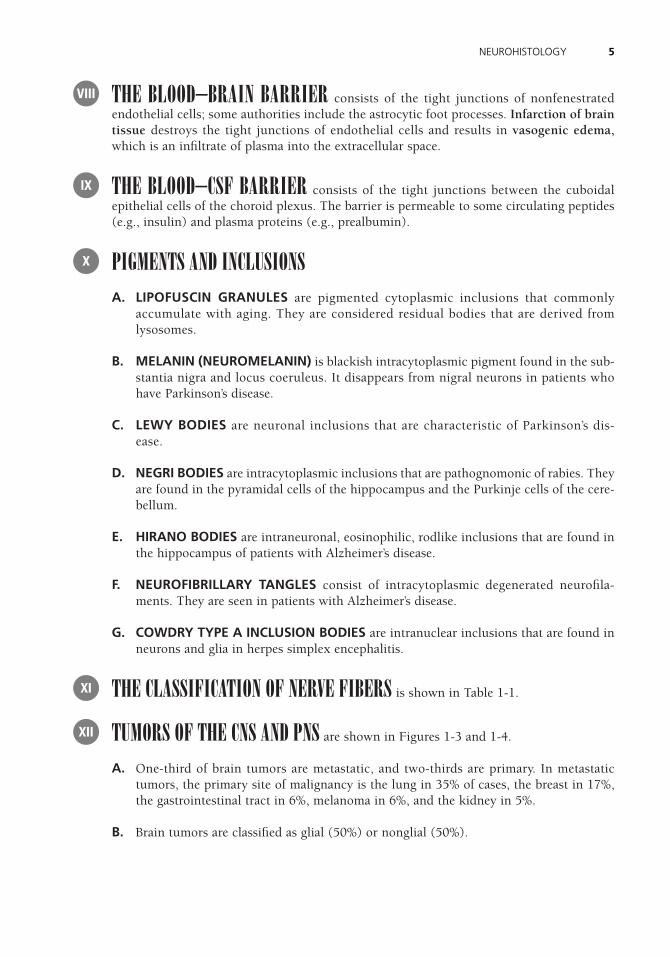
D. ARNOLD-CHIARI malformation (type 2) has a frequency of 1:1,000 (Figure 2-8). Itresults from elongation and herniation of cerebellar tonsils through foramen magnum,thereby blocking cerebrospinal fluid flow.
E. DANDY-WALKER malformation has a frequency of 1:25,000. It may result fromriboflavin inhibitors, posterior fossa trauma, or viral infection (Figure 2-9).
F. HYDROCEPHALUS is most commonly caused by stenosis of the cerebral aqueductduring development. Excessive cerebrospinal fluid accumulates in the ventricles andsubarachnoid space. This condition may result from maternal infection (cytomegalovirusand toxoplasmosis). The frequency is 1:1,000.
G. FETAL ALCOHOL SYNDROME is the most common cause of mental retardation. Itincludes microcephaly and congenital heart disease; holoprosencephaly is the mostsevere manifestation.
H. HOLOPROSENCEPHALY results from failure of midline cleavage of the embryonicforebrain. The telencephalon contains a singular ventricular cavity. Holoprosencephalyis seen in trisomy 13 (Patau syndrome); the corpus callosum may be absent. Holopros-encephaly is the most severe manifestation of the fetal alcohol syndrome.
14 CHAPTER 2
● Figure 2-7 Midsagittal section through the brain stem and diencephalon. A craniopharyngioma (arrows) lies suprasel-lar in the midline. It compresses the optic chiasm and hypothalamus. This tumor is the most common supratentorialtumor that occurs in childhood and the most common cause of hypopituitarism in children. This is a T1-weighted mag-netic resonance imaging scan.
LWBK110-3895G-C02[9-16].qxd 7/10/08 7:21 AM Page 14 Aptara Inc.
15DEVELOPMENT OF THE NERVOUS SYSTEM
● Figure 2-9 Dandy-Walker malformation. Midsagittal section. An enormous dilation of the fourth ventricle resultsfrom failure of the foramina of Luschka and Magendie to open. This condition is associated with occipital meningocele,elevation of the confluence of the sinuses (torcular Herophili), agenesis of the cerebellar vermis, and splenium of thecorpus callosum. (Reprinted from Dudek RW, Fix JD. BRS Embryology. Baltimore: Williams & Wilkins, 1997:97, with per-mission.)
● Figure 2-8 Arnold-Chiari malformation. Midsagittal section. (A) Normal cerebellum, fourth ventricle, and brainstem. (B) Abnormal cerebellum, fourth ventricle, and brain stem showing the common congenital anomalies:(1) beaking of the tectal plate, (2) aqueductal stenosis, (3) kinking and transforaminal herniation of the medulla intothe vertebral canal, and (4) herniation and unrolling of the cerebellar vermis into the vertebral canal. An accompany-ing meningomyelocele is common. (Reprinted from Fix JD. BRS Neuroanatomy. Baltimore: Williams & Wilkins, 1996:72,with permission.)
LWBK110-3895G-C02[9-16].qxd 7/10/08 7:21 AM Page 15 Aptara Inc.

I. HYDRANENCEPHALY results from bilateral hemispheric infarction secondary toocclusion of the carotid arteries. The hemispheres are replaced by hugely dilated ven-tricles.
16 CHAPTER 2
Case StudyA mother brings her newborn infant to the clinic because the infant’s “legs don’t seem to work right.” The infant was delivered at home without antenatal care. What is the most likely diagnosis?
Relevant Physical Exam Findings • Tufts of hair in the lumbosacral region • Clubfoot (Talipes equinovarus)• Chronic upper motor neuron signs, including spasticity, weakness, fatigability
Diagnosis• Spina bifida occulta results from incomplete closure of the neural tube during week 4 of
embryonic development. This type of neural tube defect often affects tissues overlying thespinal cord, including the vertebral column and skin.
LWBK110-3895G-C02[9-16].qxd 7/10/08 7:21 AM Page 16 Aptara Inc.

17
Chapter 3
Cross-Sectional Anatomy of the Brain
INTRODUCTION The illustrations in this chapter are accompanied by correspondingmagnetic resonance imaging scans. Together they represent a mini-atlas of brain slices inthe three orthogonal planes (i.e., midsagittal, coronal, and axial). An insert on each figureshows the level of the slice. The most commonly tested structures are labeled.
MIDSAGITTAL SECTION (Figures 3-1 through 3-3). The location of the structuresshown in the figures should be known.
CORONAL SECTION THROUGH THE OPTIC CHIASM (Figures 3-4 and 3-5). Thelocation of the structures shown in the figures should be known.
CORONAL SECTION THROUGH THE MAMILLARY BODIES (Figures 3-6 and3-7). The location of the structures shown in the figures should be known.
AXIAL IMAGE THROUGH THE THALAMUS AND INTERNAL CAPSULE(Figures 3-8 and 3-9). The location of the structures shown in the figures should be known.
AXIAL IMAGE THROUGH THE MIDBRAIN, MAMILLARY BODIES, ANDOPTIC TRACT (Figures 3-10 through 3-13). The location of the structures shown in thefigures should be known.
ATLAS OF THE BRAIN AND BRAIN STEM (Figures 3-14 through 3-24). Includedare midsagittal, parasagittal, coronal, and axial sections of thick, stained brain slices.
VII
VI
V
IV
III
II
I
Key ConceptsThe mini-atlas provides you with the essential examination structures labeled on computedtomography scans and magnetic resonance images.
✔
LWBK110-3895G-C03[17-29].qxd 7/10/08 7:25 AM Page 17 Aptara Inc.

18 CHAPTER 3
Cingulate gyrus
Superior frontal gyrus
Anterior cerebral artery
Crista galli
Basilar artery
Sphenoid sinus
Clivus
Nasopharynx
C2
Paracentral lobule
Precuneus
Superior sagittal sinus
Parietooccipital fissure
Great cerebral vein(of Galen)
Cuneus
Straight sinus
Calcarine sulcus
Lingual gyrus
Diploe
Cerebellar vermis
Cerebellomedullarycistern (Cistern magna)
● Figure 3-1 Midsagittal section of the brain and brain stem showing the structures surrounding the third and fourthventricles. The brain stem includes the midbrain (M), pons, and medulla oblongata.
● Figure 3-2 Midsagittal magnetic resonance imaging section through the brain and brain stem showing the impor-tant structures surrounding the third and fourth ventricles. This is a T1-weighted image. The gray matter appears gray(hypointense), whereas the white matter appears white (hyperintense).
LWBK110-3895G-C03[17-29].qxd 7/10/08 7:25 AM Page 18 Aptara Inc.
19CROSS-SECTIONAL ANATOMY OF THE BRAIN
Corpus callosum
Fornix
Lateral ventricle
Anterior cerebral artery
Optic chiasm
Hypophysis/infundibulum
Mamillary body
Cerebral aqueduct
Great cerebral vein
(of Galen)
Thalamus
Pineal gland
Superior and inferior colliculi
Fourth ventricle
Cerebellar vermis
Cerebellomedullary cistern
(Cisterna magna)
Spinal cord
Subarachnoid space
Caudate nucleus
Internal
capsule
Putamen
Claustrum
Globus pallidus
AmygdalaOptic chiasm
Hypophysis Infundibulum
Anterior commissure
Insula
Septum pellucidum
Lateral ventricle
Corpus callosum
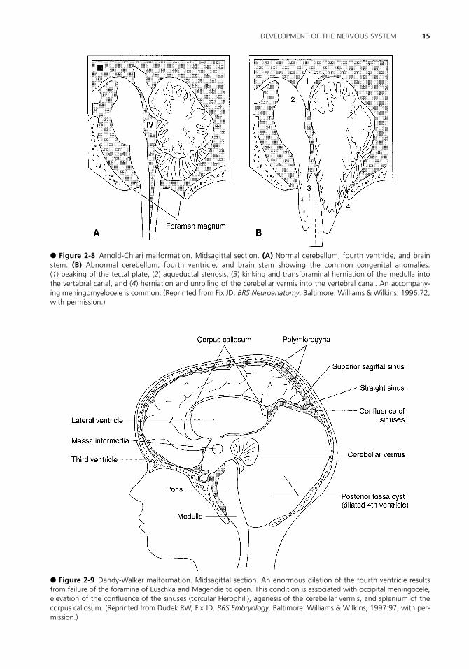
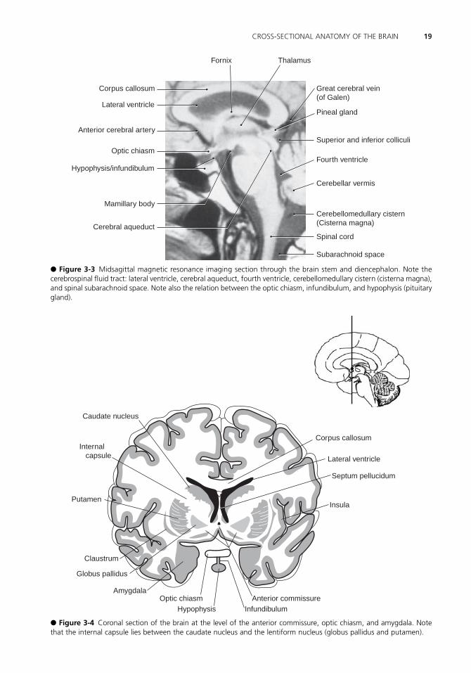
● Figure 3-3 Midsagittal magnetic resonance imaging section through the brain stem and diencephalon. Note thecerebrospinal fluid tract: lateral ventricle, cerebral aqueduct, fourth ventricle, cerebellomedullary cistern (cisterna magna),and spinal subarachnoid space. Note also the relation between the optic chiasm, infundibulum, and hypophysis (pituitarygland).
● Figure 3-4 Coronal section of the brain at the level of the anterior commissure, optic chiasm, and amygdala. Notethat the internal capsule lies between the caudate nucleus and the lentiform nucleus (globus pallidus and putamen).
LWBK110-3895G-C03[17-29].qxd 7/10/08 7:25 AM Page 19 Aptara Inc.

20 CHAPTER 3
Septum pellucidum
Internal capsule
Amygdala
Hypophysis
Cavernous sinus
Sphenoid sinus
Nasopharynx
Longitudinal cerebral fissure
Cingulate gyrus
Corpus callosum
Lateral ventricle
Caudate nucleus
Third ventricle
Optic chiasm
Infundibulum
Interior carotid artery
● Figure 3-5 Coronal magnetic resonance imaging section through the amygdala, optic chiasm, infundibulum, andinternal capsule. The cavernous sinus encircles the sella turcica and contains the following structures: cranial nerves (CN)III, IV, VI, V1, and V2; postganglionic sympathetic fibers; and the internal carotid artery. This is a T1-weighted image.
● Figure 3-6 Coronal section of the brain at the level of the thalamus, mamillary bodies, and hippocampal formation.Note that the internal capsule lies between the thalamus and the lentiform nucleus.
LWBK110-3895G-C03[17-29].qxd 7/10/08 7:25 AM Page 20 Aptara Inc.

21CROSS-SECTIONAL ANATOMY OF THE BRAIN
Corpus callosum
Caudate nucleus
Putamen
Globus pallidus
Hippocampus
Cerebral crus of the
cerebral peduncle
Thalamus
Internal capsule
Substantia nigra
Interpeduncular fossa
Base of pons
Pyramid of medulla
● Figure 3-7 Coronal magnetic resonance imaging section of the brain and brain stem at the level of the thalamus,and hippocampal formation. Note that the posterior limb of the internal capsule lies between the thalamus and thelentiform nucleus (putamen and globus pallidus). This is a T1-weighted postcontrast image.
● Figure 3-8 Axial section of the brain at the level of the internal capsule and basal nuclei (ganglia). Note that theinternal capsule has an anterior limb, a genu, and a posterior limb. Note also that the corpus callosum is sectionedthrough the genu and splenium.
LWBK110-3895G-C03[17-29].qxd 7/10/08 7:25 AM Page 21 Aptara Inc.

22 CHAPTER 3
Insula
External capsule
Velum interpositum
Superior sagittal sinus
Globus pallidus
Putamen
Septum pellucidumand fornix
Lateral ventricle
Internal capsule(posterior limb)
Thalamus and thirdventricle
Trigone (of lateral ventricle)
Corpus callosum(splenium)
Optic radiations
Visual cortex
Internal capsule (genu)
Internal capsule(anterior limb)
Caudate nucleus
Corpus callosum (genu)
● Figure 3-9 Axial magnetic resonance imaging section at the level of the internal capsule and basal nuclei (ganglia).Note that the caudate nucleus bulges into the frontal horn of the lateral ventricle. In Huntington’s disease, there is amassive loss of �-aminobutyric acid (GABA)-ergic neurons in the caudate nucleus that results in hydrocephalus ex vacuo.A lesion of the genu of the internal capsule results in a contralateral weak lower face with sparing of the upper face.This is a T1-weighted image.
● Figure 3-10 Axial section of the brain at the level of the midbrain, mamillary bodies. Note that the substantia nigraseparates the cerebral crus from the tegmentum of the midbrain.
LWBK110-3895G-C03[17-29].qxd 7/10/08 7:25 AM Page 22 Aptara Inc.

23CROSS-SECTIONAL ANATOMY OF THE BRAIN
Longitudinal cerebralfissure
Medial orbital gyrus
(Gyrus rectus)
Optic tract
Mamillary body
Red nucleus
Lateral ventricle
(trigone)
Superior colliculus
Substantia nigra
Posterior cerebral artery
Quadrigeminal cistern
Cerebellar vermis
Straight sinus
Superior sagittal sinus
Crus cerebri
Uncus / amygdala
Middle cerebral artery
Optic nerve
Optic chiasm
Optic tract
Mamillary bodies
Cerebral aqueduct
Cerebellar vermis
Superior sagittal sinus
Optic nerve
Infundibulum
Amygdala
Crus cerebri
Substantia nigra
Lateral ventricle(temporal horn)
Lateral ventricle(occipital horn)
● Figure 3-11 Axial magnetic resonance imaging (MRI) section at the level of the midbrain and mamillary bodies.Because of the high iron content, the red nuclei, mamillary bodies, and substantia nigra show a reduced MRI signal inT2-weighted images. Flowing blood in the cerebral vessels stands out as a signal void. Cerebrospinal fluid produces astrong signal in the ventricles and cisterns.
● Figure 3-12 Axial magnetic resonance imaging section at the level of the optic chiasm, mamillary bodies, and mid-brain. This patient has neurofibromatosis type 1 and an optic nerve glioma. Note the size of the right optic nerve. Theinfundibulum is postfixed. This is a T1-weighted image.
LWBK110-3895G-C03[17-29].qxd 7/10/08 7:25 AM Page 23 Aptara Inc.
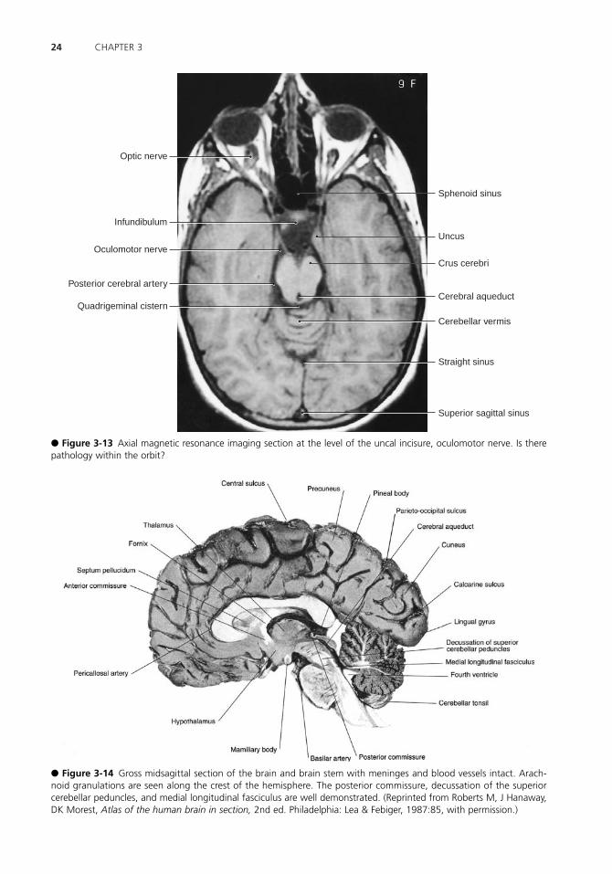
24 CHAPTER 3
Optic nerve
Infundibulum
Oculomotor nerve
Posterior cerebral artery
Quadrigeminal cistern
Sphenoid sinus
Uncus
Crus cerebri
Cerebral aqueduct
Cerebellar vermis
Straight sinus
Superior sagittal sinus
● Figure 3-13 Axial magnetic resonance imaging section at the level of the uncal incisure, oculomotor nerve. Is therepathology within the orbit?
● Figure 3-14 Gross midsagittal section of the brain and brain stem with meninges and blood vessels intact. Arach-noid granulations are seen along the crest of the hemisphere. The posterior commissure, decussation of the superiorcerebellar peduncles, and medial longitudinal fasciculus are well demonstrated. (Reprinted from Roberts M, J Hanaway,DK Morest, Atlas of the human brain in section, 2nd ed. Philadelphia: Lea & Febiger, 1987:85, with permission.)
LWBK110-3895G-C03[17-29].qxd 7/10/08 7:25 AM Page 24 Aptara Inc.

25CROSS-SECTIONAL ANATOMY OF THE BRAIN
● Figure 3-16 Gross parasagittal section through the caudate nucleus, subthalamic nucleus, substantia nigra, and den-tate nucleus. The abducent nerve (CN VI) is seen exiting the pontobulbar sulcus. Damage to the subthalamic nucleusresults in hemiballism. Parkinson’s disease results from a cell loss of the pigmented neurons in the substantia nigra.(Reprinted from M Roberts, J Hanaway, DK Morest, Atlas of the human brain in section, 2nd ed. Philadelphia: Lea &Febiger, 1987:79, with permission.)
● Figure 3-15 Gross parasagittal section through the red nucleus, medial lemniscus, and inferior olivary nucleus. Thecorticospinal fibers can be traced from the crus cerebri to the spinal cord. The abducent nerve (CN VI) is seen exitingfrom the pontonuclear sulcus. (Reprinted from M Roberts, J Hanaway, DK Morest, Atlas of the human brain in section,2nd ed. Philadelphia: Lea & Febiger, 1987:81, with permission.)
LWBK110-3895G-C03[17-29].qxd 7/10/08 7:25 AM Page 25 Aptara Inc.

26 CHAPTER 3
● Figure 3-17 Coronal section through the anterior commissure, amygdala, septal nuclei, and optic chiasm. The septalnuclei have reciprocal connections with the hippocampal formation (subiculum). (Reprinted from M Roberts, J Hanaway,DK Morest, Atlas of the human brain in section, 2nd ed. Philadelphia: Lea & Febiger, 1987:9, with permission.)
● Figure 3-18 Coronal section through the posterior limb of the internal capsule, mamillothalamic tract (MTT), mamil-lary body, and hippocampal formation. Note the MTT entering the anterior ventral nucleus. The optic tracts are visiblebilaterally. (Reprinted from M Roberts, J Hanaway, DK Morest, Atlas of the human brain in section, 2nd ed. Philadel-phia: Lea & Febiger, 1987:19, with permission.)
LWBK110-3895G-C03[17-29].qxd 7/10/08 7:25 AM Page 26 Aptara Inc.

27CROSS-SECTIONAL ANATOMY OF THE BRAIN
● Figure 3-19 Coronal section through the thalamus, ventral posteromedial nucleus (VPM), and ventral posterolateralnucleus (VPL), posterior limb of the internal capsule, substantia nigra, and red nucleus. The optic tract lies dorsal to thetemporal horn of the lateral ventricle. (Reprinted from M Roberts, J Hanaway, DK Morest, Atlas of the human brain insection, 2nd ed. Philadelphia: Lea & Febiger, 1987:23, with permission.)
● Figure 3-20 Coronal section through the lateral and medial lemnisci, lateral and medial geniculate nuclei, and hip-pocampal formation. (Reprinted from M Roberts, J Hanaway, DK Morest, Atlas of the human brain in section, 2nd ed.Philadelphia: Lea & Febiger, 1987:25, with permission.)
LWBK110-3895G-C03[17-29].qxd 7/10/08 7:25 AM Page 27 Aptara Inc.

28 CHAPTER 3
● Figure 3-21 Coronal section through the pulvinar nuclei, pineal gland (epiphysis), superior and inferior colliculi, andtrochlear nerve (CN IV). (Reprinted from M Roberts, J Hanaway, DK Morest, Atlas of the human brain in section, 2nded. Philadelphia: Lea & Febiger, 1987:29, with permission.)
● Figure 3-22 Axial section through the internal capsule, anterior commissure, and pulvinar nuclei. (Reprinted from MRoberts, J Hanaway, DK Morest, Atlas of the human brain in section, 2nd ed. Philadelphia: Lea & Febiger, 1987:51, withpermission.)
LWBK110-3895G-C03[17-29].qxd 7/10/08 7:25 AM Page 28 Aptara Inc.

29CROSS-SECTIONAL ANATOMY OF THE BRAIN
● Figure 3-23 Axial section through the mamillary nuclei and the superior colliculi. (Reprinted from M Roberts, J Han-away, DK Morest, Atlas of the human brain in section, 2nd ed. Philadelphia: Lea & Febiger, 1987:57, with permission.)
● Figure 3-24 Axial section through the mamillary nuclei, optic chiasm, and inferior colliculi. (Reprinted from M Roberts,J Hanaway, DK Morest, Atlas of the human brain in section, 2nd ed. Philadelphia: Lea & Febiger, 1987:59, with permis-sion.)
LWBK110-3895G-C03[17-29].qxd 7/10/08 7:25 AM Page 29 Aptara Inc.

30
Chapter 4
Meninges, Ventricles, and Cerebrospinal Fluid
MENINGES are three connective tissue membranes that surround the spinal cord and brain.
A. The meninges consist of the pia mater, arachnoid mater, and dura mater.1. The pia mater is a delicate, highly vascular layer of connective tissue. It closely
covers the surface of the brain and spinal cord.2. The arachnoid mater is a delicate, nonvascular connective tissue membrane. It is
located between the dura mater and the pia mater.3. The dura mater is the outer layer of meninges. It consists of dense connective tis-
sue that is divided into an outer periosteal (endosteal) layer and an inner meningeallayer. The meningeal layer forms dural folds. Dural venous sinuses are locatedbetween periosteal and meningeal layers of dura mater.
B. MENINGEAL SPACES1. The subarachnoid space (Figure 4-1) lies between the pia mater and the arach-
noid. It terminates at the level of the second sacral vertebra. It contains the cere-brospinal fluid (CSF).
2. Subdural spacea. In the cranium, the subdural space is traversed by “bridging” veins.b. In the spinal cord, it is a clinically insignificant potential space.
3. Epidural spacea. The cranial epidural space is a potential space. It contains the meningeal arter-
ies and veins.b. The spinal epidural space contains fatty areolar tissue, lymphatics, and venous
plexuses. The epidural space may be injected with a local anesthetic to pro-duce a paravertebral (“saddle”) nerve block.
C. MENINGEAL TUMORS1. Meningiomas are benign, well-circumscribed, slow-growing tumors. They
account for 15% of primary intracranial tumors and are more common in womenthan in men (3:2). Ninety percent of meningiomas are supratentorial.
I
Key ConceptsCerebrospinal fluid is produced by the choroid plexus and absorbed by the arachnoid villithat protrude into the venous sinuses. Cerebrospinal fluid pathways are well demonstratedin Figure 4-1.
✔
LWBK110-3895G-C04[30-37].qxd 7/10/08 7:40 AM Page 30 Aptara Inc.

2. Subdural and epidural hematomasa. Subdural hematoma is caused by laceration of the superior cerebral (bridg-
ing) veins.b. Epidural hematoma is caused by laceration of the middle meningeal artery.
E. MENINGITIS is inflammation of the pia–arachnoid area of the brain, the spinal cord,or both.1. Bacterial meningitis is characterized clinically by fever, headache, nuchal rigidity,
and Kernig’s sign. (With the patient supine, the examiner flexes the patient’s hip but
31MENINGES, VENTRICLES, AND CEREBROSPINAL FLUID
● Figure 4-1 The subarachnoid spaces and cisterns of the brain and spinal cord. Cerebrospinal fluid is produced in thechoroid plexuses of the ventricles. It exits the fourth ventricle, circulates in the subarachnoid space, and enters the supe-rior sagittal sinus through the arachnoid granulations. Note that the conus medullaris terminates at L-1. The lumbar cis-tern ends at S-2. (Reprinted from CR Noback, NL Strominger, R Demarest, The human nervous system, 4th ed. Balti-more: Williams & Wilkins, 1991:68, with permission.)
LWBK110-3895G-C04[30-37].qxd 7/10/08 7:40 AM Page 31 Aptara Inc.

cannot extend the knee without causing pain. It is a sign of meningeal irritation.)(Remember: Kernig � knee.) More than 70% of cases occur in children youngerthan 5 years. The disease may cause cranial nerve palsies and hydrocephalus.a. Common causes
(1) In newborns (younger than 1 month), bacterial meningitis is most fre-quently caused by group B streptococci (Streptococcus agalactiae),Escherichia coli, and Listeria monocytogenes.
(2) In older infants and young children (1 through 23 months), it is mostcommonly caused by Streptococcus pneumoniae.
(3) In young adults (2 through 18 years), it is most frequently caused byNeisseria meningitidis.
(4) In older adults (19 years and older), it is most frequently caused by Strep-tococcus pneumoniae.NB: Immunization against Hemophilis influenzae has significantly reducedthis type of meningitis.
b. CSF findings(1) Numerous polymorphonuclear leukocytes(2) Decreased glucose levels(3) Increased protein levels
2. Viral meningitis is also known as aseptic meningitis. It is characterized clinicallyby fever, headache, nuchal rigidity, and Kernig’s sign.a. Common causes. Many viruses are associated with viral meningitis, includ-
ing mumps, echovirus, Coxsackie virus, Epstein-Barr virus, and herpes sim-plex type 2.
b. CSF findings(1) Numerous lymphocytes(2) Normal glucose levels(3) Moderately increased protein levels
VENTRICULAR SYSTEMA. The choroid plexus is a specialized structure that projects into the lateral, third, and
fourth ventricles of the brain. It consists of infoldings of blood vessels of the pia materthat are covered by modified ciliated ependymal cells. It secretes the CSF. Tight junc-tions of the choroid plexus cells form the blood–CSF barrier.
B. VENTRICLES CONTAIN CSF AND CHOROID PLEXUS1. The two lateral ventricles communicate with the third ventricle through the inter-
ventricular foramina (of Monro).2. The third ventricle is located between the medial walls of the diencephalon. It
communicates with the fourth ventricle through the cerebral aqueduct.3. The cerebral aqueduct (of Sylvius) connects the third and fourth ventricles. It has
no choroid plexus. Blockage of the cerebral aqueduct results in noncommunicat-ing hydrocephalus.
4. The fourth ventricle communicates with the subarachnoid space through three outletforamina: two lateral foramina (of Luschka) and one median foramen (of Magendie).
C. HYDROCEPHALUS is dilation of the cerebral ventricles caused by blockage of the CSFpathways. It is characterized by excessive accumulation of CSF in the cerebral ventri-cles or subarachnoid space.
II
32 CHAPTER 4
LWBK110-3895G-C04[30-37].qxd 7/10/08 7:40 AM Page 32 Aptara Inc.

1. Noncommunicating hydrocephalus results from obstruction within the ventri-cles (e.g., congenital aqueductal stenosis).
2. Communicating hydrocephalus results from blockage within the subarachnoidspace (e.g., adhesions after meningitis).
3. Normal-pressure hydrocephalus occurs when the CSF is not absorbed by thearachnoid villi. It may occur secondary to posttraumatic meningeal hemorrhage.Clinically, it is characterized by the triad of progressive dementia, ataxic gait, andurinary incontinence. (Remember: wacky, wobbly, and wet.)
4. Hydrocephalus ex vacuo results from a loss of cells in the caudate nucleus (e.g.,Huntington’s disease).
5. Pseudotumor cerebri (benign intracranial hypertension) results from increasedresistance to CSF outflow at the arachnoid villi. It occurs in obese young womenand is characterized by papilledema without mass, elevated CSF pressure, and dete-riorating vision. The ventricles may be slitlike.
CEREBROSPINAL FLUID is a colorless acellular fluid. It flows through the ventriclesand into the subarachnoid space.
A. FUNCTION1. CSF supports the central nervous system (CNS) and protects it against concus-
sive injury.2. It transports hormones and hormone-releasing factors.3. It removes metabolic waste products through absorption.
B. FORMATION AND ABSORPTION. CSF is formed by the choroid plexus. Absorptionis primarily through the arachnoid villi into the superior sagittal sinus.
C. The composition of CSF is clinically relevant (Table 4-1).1. The normal number of mononuclear cells is fewer than 5/�L.2. Red blood cells in the CSF indicate subarachnoid hemorrhage (e.g., caused by
trauma or a ruptured berry aneurysm).3. CSF glucose levels are normally 50 to 75 mg/dL (66% of the blood glucose level).
Glucose levels are normal in patients with viral meningitis and decreased inpatients with bacterial meningitis.
III
33MENINGES, VENTRICLES, AND CEREBROSPINAL FLUID
Cerebrospinal Subarachnoid Bacterial Viral Fluid Normal Hemorrhage Meningitis Encephalitis
Color Clear Bloody Cloudy Clear, cloudyCell count/mm3 �5 lymphocytes Red blood cells �1,000 polymorphonuclear 25–500
present leukocytes lymphocytesProtein �45 mg/dL Normal to Elevated, Slightly
slightly elevated �100 mg/dL elevatedGlucose �66% �45 mg/dL Normal Reduced Normal
of blood (80–120 mg/dL)
CEREBROSPINAL FLUID PROFILES IN SUBARACHNOID HEMORRHAGE, BACTERIAL MENINGITIS, AND VIRAL ENCEPHALITIS
TABLE 4-1
LWBK110-3895G-C04[30-37].qxd 7/10/08 7:40 AM Page 33 Aptara Inc.

4. Total protein levels are normally between 15 and 45 mg/dL in the lumbar cistern.Protein levels are increased in patients with bacterial meningitis and normal orslightly increased in patients with viral meningitis.
5. Normal CSF pressure in the lateral recumbent position ranges from 80 to 180 mmH2O. Brain tumors and meningitis elevate CSF pressure.
HERNIATION (Figures 4-2 through 4-7)
A. TRANSTENTORIAL (UNCAL) HERNIATION is protrusion of the brain through thetentorial incisure. This may result in oculomotor paresis and contralateral hemiplegia.
B. TRANSFORAMINAL (TONSILLAR) HERNIATION is protrusion of the brain stem andcerebellum through the foramen magnum. Clinical complications include obtundationand death.
C. SUBFALCINE (CINGULATE) HERNIATION is herniation below the falx cerebri. Thiscondition does not necessarily result in severe clinical symptoms. It can present asheadache. Compression of the anterior cerebral artery may result in contralateral legweakness.
IV
34 CHAPTER 4
1
9
4 5 6
2
7
8
3
Tumor
● Figure 4-2 Coronal section of a tumor in the supratentorial compartment. (1) Anterior cerebral artery; (2) subfalcineherniation; (3) shifting of the lateral ventricles; (4) posterior cerebral artery (compression results in contralateral hemi-anopia); (5) uncal (transtentorial) herniation; (6) Kernohan’s notch (contralateral cerebral peduncle), with damaged cor-ticospinal and corticobulbar fibers; (7 ) tentorium cerebelli; (8) pyramidal cells that give rise to the corticospinal tract;(9) tonsillar (transforaminal) herniation, which damages vital medullary centers. (Adapted with permission from LeechRW, Shumann RM: Neuropathology. New York, Harper & Row, 1982, p. 16.)
LWBK110-3895G-C04[30-37].qxd 7/10/08 7:40 AM Page 34 Aptara Inc.

35MENINGES, VENTRICLES, AND CEREBROSPINAL FLUID
32
17
8
7
6
54
● Figure 4-3 Axial section through the midbrain and the herniating parahippocampal gyrus. The left oculomotor nerveis being stretched (dilated pupil). The left posterior cerebral artery is compressed, resulting in a contralateral hemianopia.The right crus cerebri is damaged (Kernohan’s notch) by the free edge of the tentorial incisure, resulting in a contralat-eral hemiparesis. Kernohan’s notch results in a false localizing sign. The caudal displacement of the brain stem causesrupture of the paramedian arteries of the basilar artery. Hemorrhage into the midbrain and rostral pontine tegmentumis usually fatal (Duret’s hemorrhages). The posterior cerebral arteries lie superior to the oculomotor nerves. (1) Parahip-pocampal gyrus; (2) crus cerebri; (3) posterior cerebral artery; (4) optic nerve; (5) optic chiasma; (6) oculomotor nerve;(7) free edge of tentorium; (8) Kernohan’s notch. (Adapted from RW Leech, RM Shumann, Neuropathology. New York:Harper & Row, 1982:19, with permission.)
A
B
C
D
E
● Figure 4-4 Magnetic resonance imaging scan showing brain trauma. (A) Internal capsule; (B) subdural hematoma;(C ) subdural hematoma; (D) thalamus; (E ) epidural hematoma. Epidural hematomas may cross dural attachments. Sub-dural hematomas do not cross dural attachments. The hyperintense signals are caused by methemoglobin. This is a T1-weighted image.
LWBK110-3895G-C04[30-37].qxd 7/10/08 7:40 AM Page 35 Aptara Inc.

36 CHAPTER 4
A
B
C
D
● Figure 4-5 Computed tomography axial section showing an intraparenchymal hemorrhage in the left frontal lobe.(A) Intraparenchymal hemorrhage; (B) lateral ventricle; (C ) internal capsule; (D) calcified glomus in the trigone region ofthe lateral ventricle.
A
B
C
D
● Figure 4-6 Computed tomography axial section showing an epidural hematoma and a skull fracture. (A) Epiduralhematoma; (B) skull fracture; (C ) calcified pineal gland; (D) calcified glomus in the trigone region of the lateral ventricle.The epidural hematoma is a classic biconvex, or lentiform, shape.
LWBK110-3895G-C04[30-37].qxd 7/10/08 7:40 AM Page 36 Aptara Inc.

37MENINGES, VENTRICLES, AND CEREBROSPINAL FLUID
AB
● Figure 4-7 Computed tomography (CT) axial section showing a skull fracture (A) on the left side. An epiduralhematoma (B) underlies the fracture. The CT scan shows a bone window.
Case StudyThe patient is a 68-year-old man with alcoholic cirrhosis. He fell 4 weeks ago. He has a his-tory of progressive weakness on the right side. What is the most likely diagnosis?
Relevant Physical Exam Findings• Hemiparesis • Reflex asymmetry
Relevant Lab Findings• Computed tomography scan of the head showed a crescent-shaped hypodense area
between the cortex and skull. This mass has resulted in massive midline shift to the right,with subfalcine and uncal herniation.
Diagnosis• Subdural hematoma results from bleeding between the dura mater and the arachnoid
membrane from the bridging veins that connect the cerebral cortex to the dural venoussinuses. Subdural hematoma is common after acute deceleration injury from a fall or motorvehicle accident but rarely is associated with skull fracture.
LWBK110-3895G-C04[30-37].qxd 7/10/08 7:40 AM Page 37 Aptara Inc.

38
Chapter 5
Blood Supply
THE SPINAL CORD AND LOWER BRAIN STEM are supplied with blood throughthe anterior spinal artery (Figure 5-1).
A. The anterior spinal artery supplies the anterior two-thirds of the spinal cord.
B. In the medulla, the anterior spinal artery supplies the pyramid, medial lemniscus, androot fibers of cranial nerve (CN) XII.
THE INTERNAL CAROTID SYSTEM (see Figure 5-1) consists of the internal carotidartery and its branches.
A. The ophthalmic artery enters the orbit with the optic nerve (CN II). The central arteryof the retina is a branch of the ophthalmic artery. Occlusion results in blindness.
B. The posterior communicating artery irrigates the hypothalamus and ventral thalamus.An aneurysm of this artery is the second most common aneurysm of the circle of Willis.It commonly results in third-nerve palsy.
C. The anterior choroidal artery arises from the internal carotid artery. It is not part ofthe circle of Willis. It perfuses the lateral geniculate body, globus pallidus, and poste-rior limb of the internal capsule.
D. The anterior cerebral artery (Figure 5-2) supplies the medial surface of the hemispherefrom the frontal pole to the parietooccipital sulcus.1. The anterior cerebral artery irrigates the paracentral lobule, which contains the
leg-foot area of the motor and sensory cortices.2. The anterior communicating artery connects the two anterior cerebral arteries. It
is the most common site of aneurysm of the circle of Willis, which may causebitemporal lower quadrantanopia.
II
I
Key Concepts1) The essential arteries supplying the brain and spinal cord and the functional areas that
they irrigate 2) The carotid and vertebral angiograms and the epidural and subdural hematomas in
computed tomography scans and magnetic resonance images
✔
LWBK110-3895G-C05[38-48].qxd 7/10/08 7:43 AM Page 38 Aptara Inc.
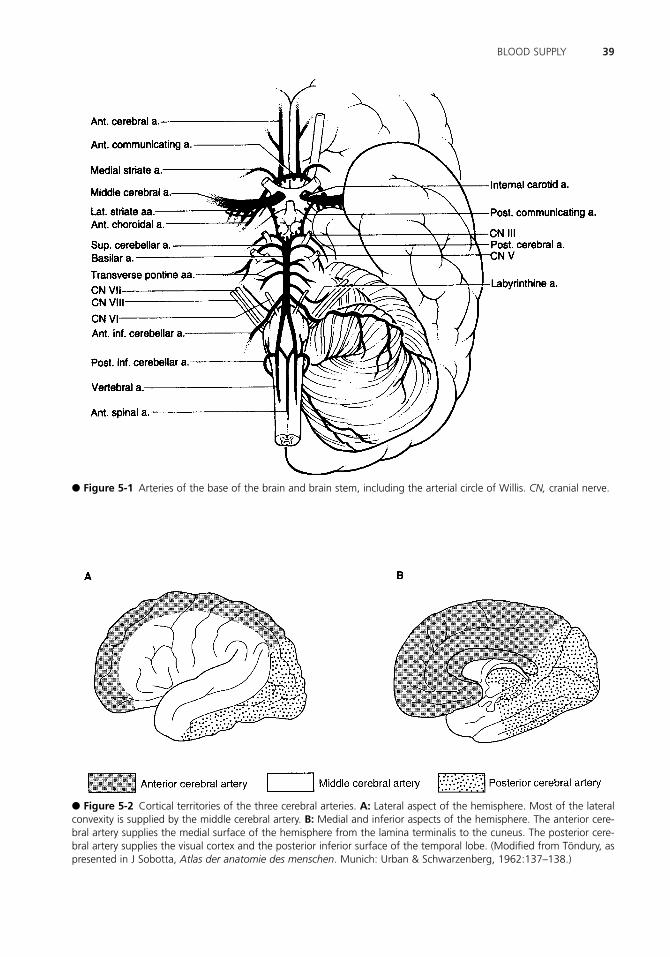
39BLOOD SUPPLY
● Figure 5-1 Arteries of the base of the brain and brain stem, including the arterial circle of Willis. CN, cranial nerve.
● Figure 5-2 Cortical territories of the three cerebral arteries. A: Lateral aspect of the hemisphere. Most of the lateralconvexity is supplied by the middle cerebral artery. B: Medial and inferior aspects of the hemisphere. The anterior cere-bral artery supplies the medial surface of the hemisphere from the lamina terminalis to the cuneus. The posterior cere-bral artery supplies the visual cortex and the posterior inferior surface of the temporal lobe. (Modified from Töndury, aspresented in J Sobotta, Atlas der anatomie des menschen. Munich: Urban & Schwarzenberg, 1962:137–138.)
LWBK110-3895G-C05[38-48].qxd 7/10/08 7:43 AM Page 39 Aptara Inc.

40 CHAPTER 5
● Figure 5-3 Coronal section through the cerebral hemisphere at the level of the internal capsule and thalamus, show-ing the major vascular territories.
3. The medial striate arteries (see Figure 5-1) are the penetrating arteries of the ante-rior cerebral artery. They supply the anterior portion of the putamen and caudatenucleus and the anteroinferior part of the internal capsule.
E. The middle cerebral artery (see Figure 5-2)1. This middle cerebral artery supplies the lateral convexity of the hemisphere, including
a. Broca’s and Wernicke’s speech areas.b. The face and arm areas of the motor and sensory cortices.c. The frontal eye field.
2. The lateral striate arteries (Figure 5-3) are the penetrating branches of the mid-dle cerebral artery. They are the arteries of stroke, and they supply the internalcapsule, caudate nucleus, putamen, and globus pallidus.
THE VERTIBROBASILAR SYSTEM See Figure 5-1.
A. The vertebral artery is a branch of the subclavian artery. It gives rise to the anteriorspinal artery (see I.) and the posterior inferior cerebellar artery (PICA), which sup-plies the dorsolateral quadrant of the medulla. This quadrant includes the nucleusambiguus (CN IX, X, and XI) and the inferior surface of the cerebellum.
III
LWBK110-3895G-C05[38-48].qxd 7/10/08 7:43 AM Page 40 Aptara Inc.

B. The basilar artery is formed by the two vertebral arteries. It gives rise to the followingarteries:1. The paramedian branches of the pontine arteries supply the base of the pons,
which includes the corticospinal fibers and the exiting root fibers of the abducentnerve (CN VI).
2. The labyrinthine artery arises from the basilar artery in 15% of people. It arisesfrom the anterior inferior cerebellar artery in 85% of people.
3. The anterior inferior cerebellar artery (AICA) supplies the caudal lateral pontinetegmentum, including CN VII, the spinal trigeminal tract of CN V, and the inferiorsurface of the cerebellum.
4. The superior cerebellar artery supplies the dorsolateral tegmentum of the rostralpons (i.e., rostral to the motor nucleus of CN V), the superior cerebellar pedun-cle, the superior surface of the cerebellum and cerebellar nuclei, and the cochlearnuclei.
5. The posterior cerebral artery (see Figures 5-1 through 5-3) is connected to thecarotid artery through the posterior communicating artery. It provides the majorblood supply to the midbrain. It also supplies the thalamus, lateral and medialgeniculate bodies, and occipital lobe (which includes the visual cortex and the infe-rior surface of the temporal lobe, including the hippocampal formation). Occlu-sion of this artery results in a contralateral hemianopia with macular sparing.
THE BLOOD SUPPLY OF THE INTERNAL CAPSULE comes primarily from the lat-eral striate arteries of the middle cerebral artery and the anterior choroidal artery.
VEINS OF THE BRAINA. The superior cerebral (“bridging”) veins drain into the superior sagittal sinus. Lacer-
ation results in a subdural hematoma.
B. The great cerebral vein (of Galen) drains the deep cerebral veins into the straight sinus.
VENOUS DURAL SINUSESA. The superior sagittal sinus receives blood from the bridging veins and emissary veins
(a potential route for transmission of extracranial infection into brain). The superiorsagittal sinus also receives cerebrospinal fluid (CSF) through the arachnoid villi.
B. The cavernous sinus contains CN III, IV, V-1 and V-2, and VI and the postganglionicsympathetic fibers. It also contains the siphon of the internal carotid artery (Figure 5-4).
ANGIOGRAPHYA. CAROTID ANGIOGRAPHY. Figures 5-5A and B show the internal carotid artery, ante-
rior cerebral artery, and middle cerebral artery.
B. VERTEBRAL ANGIOGRAPHY. Figures 5-5C and D show the vertebral artery, PICAand AICA, basilar artery, superior cerebellar artery, and posterior cerebral artery(Figures 5-6 and 5-7).
VII
VI
V
IV
41BLOOD SUPPLY
LWBK110-3895G-C05[38-48].qxd 7/10/08 7:43 AM Page 41 Aptara Inc.

42 CHAPTER 5
Superior sagittal sinus
Anterior cerebral artery
Cavernous part of ICA
Petrosal part of ICA
Basilar artery
Sigmoid sinus
Superior cerebral veins
Superior sagittal sinus
Branches of MCA
PCA
Straight sinus
Confluence of thesinuses
Transverse sinus
Vertebral artery
● Figure 5-4 Magnetic resonance angiogram, lateral projection, showing the major venous sinuses and arteries. Notethe bridging veins entering the superior sagittal sinus. ICA, internal carotid artery; MCA, middle cerebral artery; PCA,posterior cerebral artery.
● Figure 5-5 A: Carotid angiogram, lateral projection. B: Carotid angiogram, anteroposterior projection. C: Vertebralangiogram, lateral projection. D: Vertebral angiogram, anteroposterior projection.
LWBK110-3895G-C05[38-48].qxd 7/10/08 7:43 AM Page 42 Aptara Inc.

43BLOOD SUPPLY
B
A C
● Figure 5-6 Arterial anatomy of a magnetic resonance section; axial (A), sagittal (B, C). ACAcm, anterior cerebralartery, callosal marginal branch; A2 and A1, branches of the anterior cerebral artery; ACApc, pericallosal branch of theanterior cerebral artery; ACoA, anterior communicating artery; AICA, anterior inferior cerebellar artery; M1 and M2,segments of the middle cerebral artery (MCA); MCAb � bifurcation; ICAs, internal carotid artery siphon; ICAc, internalcarotoid artery cavernous PCA, posterior cerebral arterior, PCoA, posterior communicating artery; BA, basilar artery; SCA,superior cerebellar artery. (Reprinted from CB Grossman, Magenetic resonance imaging and computed tomography ofthe head and spine, 2nd ed. Philadelphia: Williams & Wilkins, 1996:124, with permission.)
C. VEINS AND DURAL SINUSES. Figure 5-8 shows the internal cerebral vein, superiorcerebral veins, great cerebral vein, superior ophthalmic vein, and major dural sinuses.
D. DIGITAL SUBTRACTION ANGIOGRAPHY. See Figures 5-9 through 5-12.
THE MIDDLE MENINGEAL ARTERY, a branch of the maxillary artery, enters the cra-nium through the foramen spinosum. It supplies most of the dura, including its calvarialportion. Laceration results in epidural hemorrhage (hematoma) (Figures 5-13 and 5-14).
VIII
LWBK110-3895G-C05[38-48].qxd 7/10/08 7:43 AM Page 43 Aptara Inc.

44 CHAPTER 5
● Figure 5-7 Venous anatomy of a magnetic resonance section; midsagittal (A), lateral (B), submental-vertex (C). IhV,interhemispheric veins; SSS, superior sagittal sinus; ISS, inferior sagittal sinus; TsV, thalmostriate veins; StS, straight sinus;PcCbV, precentral cerebellar vein; To, torcula; VG, vein of anterior pontomesencephalic veins. (Reprinted from CB Gross-man, Magnetic resonance imaging and computed tomography of the head and spine, 2nd ed. Philadelphia: Williams &Wilkins, 1996:125, with permission.)
● Figure 5-8 Carotid angiogram, venous phase, showing the cerebral veins and venous sinuses.
LWBK110-3895G-C05[38-48].qxd 7/10/08 7:43 AM Page 44 Aptara Inc.

45BLOOD SUPPLY
Pericallosal arteryof ACA
Frontopolar branchof ACA
Ophthalmic artery
Cervical ICA
Petrous ICA
Cavernous ICA
Callosomarginal arteryof ACA
MCA M1 segment
PCoM
ACA Cortical branches of MCA
Lateral striate branchesof MCA
M1 segment of MCA
Supraclinoid part of ICA
Petrous part of ICA
Cervical part of ICA
A1 segment of ACA
ACoM
Cavernous part of ICA
● Figure 5-9 Carotid angiogram, lateral projection. Identify the cortical branches of the anterior cerebral artery (ACA)and middle cerebral artery (MCA). Follow the course of the internal carotid artery (ICA). Remember that aneurysms ofthe posterior communicating artery (PCoM ) may result in third-nerve palsy. The paracentral lobule is irrigated by thecallosomarginal artery.
● Figure 5-10 Carotid angiogram, anteroposterior projection. Identify the anterior cerebral artery (ACA), middle cere-bral artery (MCA), and internal carotid artery (ICA). The horizontal branches of the MCA perfuse the basal nuclei andinternal capsule. ACoM, anterior communicating artery.
LWBK110-3895G-C05[38-48].qxd 7/10/08 7:43 AM Page 45 Aptara Inc.
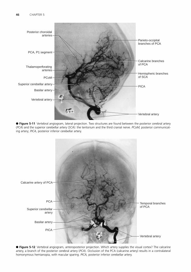
46 CHAPTER 5
Posterior choroidalarteries
Superior cerebellar artery
Basilar artery
Vertebral artery
PCoM
Thalamoperforatingarteries
PCA, P1 segment
Parieto-occipitalbranches of PCA
Calcarine branchesof PCA
Hemispheric branchesof SCA
PICA
Vertebral artery
● Figure 5-11 Vertebral angiogram, lateral projection. Two structures are found between the posterior cerebral artery(PCA) and the superior cerebellar artery (SCA): the tentorium and the third cranial nerve. PCoM, posterior communicat-ing artery; PICA, posterior inferior cerebellar artery.
Calcarine artery of PCA
Temporal branchesof PCA
Vertebral artery
PCA
Superior cerebellarartery
Basilar artery
PICA
● Figure 5-12 Vertebral angiogram, anteroposterior projection. Which artery supplies the visual cortex? The calcarineartery, a branch of the posterior cerebral artery (PCA). Occlusion of the PCA (calcarine artery) results in a contralateralhomonymous hemianopia, with macular sparing. PICA, posterior inferior cerebellar artery.
LWBK110-3895G-C05[38-48].qxd 7/10/08 7:43 AM Page 46 Aptara Inc.

47BLOOD SUPPLY
Outer table
Diploë
Dura
Periosteum(of inner table)
Arachnoid
Dura
● Figure 5-13 An epidural hematoma results from laceration of the middle meningeal artery. Arterial bleeding into theepidural space forms a biconvex clot. The classic “lucid interval” is seen in 50% of cases. Skull fractures are usuallyfound. Epidural hematomas rarely cross sutural lines. (Reprinted from AG Osburn, KA Tong, Handbook of neuroradiol-ogy: brain and skull. St. Louis: Mosby, 1996:191, with permission.)
● Figure 5-14 A subdural hematoma (SDH) results from lacerated bridging veins. SDHs are frequently accompanied bytraumatic subarachnoid hemorrhages and cortical contusions. Sudden deceleration of the head causes tearing of thesuperior cerebral veins. The SDH extends over the crest of the convexity into the interhemispheric fissure but does notcross the dural attachment of the falx cerebri. The clot can be crescent-shaped, biconvex, or multiloculated. SDHs aremore common than epidural hematomas and always cause brain damage. (Reprinted from AG Osburn, KA Tong, Hand-book of neuroradiology: brain and skull. St. Louis: Mosby, 1996:192, with permission.)
LWBK110-3895G-C05[38-48].qxd 7/10/08 7:43 AM Page 47 Aptara Inc.

48 CHAPTER 5
Case Study A 62-year-old man comes to the clinic complaining of problems with his vision and a horribleheadache that began earlier in the day. He reports bumping into objects and not being able toread half the printed page of the newspaper. He has a history of hypertension and diabetesmellitus. What is the most likely diagnosis?
Relevant Physical Exam Findings• Complete hemianopia • Contralateral face and limb sensory loss
Relevant Lab Findings • Computed tomography scan of the brain determined the presence of ischemic infarction
with hemorrhagic change.
Diagnosis• Posterior cerebral artery infarct
LWBK110-3895G-C05[38-48].qxd 7/10/08 7:43 AM Page 48 Aptara Inc.

49
Chapter 6
Spinal Cord
GRAY AND WHITE RAMI COMMUNICANS (Figure 6-1)
A. Gray rami communicans contain unmyelinated postganglionic sympathetic fibers. Theyare found at all levels of the spinal cord.
B. White rami communicans contain myelinated preganglionic sympathetic fibers. Theyare found from T-1 to L-3 (the extent of the lateral horn and the intermediolateral cellcolumn).
TERMINATION OF THE CONUS MEDULLARIS (see Figure 4-1) occurs in the new-born at the level of the body of the third lumbar vertebra (L-3). In the adult, it occurs atthe level of the lower border of the first lumbar vertebra (L-1). This is clinically relevant indetermining the appropriate position for performing lumbar puncture in children andadults.
LOCATION OF THE MAJOR MOTOR AND SENSORY NUCLEI OF THE SPINALCORD (Figure 6-2)
A. The ciliospinal center of Budge, from C-8 to T-2, mediates the sympathetic innerva-tion of the eye.
B. The intermediolateral nucleus (cell column) of the lateral horn, from C-8 to L-3, medi-ates the entire sympathetic innervation of the body.
C. The nucleus dorsalis of Clark, from C-8 to L-3, gives rise to the dorsal spinocerebellartract.
D. The sacral parasympathetic nucleus, from S-2 to S-4
III
II
I
Key Concepts1) The adult spinal cord terminates (conus terminalis) at the lower border of the first lum-
bar vertebra. 2) The newborn’s spinal cord extends to the third lumbar vertebra. In adults, the cauda
equina extends from vertebral level L-2 to coccygeal vertebra (Co).
✔
LWBK110-3895G-C06[49-52].qxd 7/10/08 7:44 AM Page 49 Aptara Inc.

E. The nucleus of the accessory nerve, from C-1 to C-6
F. The phrenic nucleus, from C-3 to C-6
THE CAUDA EQUINA Motor and sensory roots that are found in the subarachnoidspace below the conus medullaris form the cauda equina. They exit the vertebral canalthrough the lumbar intervertebral and sacral foramina.
THE MYOTATIC REFLEX (see Figure 6-1) is a monosynaptic and ipsilateral musclestretch reflex (MSR). Like all reflexes, the myotatic reflex has an afferent and an efferentlimb. Interruption of either limb results in areflexia.
A. The afferent limb includes a muscle spindle (receptor) and a dorsal root ganglion neu-ron and its Ia fiber.
B. The efferent limb includes a ventral horn motor neuron that innervates striated mus-cle (effector).
C. The five most commonly tested MSRs are listed in Table 6-1.
V
IV
50 CHAPTER 6
Muscle Stretch Reflex Cord Segment Muscle
Ankle jerk S-1 GastrocnemiusKnee jerk L-2–L-4 QuadricepsBiceps jerk C-5 and C-6 BicepsForearm jerk C-5 and C-6 BrachioradialisTriceps jerk C-7 and C-8 Triceps
THE FIVE MOST COMMONLY TESTED MUSCLE STRETCH REFLEXESTABLE 6-1
● Figure 6-1 The four functional components of the thoracic spinal nerve: general visceral afferent (GVA), generalsomatic afferent (GSA), general somatic efferent (GSE), and general visceral efferent (GVE ). Proprioceptive, cutaneous,and visceral reflex arcs are shown. The muscle stretch (myotatic) reflex includes the muscle spindle, GSA dorsal rootganglion cell, GSE ventral horn motor neuron, and skeletal muscle.
LWBK110-3895G-C06[49-52].qxd 7/10/08 7:44 AM Page 50 Aptara Inc.

51SPINAL CORD
Spinal cord
Segment C1
Segment C4
Segment C8
Segment T2
Segment T12
Segment L4
Segment S3
Lateral horn(intermediolateral
cell column)
● Figure 6-2 Spinal cord segments. Cervical segments are the largest segments. The thoracic and sacral segments arerelatively small. Note the presence of an intermediolateral cell column in the thoracic and sacral segments. (Reprintedfrom A Siegel, HN Sapru, Essential neuroscience. Baltimore: Lippincott Williams & Wilkins, 2006:142, with permission.)
LWBK110-3895G-C06[49-52].qxd 7/10/08 7:44 AM Page 51 Aptara Inc.

52 CHAPTER 6
Case Study A 46-year-old man was admitted with complaints of lower back pain that radiated down tohis foot over the last 2 months. The pain was not relieved with medical therapy. What is yourdiagnosis?
Relevant Physical Exam Findings• Absent right ankle jerk • Weakness of dorsiflexion and plantar flexion • Decreased pinprick over the dorsum of the foot
Relevant Lab Findings • An anteroposterior myelogram demonstrated compression of the first sacral nerve root on
the left at the level of the L5-S1 vertebrae.
Diagnosis• Lumbar intervertebral disc herniation
LWBK110-3895G-C06[49-52].qxd 7/10/08 7:44 AM Page 52 Aptara Inc.

53
Chapter 7
Autonomic Nervous System
INTRODUCTION The autonomic nervous system (ANS) is a general visceral efferentmotor system that controls and regulates smooth muscle, cardiac muscle, and glands.
A. The ANS consists of two types of projection neurons:1. Preganglionic neurons.2. Postganglionic neurons. Sympathetic ganglia have interneurons.
B. AUTONOMIC OUTPUT is controlled by the hypothalamus.
C. The ANS has three divisions:1. Sympathetic. Figure 7-1 shows the sympathetic innervation of the ANS.2. Parasympathetic. Figure 7-2 shows the parasympathetic innervation of the ANS.
Table 7-1 compares the effects of sympathetic and parasympathetic activity onorgan systems.
3. Enteric. The enteric division includes the intramural ganglia of the gastrointesti-nal tract, submucosal plexus, and myenteric plexus.
CRANIAL NERVES (CN) WITH PARASYMPATHETIC COMPONENTS include thefollowing:
A. CN III (ciliary ganglion).
B. CN VII (pterygopalatine and submandibular ganglia).
C. CN IX (otic ganglion).
D. CN X [terminal (intramural) ganglia].
COMMUNICATING RAMI of the ANS include the following:
A. WHITE RAMI COMMUNICANTES, which are found between T-1 and L-2 and aremyelinated.
III
II
I
Key ConceptsUnderstand the etiology of Horner’s syndrome. Make sure you know the actions of the auto-nomic nervous system on the organ systems (Table 7.1).
✔
LWBK110-3895G-C07[53-57].qxd 7/10/08 7:46 AM Page 53 Aptara Inc.

54 CHAPTER 7
● Figure 7-1 The sympathetic (thoracolumbar) innervation of the autonomic nervous system. The entire sympatheticinnervation of the head is through the superior cervical ganglion. Gray communicating rami are found at all spinal cordlevels. White communicating rami are found only in spinal segments T-1 through L-2.
LWBK110-3895G-C07[53-57].qxd 7/10/08 7:46 AM Page 54 Aptara Inc.

55AUTONOMIC NERVOUS SYSTEM
● Figure 7-2 The parasympathetic (craniosacral) innervation of the autonomic nervous system. Sacral outflow includessegments S-2 through S-4. Cranial outflow is mediated through cranial nerves (CN) III, VII, IX, and X.
LWBK110-3895G-C07[53-57].qxd 7/10/08 7:46 AM Page 55 Aptara Inc.

56 CHAPTER 7
Structure Sympathetic Function Parasympathetic Function
EyeRadial muscle of iris Dilation of pupil (mydriasis)Circular muscle of iris Constriction of pupil (miosis)Ciliary muscle of ciliary body Contraction for near vision
Lacrimal gland Stimulation of secretionSalivary glands Viscous secretion Watery secretionSweat glands
Thermoregulatory IncreaseApocrine (stress) Increase
HeartSinoatrial node Acceleration Deceleration (vagal arrest)Atrioventricular node Increase in conduction velocity Decrease in conduction velocityContractility Increase Decrease (atria)
Vascular smooth muscleSkin, splanchnic vessels ContractionSkeletal muscle vessels Relaxation
Bronchiolar smooth muscle Relaxation ContractionGastrointestinal tract
Smooth muscleWalls Relaxation ContractionSphincters Contraction RelaxationSecretions and motility Decrease Increase
Genitourinary tractSmooth muscleBladder wall Little or no effect ContractionSphincter Contraction RelaxationPenis, seminal vesicles Ejaculationa Erectiona
Adrenal medulla Secretion of epinephrine and norepinephrine
Metabolic functionsLiver Gluconeogenesis and glycogenolysisFat cells LipolysisKidney Renin release
aNote erection versus ejaculation: Remember point and shoot, where p � parasympathetic and s � sympathetic.Reprinted from J Fix, BRS neuroanatomy. Media, PA: Williams & Wilkins, 1991, with permission.
SYMPATHETIC AND PARASYMPATHETIC ACTIVITY ON ORGAN SYSTEMSTABLE 7-1
B. GRAY RAMI COMMUNICANTES, which are found at all spinal levels and areunmyelinated.
NEUROTRANSMITTERS of the ANS include
A. ACETYLCHOLINE, which is the neurotransmitter of the preganglionic neurons.
B. NOREPINEPHRINE, which is the neurotransmitter of the postganglionic neurons, withthe exception of sweat glands and some blood vessels that receive cholinergic sympa-thetic innervation.
IV
LWBK110-3895G-C07[53-57].qxd 7/10/08 7:46 AM Page 56 Aptara Inc.

C. DOPAMINE, which is the neurotransmitter of the small, intensely fluorescent (SIF)cells, which are interneurons of the sympathetic ganglia.
D. VASOACTIVE INTESTINAL POLYPEPTIDE (VIP), a vasodilator that is colocalized withacetylcholine in some postganglionic parasympathetic fibers.
E. NITRIC OXIDE (NO), a newly discovered neurotransmitter that is responsible for therelaxation of smooth muscle. It is also responsible for penile erection (see Chapter 23).
CLINICAL CORRELATIONA. MEGACOLON (HIRSCHSPRUNG’S DISEASE, OR CONGENITAL AGANGLIONIC
MEGACOLON) is characterized by extreme dilation and hypertrophy of the colon, withfecal retention, and by the absence of ganglion cells in the myenteric plexus. It occurswhen neural crest cells do not migrate into the colon.
B. FAMILIAL DYSAUTONOMIA (RILEY-DAY SYNDROME) predominantly affects Jew-ish children. It is an autosomal recessive trait that is characterized by abnormal sweat-ing, unstable blood pressure (e.g., orthostatic hypotension), difficulty in feeding (as aresult of inadequate muscle tone in the gastrointestinal tract), and progressive sensoryloss. It results in the loss of neurons in the autonomic and sensory ganglia.
C. RAYNAUD’S DISEASE is a painful disorder of the terminal arteries of the extremities.It is characterized by idiopathic paroxysmal bilateral cyanosis of the digits (as a resultof arterial and arteriolar constriction because of cold or emotion). It may be treated bypreganglionic sympathectomy.
D. PEPTIC ULCER DISEASE results from excessive production of hydrochloric acidbecause of increased parasympathetic (tone) stimulation.
E. HORNER’S SYNDROME (see Chapter 15) is oculosympathetic paralysis.
F. SHY-DRAGER SYNDROME involves preganglionic sympathetic neurons from theintermediolateral cell column. It is characterized by orthostatic hypotension, anhidro-sis, impotence, and bladder atonicity.
G. BOTULISM. The toxin of Clostridium botulinum blocks the release of acetylcholine andresults in paralysis of all striated muscles. Autonomic effects include dry eyes, drymouth, and gastrointestinal ileus (bowel obstruction).
H. LAMBERT-EATON MYASTHENIC SYNDROME is a presynaptic disorder of neuro-muscular transmission in which acetylcholine release is impaired, resulting in auto-nomic dysfunction (such as dry mouth) as well as proximal muscle weakness andabnormal tendon reflexes.
V
57AUTONOMIC NERVOUS SYSTEM
LWBK110-3895G-C07[53-57].qxd 7/10/08 7:46 AM Page 57 Aptara Inc.

58
Chapter 8
Tracts of the Spinal Cord
INTRODUCTION Figure 8-1 shows the ascending and descending tracts of the spinalcord. This chapter covers four of the major tracts.
POSTERIOR (DORSAL) COLUMN–MEDIAL LEMNISCUS PATHWAY (Figure 8-1and 8-2; see also Figure 9-1)
A. FUNCTION. The posterior (dorsal) column—medial lemniscus pathway mediates tac-tile discrimination, vibration sensation, form recognition, and joint and muscle sensa-tion (conscious proprioception).
B. RECEPTORS include Pacinian and Meissner’s tactile corpuscles, joint receptors, mus-cle spindles, and Golgi tendon organs.
C. FIRST-ORDER NEURONS are located in the spinal (dorsal root) ganglia at all levels.They project axons to the spinal cord through the medial root entry zone. First-orderneurons give rise to1. The gracile fasciculus from the lower extremity.2. The cuneate fasciculus from the upper extremity.3. The collaterals for spinal reflexes (e.g., myotatic reflex).4. The axons that ascend in the dorsal columns and terminate in the gracile and
cuneate nuclei of the caudal medulla.
D. SECOND-ORDER NEURONS are located in the gracile and cuneate nuclei of the cau-dal medulla. They give rise to axons and internal arcuate fibers that decussate and forma compact fiber bundle (i.e., medial lemniscus). The medial lemniscus ascends throughthe contralateral brain stem and terminates in the ventral posterolateral (VPL) nucleusof the thalamus.
E. THIRD-ORDER NEURONS are located in the VPL nucleus of the thalamus. They pro-ject through the posterior limb of the internal capsule to the postcentral gyrus, whichis the primary somatosensory cortex (Brodmann’s areas 3, 1, and 2).
II
I
Key ConceptsThe most important tracts of the spinal cord are corticospinal (pyramidal), posterior (dor-sal) columns, pain and temperature. Know them cold!
✔
LWBK110-3895G-C08[58-64].qxd 7/10/08 7:47 AM Page 58 Aptara Inc.
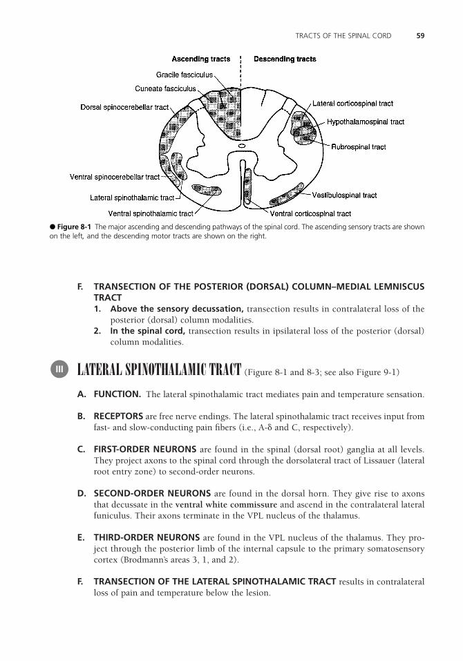
59TRACTS OF THE SPINAL CORD
F. TRANSECTION OF THE POSTERIOR (DORSAL) COLUMN–MEDIAL LEMNISCUSTRACT1. Above the sensory decussation, transection results in contralateral loss of the
posterior (dorsal) column modalities.2. In the spinal cord, transection results in ipsilateral loss of the posterior (dorsal)
column modalities.
LATERAL SPINOTHALAMIC TRACT (Figure 8-1 and 8-3; see also Figure 9-1)
A. FUNCTION. The lateral spinothalamic tract mediates pain and temperature sensation.
B. RECEPTORS are free nerve endings. The lateral spinothalamic tract receives input fromfast- and slow-conducting pain fibers (i.e., A-� and C, respectively).
C. FIRST-ORDER NEURONS are found in the spinal (dorsal root) ganglia at all levels.They project axons to the spinal cord through the dorsolateral tract of Lissauer (lateralroot entry zone) to second-order neurons.
D. SECOND-ORDER NEURONS are found in the dorsal horn. They give rise to axonsthat decussate in the ventral white commissure and ascend in the contralateral lateralfuniculus. Their axons terminate in the VPL nucleus of the thalamus.
E. THIRD-ORDER NEURONS are found in the VPL nucleus of the thalamus. They pro-ject through the posterior limb of the internal capsule to the primary somatosensorycortex (Brodmann’s areas 3, 1, and 2).
F. TRANSECTION OF THE LATERAL SPINOTHALAMIC TRACT results in contralateralloss of pain and temperature below the lesion.
III
● Figure 8-1 The major ascending and descending pathways of the spinal cord. The ascending sensory tracts are shownon the left, and the descending motor tracts are shown on the right.
LWBK110-3895G-C08[58-64].qxd 7/10/08 7:47 AM Page 59 Aptara Inc.

60 CHAPTER 8
Thalamus
Internal capsule
Lentiform nucleus
Trigeminal nerve
Nucleus gracilis
Nucleus cuneatus
Internal arcuate fibers
(neuron II)
Cuneate fasciculus
Spinal ganglion cell
(neuron I)
Pacinian
corpuscle
Leg area
Postcentral gyrus
Midbrain
Pons
Medulla
Cervical spinal cord
Lumbosacral spinal cord
Trunk area
Arm area
Head area
Face area
Ventral posterolateral
nucleus of thalamus
(neuron III)
Medial lemniscus
Medial lemniscus
Medial lemniscus
Spinal trigeminal nucleus
Decussation of
medial lemniscus
Gracile fasciculus
Meissner’s
corpuscle
Som
esth
etic cortex
Gracile fasciculus
Cuneate fasciculus
● Figure 8-2 The dorsal column–medial lemniscus pathway. Impulses conducted by this pathway mediate discrimina-tory tactile sense (e.g., touch, vibration, pressure) and kinesthetic sense (e.g., position, movement). The posterior (dor-sal) column system mediates conscious proprioception. (Adapted from MB Carpenter, J Sutin, Human neuroanatomy.Baltimore: Williams & Wilkins, 1983:266, with permission.)
LWBK110-3895G-C08[58-64].qxd 7/10/08 7:47 AM Page 60 Aptara Inc.

61TRACTS OF THE SPINAL CORD
● Figure 8-3 The lateral spinothalamic tract. Impulses conducted by this tract mediate pain and thermal sense. Numer-ous collaterals are distributed to the brain stem reticular formation. (Adapted from MB Carpenter, J Sutin, Human neu-roanatomy. Baltimore: Williams & Wilkins, 1983:274, with permission.)
LWBK110-3895G-C08[58-64].qxd 7/10/08 7:47 AM Page 61 Aptara Inc.

62 CHAPTER 8
● Figure 8-4 The lateral and ventral corticospinal (pyramidal) tracts. These major descending motor pathways mediatevolitional motor activity. The cells of origin are located in the premotor, the motor, and the sensory cortices. CN, cranialnerve. (Adapted from MB Carpenter, J Sutin, Human neuroanatomy. Baltimore: Williams & Wilkins, 1983:285, with per-mission.)
LWBK110-3895G-C08[58-64].qxd 7/10/08 7:47 AM Page 62 Aptara Inc.

LATERAL CORTICOSPINAL TRACT (Figure 8-1 and 8-4; see also Figure 9-1)
A. FUNCTION. The lateral corticospinal tract mediates voluntary skilled motor activity,primarily of the upper limbs. It is not fully myelinated until the end of the second year(Babinski’s sign).
B. FIBER CALIBER. Approximately 90% of the fibers lie between 1 and 4 �, and 4% lieabove 20 �m (from the giant cells of Betz).
C. ORIGIN AND TERMINATION1. Origin. The lateral corticospinal tract arises from layer V of the cerebral cortex
from three cortical areas in equal aliquots:a. The premotor cortex (Brodmann’s area 6).b. The primary motor cortex, or precentral gyrus (Brodmann’s area 4).
Arm, face, and foot areas. The arm and face areas of the motor homunculusarise from the lateral convexity; the foot region of the motor homunculus isfound in the paracentral lobule (see Figure 23-2).
c. The primary sensory cortex, or postcentral gyrus (Brodmann’s areas 3, 1, and 2).2. Termination. The lateral corticospinal tract terminates contralaterally, through
interneurons, on ventral horn motor neurons.
D. COURSE of the lateral corticospinal tract1. Telencephalon. The lateral corticospinal tract runs in the posterior limb of the
internal capsule in the telencephalon.2. Midbrain. The lateral corticospinal tract runs in the middle three-fifths of the crus
cerebri in the midbrain.3. Pons. The lateral corticospinal tract runs in the base of the pons.4. Medulla. The lateral corticospinal tract runs in the medullary pyramids. Between
85% and 90% of the corticospinal fibers cross in the pyramidal decussation as thelateral corticospinal tract. The remaining 10% to 15% of the fibers continue as theanterior corticospinal tract.
5. Spinal cord. The lateral corticospinal tract runs in the dorsal quadrant of the lat-eral funiculus.
E. TRANSECTION OF THE LATERAL CORTICOSPINAL TRACT1. Above the motor decussation, transection results in contralateral spastic paresis
and Babinski’s sign (upward fanning of the toes).2. In the spinal cord, transection results in ipsilateral spastic paresis and Babinski’s
sign.
HYPOTHALAMOSPINAL TRACT (Figure 8-5)
A. ANATOMIC LOCATION. The hypothalamospinal tract projects without interruptionfrom the hypothalamus to the ciliospinal center of the intermediolateral cell column atT-1 to T-2. It is found in the spinal cord at T-1 or above in the dorsolateral quadrant ofthe lateral funiculus. It is also found in the lateral tegmentum of the medulla, pons,and midbrain.
V
IV
63TRACTS OF THE SPINAL CORD
LWBK110-3895G-C08[58-64].qxd 7/10/08 7:47 AM Page 63 Aptara Inc.

B. CLINICAL FEATURES. Interruption of this tract at any level results in Horner’s syn-drome (i.e., miosis, ptosis, hemianhidrosis, and apparent enophthalmos). The signs arealways ipsilateral.
64 CHAPTER 8
● Figure 8-5 The oculosympathetic pathway. Hypothalamic fibers project to the ipsilateral ciliospinal center of the inter-mediolateral cell column at T-1. The ciliospinal center projects preganglionic sympathetic fibers to the superior cervicalganglion. The superior cervical ganglion projects perivascular postganglionic sympathetic fibers through the tympaniccavity, cavernous sinus, and superior orbital fissure to the dilator muscle of the iris. Interruption of this pathway at anylevel results in Horner’s syndrome. CN, cranial nerve.
Case Study A 17-year-old man complained of pain on the left side of his chest and progressive weaknessof his left lower limb for 2 months before coming to the clinic. What is the most likely diag-nosis?
Relevant Physical Exam Findings• Neurologic evaluation revealed weakness in the left lower limb; spasticity and hyper-
reflexia at the knee and ankle were also observed. • On the left side, a loss of two-point discrimination, vibratory sense, and proprioception
below the hip was observed. A loss of pain and temperature sensation below the T7 der-matome was observed on the right side.
Diagnosis• Brown-Séquard syndrome, resulting from an upper motor neuron lesion (tumor) at T5-T6
spinal cord levels, represents an incomplete spinal cord lesion characterized by symptomsindicative of hemisection of the spinal cord. It involves ipsilateral hemiplegia with con-tralateral pain and temperature deficits due to decussating fibers of the spinothalamic tract.
LWBK110-3895G-C08[58-64].qxd 7/10/08 7:47 AM Page 64 Aptara Inc.

65
Chapter 9
Lesions of the Spinal Cord
DISEASES OF THE MOTOR NEURONS AND CORTICOSPINAL TRACTS(Figures 9-1 and 9-2)
A. UPPER MOTOR NEURON (UMN) LESIONS ARE CAUSED BY TRANSECTION OFTHE CORTICOSPINAL TRACT OR DESTRUCTION OF THE CORTICAL CELLS OFORIGIN. THEY RESULT IN SPASTIC PARESIS with pyramidal signs (Babinski’s sign).
B. LOWER MOTOR NEURON (LMN) LESIONS ARE CAUSED BY DAMAGE TO THEMOTOR NEURONS. THEY RESULT IN FLACCID PARALYSIS, areflexia, atrophy, fas-ciculations, and fibrillations. Poliomyelitis or Werdnig-Hoffmann disease (Figure9-2A) results from damage to the motor neurons.
C. An example of a combined UMN and LMN disease is amyotrophic lateral sclerosis(ALS, or Lou Gehrig’s disease) (Figure 9-2D). ALS is caused by damage to the corti-cospinal tracts, with pyramidal signs, and by damage to the LMNs, with LMN symp-toms. Patients with ALS have no sensory deficits.
SENSORY PATHWAY LESIONS An example of a condition caused by these lesionsis dorsal column disease (tabes dorsalis) (Figure 9-2C). This disease is seen in patientswith neurosyphilis. It is characterized by a loss of tactile discrimination and position andvibration sensation. Irritative involvement of the dorsal roots results in pain and paresthe-sias. Patients have a Romberg sign. (Subject stands with his feet together and, when hecloses his eyes, loses his balance. This is a sign of dorsal column ataxia.)
COMBINED MOTOR AND SENSORY LESIONSA. SPINAL CORD HEMISECTION (BROWN-SÉQUARD SYNDROME) (Figure 9-2E) is
caused by damage to the following structures:1. The dorsal columns [gracile (leg) and cuneate (arm) fasciculi]. Damage results
in ipsilateral loss of tactile discrimination and position and vibration sensation.
III
II
I
Key ConceptsStudy the eight classic national board lesions of the spinal cord. Four heavy hitters are Brown-Siquard syndrome, B12 avitaminosis (subacute combined degeneration), syringomyelia, andamyotrophic lateral sclerosis (ALS, or Lou Gehrig’s disease).
✔
LWBK110-3895G-C09[65-69].qxd 7/10/08 7:48 AM Page 65 Aptara Inc.

2. The lateral corticospinal tract. Damage results in ipsilateral spastic paresis withpyramidal signs below the lesion.
3. The lateral spinothalamic tract. Damage results in contralateral loss of pain andtemperature sensation one segment below the lesion.
4. The hypothalamospinal tract at T-1 and above. Damage results in ipsilateral Horner’ssyndrome (i.e., miosis, ptosis, hemianhidrosis, and apparent enophthalmos).
5. The ventral (anterior) horn. Damage results in ipsilateral flaccid paralysis of inner-vated muscles.
B. VENTRAL SPINAL ARTERY OCCLUSION (Figure 9-2F) causes infarction of the ante-rior two-thirds of the spinal cord but spares the dorsal columns and horns. It results indamage to the following structures:1. The lateral corticospinal tracts. Damage results in bilateral spastic paresis with
pyramidal signs below the lesion.2. The lateral spinothalamic tracts. Damage results in bilateral loss of pain and tem-
perature sensation below the lesion.3. The hypothalamospinal tract at T-2 and above. Damage results in bilateral Horner’s
syndrome.4. The ventral (anterior) horns. Damage results in bilateral flaccid paralysis of the
innervated muscles.5. The corticospinal tracts to the sacral parasympathetic centers at S-2 to S-4. Dam-
age results in bilateral damage and loss of voluntary bladder and bowel control.
66 CHAPTER 9
● Figure 9-1 Transverse section of the cervical spinal cord. The clinically important ascending and descending path-ways are shown on the left. Clinical deficits that result from the interruption of these pathways are shown on the right.Destructive lesions of the posterior (dorsal) horns result in anesthesia and areflexia. Destruction of the ventral whitecommissure interrupts the central transmission of pain and temperature impulses bilaterally through the lateral spinothal-amic tracts.
LWBK110-3895G-C09[65-69].qxd 7/10/08 7:48 AM Page 66 Aptara Inc.
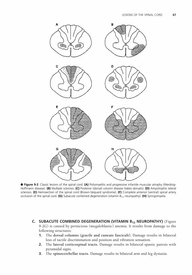
C. SUBACUTE COMBINED DEGENERATION (VITAMIN B12 NEUROPATHY) (Figure9-2G) is caused by pernicious (megaloblastic) anemia. It results from damage to thefollowing structures:1. The dorsal columns (gracile and cuneate fasciculi). Damage results in bilateral
loss of tactile discrimination and position and vibration sensation.2. The lateral corticospinal tracts. Damage results in bilateral spastic paresis with
pyramidal signs.3. The spinocerebellar tracts. Damage results in bilateral arm and leg dystaxia.
67LESIONS OF THE SPINAL CORD
● Figure 9-2 Classic lesions of the spinal cord. (A) Poliomyelitis and progressive infantile muscular atrophy (Werdnig-Hoffmann disease). (B) Multiple sclerosis. (C) Posterior (dorsal) column disease (tabes dorsalis). (D) Amyotrophic lateralsclerosis. (E) Hemisection of the spinal cord (Brown-Séquard syndrome). (F) Complete anterior (ventral) spinal arteryocclusion of the spinal cord. (G) Subacute combined degeneration (vitamin B12 neuropathy). (H) Syringomyelia.
LWBK110-3895G-C09[65-69].qxd 7/10/08 7:48 AM Page 67 Aptara Inc.

D. SYRINGOMYELIA (Figure 9-2H) is a central cavitation of the cervical cord ofunknown etiology. It results in damage to the following structures:1. The ventral white commissure. Damage to decussating lateral spinothalamic axons
causes bilateral loss of pain and temperature sensation.2. The ventral horns. LMN lesions result in flaccid paralysis of the intrinsic muscles
of the hands.
E. FRIEDREICH’S ATAXIA HAS THE SAME SPINAL CORD PATHOLOGY AND SYMP-TOMS AS SUBACUTE COMBINED DEGENERATION.
F. In multiple sclerosis (see Figure 9-2B), the plaques primarily involve the white matterof the cervical segments of the spinal cord. The lesions are random and asymmetric.
PERIPHERAL NERVOUS SYSTEM (PNS) LESIONS An example of a PNS lesionis Guillain-Barré syndrome (acute idiopathic polyneuritis, or postinfectious polyneuritis).It primarily affects the motor fibers of the ventral roots and peripheral nerves, and it pro-duces LMN symptoms (i.e., muscle weakness, ascending flaccid paralysis, and areflexia.)Guillain-Barré syndrome has the following features:
A. It is characterized by demyelination and edema.
B. Upper cervical root (C4) involvement and respiratory paralysis are common.
C. Caudal cranial nerve involvement with facial diplegia is present in 50% of cases.
D. Elevated protein levels may cause papilledema.
E. To a lesser degree, sensory fibers are affected, resulting in paresthesias.
F. The protein level in the cerebrospinal fluid is elevated but without pleocytosis (albu-minocytologic dissociation).
INTERVERTEBRAL DISK HERNIATION is seen at the L-4 to L-5 or L-5 to S-1 inter-space in 90% of cases. It appears at the C-5 to C-6 or C-6 to C-7 interspace in 10% of cases.
A. Intervertebral disk herniation consists of prolapse, or herniation, of the nucleus pul-posus through the defective anulus fibrosus and into the vertebral canal.
B. The nucleus pulposus impinges on the spinal roots, resulting in spinal root symptoms(i.e., paresthesias, pain, sensory loss, hyporeflexia, and muscle weakness).
CAUDA EQUINA SYNDROME (SPINAL ROOTS L3 TO C0) results usually from anerve root tumor, an ependymoma, or a dermoid tumor, or from a lipoma of the terminalcord. It is characterized by
A. Severe radicular unilateral pain.
B. Sensory distribution in a unilateral saddle-shaped area.
VI
V
IV
68 CHAPTER 9
LWBK110-3895G-C09[65-69].qxd 7/10/08 7:48 AM Page 68 Aptara Inc.

C. Unilateral muscle atrophy and absent quadriceps (L3) and ankle jerks (S1).
D. Unremarkable incontinence and sexual functions.
E. Gradual and unilateral onset.
CONUS MEDULLARIS SYNDROME (CORD SEGMENTS S3 TO C0) usually resultsfrom an intramedullary tumor (e.g., ependymoma). It is characterized by
A. Pain, usually bilateral and not severe.
B. Sensory distribution in a bilateral saddle-shaped area.
C. Unremarkable muscle changes; normal quadriceps and ankle reflexes.
D. Severely impaired incontinence and sexual functions.
E. Sudden and bilateral onset.
VII
69LESIONS OF THE SPINAL CORD
LWBK110-3895G-C09[65-69].qxd 7/10/08 7:48 AM Page 69 Aptara Inc.

70
Chapter 10
Brain Stem
INTRODUCTION The brain stem includes the medulla, pons, and midbrain. It extendsfrom the pyramidal decussation to the posterior commissure. The brain stem receives itsblood supply from the vertebrobasilar system. It contains cranial nerves (CN) III to XII(except the spinal part of CN XI). Figures 10-1 and 10-2 show its surface anatomy.
CROSS SECTION THROUGH THE MEDULLA (Figure 10-3)
A. MEDIAL STRUCTURES1. The hypoglossal nucleus of CN XII2. The medial lemniscus, which contains crossed fibers from the gracile and cuneate
nuclei3. The pyramid (corticospinal fibers)
B. LATERAL STRUCTURES1. The nucleus ambiguus (CN IX, X, and XI)2. The vestibular nuclei (CN VIII)3. The inferior cerebellar peduncle, which contains the dorsal spinocerebellar,
cuneocerebellar, and olivocerebellar tracts4. The lateral spinothalamic tract (spinal lemniscus)5. The spinal nucleus and tract of trigeminal nerve
CROSS SECTION THROUGH THE PONS (Figure 10-4). The pons has a dorsaltegmentum and a ventral base.
A. MEDIAL STRUCTURES1. Medial longitudinal fasciculus (MLF)2. Abducent nucleus of CN VI (underlies facial colliculus)3. Genu (internal) of CN VII (underlies facial nerve) (facial colliculus)
III
II
I
Key Concepts1) Study the transverse sections of the brain stem and localize the cranial nerve nuclei. 2) Study the ventral surface of the brain stem and identify the exiting and entering cranial
nerves. 3) On the dorsal surface of the brain stem, identify the only exiting cranial nerve, the
trochlear nerve.
✔
LWBK110-3895G-C10[70-73].qxd 7/10/08 7:49 AM Page 70 Aptara Inc.

71BRAIN STEM
● Figure 10-1 The dorsal surface of the brain stem. The three cerebellar peduncles have been removed to expose therhomboid fossa. The trochlear nerve is the only nerve to exit the brain stem from the dorsal surface. The facial colliculussurmounts the genu of the facial nerve and the abducent nucleus. CN, cranial nerve.
● Figure 10-2 The ventral surface of the brain stem and the attached cranial nerves (CN).
4. Abducent fibers of CN VI5. Medial lemniscus6. Corticospinal tract (in the base of the pons)
B. LATERAL STRUCTURES1. Facial nucleus (CN VII)2. Facial (intraaxial) nerve fibers3. Spinal nucleus and tract of trigeminal nerve (CN V)
LWBK110-3895G-C10[70-73].qxd 7/10/08 7:49 AM Page 71 Aptara Inc.

4. Lateral spinothalamic tract (spinal lemniscus)5. Vestibular nuclei of CN VIII6. Cochlear nuclei of CN VIII
CROSS SECTION THROUGH THE ROSTRAL MIDBRAIN (Figure 10-5). The mid-brain has a dorsal tectum, an intermediate tegmentum, and a base. The aqueduct liesbetween the tectum and the tegmentum.
A. DORSAL STRUCTURES include the superior colliculi.
B. TEGMENTUM1. Oculomotor nucleus (CN III)2. Medial longitudinal fasciculus (MLF)
IV
72 CHAPTER 10
● Figure 10-3 Transverse section of the medulla at the midolivary level. The vagal nerve [cranial nerve (CN) X], hypoglos-sal nerve (CN XII), and vestibulocochlear nerve (CN VIII) are prominent in this section. The nucleus ambiguus gives riseto special visceral efferent fibers to CN IX, X, and XI.
● Figure 10-4 Transverse section of the pons at the level of the abducent nucleus of cranial nerve (CN) VI and the facialnucleus of CN VII. MLF, medial longitudinal fasciculus.
LWBK110-3895G-C10[70-73].qxd 7/10/08 7:49 AM Page 72 Aptara Inc.

73BRAIN STEM
3. Red nucleus4. Substantia nigra5. Dentatothalamic tract (crossed)6. Medial lemniscus7. Lateral spinothalamic tract (in the spinal lemniscus)
C. CRUS CEREBRI (basis pedunculi cerebri, or cerebral peduncle). The corticospinal tractlies in the middle three-fifths of the crus cerebri.
CORTICONUCLEAR FIBERS project bilaterally to all motor cranial nerve nuclei exceptthe facial nucleus. The division of the facial nerve nucleus that innervates the upper face(the orbicularis oculi muscle and above) receives bilateral corticonuclear input. The divi-sion of the facial nerve nucleus that innervates the lower face receives only contralateralcorticonuclear input.
V
● Figure 10-5 Transverse section of the midbrain at the level of the superior colliculus, oculomotor nucleus of cranialnerve (CN) III, and red nucleus. MLF, medial longitudinal fasciculus.
LWBK110-3895G-C10[70-73].qxd 7/10/08 7:49 AM Page 73 Aptara Inc.

74
Chapter 11
Cranial Nerves
THE OLFACTORY NERVE, the first cranial nerve (CN I) (Figure 11-1), mediates olfac-tion (smell). It is the only sensory system that has no precortical relay in the thalamus. Theolfactory nerve is a special visceral afferent (SVA) nerve; see Appendix I. It consists ofunmyelinated axons of bipolar neurons that are located in the nasal mucosa, the olfactoryepithelium. It enters the cranial cavity through the cribriform plate of the ethmoid bone(see Appendix I).
A. OLFACTORY PATHWAY1. Olfactory receptor cells are first-order neurons that project to the mitral cells of
the olfactory bulb.2. Mitral cells are the principal cells of the olfactory bulb. They are excitatory and
glutaminergic. They project through the olfactory tract and lateral olfactory striato the primary olfactory cortex and amygdala.
3. The primary olfactory cortex (Brodmann’s area 34) consists of the piriform cor-tex that overlies the uncus.
B. LESIONS OF THE OLFACTORY PATHWAY result from trauma (e.g., skull fracture)and often from olfactory groove meningiomas. These lesions cause ipsilateral anosmia(localizing value). Lesions that involve the parahippocampal uncus may cause olfac-tory hallucinations [uncinate fits (seizures) with déjà vu].
C. FOSTER KENNEDY SYNDROME consists of ipsilateral anosmia, ipsilateral optic atro-phy, and contralateral papilledema. It is usually caused by an anterior fossa menin-gioma.
THE OPTIC NERVE (CN II) is a special somatic afferent (SSA) nerve that subservesvision and pupillary light reflexes (afferent limb; see Chapter 15). It enters the cranial cav-ity through the optic canal of the sphenoid bone. It is not a true peripheral nerve but is atract of the diencephalon. A transected optic nerve cannot regenerate.
II
I
Key Concepts1) This chapter is pivotal. It spawns more neuroanatomy examination questions than any
other chapter. Carefully study all of the figures and legends. 2) The seventh cranial nerve deserves special consideration (see Figures 11-5 and 11-6).
Understand the difference between an upper motor neuron and a lower motor neuron(Bell’s palsy).
✔
LWBK110-3895G-C11[74-87].qxd 7/10/08 7:50 AM Page 74 Aptara Inc.

THE OCULOMOTOR NERVE (CN III) is a general somatic efferent (GSE), general vis-ceral efferent (GVE) nerve.
A. GENERAL CHARACTERISTICS. The oculomotor nerve moves the eye, constricts thepupil, accommodates, and converges. It exits the brain stem from the interpeduncularfossa of the midbrain, passes through the cavernous sinus, and enters the orbit throughthe superior orbital fissure.1. The GSE component arises from the oculomotor nucleus of the rostral midbrain.
It innervates four extraocular muscles and the levator palpebrae muscle. (Remem-ber the mnemonic SIN: superior muscles are intorters of the globe.)a. The medial rectus muscle adducts the eye. With its opposite partner, it con-
verges the eyes.b. The superior rectus muscle elevates, intorts, and adducts the eye.c. The inferior rectus muscle depresses, extorts, and adducts the eye.d. The inferior oblique muscle elevates, extorts, and abducts the eye.e. The levator palpebrae muscle elevates the upper eyelid.
2. The GVE component consists of preganglionic parasympathetic fibers.a. The accessory nucleus of the oculomotor nerve (Edinger-Westphal nucleus)
projects preganglionic parasympathetic fibers to the ciliary ganglion of theorbit through CN III.
b. The ciliary ganglion projects postganglionic parasympathetic fibers to thesphincter pupillae (miosis) and the ciliary muscle (accommodation).
B. CLINICAL CORRELATION1. Oculomotor paralysis (palsy) is seen with transtentorial herniation (e.g., tumor,
subdural or epidural hematoma).a. Denervation of the levator palpebrae muscle causes ptosis (i.e., drooping of
the upper eyelid).b. Denervation of the extraocular muscles innervated by CN III causes the
affected eye to look “down and out” as a result of the unopposed action of the
III
75CRANIAL NERVES
● Figure 11-1 The base of the brain with attached cranial nerves (CN). (Reprinted from RC Truex, CE Kellner. Detailedatlas of the head and neck. New York: Oxford University Press, 1958:34, with permission.)
LWBK110-3895G-C11[74-87].qxd 7/10/08 7:50 AM Page 75 Aptara Inc.

lateral rectus and superior oblique muscles. The superior oblique and lateralrectus muscles are innervated by CN IV and CN VI, respectively. Oculomotorpalsy results in diplopia (double vision) when the patient looks in the direc-tion of the paretic muscle.
c. Interruption of parasympathetic innervation (internal ophthalmoplegia)results in a dilated, fixed pupil and paralysis of accommodation (cycloplegia).
2. Other conditions associated with CN III impairmenta. Transtentorial (uncal) herniation. Increased supratentorial pressure (e.g.,
from a tumor) forces the hippocampal uncus through the tentorial notch andcompresses or stretches the oculomotor nerve.(1) Sphincter pupillae fibers are affected first, resulting in a dilated, fixed pupil.(2) Somatic efferent fibers are affected later, resulting in external strabis-
mus (exotropia).b. Aneurysms of the carotid and posterior communicating arteries often com-
press CN III within the cavernous sinus or interpeduncular cistern. They usu-ally affect the peripheral pupilloconstrictor fibers first (e.g., uncal herniation).
c. Diabetes mellitus (diabetic oculomotor palsy) often affects the oculomotornerve. It damages the central fibers and spares the sphincter pupillae fibers.
THE TROCHLEAR NERVE (CN IV) is a GSE nerve.
A. GENERAL CHARACTERISTICS. The trochlear nerve is a pure motor nerve that inner-vates the superior oblique muscle. This muscle depresses, intorts, and abducts the eye.(Figure 11-2.)1. It arises from the contralateral trochlear nucleus of the caudal midbrain.2. It decussates beneath the superior medullary velum of the midbrain and exits the
brain stem on its dorsal surface, caudal to the inferior colliculus.3. It encircles the midbrain within the subarachnoid space, passes through the cav-
ernous sinus, and enters the orbit through the superior orbital fissure.
B. CLINICAL CORRELATION. Because of its course around the midbrain, the trochlearnerve is particularly vulnerable to head trauma. The trochlear decussation under-lies the superior medullary velum. Trauma at this site often results in bilateralfourth-nerve palsies. Pressure against the free border of the tentorium (herniation)may injure the nerve (Figure 11-2). CN IV paralysis results in the following condi-tions:1. Extorsion of the eye and weakness of downward gaze.2. Vertical diplopia, which increases when looking down.3. Head tilting to compensate for extorsion (may be misdiagnosed as idiopathic tor-
ticollis).
THE TRIGEMINAL NERVE (CN V) is a special visceral efferent (SVE), general somaticafferent (GSA) nerve (Figure 11-3).
A. GENERAL CHARACTERISTICS. The trigeminal nerve is the nerve of pharyngeal(branchial) arch 1 (mandibular). It has three divisions: ophthalmic (CN V-1), maxil-lary (CN V-2), and mandibular (CN V-3).1. The SVE component arises from the motor nucleus of trigeminal nerve that is found
in the lateral midpontine tegmentum. It innervates the muscles of mastication (i.e.,
V
IV
76 CHAPTER 11
LWBK110-3895G-C11[74-87].qxd 7/10/08 7:50 AM Page 76 Aptara Inc.

77CRANIAL NERVES
A B
● Figure 11-2 Paralysis of the right superior oblique muscle. (A) A pair of eyes with normal extorsion and intorsionmovements. Tilting the chin to the right side results in compensatory intorsion of the left eye and extorsion of the righteye. (B) Paralysis of the right superior oblique muscle results in extorsion of the right eye, causing diplopia. Tilting thechin to the right side results in compensatory intorsion of the left eye, thus permitting binocular alignment. (Reprintedfrom JD Fix. BRS neuroanatomy, 3rd ed. Baltimore: Williams & Wilkins, 1996:220, with permission.)
● Figure 11-3 Jaw jerk (masseter reflex) pathway showing two neurons. Note that the first-order (sensory) neuron isfound in the mesencephalic nucleus of the pons and midbrain, not in the trigeminal ganglion. CN, cranial nerve.(Reprinted from JD Fix. BRS neuroanatomy, 3rd ed. Baltimore: Williams & Wilkins, 1996:220, with permission.)
LWBK110-3895G-C11[74-87].qxd 7/10/08 7:50 AM Page 77 Aptara Inc.

temporalis, masseter, lateral, and medial pterygoids), the tensors tympani and velipalatini, the mylohyoid muscle, and the anterior belly of the digastric muscle.
2. The GSA component provides sensory innervation to the face, mucous mem-branes of the nasal and oral cavities and frontal sinus, hard palate, and deep struc-tures of the head (proprioception from muscles and the temporomandibular joint).It innervates the dura of the anterior and middle cranial fossae (supratentorialdura).
B. CLINICAL CORRELATION. Lesions result in the following neurologic deficits:1. Loss of general sensation (hemianesthesia) from the face and mucous mem-
branes of the oral and nasal cavities.2. Loss of the corneal reflex (afferent limb, CN V-1; Figure 11-4).3. Flaccid paralysis of the muscles of mastication.4. Deviation of the jaw to the weak side as a result of the unopposed action of the
opposite lateral pterygoid muscle.5. Paralysis of the tensor tympani muscle, which leads to hypoacusis (partial deaf-
ness to low-pitched sounds).6. Trigeminal neuralgia (tic douloureux), which is characterized by recurrent parox-
ysms of sharp, stabbing pain in one or more branches of the nerve.
78 CHAPTER 11
Genu of CN VII
Tertiary neuron
CN VII
Facial nucleus
Decussating
corneal reflex fiber
To orbicularis
oculi m
uscle
From cornea
Trigeminothalamic
pain fiber
Secondary neuron
Primary neuron
V-1
V-2
V-3
V-3 (motor)
CN VII
Principal sensory of
nucleus (CN V)
Spinal nucleus of trigeminal nerve
Spinal tract of trigeminal nerve
Afferent limb ofcorneal reflex
Efferent limb ofcorneal reflex
● Figure 11-4 The corneal reflex pathway showing the three neurons and decussation. This reflex is consensual, likethe pupillary light reflex. Second-order pain neurons are found in the caudal division of the spinal nucleus of trigeminalnerve. Second-order corneal reflex neurons are found at more rostral levels. CN, cranial nerve.
LWBK110-3895G-C11[74-87].qxd 7/10/08 7:50 AM Page 78 Aptara Inc.

THE ABDUCENT NERVE (CN VI)A. GENERAL CHARACTERISTICS. The abducent nerve is a pure GSE nerve that inner-
vates the lateral rectus muscle, which abducts the eye.1. It arises from the abducent nucleus that is found in the dorsomedial tegmentum
of the caudal pons.2. Exiting intraaxial fibers pass through the corticospinal tract. A lesion results in
alternating abducent hemiparesis.3. It passes through the pontine cistern and cavernous sinus and enters the orbit
through the superior orbital fissure.
B. CLINICAL CORRELATION. CN VI PARALYSIS is the most common isolated palsy thatresults from the long peripheral course of the nerve. It is seen in patients with menin-gitis, subarachnoid hemorrhage, late-stage syphilis, and trauma. Abducent nerve paral-ysis results in the following defects:1. Convergent (medial) strabismus (esotropia) with inability to abduct the eye.2. Horizontal diplopia with maximum separation of the double images when look-
ing toward the paretic lateral rectus muscle.
THE FACIAL NERVE (CN VII)A. GENERAL CHARACTERISTICS. The facial nerve is a GSA, general visceral afferent
(GVA), SVA, GVE, and SVE nerve (Figures 11-5 and 11-6). It mediates facial move-ments, taste, salivation, lacrimation, and general sensation from the external ear. Itis the nerve of the pharyngeal (branchial) arch 2 (hyoid). It includes the facial nerveproper (motor division), which contains the SVE fibers that innervate the muscles offacial (mimetic) expression. CN VII includes the intermediate nerve, which contains
VII
VI
79CRANIAL NERVES
● Figure 11-5 The functional components of the facial nerve (cranial nerve [CN] VII).
LWBK110-3895G-C11[74-87].qxd 7/10/08 7:50 AM Page 79 Aptara Inc.

GSA, SVA, and GVE fibers. All first-order sensory neurons are found in the geniculateganglion within the temporal bone.1. Anatomy. The facial nerve exits the brain stem in the cerebellopontine angle. It
enters the internal auditory meatus and the facial canal. It then exits the facial canaland skull through the stylomastoid foramen.
2. The GSA component has cell bodies located in the geniculate ganglion. It inner-vates the posterior surface of the external ear through the posterior auricularbranch of CN VII. It projects centrally to the spinal tract and nucleus of trigemi-nal nerve.
3. The GVA component has no clinical significance. The cell bodies are located inthe geniculate ganglion. Fibers innervate the soft palate and the adjacent pharyn-geal wall.
80 CHAPTER 11
● Figure 11-6 Corticonuclear innervation of the facial nerve (cranial nerve [CN] VII) nucleus. An upper motor neuron(UMN) lesion (e.g., stroke involving the internal capsule) results in contralateral weakness of the lower face, with spar-ing of the upper face. A lower motor neuron (LMN) lesion (e.g., Bell’s palsy) results in paralysis of the facial muscles inboth the upper and lower face. (Redrawn from WE DeMyer, Technique of the neurological examination: A programmedtext, 4th ed. New York: McGraw-Hill, 1994:177, with permission.)
LWBK110-3895G-C11[74-87].qxd 7/10/08 7:50 AM Page 80 Aptara Inc.

4. The SVA component (taste) has cell bodies located in the geniculate ganglion. Itprojects centrally to the solitary tract and nucleus. It innervates the taste buds fromthe anterior two-thirds of the tongue through:a. The intermediate nerve.b. The chorda tympani, which is located in the tympanic cavity medial to the
tympanic membrane and malleus. It contains the SVA and GVE (parasympa-thetic) fibers.
c. The lingual nerve (a branch of CN V-3).d. The central gustatory pathway (see Figure 11-5). Taste fibers from CN VII,
CN IX, and CN X project through the solitary tract to the solitary nucleus.The solitary nucleus projects through the central tegmental tract to the ven-tral posteromedial nucleus (VPM) of the thalamus. The VPM projects to thegustatory cortex of the parietal lobe (parietal operculum).
5. The GVE component is a parasympathetic component that innervates thelacrimal, submandibular, and sublingual glands. It contains preganglionicparasympathetic neurons that are located in the superior salivatory nucleus ofthe caudal pons.a. Lacrimal pathway (see Figure 11-5). The superior salivatory nucleus projects
through the intermediate and greater petrosal nerves to the pterygopalatine(sphenopalatine) ganglion. The pterygopalatine ganglion projects to thelacrimal gland of the orbit.
b. Submandibular pathway (see Figure 11-5). The superior salivatory nucleusprojects through the intermediate nerve and chorda tympani to the sub-mandibular ganglion. The submandibular ganglion projects to and innervatesthe submandibular and sublingual glands.
6. The SVE component arises from the facial nucleus, loops around the abducentnucleus of the caudal pons, and exits the brain stem in the cerebellopontineangle. It enters the internal auditory meatus, traverses the facial canal, sends abranch to the stapedius muscle of the middle ear, and exits the skull throughthe stylomastoid foramen. It innervates the muscles of facial expression, the sty-lohyoid muscle, the posterior belly of the digastric muscle, and the stapediusmuscle.
B. CLINICAL CORRELATION. Lesions cause the following conditions:1. Flaccid paralysis of the muscles of facial expression (upper and lower face).2. Loss of the corneal reflex (efferent limb), which may lead to corneal ulcera-
tion.3. Loss of taste (ageusia-gustatory anesthesia) from the anterior two-thirds of the
tongue, which may result from damage to the chorda tympani.4. Hyperacusis (increased acuity to sounds) as a result of stapedius paralysis.5. Bell’s palsy (peripheral facial paralysis), which is caused by trauma or infection
and involves the upper and lower face.6. Crocodile tears syndrome (lacrimation during eating), which is a result of aber-
rant regeneration of SVE fibers after trauma.7. Supranuclear (central) facial palsy, which results in contralateral weakness of the
lower face, with sparing of the upper face (see Figure 11-6).8. Bilateral facial nerve palsies, which occur in Guillain-Barré syndrome. 9. Möbius’ syndrome, which consists of congenital facial diplegia (CN VII) and con-
vergent strabismus (CN VI).
81CRANIAL NERVES
LWBK110-3895G-C11[74-87].qxd 7/10/08 7:50 AM Page 81 Aptara Inc.

THE VESTIBULOCOCHLEAR NERVE (CN VIII) is an SSA nerve. It has two func-tional divisions: the vestibular nerve, which maintains equilibrium and balance, and thecochlear nerve, which mediates hearing. It exits the brain stem at the cerebellopontineangle and enters the internal auditory meatus. It is confined to the temporal bone.
A. VESTIBULAR NERVE (see Figure 14-1)1. General characteristics
a. It is associated functionally with the cerebellum (flocculonodular lobe) andocular motor nuclei.
b. It regulates compensatory eye movements.c. Its first-order sensory bipolar neurons are located in the vestibular ganglion in
the fundus of the internal auditory meatus.d. It projects its peripheral processes to the hair cells of the cristae of the semi-
circular ducts and the hair cells of the utricle and saccule.e. It projects its central processes to the four vestibular nuclei of the brain stem
and the flocculonodular lobe of the cerebellum.f. It conducts efferent fibers to the hair cells from the brain stem.
2. Clinical correlation. Lesions result in disequilibrium, vertigo, and nystagmus.
B. COCHLEAR NERVE (see Figure 13-1)1. General characteristics
a. Its first-order sensory bipolar neurons are located in the spiral (cochlear) gan-glion of the modiolus of the cochlea, within the temporal bone.
b. It projects its peripheral processes to the hair cells of the organ of Corti.c. It projects its central processes to the dorsal and ventral cochlear nuclei of the
brain stem.d. It conducts efferent fibers to the hair cells from the brain stem.
2. Clinical correlation. Destructive lesions cause hearing loss (sensorineural deaf-ness). Irritative lesions can cause tinnitus (ear ringing). An acoustic neuroma(schwannoma) is a Schwann cell tumor of the cochlear nerve that causesdeafness.
THE GLOSSOPHARYNGEAL NERVE (CN IX) is a GSA, GVA, SVA, SVE, and GVEnerve (Figure 11-7).
A. GENERAL CHARACTERISTICS. The glossopharyngeal nerve is primarily a sensorynerve. Along with CN X, CN XI, and CN XII, it mediates taste, salivation, andswallowing. It mediates input from the carotid sinus, which contains barorecep-tors that monitor arterial blood pressure. It also mediates input from the carotidbody, which contains chemoreceptors that monitor the CO2 and O2 concentrationof the blood.1. Anatomy. CN IX is the nerve of pharyngeal (branchial) arch 3. It exits the brain
stem (medulla) from the postolivary sulcus with CN X and CN XI. It exits the skullthrough the jugular foramen with CN X and CN XI.
2. The GSA component innervates part of the external ear and the external auditorymeatus through the auricular branch of the vagus nerve. It has cell bodies in thesuperior ganglion. It projects its central processes to the spinal tract and nucleusof trigeminal nerve.
IX
VIII
82 CHAPTER 11
LWBK110-3895G-C11[74-87].qxd 7/10/08 7:50 AM Page 82 Aptara Inc.

3. The GVA component innervates structures that are derived from the endoderm(e.g., pharynx). It innervates the mucous membranes of the posterior one-thirdof the tongue, tonsil, upper pharynx, tympanic cavity, and auditory tube. It alsoinnervates the carotid sinus (baroreceptors) and carotid body (chemoreceptors)through the sinus nerve. It has cell bodies in the inferior (petrosal) ganglion. It isthe afferent limb of the gag reflex and the carotid sinus reflex.
4. The SVA component innervates the taste buds of the posterior one-third of thetongue. It has cell bodies in the inferior (petrosal) ganglion. It projects its centralprocesses to the solitary tract and nucleus. (For a discussion of the central path-way, see VII.A.4.d.)
83CRANIAL NERVES
UMNUMNMotor cortex
Corticonuclear tract
Decussation
Nucleus ambiguus
CN X (vagal nerve)
LMN
Levator veli palatini
and palatal arches
Corticonuclear tract
UMN lesion
LMN lesion
LMN
Medulla
Medial lemniscus
Pyramid
A B
● Figure 11-7 Innervation of the palatal arches and uvula. Sensory innervation is mediated by the glossopharyngealnerve [cranial nerve (CN) IX]. Motor innervation of the palatal arches and uvula is mediated by the vagus nerve (CN X).(A) A normal palate and uvula in a person who is saying “Ah.” (B) A patient with an upper motor neuron (UMN) lesion(left) and a lower motor neuron (LMN) lesion (right). When this patient says “Ah,” the palatal arches sag. The uvuladeviates toward the intact (left) side. (Modified from WE DeMyer, Technique of the neurological examination: a pro-grammed text, 4th ed. New York: McGraw-Hill, 1994:191, with permission.)
LWBK110-3895G-C11[74-87].qxd 7/10/08 7:50 AM Page 83 Aptara Inc.

5. The SVE component innervates only the stylopharyngeus muscle. It arises fromthe nucleus ambiguus of the lateral medulla.
6. The GVE component is a parasympathetic component that innervates theparotid gland. Preganglionic parasympathetic neurons are located in the infe-rior salivatory nucleus of the medulla. They project through the tympanic andlesser petrosal nerves to the otic ganglion. Postganglionic fibers from the oticganglion project to the parotid gland through the auriculotemporal nerve (CNV-3).
B. CLINICAL CORRELATION. Lesions cause the following conditions:1. Loss of the gag (pharyngeal) reflex (interruption of the afferent limb).2. Hypersensitive carotid sinus reflex (syncope).3. Loss of general sensation in the pharynx, tonsils, fauces, and back of the tongue.4. Loss of taste from the posterior one-third of the tongue.5. Glossopharyngeal neuralgia, which is characterized by severe stabbing pain in
the root of the tongue.
THE VAGAL NERVE (CN X) is a GSA, GVA, SVA, SVE, and GVE nerve (see Figure11-7).
A. GENERAL CHARACTERISTICS. The vagal nerve mediates phonation, swallowing(with CN IX, CN XI, and CN XII), elevation of the palate, taste, and cutaneous sensa-tion from the ear. It innervates the viscera of the neck, thorax, and abdomen. 1. Anatomy. The vagal nerve is the nerve of pharyngeal (branchial) arches 4 and 6.
Pharyngeal arch 5 is either absent or rudimentary. It exits the brain stem (medulla)from the postolivary sulcus. It exits the skull through the jugular foramen withCN IX and CN XI.
2. The GSA component innervates the infratentorial dura, external ear, external audi-tory meatus, and tympanic membrane. It has cell bodies in the superior (jugular)ganglion, and it projects its central processes to the spinal tract and nucleus oftrigeminal nerve.
3. The GVA component innervates the mucous membranes of the pharynx, larynx,esophagus, trachea, and thoracic and abdominal viscera (to the left colic flexure).It has cell bodies in the inferior (nodose) ganglion. It projects its central processesto the solitary tract and nucleus.
4. The SVA component innervates the taste buds in the epiglottic region. It has cellbodies in the inferior (nodose) ganglion. It projects its central processes to thesolitary tract and nucleus. (For a discussion of the central pathway, seeVII.A.4.d.)
5. The SVE component innervates the pharyngeal (brachial) arch muscles of the lar-ynx and pharynx, the striated muscle of the upper esophagus, the muscle of theuvula, and the levator veli palatini and palatoglossus muscles. It receives SVE inputfrom the cranial division of the spinal accessory nerve (CN XI). It arises from thenucleus ambiguus in the lateral medulla. The SVE component provides the effer-ent limb of the gag reflex.
6. The GVE component innervates the viscera of the neck and the thoracic (heart)and abdominal cavities as far as the left colic flexure. Preganglionic parasympa-thetic neurons that are located in the dorsal motor nucleus of the medulla projectto the terminal (intramural) ganglia of the visceral organs.
X
84 CHAPTER 11
LWBK110-3895G-C11[74-87].qxd 7/10/08 7:50 AM Page 84 Aptara Inc.

B. CLINICAL CORRELATION. LESIONS and reflexes cause the following conditions:1. Ipsilateral paralysis of the soft palate, pharynx, and larynx that leads to dyspho-
nia (hoarseness), dyspnea, dysarthria, and dysphagia.2. Loss of the gag (palatal) reflex (efferent limb).3. Anesthesia of the pharynx and larynx that leads to unilateral loss of the cough
reflex.4. Aortic aneurysms and tumors of the neck and thorax that frequently compress
the vagal nerve and can lead to cough, dyspnea, dysphagia, hoarseness, andchest/back pain.
5. Complete laryngeal paralysis, which can be rapidly fatal if it is bilateral(asphyxia).
6. Parasympathetic (vegetative) disturbances, including bradycardia (irritativelesion), tachycardia (destructive lesion), and dilation of the stomach.
7. The oculocardiac reflex, in which pressure on the eye slows the heart rate (affer-ent limb of CN V-1 and efferent limb of CN X).
8. The carotid sinus reflex, in which pressure on the carotid sinus slows the heartrate (bradycardia; efferent limb of CN X).
THE ACCESSORY NERVE (CN XI), or spinal accessory nerve, is an SVE nerve (Figure11-8).
A. GENERAL CHARACTERISTICS. The accessory nerve mediates head and shouldermovement and innervates the laryngeal muscles. It has the following divisions:
XI
85CRANIAL NERVES
CN XICN IX
CN IX
CN X
Jugular foramen
Ambiguus nucleus
in medulla
Superior laryngeal nerve
Stylopharyngeus nerve
Inferior laryngeal nerve
Foramen magnum
Accessory nucleus
in spinal cord (C1-C5)
Facial nucleus in pons
● Figure 11-8 The cranial and spinal divisions of the accessory nerve [cranial nerve (CN) XI]. The cranial division hitch-hikes a ride with the accessory nerve, then joins the vagal nerve to become the inferior (recurrent) laryngeal nerve. Therecurrent laryngeal nerve innervates the intrinsic muscles of the larynx, except for the cricothyroid muscle. The spinaldivision innervates the trapezoid and sternocleidomastoid muscles. Three nerves pass through the jugular foramen (glo-mus jugulare tumor). CN, cranial nerve.
LWBK110-3895G-C11[74-87].qxd 7/10/08 7:50 AM Page 85 Aptara Inc.
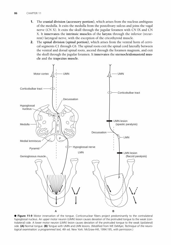
86 CHAPTER 11
UMNUMNMotor cortex
Corticobulbar tract
Decussation
Hypoglossal nerve
LMN
Corticobulbar tract
UMN lesion
(spastic paralysis)
LMN lesion
(flaccid paralysis)
LMN
Medulla
Decussation
Hypoglossal
nucleus
Medial lemniscus
Pyramid
P
G
C
Genioglossus muscle
A B
● Figure 11-9 Motor innervation of the tongue. Corticonuclear fibers project predominantly to the contralateralhypoglossal nucleus. An upper motor neuron (UMN ) lesion causes deviation of the protruded tongue to the weak (con-tralateral) side. A lower motor neuron (LMN ) lesion causes deviation of the protruded tongue to the weak (ipsilateral)side. (A) Normal tongue. (B) Tongue with UMN and LMN lesions. (Modified from WE DeMyer, Technique of the neuro-logical examination: a programmed text, 4th ed. New York: McGraw-Hill, 1994:195, with permission.)
1. The cranial division (accessory portion), which arises from the nucleus ambiguusof the medulla. It exits the medulla from the postolivary sulcus and joins the vagalnerve (CN X). It exits the skull through the jugular foramen with CN IX and CNX. It innervates the intrinsic muscles of the larynx through the inferior (recur-rent) laryngeal nerve, with the exception of the cricothyroid muscle.
2. The spinal division (spinal portion), which arises from the ventral horn of cervi-cal segments C1 through C6. The spinal roots exit the spinal cord laterally betweenthe ventral and dorsal spinal roots, ascend through the foramen magnum, and exitthe skull through the jugular foramen. It innervates the sternocleidomastoid mus-cle and the trapezius muscle.
LWBK110-3895G-C11[74-87].qxd 7/10/08 7:50 AM Page 86 Aptara Inc.

87CRANIAL NERVES
B. CLINICAL CORRELATION. Lesions cause the following conditions:1. Paralysis of the sternocleidomastoid muscle that results in difficulty in turning
the head to the contralateral side.2. Paralysis of the trapezius muscle that results in shoulder droop and inability to
shrug the shoulder.3. Paralysis and anesthesia of the larynx if the cranial root is involved.
THE HYPOGLOSSAL NERVE (CN XII) is a GSE nerve (Figure 11-9).
A. GENERAL CHARACTERISTICS. The hypoglossal nerve mediates tongue movement.It arises from the hypoglossal nucleus of the medulla and exits the medulla in the pre-olivary sulcus. It exits the skull through the hypoglossal canal, and it innervates theintrinsic and extrinsic muscles of the tongue. Extrinsic muscles are the genioglossus,styloglossus, and hyoglossus.
B. CLINICAL CORRELATION1. Transection results in hemiparalysis of the tongue.2. Protrusion causes the tongue to point toward the lesioned (weak) side because of
the unopposed action of the opposite genioglossus muscle (Figure 11-10).
XII
A
B
C
D
E
H
G
F
● Figure 11-10 The basis cerebri showing cranial nerves and the floor of the hypothalamus: olfactory tract (A); opticnerve (B); optic chiasm (C); optic tract (D); mamillary body (E); trochlear nerve (F); oculomotor nerve (G); infundibulum(H). (Reprinted from JD Fix, BRS neuroanatomy, 3rd ed. Baltimore: Williams & Wilkins, 1996:293, with permission.)
LWBK110-3895G-C11[74-87].qxd 7/10/08 7:50 AM Page 87 Aptara Inc.

88
Chapter 12
Trigeminal System
INTRODUCTION The trigeminal system provides sensory innervation to the face, oralcavity, and supratentorial dura through general somatic afferent (GSA) fibers. It also inner-vates the muscles of mastication through special visceral efferent (SVE) fibers.
THE TRIGEMINAL GANGLION (semilunar or gasserian) contains pseudounipolar gan-glion cells. It has three divisions:
A. The ophthalmic nerve [cranial nerve (CN) V-1] lies in the wall of the cavernous sinus.It enters the orbit through the superior orbital fissure and innervates the forehead, dor-sum of the nose, upper eyelid, orbit (cornea and conjunctiva), and cranial dura. Theophthalmic nerve mediates the afferent limb of the corneal reflex.
B. The maxillary nerve (CN V-2) lies in the wall of the cavernous sinus and innervatesthe upper lip and cheek, lower eyelid, anterior portion of the temple, oral mucosa ofthe upper mouth, nose, pharynx, gums, teeth and palate of the upper jaw, and cranialdura. It exits the skull through the foramen rotundum.
C. The mandibular nerve (CN V-3) exits the skull through the foramen ovale. Its sensory(GSA) component innervates the lower lip and chin, posterior portion of the temple,external auditory meatus, and tympanic membrane, external ear, teeth of the lower jaw,oral mucosa of the cheeks and floor of the mouth, anterior two-thirds of the tongue,temporomandibular joint, and cranial dura.
D. The motor (SVE) component of CN V accompanies the mandibular nerve (CN V-3)through the foramen ovale. It innervates the muscles of mastication, mylohyoid, ante-rior belly of the digastric, and tensores tympani and veli palatini. It innervates the mus-cles that move the jaw, the lateral and medial pterygoids (Figure 12-1).
TRIGEMINOTHALAMIC PATHWAYS (Figure 12-2)
A. The ventral trigeminothalamic tract mediates pain and temperature sensation from theface and oral cavity.
III
II
I
Key Concepts1) Cranial nerve (CN) V-1 is the afferent limb of the corneal reflex. 2) CN V-1, V-2, III, IV, and VI and the postganglionic sympathetic fibers are all found in
the cavernous sinus.
✔
LWBK110-3895G-C12[88-92].qxd 7/10/08 7:50 AM Page 88 Aptara Inc.

89TRIGEMINAL SYSTEM
1. First-order neurons are located in the trigeminal (gasserian) ganglion. They giverise to axons that descend in the spinal tract of trigeminal nerve and synapse withsecond-order neurons in the spinal nucleus of trigeminal nerve.
2. Second-order neurons are located in the spinal trigeminal nucleus. They give riseto decussating axons that terminate in the contralateral ventral posteromedial(VPM) nucleus of the thalamus.
3. Third-order neurons are located in the VPM nucleus of the thalamus. They pro-ject through the posterior limb of the internal capsule to the face area of thesomatosensory cortex (Brodmann’s areas 3, 1, and 2).
B. The dorsal trigeminothalamic tract mediates tactile discrimination and pressure sen-sation from the face and oral cavity. It receives input from Meissner’s and Pacinian cor-puscles.1. First-order neurons are located in the trigeminal (gasserian) ganglion. They
synapse in the principal sensory nucleus of CN V.
Superior cerebellar peduncle
Principal sensory nucleus of CN V
Motor nucleus CN V
Medial lemniscus
Corticospinal tract
Motor cortex
Condyloid process
LMN
UMN
4th ventricle
CN V motor
Pons
Lateral pterygoid muscle
● Figure 12-1 Function and innervation of the lateral pterygoid muscles (LPMs). The LPM receives its innervation fromthe motor nucleus of the trigeminal nerve found in the rostral pons. Bilateral innervation of the LPMs results in protru-sion of the tip of the mandible in the midline. The LPMs also open the jaw. Denervation of one LPM results in deviationof the mandible to the ipsilateral or weak side. The trigeminal motor nucleus receives bilateral corticonuclear input. CN,cranial nerve; LMN, lower motor neuron; UMN, upper motor neuron. (Modified from WE DeMyer, Technique of theneurological examination: A programmed text, 4th ed. New York: McGraw-Hill, 1994:174, with permission.)
LWBK110-3895G-C12[88-92].qxd 7/10/08 7:50 AM Page 89 Aptara Inc.
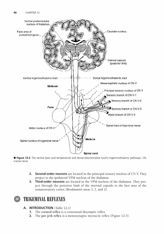
2. Second-order neurons are located in the principal sensory nucleus of CN V. Theyproject to the ipsilateral VPM nucleus of the thalamus.
3. Third-order neurons are located in the VPM nucleus of the thalamus. They pro-ject through the posterior limb of the internal capsule to the face area of thesomatosensory cortex (Brodmann’s areas 3, 1, and 2).
TRIGEMINAL REFLEXESA. INTRODUCTION (Table 12-1)
1. The corneal reflex is a consensual disynaptic reflex.2. The jaw jerk reflex is a monosynaptic myotactic reflex (Figure 12-3).
IV
90 CHAPTER 12
● Figure 12-2 The ventral (pain and temperature) and dorsal (discriminative touch) trigeminothalamic pathways. CN,cranial nerve.
LWBK110-3895G-C12[88-92].qxd 7/10/08 7:50 AM Page 90 Aptara Inc.

91TRIGEMINAL SYSTEM
3. The tearing (lacrimal) reflex occurs as a result of corneal or conjunctival irritation.4. The oculocardiac reflex occurs when pressure on the globe results in bradycardia.
B. CLINICAL CORRELATION. Trigeminal Neuralgia (tic douloureux) is characterized byrecurrent paroxysms of sharp, stabbing pain in one or more branches of the trigeminalnerve on one side of the face. It usually occurs in people older than 50 years, and it ismore common in women than in men. Carbamazepine is the drug of choice foridiopathic trigeminal neuralgia.
Reflex Afferent Limb Efferent Limb
Corneal reflex Ophthalmic nerve (CN V-1) Facial nerve (CN VII)Jaw jerk Mandibular nerve (CN V-3)a Mandibular nerve (CN V-3)Tearing (lacrimal) reflex Ophthalmic nerve (CN V-1) Facial nerve (CN VII)Oculocardiac reflex Ophthalmic nerve (CN V-1) Vagal nerve (CN X)
CN, cranial nerve.aThe cell bodies are found in the mesencephalic nucleus of CN V.
THE TRIGEMINAL REFLEXESTABLE 12-1
Motor division CN V
Masseter muscle
Principal sensory nucleus of CN V
Spinal trigeminal nucleus
V-3
Mesencephalic nucleus
with primary neuron
Muscle spindle
from masseter muscle
Motor nucleus CN V
with secondary neuron
● Figure 12-3 The jaw jerk (masseter) reflex. The afferent limb is V-3, and the efferent limb is the motor root thataccompanies V-3. First-order sensory neurons are located in the mesencephalic nucleus. The jaw jerk reflex, like all mus-cle stretch reflexes, is a monosynaptic myotactic reflex. Hyperreflexia indicates an upper motor neuron lesion. CN, cra-nial nerve.
LWBK110-3895G-C12[88-92].qxd 7/10/08 7:50 AM Page 91 Aptara Inc.

THE CAVERNOUS SINUS (Figure 12-4) contains the following structures:
A. INTERNAL CAROTID ARTERY (siphon)
B. CN III, IV, V-1, V-2, and VI
C. POSTGANGLIONIC SYMPATHETIC FIBERS en route to the orbit
V
92 CHAPTER 12
● Figure 12-4 The contents of the cavernous sinus. The wall of the cavernous sinus contains the ophthalmic cranialnerve (CN ) V-1 and maxillary (CN V-2) divisions of the trigeminal nerve (CN V) and the trochlear (CN IV) and oculomotor(CN III) nerves. The siphon of the internal carotid artery and the abducent nerve (CN VI), along with postganglionic sym-pathetic fibers, lies within the cavernous sinus.
Case Study A 50-year-old woman complains of sudden onset of pain over the left side of her lower face,with the attacks consisting of brief shocks of pain that last only a few seconds at a time.Between episodes, she has no pain. Usually, the attacks are triggered by brushing her teeth,and they extend from her ear to her chin. What is the most likely diagnosis?
Relevant Physical Exam Findings • Neurologic exam was normal to motor, sensory, and reflex testing. Magnetic resonance
imaging findings were normal as well.
Diagnosis • Trigeminal neuralgia (tic douloureux)
LWBK110-3895G-C12[88-92].qxd 7/10/08 7:50 AM Page 92 Aptara Inc.
93
Chapter 13
Auditory System
INTRODUCTION The auditory system is an exteroceptive special somatic afferent sys-tem that can detect sound frequencies from 20 Hz to 20,000 Hz. It is derived from the oticvesicle, which is a derivative of the otic placode, a thickening of the surface ectoderm.
THE AUDITORY PATHWAY (Figure 13-1) consists of the following structures:
A. The hair cells of the organ of Corti are innervated by the peripheral processes of bipo-lar cells of the spiral ganglion. They are stimulated by vibrations of the basilar mem-brane. 1. Inner hair cells (IHCs) are the chief sensory elements; they synapse with dendrites
of myelinated neurons whose axons make up 90% of the cochlear nerve.2. Outer hair cells (OHCs) synapse with dendrites of unmyelinated neurons whose
axons make up 10% of the cochlear nerve. The OHCs reduce the threshold of theIHCs.
B. The bipolar cells of the spiral (cochlear) ganglion project peripherally to the hair cellsof the organ of Corti. They project centrally as the cochlear nerve to the cochlear nuclei.
C. The cochlear nerve [cranial nerve (CN) VIII] extends from the spiral ganglion to thecerebellopontine angle, where it enters the brain stem.
D. The cochlear nuclei receive input from the cochlear nerve. They project contralater-ally to the superior olivary nucleus and lateral lemniscus.
E. The superior olivary nucleus, which plays a role in sound localization, receives bilat-eral input from the cochlear nuclei. It projects to the lateral lemniscus.
F. The trapezoid body is located in the pons. It contains decussating fibers from the ven-tral cochlear nuclei.
II
I
Key Concepts1) Figure 13-1 shows an important overview of the auditory pathway.2) What are the causes of conduction and sensorineural deafness? 3) Describe the Weber and Rinne tuning fork tests. 4) Remember that the auditory nerve and the organ of Corti are derived from the otic placode.
✔
LWBK110-3895G-C13[93-96].qxd 7/10/08 7:52 AM Page 93 Aptara Inc.

G. The lateral lemniscus receives input from the contralateral cochlear nuclei and supe-rior olivary nuclei.
H. The nucleus of inferior colliculus receives input from the lateral lemniscus. It projectsthrough the brachium of the inferior colliculus to the medial geniculate body.
I. The medial geniculate body receives input from the nucleus of the inferior colliculus.It projects through the internal capsule as the auditory radiation to the primary audi-tory cortex, the transverse temporal gyri of Heschl.
94 CHAPTER 13
● Figure 13-1 Peripheral and central connections of the auditory system. This system arises from the hair cells of theorgan of Corti and terminates in the transverse temporal gyri of Heschl of the superior temporal gyrus. It is character-ized by the bilaterality of projections and the tonotopic localization of pitch at all levels. For example, high pitch (20,000 Hz)is localized at the base of the cochlea and in the posteromedial part of the transverse temporal gyri. CN, cranial nerve.
LWBK110-3895G-C13[93-96].qxd 7/10/08 7:52 AM Page 94 Aptara Inc.

J. The transverse temporal gyri of Heschl contain the primary auditory cortex (Brod-mann’s areas 41 and 42). The gyri are located in the depths of the lateral sulcus.
HEARING DEFECTSA. CONDUCTION DEAFNESS is caused by interruption of the passage of sound waves
through the external or middle ear. It may be caused by obstruction (e.g., wax), oto-sclerosis, or otitis media and is often reversible.
B. NERVE DEAFNESS (sensorineural, or perceptive, deafness) is typically permanentand is caused by disease of the cochlea, cochlear nerve (acoustic neuroma), or centralauditory connections. It is usually caused by presbycusis that results from degenera-tive disease of the organ of Corti in the first few millimeters of the basal coil of thecochlea (high-frequency loss of 4,000 to 8,000 Hz).
AUDITORY TESTSA. TUNING FORK TESTS (Table 13-1)
1. Weber’s test is performed by placing a vibrating tuning fork on the vertex of theskull. Normally, a patient hears equally on both sides.a. A patient who has unilateral conduction deafness hears the vibration more
loudly in the affected ear.b. A patient who has unilateral partial nerve deafness hears the vibration more
loudly in the normal ear.2. The Rinne test compares air and bone conduction. It is performed by placing a
vibrating tuning fork on the mastoid process until the vibration is no longer heard;then the fork is held in front of the ear. Normally, a patient hears the vibration inthe air after bone conduction is gone. Note that a positive Rinne test means thatsound conduction is normal [air conduction (AC) is greater than bone conduction(BC)], whereas a negative Rinne test indicates conduction loss, with BC greaterthan AC (Table 13-1).a. A patient who has unilateral conduction deafness does not hear the vibration
in the air after bone conduction is gone.b. A patient who has unilateral partial nerve deafness hears the vibration in the
air after bone conduction is gone.
IV
III
95AUDITORY SYSTEM
Otologic Finding Weber Test Rinne Test
Conduction deafness (left ear) Lateralizes to left ear BC �AC on leftAC �BC on right
Conduction deafness (right ear) Lateralizes to right ear BC �AC on rightAC �BC on left
Nerve deafness (left ear) Lateralizes to right ear AC �BC both earsNerve deafness (right ear) Lateralizes to left ear AC �BC both earsNormal ears No lateralization AC �BC both ears
AC, air conduction; BC, bone conduction.
TUNING FORK TEST RESULTSTABLE 13-1
LWBK110-3895G-C13[93-96].qxd 7/10/08 7:52 AM Page 95 Aptara Inc.

B. BRAIN STEM AUDITORY EVOKED POTENTIALS (BAEPS)1. Testing method. Clicks are presented to one ear, then to the other. Scalp elec-
trodes and a computer generate a series of seven waves. The waves are associatedwith specific areas of the auditory pathway.
2. Diagnostic value. This method is valuable for diagnosing brain stem lesions (mul-tiple sclerosis) and posterior fossa tumors (acoustic neuromas). It is also usefulfor assessing hearing in infants. Approximately 50% of patients with multiple scle-rosis have abnormal BAEPs.
96 CHAPTER 13
Case Study A 45-year-old woman presents with a 10-year history of auditory decline in her left ear. Theproblem began after her first pregnancy. There is no history of otologic infection or trauma.What is the most likely diagnosis?
Relevant Physical Exam Findings • The external auditory meatus and tympanic membrane were benign bilaterally. • The Weber test lateralized to the left side at 512 Hz, and the Rinne test was negative at
512 Hz on the left and was positive on the right.
Diagnosis• Otosclerosis
LWBK110-3895G-C13[93-96].qxd 7/10/08 7:52 AM Page 96 Aptara Inc.

97
Chapter 14
Vestibular System
INTRODUCTION Like the auditory system, the vestibular system is derived from theotic vesicle. The otic vesicle is a derivative of the otic placode, which is a thickening ofthe surface ectoderm. This system maintains posture and equilibrium and coordinateshead and eye movements.
THE LABYRINTH A. KINETIC LABYRINTH
1. Three semicircular ducts lie within the three semicircular canals (i.e., superior,lateral, and posterior).
2. These ducts respond to angular acceleration and deceleration of the head.a. They contain hair cells in the crista ampullaris. The hair cells respond to
endolymph flow.b. Endolymph flow toward the ampulla (ampullopetal) or utricle (utriculopetal)
is a stronger stimulus than is endolymph flow in the opposite direction.
B. STATIC LABYRINTH1. The utricle and saccule respond to the position of the head with respect to linear
acceleration and the pull of gravity.2. The utricle and saccule contain hair cells whose cilia are embedded in the otolithic
membrane. When hair cells are bent toward the longest cilium (kinocilium), thefrequency of sensory discharge increases.
THE VESTIBULAR PATHWAYS (Figures 14-1 and 14-2) consist of the followingstructures:
A. HAIR CELLS OF THE SEMICIRCULAR DUCTS, SACCULE, AND UTRICLE are inner-vated by peripheral processes of bipolar cells of the vestibular ganglion.
B. The vestibular ganglion is located in the fundus of the internal auditory meatus.1. Bipolar neurons project through their peripheral processes to the hair cells.
III
II
I
Key Concepts1) This chapter describes the two types of vestibular nystagmus: postrotational and caloric
(COWS acronym). 2) Vestibuloocular reflexes in the unconscious patient are also discussed (see Figure 14-3).
✔
LWBK110-3895G-C14[97-100].qxd 7/10/08 7:53 AM Page 97 Aptara Inc.
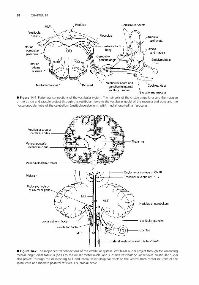
98 CHAPTER 14
● Figure 14-1 Peripheral connections of the vestibular system. The hair cells of the cristae ampullares and the maculaeof the utricle and saccule project through the vestibular nerve to the vestibular nuclei of the medulla and pons and theflocculonodular lobe of the cerebellum (vestibulocerebellum). MLF, medial longitudinal fasciculus.
● Figure 14-2 The major central connections of the vestibular system. Vestibular nuclei project through the ascendingmedial longitudinal fasciculi (MLF ) to the ocular motor nuclei and subserve vestibuloocular reflexes. Vestibular nucleialso project through the descending MLF and lateral vestibulospinal tracts to the ventral horn motor neurons of thespinal cord and mediate postural reflexes. CN, cranial nerve.
LWBK110-3895G-C14[97-100].qxd 7/10/08 7:53 AM Page 98 Aptara Inc.

2. Bipolar neurons project their central processes as the vestibular nerve [cranialnerve (CN) VIII] to the vestibular nuclei and to the flocculonodular lobe of thecerebellum.
C. VESTIBULAR NUCLEI1. These nuclei receive input from
a. The semicircular ducts, saccule, and utricle.b. The flocculonodular lobe of the cerebellum.
2. The nuclei project fibers toa. The flocculonodular lobe of the cerebellum.b. CN III, IV, and VI through the medial longitudinal fasciculus (MLF).c. The spinal cord through the lateral vestibulospinal tract.d. The ventral posteroinferior and posterolateral nuclei of the thalamus, both of
which project to the postcentral gyrus.
VESTIBULOOCULAR REFLEXES are mediated by the vestibular nuclei, MLF, ocularmotor nuclei, and CN III, IV, and VI.
A. VESTIBULAR (HORIZONTAL) NYSTAGMUS1. The fast phase of nystagmus is in the direction of rotation.2. The slow phase of nystagmus is in the opposite direction.
B. POSTROTATORY (HORIZONTAL) NYSTAGMUS1. The fast phase of nystagmus is in the opposite direction of rotation.2. The slow phase of nystagmus is in the direction of rotation.3. The patient past-points and falls in the direction of previous rotation.
C. CALORIC NYSTAGMUS (STIMULATION OF HORIZONTAL DUCTS) in normal sub-jects1. Cold water irrigation of the external auditory meatus results in nystagmus to the
opposite side.2. Warm water irrigation of the external auditory meatus results in nystagmus to
the same side.3. Remember the mnemonic COWS: cold, opposite, warm, same.
D. Test results in unconscious subjects (Figure 14-3)
IV
99VESTIBULAR SYSTEM
Cold H2O Cold H2OCold H2OCold H2O
Normal conscious subject Brain stem intact MLF (bilateral) lesion Low brain stem lesion
● Figure 14-3 Cold caloric responses in the unconscious patient. When the brain stem is intact, the eyes deviate towardthe irrigated side; with bilateral transection of the medial longitudinal fasciculi (MLF ), the eye deviates to the abductedside. Destruction of the caudal brain stem results in no deviation of the eyes. Double-headed arrows indicate nystag-mus; single-headed arrows indicate deviation of the eyes to one side.
LWBK110-3895G-C14[97-100].qxd 7/10/08 7:53 AM Page 99 Aptara Inc.

1. No nystagmus is seen in normal conscious subjects.2. When the brain stem is intact, there is deviation of the eyes to the side of the cold
irrigation in unconscious subjects.3. With bilateral MLF transaction in unconscious subjects, there is deviation of the
abducting eye to the side of the cold irrigation.4. With lower brain stem damage to the vestibular nuclei, there is no deviation of the
eyes in unconscious subjects.
100 CHAPTER 14
Case Study A 60-year-old woman came to the clinic with complaints of progressive hearing loss, facialweakness, and headaches on the right side. She also said that she had become more unsteadyin walking, with weakness and numbness of the right side of the face. No nausea or vomit-ing was noted. What is the most likely diagnosis?
Relevant Physical Exam Findings • Reduced pain and touch sensation in right face • Right facial weakness • Absent right corneal reflex• Hearing loss on right side • No response to caloric test stimulation on right side • Bilateral papilledema
Diagnosis• Acoustic neuroma
LWBK110-3895G-C14[97-100].qxd 7/10/08 7:53 AM Page 100 Aptara Inc.

101
Chapter 15
Visual System
INTRODUCTION The visual system is served by the optic nerve, which is a specialsomatic afferent nerve.
THE VISUAL PATHWAY (Figure 15-1) includes the following structures:
A. GANGLION CELLS OF THE RETINA form the optic nerve [cranial nerve (CN) II].They project from the nasal hemiretina to the contralateral lateral geniculate body andfrom the temporal hemiretina to the ipsilateral lateral geniculate body.
B. The optic nerve projects from the lamina cribrosa of the scleral canal, through the opticcanal, to the optic chiasm (Figure 15-2).1. Transection causes ipsilateral blindness, with no direct pupillary light reflex.2. The section of the optic nerve at the optic chiasm transects all fibers from the ipsi-
lateral retina and fibers from the contralateral inferior nasal quadrant that loop intothe optic nerve. This lesion causes ipsilateral blindness and a contralateral uppertemporal quadrant defect (junction scotoma).
C. The optic chiasm contains decussating fibers from the two nasal hemiretinas. It con-tains noncrossing fibers from the two temporal hemiretinas and projects fibers to thesuprachiasmatic nucleus of the hypothalamus.1. Midsagittal transection or pressure (often from a pituitary tumor) causes bitem-
poral hemianopia.2. Bilateral lateral compression causes binasal hemianopia (calcified internal carotid
arteries).
D. The optic tract contains fibers from the ipsilateral temporal hemiretina and the con-tralateral nasal hemiretina. It projects to the ipsilateral lateral geniculate body, pretec-tal nuclei, and superior colliculus. Transection causes contralateral hemianopia.
II
I
Key Concepts1) Know the lesions of the visual system. 2) How are quadrantanopias created? 3) There are two major lesions of the optic chiasm. Know them! 4) What is Meyer’s loop?
✔
LWBK110-3895G-C15[101-108].qxd 7/10/08 7:54 AM Page 101 Aptara Inc.

E. The lateral geniculate body is a six-layered nucleus. Layers 1, 4, and 6 receive crossedfibers; layers 2, 3, and 5 receive uncrossed fibers. The lateral geniculate body receives inputfrom layer VI of the striate cortex (Brodmann’s area 17). It also receives fibers from theipsilateral temporal hemiretina and the contralateral nasal hemiretina. It projects throughthe geniculocalcarine tract to layer IV of the primary visual cortex (Brodmann’s area 17).
F. The geniculocalcarine tract (visual radiation) projects through two divisions to thevisual cortex.1. The upper division (Figure 15-3) projects to the upper bank of the calcarine sul-
cus, the cuneus. It contains input from the superior retinal quadrants, which rep-resent the inferior visual-field quadrants.a. Transection causes a contralateral lower quadrantanopia.b. Lesions that involve both cunei cause a lower altitudinal hemianopia (alti-
tudinopia).2. The lower division (see Figure 15-3) loops from the lateral geniculate body ante-
riorly (Meyer’s loop), then posteriorly, to terminate in the lower bank of the cal-carine sulcus, the lingual gyrus. It contains input from the inferior retinal quad-rants, which represent the superior visual field quadrants.a. Transection causes a contralateral upper quadrantanopia (“pie in the sky”).b. Transection of both lingual gyri causes an upper altitudinal hemianopia
(altitudinopia).
102 CHAPTER 15
8
9
10
11
● Figure 15-1 The visual pathway from the retina to the visual cortex showing visual field defects. (1) Ipsilateral blind-ness. (2) Binasal hemianopia. (3) Bitemporal hemianopia. (4) Right hemianopia. (5) Right upper quadrantanopia. (6) Rightlower quadrantanopia. (7) Right hemianopia with macular sparing. (8) Left constricted field as a result of end-stageglaucoma. Bilateral constricted fields may be seen in hysteria. (9) Left central scotoma as seen in optic (retrobulbar) neu-ritis in multiple sclerosis. (10) Upper altitudinal hemianopia as a result of bilateral destruction of the lingual gyri. (11) Lower altitudinal hemianopia as a result of bilateral destruction of the cunei.
LWBK110-3895G-C15[101-108].qxd 7/10/08 7:54 AM Page 102 Aptara Inc.
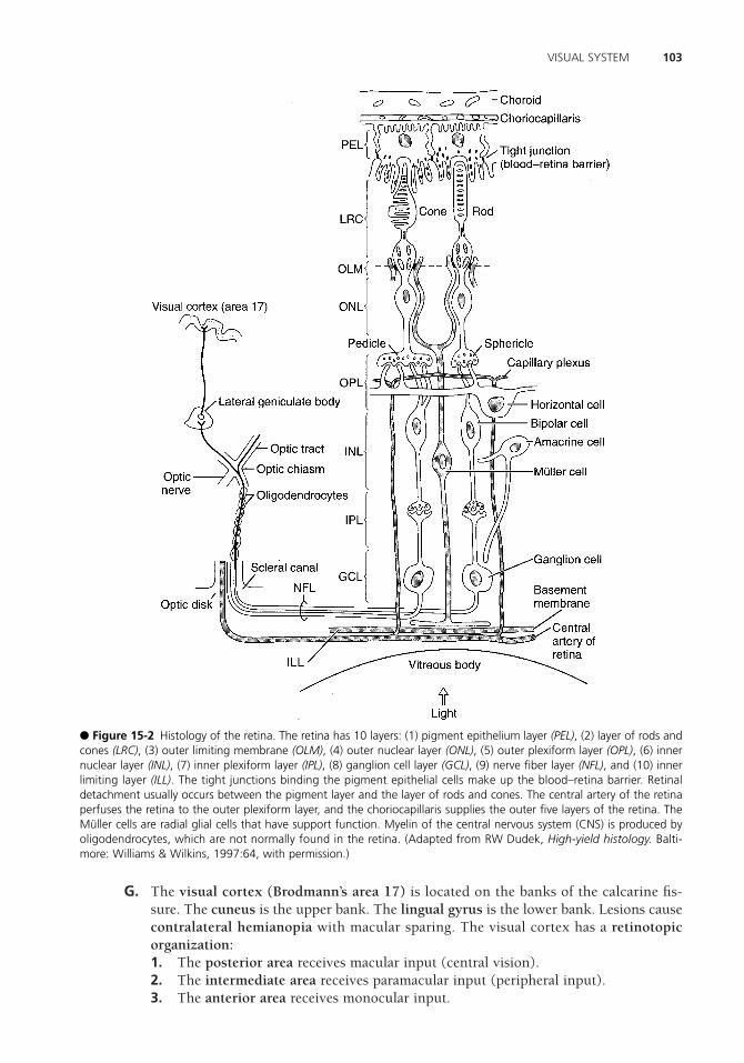
G. The visual cortex (Brodmann’s area 17) is located on the banks of the calcarine fis-sure. The cuneus is the upper bank. The lingual gyrus is the lower bank. Lesions causecontralateral hemianopia with macular sparing. The visual cortex has a retinotopicorganization:1. The posterior area receives macular input (central vision).2. The intermediate area receives paramacular input (peripheral input).3. The anterior area receives monocular input.
103VISUAL SYSTEM
● Figure 15-2 Histology of the retina. The retina has 10 layers: (1) pigment epithelium layer (PEL), (2) layer of rods andcones (LRC), (3) outer limiting membrane (OLM), (4) outer nuclear layer (ONL), (5) outer plexiform layer (OPL), (6) innernuclear layer (INL), (7) inner plexiform layer (IPL), (8) ganglion cell layer (GCL), (9) nerve fiber layer (NFL), and (10) innerlimiting layer (ILL). The tight junctions binding the pigment epithelial cells make up the blood–retina barrier. Retinaldetachment usually occurs between the pigment layer and the layer of rods and cones. The central artery of the retinaperfuses the retina to the outer plexiform layer, and the choriocapillaris supplies the outer five layers of the retina. TheMüller cells are radial glial cells that have support function. Myelin of the central nervous system (CNS) is produced byoligodendrocytes, which are not normally found in the retina. (Adapted from RW Dudek, High-yield histology. Balti-more: Williams & Wilkins, 1997:64, with permission.)
LWBK110-3895G-C15[101-108].qxd 7/10/08 7:54 AM Page 103 Aptara Inc.

104 CHAPTER 15
● Figure 15-3 Relations of the left upper and left lower divisions of the geniculocalcarine tract to the lateral ventricleand calcarine sulcus. Transection of the upper division (A) results in right lower homonymous quadrantanopia. Transec-tion of the lower division (B) results in right upper homonymous quadrantanopia. (Reprinted from JD Fix, BRS neu-roanatomy. Baltimore: Williams & Wilkins, 1997:261, with permission.)
● Figure 15-4 The pupillary light pathway. Light shined into one eye causes both pupils to constrict. The response inthe stimulated eye is called the direct pupillary light reflex. The response in the opposite eye is called the consensualpupillary light reflex. CN, cranial nerve.
LWBK110-3895G-C15[101-108].qxd 7/10/08 7:54 AM Page 104 Aptara Inc.

105VISUAL SYSTEM
THE PUPILLARY LIGHT REFLEX PATHWAY (Figure 15-4) has an afferent limb(CN II) and an efferent limb (CN III). It includes the following structures:
A. GANGLION CELLS OF THE RETINA, which project bilaterally to the pretectal nuclei.
B. The pretectal nucleus of the midbrain, which projects (through the posterior com-missure) crossed and uncrossed fibers to the accessory oculomotor (Edinger-Westphal)nucleus.
C. The accessory oculomotor (Edinger-Westphal) nucleus of CN III, which gives rise topreganglionic parasympathetic fibers. These fibers exit the midbrain with CN III andsynapse with postganglionic parasympathetic neurons of the ciliary ganglion.
D. The ciliary ganglion, which gives rise to postganglionic parasympathetic fibers. Thesefibers innervate the sphincter muscle of the iris.
THE PUPILLARY DILATION PATHWAY (Figure 15-5) is mediated by the sympa-thetic division of the autonomic nervous system. Interruption of this pathway at any levelcauses ipsilateral Horner’s syndrome. It includes the following structures:
IV
III
A
Flashlight swung from right eye to left eye Looking straight ahead
C
Looking right
B
D
Eyes of a comatose patient
E F
Looking right
H I
Looking straight ahead
J
No reaction to light
Looking up Eyes converged
G
Looking left and down
Looking left Looking straight aheadEyes converged
Eyes converged
● Figure 15-5 Ocular motor palsies and pupillary syndromes. (A) Relative afferent (Marcus Gunn) pupil, left eye. (B) Horner’s syndrome, left eye. (C) Internuclear ophthalmoplegia, right eye. (D) Third-nerve palsy, left eye. (E) Sixth-nervepalsy, right eye. (F) Paralysis of upward gaze and convergence (Parinaud’s syndrome). (G) Fourth-nerve palsy, right eye.(H) Argyll Robertson pupil. (I) Destructive lesion of the right frontal eye field. (J) Third-nerve palsy with ptosis, right eye.
LWBK110-3895G-C15[101-108].qxd 7/10/08 7:54 AM Page 105 Aptara Inc.

A. The hypothalamus. Hypothalamic neurons of the paraventricular nucleus projectdirectly to the ciliospinal center (T1–T2) of the intermediolateral cell column of thespinal cord.
B. The ciliospinal center of Budge (T1–T2) projects preganglionic sympathetic fibersthrough the sympathetic trunk to the superior cervical ganglion.
C. The superior cervical ganglion projects postganglionic sympathetic fibers through theperivascular plexus of the carotid system to the dilator muscle of the iris. Postgan-glionic sympathetic fibers pass through the tympanic cavity and cavernous sinus andenter the orbit through the superior orbital fissure.
THE NEAR REFLEX AND ACCOMMODATION PATHWAYA. The cortical visual pathway projects from the primary visual cortex (Brodmann’s area
17) to the visual association cortex (Brodmann’s area 19).
B. The visual association cortex (Brodmann’s area 19) projects through the corticotectaltract to the superior colliculus and pretectal nucleus.
C. The superior colliculus and pretectal nucleus project to the oculomotor complex ofthe midbrain. This complex includes the following structures:1. The rostral accessory oculomotor (Edinger-Westphal) nucleus, which mediates
pupillary constriction through the ciliary ganglion.2. The caudal accessory oculomotor (Edinger-Westphal) nucleus, which mediates
contraction of the ciliary muscle. This contraction increases the refractive powerof the lens.
3. The medial rectus subnucleus of CN III, which mediates convergence.
CORTICAL AND SUBCORTICAL CENTERS FOR OCULAR MOTILITYA. The frontal eye field is located in the posterior part of the middle frontal gyrus (Brod-
mann’s area 8). It regulates voluntary (saccadic) eye movements.1. Stimulation (e.g., from an irritative lesion) causes contralateral deviation of the
eyes (i.e., away from the lesion).2. Destruction causes transient ipsilateral conjugate deviation of the eyes (i.e.,
toward the lesion).
B. OCCIPITAL EYE FIELDS are located in Brodmann’s areas 18 and 19 of the occipitallobes. These fields are cortical centers for involuntary (smooth) pursuit and trackingmovements. Stimulation causes contralateral conjugate deviation of the eyes.
C. The subcortical center for lateral conjugate gaze is located in the abducent nucleus ofthe pons (Figure 15-6). Some authorities place the “center” in the paramedian pontinereticular formation.1. It receives input from the contralateral frontal eye field.2. It projects to the ipsilateral lateral rectus muscle and through the medial longitu-
dinal fasciculus (MLF) to the contralateral medial rectus subnucleus of the oculo-motor complex.
VI
V
106 CHAPTER 15
LWBK110-3895G-C15[101-108].qxd 7/10/08 7:54 AM Page 106 Aptara Inc.

D. The subcortical center for vertical conjugate gaze is located in the midbrain at thelevel of the posterior commissure. It is called the rostral interstitial nucleus of the MLFand is associated with Parinaud’s syndrome (see Figures 15-5F and 16-3A).
CLINICAL CORRELATIONA. In MLF syndrome, or internuclear ophthalmoplegia (see Figure 15-5), there is dam-
age (demyelination) to the MLF between the abducent and oculomotor nuclei. It causesmedial rectus palsy on attempted lateral conjugate gaze and monocular horizontalnystagmus in the abducting eye. (Convergence is normal.) This syndrome is most com-monly seen in multiple sclerosis.
B. ONE-AND-A-HALF SYNDROME consists of bilateral lesions of the MLF and a unilat-eral lesion of the abducent nucleus. On attempted lateral conjugate gaze, the only mus-cle that functions is the intact lateral rectus.
VII
107VISUAL SYSTEM
A
Bilateral MLF syndrome
Left
Patient with MLF syndrome cannot
adduct the eye on attempted lateral
conjugate gaze and has nystagmus
in abducting eye. The nystagmus is
in the direction of the large arrow-
head. Convergence remains intact.
B
C
Right
Convergence
● Figure 15-6 Connections of the pontine center for lateral conjugate gaze. Lesions of the medial longitudinal fasci-culus (MLF ) between the abducent and oculomotor nuclei result in medial rectus palsy on attempted lateral conjugategaze and horizontal nystagmus in the abducting eye. Convergence remains intact (inset). A unilateral MLF lesion wouldaffect only the ipsilateral medial rectus. CN, cranial nerve.
LWBK110-3895G-C15[101-108].qxd 7/10/08 7:54 AM Page 107 Aptara Inc.

C. ARGYLL ROBERTSON PUPIL (pupillary light–near dissociation) is the absence of a mioticreaction to light, both direct and consensual, with the preservation of a miotic reaction tonear stimulus (accommodation–convergence). It occurs in syphilis and diabetes.
D. HORNER’S SYNDROME is caused by transection of the oculosympathetic pathway atany level (see IV). This syndrome consists of miosis, ptosis, apparent enophthalmos,and hemianhidrosis.
E. RELATIVE AFFERENT (MARCUS GUNN) PUPIL results from a lesion of the optic nerve,the afferent limb of the pupillary light reflex (e.g., retrobulbar neuritis seen in multiplesclerosis). The diagnosis can be made with the swinging flashlight test (see Figure 15-5A).
F. TRANSTENTORIAL (UNCAL) HERNIATION occurs as a result of increased supraten-torial pressure, which is commonly caused by a brain tumor or hematoma (subduralor epidural).1. The pressure cone forces the parahippocampal uncus through the tentorial
incisure.2. The impacted uncus forces the contralateral crus cerebri against the tentorial edge
(Kernohan’s notch) and puts pressure on the ipsilateral CN III and posterior cere-bral artery. As a result, the following neurologic defects occur.a. Ipsilateral hemiparesis occurs as a result of pressure on the corticospinal
tract, which is located in the contralateral crus cerebri.b. A fixed and dilated pupil, ptosis, and a “down-and-out” eye are caused by
pressure on the ipsilateral oculomotor nerve.c. Contralateral homonymous hemianopia is caused by compression of the
posterior cerebral artery, which irrigates the visual cortex.
G. PAPILLEDEMA (CHOKED DISK) is noninflammatory congestion of the optic disk asa result of increased intracranial pressure. It is most commonly caused by brain tumors,subdural hematoma, or hydrocephalus. It usually does not alter visual acuity, but itmay cause bilateral enlarged blind spots. It is often asymmetric and is greater on theside of the supratentorial lesion.
H. ADIE’S PUPIL is a large tonic pupil that reacts slowly to light but does react to near(light–near dissociation). It is frequently seen in women with absent knee or ankle jerks.
108 CHAPTER 15
Case Study A 40-year-old man comes to the clinic with a severe unilateral headache on the left side witha drooping left upper eyelid. He experienced mild head trauma 1 week ago. He does not com-plain of blurred or double vision. What is the most likely diagnosis?
Relevant Physical Exam Findings • The right pupil is 4 mm and normally reactive, and the left pupil is 2 mm and normally
reactive.• The left pupil dilates poorly. • There is 2- to 3-mm ptosis of the left upper eyelid.
Diagnosis• Horner’s syndrome.
LWBK110-3895G-C15[101-108].qxd 7/10/08 7:54 AM Page 108 Aptara Inc.

109
Chapter 16
Lesions of the Brain Stem
LESIONS OF THE MEDULLA (Figure 16-1)
A. MEDIAL MEDULLARY SYNDROME (ANTERIOR SPINAL ARTERY SYNDROME).Affected structures and resultant deficits include1. The corticospinal tract (medullary pyramid). Lesions result in contralateral spas-
tic hemiparesis.2. The medial lemniscus. Lesions result in contralateral loss of tactile and vibration
sensation from the trunk and extremities.3. The hypoglossal nucleus or intraaxial root fibers [cranial nerve (CN) XII].
Lesions result in ipsilateral flaccid hemiparalysis of the tongue. When protruded,the tongue points to the side of the lesion (i.e., the weak side). See Figure 11-9.
B. LATERAL MEDULLARY [WALLENBERG; POSTERIOR INFERIOR CEREBELLARARTERY (PICA)] SYNDROME is characterized by dissociated sensory loss (seeI.B.6–7). Affected structures and resultant deficits include1. The vestibular nuclei. Lesions result in nystagmus, nausea, vomiting, and vertigo.2. The inferior cerebellar peduncle. Lesions result in ipsilateral cerebellar signs [e.g.,
dystaxia, dysmetria (past pointing), dysdiadochokinesia].3. The nucleus ambiguus of CN IX, CN X, and CN XI. Lesions result in ipsilateral
laryngeal, pharyngeal, and palatal hemiparalysis [i.e., loss of the gag reflex (effer-ent limb), dysarthria, dysphagia, and dysphonia (hoarseness)].
4. The glossopharyngeal nerve roots. Lesions result in loss of the gag reflex (afferentlimb).
5. The vagal nerve roots. Lesions result in the same deficits as seen in lesions involv-ing the nucleus ambiguus (see I.B.3).
6. The spinothalamic tracts (spinal lemniscus). Lesions result in contralateral lossof pain and temperature sensation from the trunk and extremities.
7. The spinal trigeminal nucleus and tract. Lesions result in ipsilateral loss of painand temperature sensation from the face (facial hemianesthesia).
8. The descending sympathetic tract. Lesions result in ipsilateral Horner’s syndrome(i.e., ptosis, miosis, hemianhidrosis, and apparent enophthalmos).
I
Key Concepts1) The three most important lesions of the brain stem are occlusion of the anterior spinal
artery (Figure 16-1), occlusion of the posterior inferior cerebellar artery (Figure 16-1),and medial longitudinal fasciculus syndrome (Figure 16-2).
2) Weber’s syndrome is the most common midbrain lesion (Figure 16-3).
✔
LWBK110-3895G-C16[109-115].qxd 7/10/08 7:55 AM Page 109 Aptara Inc.

LESIONS OF THE PONS (Figure 16-2A)
A. MEDIAL INFERIOR PONTINE SYNDROME results from occlusion of the paramedianbranches of the basilar artery. Affected structures and resultant deficits include1. The corticospinal tract. Lesions result in contralateral spastic hemiparesis.2. The medial lemniscus. Lesions result in contralateral loss of tactile sensation from
the trunk and extremities.3. The abducent nerve roots. Lesions result in ipsilateral lateral rectus paralysis.
B. LATERAL INFERIOR PONTINE SYNDROME (anterior inferior cerebellar artery syn-drome; Figure 16-2B). Affected structures and resultant deficits include1. The facial nucleus and intraaxial nerve fibers. Lesions result in
a. Ipsilateral facial nerve paralysis.b. Ipsilateral loss of taste from the anterior two-thirds of the tongue.c. Ipsilateral loss of lacrimation and reduced salivation.d. Loss of corneal and stapedial reflexes (efferent limbs).
2. The cochlear nuclei and intraaxial nerve fibers. Lesions result in unilateral cen-tral deafness.
3. The vestibular nuclei and intraaxial nerve fibers. Lesions result in nystagmus,nausea, vomiting, and vertigo.
4. The spinal nucleus and tract of trigeminal nerve. Lesions result in ipsilateral lossof pain and temperature sensation from the face (facial hemianesthesia).
5. The middle and inferior cerebellar peduncles. Lesions result in ipsilateral limband gait dystaxia.
6. The spinothalamic tracts (spinal lemniscus). Lesions result in contralateral lossof pain and temperature sensation from the trunk and extremities.
7. The descending sympathetic tract. Lesions result in ipsilateral Horner’s syndrome.
II
110 CHAPTER 16
Figure 16-1 Vascular lesions of the medulla at the level of the hypoglossal nucleus of cranial nerve (CN) XII and thedorsal motor nucleus of CN X. (A) Medial medullary syndrome (anterior spinal artery). (B) Lateral medullary (posteriorinferior cerebellar artery) syndrome.
LWBK110-3895G-C16[109-115].qxd 7/10/08 7:55 AM Page 110 Aptara Inc.

C. MEDIAL LONGITUDINAL FASCICULUS (MLF) SYNDROME (INTERNUCLEAROPHTHALMOPLEGIA) (Figure 16-2C) interrupts fibers from the contralateral abdu-cent nucleus that project through the MLF to the ipsilateral medial rectus subnucleusof CN III. It causes medial rectus palsy on attempted lateral conjugate gaze and nys-tagmus in the abducting eye. Convergence remains intact. This syndrome is often seenin patients with multiple sclerosis.
D. FACIAL COLLICULUS SYNDROME usually results from a pontine glioma or a vascu-lar accident. The internal genu of CN VII and the nucleus of CN VI underlie the facialcolliculus.1. Lesions of the internal genu of the facial nerve cause
a. Ipsilateral facial paralysis.b. Ipsilateral loss of the corneal reflex.
2. Lesions of the abducent nucleus causea. Lateral rectus paralysis.b. Medial (convergent) strabismus.c. Horizontal diplopia.
LESIONS OF THE MIDBRAIN (Figure 16-3)
A. DORSAL MIDBRAIN (PARINAUD’S) SYNDROME (see Figure 16-3A) is often theresult of a pinealoma or germinoma of the pineal region. Affected structures and result-ant deficits include1. The superior colliculus and pretectal area. Lesions cause paralysis of upward and
downward gaze, pupillary disturbances, and absence of convergence.2. The cerebral aqueduct. Compression causes noncommunicating hydrocephalus.
B. PARAMEDIAN MIDBRAIN (BENEDIKT’S) SYNDROME (see Figure 16-3B). Affectedstructures and resultant deficits include1. The oculomotor nerve roots (intraaxial fibers). Lesions cause complete ipsilateral
oculomotor paralysis. Eye abduction and depression is caused by the intact lateral
III
111LESIONS OF THE BRAIN STEM
Figure 16-2 Vascular lesions of the caudal pons at the level of the abducent nucleus of cranial nerve (CN) VI and thefacial nucleus of CN VII. (A) Medial inferior pontine syndrome. (B) Lateral inferior pontine syndrome (anterior inferiorcerebellar artery syndrome). (C) Medial longitudinal fasciculus (MLF) syndrome.
LWBK110-3895G-C16[109-115].qxd 7/10/08 7:55 AM Page 111 Aptara Inc.

rectus (CN VI) and superior oblique (CN IV) muscles. Ptosis (paralysis of the lev-ator palpebrae superioris muscle) and fixation and dilation of the ipsilateral pupil(complete internal ophthalmoplegia) also occur.
2. The dentatothalamic fibers. Lesions cause contralateral cerebellar dystaxia withintention tremor.
3. The medial lemniscus. Lesions result in contralateral loss of tactile sensation fromthe trunk and extremities.
C. MEDIAL MIDBRAIN (WEBER’S) SYNDROME (see Figure 16-3C). Affected structuresand resultant deficits include1. The oculomotor nerve roots (intraaxial fibers). Lesions cause complete ipsilateral
oculomotor paralysis. Eye abduction and depression are caused by intact lateralrectus (CN VI) and superior oblique (CN IV) muscles. Ptosis and fixation and dila-tion of the ipsilateral pupil also occur.
2. The corticospinal tracts. Lesions result in contralateral spastic hemiparesis.3. The corticonuclear fibers. Lesions cause contralateral weakness of the lower face
(CN VII), tongue (CN XII), and palate (CN X). The upper face division of the facialnucleus receives bilateral corticonuclear input. The uvula and pharyngeal wall arepulled toward the normal side (CN X), and the protruded tongue points to theweak side.
ACOUSTIC NEUROMA (SCHWANNOMA) (Figure 16-4) is a benign tumor of Schwanncells that affects the vestibulocochlear nerve (CN VIII). It accounts for 8% of all intracra-nial tumors. It is a posterior fossa tumor of the internal auditory meatus and cerebellopon-tine angle. The neuroma often compresses the facial nerve (CN VII), which accompaniesCN VIII in the cerebellopontine angle and internal auditory meatus. It may impinge on thepons and affect the spinal trigeminal tract (CN V). Schwannomas occur twice as often inwomen as in men. Affected structures and resultant deficits include
A. The cochlear nerve of CN VIII. Damage results in tinnitus and unilateral nerve deafness.
B. The vestibular nerve of CN VIII. Damage results in vertigo, nystagmus, nausea, vom-iting, and unsteadiness of gait.
IV
112 CHAPTER 16
Figure 16-3 Lesions of the rostral midbrain at the level of the superior colliculus and oculomotor nucleus of cranialnerve (CN) III. (A) Dorsal midbrain (Parinaud’s) syndrome. (B) Paramedian midbrain (Benedikt’s) syndrome. (C) Medialmidbrain (Weber’s) syndrome.
LWBK110-3895G-C16[109-115].qxd 7/10/08 7:55 AM Page 112 Aptara Inc.

C. The facial nerve (CN VII). Damage results in facial weakness and loss of the cornealreflex (efferent limb).
D. The spinal tract of trigeminal nerve (CN V). Damage results in paresthesia, anesthe-sia of the ipsilateral face, and loss of the corneal reflex (afferent limb).
E. NEUROFIBROMATOSIS TYPE 2. This disorder often occurs with bilateral acousticneuromas.
JUGULAR FORAMEN SYNDROME usually results from a posterior fossa tumor (e.g.,glomus jugulare tumor, the most common inner ear tumor) that compresses CN IX, X, andXI. Affected structures and resultant deficits include
A. The glossopharyngeal nerve (CN IX). Damage results in1. Ipsilateral loss of the gag reflex.2. Ipsilateral loss of pain, temperature, and taste in the tongue.
B. The vagal nerve (CN X). Damage results in1. Ipsilateral paralysis of the soft palate and larynx.2. Ipsilateral loss of the gag reflex.
C. The accessory nerve (CN XI). Damage results in1. Paralysis of the sternocleidomastoid muscle, which results in the inability to turn
the head to the opposite side.2. Paralysis of the trapezius muscle, which causes shoulder droop and inability to
shrug the shoulder.
V
113LESIONS OF THE BRAIN STEM
Figure 16-4 Magnetic resonance image of an acoustic neuroma. This coronal section shows dilation of the ventricles.The vestibulocochlear nerve is visible in the left internal auditory meatus. The tumor indents the lateral pons. Cranialnerve (CN) palsies include CN V, VII, and VIII. Symptoms include unilateral deafness, facial anesthesia and weakness,and an absent coronal reflex. This is a T1-weighted image.
LWBK110-3895G-C16[109-115].qxd 7/10/08 7:55 AM Page 113 Aptara Inc.

“LOCKED-IN” SYNDROME is a lesion of the base of the pons as the result of infarc-tion, trauma, tumor, or demyelination. The corticospinal and corticonuclear tracts areaffected bilaterally. The oculomotor and trochlear nerves are not injured. Patients are con-scious and may communicate through vertical eye movements.
CENTRAL PONTINE MYELINOLYSIS is a lesion of the base of the pons that affectsthe corticospinal and corticobulbar tracts. More than 75% of cases are associated with alco-holism or rapid correction of hyponatremia. Symptoms include spastic quadriparesis,pseudobulbar palsy, and mental changes. This condition may become the locked-in syn-drome.
VII
VI
114 CHAPTER 16
Thrombosis
BA
Left SCLA Left SCLA
Thrombosis
CCAVA
BCT
Aorta Aorta
ECA
ICA
PCOM
PCA
SCA
AICA
PICA
MCA
ACA
ACOMACOM
ASA
BA
CCAVA
BCT
ECA
ICA
PCOM
PCA
SCA
AICA
PICA
MCA
ACA
ASA
A BFigure 16-5 Anatomy of the subclavian steal syndrome. Thrombosis of the proximal part of the subclavian artery (left)results in retrograde blood flow through the ipsilateral vertebral artery and into the left subclavian artery. Blood can beshunted from the right vertebral artery and down the left vertebral artery (A). Blood may also reach the left vertebralartery through the carotid circulation (B). ACA, anterior cerebellar artery; ACOM, anterior communicating artery; AICA,anterior inferior cerebellar artery; ASA, anterior spinal artery; BA, basilar artery; BCT, brachiocephalic trunk; CCA, com-mon carotid artery; ECA, external carotid artery; ICA, internal carotid artery; MCA, middle cerebral artery; PCA, poste-rior cerebral artery; PCOM, posterior communicating artery; PICA, posterior inferior cerebellar artery; SCA, superior com-municating artery; SCLA, subclavian artery; VA, vertebral artery.
LWBK110-3895G-C16[109-115].qxd 7/10/08 7:55 AM Page 114 Aptara Inc.

“TOP OF THE BASILAR” SYNDROME results from embolic occlusion of the rostralbasilar artery. Neurologic signs include optic ataxia and psychic paralysis of fixation of gaze(Balint’s syndrome), ectopic pupils, somnolence, and cortical blindness, with or withoutvisual anosognosia (Anton’s syndrome).
SUBCLAVIAN STEAL SYNDROME (Figure 16-5) results from thrombosis of the leftsubclavian artery proximal to the vertebral artery. Blood is shunted retrograde down the leftvertebral artery and into the left subclavian artery. Clinical signs include transient weak-ness and claudication of the left arm on exercise and vertebrobasilar insufficiency (i.e., ver-tigo, dizziness).
THE CEREBELLOPONTINE ANGLE is the junction of the medulla, pons, and cere-bellum. CN VII and VIII are found there. Five brain tumors, including a cyst, are oftenlocated in the cerebellopontine angle cistern. Remember the acronym SAME: schwannoma(75%), arachnoid cyst (1%), meningioma (10%), and ependymoma (1%) and epidermoid(5%). The percentages refer to cerebellopontine angle tumors.
X
IX
VIII
115LESIONS OF THE BRAIN STEM
Case Study For several weeks, a 60-year-old hypertensive, diabetic man has experienced sudden dizzi-ness, facial pain, double vision, and difficulty in walking. He is also having problems in swal-lowing and speaking. What is the most likely diagnosis?
Relevant Physical Exam Findings • Decreased temperature and pain sense below the left T-4 level• Horner’s syndrome on the right side• Decreased position sensation in the right fingers and toes• Ataxia and mild weakness in the right limbs
Relevant Lab Finding• An infarct involving the right lateral part of the lower medulla and the cerebellum seen
on brain magnetic resonance image
Diagnosis• Wallenberg’s syndrome is an infarction involving the lateral or medial branches of the
posterior inferior cerebellar artery.
LWBK110-3895G-C16[109-115].qxd 7/10/08 7:55 AM Page 115 Aptara Inc.

116
Chapter 17
Thalamus
INTRODUCTION The thalamus is the largest division of the diencephalon. It plays animportant role in the integration of the sensory and motor systems.
MAJOR THALAMIC NUCLEI AND THEIR CONNECTIONS (Figure 17-1)
A. The anterior nucleus receives hypothalamic input from the mamillary nucleus throughthe mamillothalamic tract. It projects to the cingulate gyrus and is part of the Papezcircuit of emotion of the limbic system.
B. The mediodorsal (dorsomedial) nucleus is reciprocally connected to the prefrontalcortex. It has abundant connections with the intralaminar nuclei. It receives input from the amygdale, substantia nigra, and temporal neocortex. When it isdestroyed, memory loss occurs (Wernicke-Korsakoff syndrome). The mediodorsalnucleus plays a role in the expression of affect, emotion, and behavior (limbic function).
C. The centromedian nucleus is the largest intralaminar nucleus. It is reciprocally con-nected to the motor cortex (Brodmann’s area 4). The centromedian nucleus receivesinput from the globus pallidus. It projects to the striatum (caudate nucleus and puta-men) and projects diffusely to the entire neocortex.
D. The pulvinar is the largest thalamic nucleus. It has reciprocal connections with theassociation cortex of the occipital, parietal, and posterior temporal lobes. It receivesinput from the lateral and medial geniculate bodies and the superior colliculus. It playsa role in the integration of visual, auditory, and somesthetic input. Destruction of thedominant pulvinar may result in sensory dysphasia.
E. VENTRAL TIER NUCLEI1. The ventral anterior nucleus receives input from the globus pallidus and substan-
tia nigra. It projects diffusely to the prefrontal cortex, orbital cortex, and premotorcortex (Brodmann’s area 6).
II
I
Key Concepts1) Key entry and exit points into and out of the thalamus (demonstrated by Figure 17-1). 2) Anatomy of the internal capsule. (It will be on the examination.) What is the blood
supply of the internal capsule (stroke)?
✔
LWBK110-3895G-C17[116-119].qxd 7/10/08 7:56 AM Page 116 Aptara Inc.

2. The ventral lateral nucleus receives input from the cerebellum (dentate nucleus),globus pallidus, and substantia nigra. It projects to the motor cortex (Brodmann’sarea 4) and the supplementary motor cortex (Brodmann’s area 6).
3. The ventral posterior nucleus (ventrobasal complex) is the nucleus of termina-tion of general somatic afferent (touch, pain, and temperature) and special visceralafferent (taste) fibers. It has two subnuclei. a. The ventral posterolateral nucleus receives the spinothalamic tracts and the
medial lemniscus. It projects to the somesthetic (sensory) cortex (Brodmann’sareas 3, 1, and 2).
b. The ventral posteromedial (VPM) nucleus receives the trigeminothalamictracts and projects to the somesthetic (sensory) cortex (Brodmann’s areas 3, 1,and 2). The gustatory (taste) pathway originates in the solitary nucleus andprojects via the central tegmental tract to the VPM and thence to the gusta-tory cortex of the postcentral gyrus (Brodmann’s area 3b), of the frontal oper-culum, and of the insular cortex. The taste pathway is ipsilateral.
117THALAMUS
Figure 17-1 Major thalamic nuclei and their connections. (A) Dorsolateral aspect and major nuclei. (B) Major afferentand efferent connections. VA, ventral anterior nucleus; VL, ventral lateral nucleus; VPL, ventral posterior lateral nucleus;VPM, ventral posterior medial nucleus.
LWBK110-3895G-C17[116-119].qxd 7/10/08 7:56 AM Page 117 Aptara Inc.

F. METATHALAMUS1. The lateral geniculate body is a visual relay nucleus. It receives retinal input through
the optic tract and projects to the primary visual cortex (Brodmann’s area 17).2. The medial geniculate body is an auditory relay nucleus. It receives auditory input
through the brachium of the inferior colliculus and projects to the primary audi-tory cortex (Brodmann’s areas 41 and 42).
G. The reticular nucleus of the thalamus surrounds the thalamus as a thin layer of �-aminobutyric acid (GABA)-ergic neurons. It lies between the external medullary laminaand the internal capsule. It receives excitatory collateral input from corticothalamic andthalamocortical fibers. It projects inhibitory fibers to thalamic nuclei, from which itreceives input. It is thought to play a role in normal electroencephalogram readings.
BLOOD SUPPLY The thalamus is irrigated by three arteries (see Figure 5-1):
A. The posterior communicating artery.
B. The posterior cerebral artery.
C. The anterior choroidal artery (lateral geniculate body).
THE INTERNAL CAPSULE (Figure 17-2) is a layer of white matter (myelinated axons)that separates the caudate nucleus and the thalamus medially from the lentiform nucleuslaterally.
IV
III
118 CHAPTER 17
Figure 17-2 Horizontal section of the right internal capsule showing the major fiber projections. Clinically importanttracts lie in the genu and posterior limb. Lesions of the internal capsule cause contralateral hemiparesis and contralat-eral hemianopia. VP, ventral posterior.
LWBK110-3895G-C17[116-119].qxd 7/10/08 7:56 AM Page 118 Aptara Inc.

A. The anterior limb is located between the caudate nucleus and the lentiform nucleus(globus pallidus and putamen).
B. The genu contains the corticonuclear fibers.
C. The posterior limb is located between the thalamus and the lentiform nucleus. It con-tains corticospinal (pyramid) fibers as well as sensory (pain, temperature, and touch),visual, and auditory radiations.
D. BLOOD SUPPLY1. The anterior limb is irrigated by the medial striate branches of the anterior cere-
bral artery and the lateral striate (lenticulostriate) branches of the middle cerebralartery.
2. The genu is perfused either by direct branches from the internal carotid artery orby pallidal branches of the anterior choroidal artery.
3. The posterior limb is supplied by branches of the anterior choroidal artery andlenticulostriate branches of the middle cerebral arteries.
119THALAMUS
Case Study A 90-year-old woman complains of an intense burning sensation on the left side of her neckand upper limb. The patient has a history of high blood pressure and diabetes. What is themost likely diagnosis?
Differentials• Hypoglycemia; middle cerebral artery stroke; migraine
Relevant Physical Exam Findings• Unilateral sensory loss is observed. Though the patient may complain of weakness on the
affected side, no weakness is found on examination.
Relevant Lab Findings • Normal serum glucose levels • Thrombocytopenia • Ischemic infarction of posterior cerebral artery seen on computed tomography scan
Diagnosis• Infarction of the ventral posterolateral nucleus of the thalamus results in pure hemisen-
sory loss contralateral to the lesion.
LWBK110-3895G-C17[116-119].qxd 7/10/08 7:56 AM Page 119 Aptara Inc.

120
Chapter 18
Hypothalamus
INTRODUCTIONA. GENERAL STRUCTURE AND FUNCTION. The hypothalamus is a division of the dien-
cephalon that subserves three systems: the autonomic nervous system, the endocrinesystem, and the limbic system. The hypothalamus helps to maintain homeostasis.
B. MAJOR HYPOTHALAMIC NUCLEI AND THEIR FUNCTIONS1. The medial preoptic nucleus (Figure 18-1) regulates the release of gonadotropic
hormones from the adenohypophysis. It contains the sexually dimorphic nucleus,the development of which depends on testosterone levels.
2. The suprachiasmatic nucleus receives direct input from the retina. It plays a rolein the regulation of circadian rhythms.
3. The anterior nucleus plays a role in temperature regulation. It stimulates theparasympathetic nervous system. Destruction results in hyperthermia.
4. The paraventricular nucleus (Figure 18-2) synthesizes antidiuretic hormone(ADH), oxytocin, and corticotropin-releasing hormone. It gives rise to thesupraopticohypophyseal tract, which projects to the neurohypophysis. It regu-lates water balance (conservation) and projects directly to the autonomic nucleiof the brain stem and all levels of the spinal cord. Destruction results in diabetesinsipidus.
5. The supraoptic nucleus synthesizes ADH and oxytocin (similar to the paraven-tricular nucleus).
6. The dorsomedial nucleus, when stimulated in animals, results in savage behavior.7. The ventromedial nucleus is considered a satiety center. When stimulated, it
inhibits the urge to eat. Bilateral destruction results in hyperphagia, obesity, andsavage behavior.
8. The arcuate (infundibular) nucleus contains neurons that produce factors thatstimulate or inhibit the action of the hypothalamus. This nucleus gives rise to thetuberohypophysial tract, which terminates in the hypophyseal portal system (seeFigure 18-2) of the infundibulum (medium eminence). It contains neurons thatproduce dopamine (i.e., prolactin-inhibiting factor).
I
Key Concepts1) Figures 18-1 and 18-2 show that the paraventricular and supraoptic nuclei synthesize
and release antidiuretic hormone and oxytocin. 2) The suprachiasmatic nucleus receives direct input from the retina and plays a role in
the regulation of circadian rhythms.
✔
LWBK110-3895G-C18[120-124].qxd 7/10/08 7:57 AM Page 120 Aptara Inc.

121HYPOTHALAMUS
Paraventricular and supraoptic nuclei• regulate water balance
• produce ADH and oxytocin
• destruction causes diabetes insipidus
• paraventricular nucleus projects to
autonomic nuclei of brain stem and
spinal cord
Dorsomedial nucleus• stimulation results in obesity and savage behavior
Posterior nucleus• thermal regulation (conservation of heat)
• destruction results in inability to thermoregulate
• stimulates the sympathetic NS
Lateral nucleus• stimulation induces eating
• destruction results in starvation
Midbrain
Pons
CN III
Mamillary body• receives input from
hippocampal formation
via fornix
• projects to anterior nucleus
of thalamus
• contains hemorrhagic lesions
in Wernicke’s encephalopathy
Ventromedial nucleus• satiety center
• destruction results in obesity
and savage behaviorArcuate nucleus• produces hypothalamic releasing factors
• contains DOPA-ergic neurons that inhibit prolactin release
Anterior nucleus• thermal regulation
(dissipation of heat)
• stimulates parasympathetic NS
• destruction results in hyperthermia
Preoptic area• contains sexual dimorphic nucleus
• regulates release of gonadotropic
hormones
Suprachiasmatic nucleus• receives input from retina
• controls circadian rhythms
Figure 18-1 Major hypothalamic nuclei and their functions. ADH, antidiuretic hormone; CN, cranial nerve; DOPA,dopamine; NS, nervous system.
Figure 18-2 The hypophyseal portal system. The paraventricular and supraoptic nuclei produce antidiuretic hormone (ADH)and oxytocin and transport them through the supraopticohypophysial tract to the capillary bed of the neurohypophysis. Thearcuate nucleus of the infundibulum transports hypothalamic-stimulating hormones through the tuberohypophysial tract tothe sinusoids of the infundibular stem. These sinusoids then drain into the secondary capillary plexus in the adenohypophysis.
LWBK110-3895G-C18[120-124].qxd 7/10/08 7:57 AM Page 121 Aptara Inc.

9. The mamillary nucleus receives input from the hippocampal formation throughthe postcommissural fornix. It projects to the anterior nucleus of the thalamusthrough the mamillothalamic tract (part of the Papez circuit). Patients with Wer-nicke’s encephalopathy, which is a thiamine (vitamin B1) deficiency, have lesionsin the mamillary nucleus. Lesions are also associated with alcoholism.
10. The posterior hypothalamic nucleus plays a role in thermal regulation (i.e., con-servation and increased production of heat). Lesions result in poikilothermia (i.e.,inability to thermoregulate).
11. The lateral hypothalamic nucleus induces eating when stimulated. Lesions causeanorexia and starvation.
C. MAJOR FIBER SYSTEMS OF THE HYPOTHALAMUS1. The fornix is the largest projection to the hypothalamus. It projects from the hip-
pocampal formation to the mamillary nucleus, anterior nucleus of the thalamus,and septal area. The fornix then projects from the septal area to the hippocampalformation.
2. The medial forebrain bundle traverses the entire lateral hypothalamic area. It inter-connects the orbitofrontal cortex, septal area, hypothalamus, and midbrain.
3. The mamillothalamic tract projects from the mamillary nuclei to the anteriornucleus of the thalamus (part of the Papez circuit).
4. The stria terminalis is the major pathway from the amygdala. It interconnects theseptal area, hypothalamus, and amygdala.
5. The supraopticohypophysial tract conducts fibers from the supraoptic and par-aventricular nuclei to the neurohypophysis, which is the release site for ADH andoxytocin.
6. The tuberohypophysial (tuberoinfundibular) tract conducts fibers from the arcu-ate nucleus to the hypophyseal portal system (see Figure 18-2).
7. The hypothalamospinal tract contains direct descending autonomic fibers.These fibers influence the preganglionic sympathetic neurons of the intermedi-olateral cell column and preganglionic neurons of the sacral parasympatheticnucleus. Interruption above the first thoracic segment (T-1) causes Horner’ssyndrome.
FUNCTIONS
A. AUTONOMIC FUNCTION1. The anterior hypothalamus has an excitatory effect on the parasympathetic nerv-
ous system.2. The posterior hypothalamus has an excitatory effect on the sympathetic nervous
system.
B. TEMPERATURE REGULATION1. The anterior hypothalamus regulates and maintains body temperature. Destruc-
tion causes hyperthermia.2. The posterior hypothalamus helps to produce and conserve heat. Destruction
causes the inability to thermoregulate.
C. WATER BALANCE REGULATION. The paraventricular nucleus synthesizes ADH,which controls water excretion by the kidneys.
II
122 CHAPTER 18
LWBK110-3895G-C18[120-124].qxd 7/10/08 7:57 AM Page 122 Aptara Inc.

D. FOOD INTAKE REGULATION. Two hypothalamic nuclei play a role in the control ofappetite.1. When stimulated, the ventromedial nucleus inhibits the urge to eat. Bilateral
destruction results in hyperphagia, obesity, and savage behavior.2. When stimulated, the lateral hypothalamic nucleus induces the urge to eat.
Destruction causes starvation and emaciation.
123HYPOTHALAMUS
Figure 18-3 Coronal section through the hypothalamus at the level of the dorsomedial, ventromedial, and lateral hypo-thalamic nuclei. Lesions or stimulation of these nuclei result in obesity, cachexia, and rage. The column of the fornixseparates the medial from the lateral hypothalamic zones. A lesion of the optic tract results in a contralateral hemi-anopia. FX, fornix; DM, medial dorsal nucleus of thalamus; OT, optic tract; VL, ventral lateral nucleus of thalamus; VP,ventral posterior nucleus of thalamus. (Reprinted from JD Fix, BRS neuroanatomy, 3rd ed. Baltimore: Williams & Wilkins,1996:313, with permission.)
LWBK110-3895G-C18[120-124].qxd 7/10/08 7:57 AM Page 123 Aptara Inc.

CLINICAL CORRELATIONA. DIABETES INSIPIDUS, which is characterized by polyuria and polydipsia, is the best-
known hypothalamic syndrome. It results from lesions of the ADH pathways to theposterior lobe of the pituitary gland.
B. The syndrome of inappropriate ADH secretion is usually caused by lung tumors ordrug therapy (e.g., carbamazepine, chlorpromazine).
C. CRANIOPHARYNGIOMA is a congenital tumor that originates from remnants ofRathke’s pouch (see Chapter 1). This tumor is usually calcified. It is the most commonsupratentorial tumor in children and the most common cause of hypopituitarism inchildren.1. Pressure on the chiasma results in bitemporal hemianopia.2. Pressure on the hypothalamus causes hypothalamic syndrome (i.e., adiposity,
diabetes insipidus, disturbance of temperature regulation, and somnolence).
D. PITUITARY ADENOMAS account for 15% of clinical symptomatic intracranial tumors.They are rarely seen in children. When pituitary adenomas are endocrine-active, theycause endocrine abnormalities (e.g., amenorrhea and galactorrhea from a prolactin-secreting adenoma, the most common type).1. Pressure on the chiasma results in bitemporal hemianopia.2. Pressure on the hypothalamus may cause hypothalamus syndrome (Figure 18-3).
III
124 CHAPTER 18
Case Study A 65-year-old diabetic man was hospitalized after an auto accident with lethargy andprogressive confusion. Laboratory tests results revealed low sodium levels. The patient wasdischarged after serum sodium levels were elevated to 130 mmol/L.
Differentials• Diabetes
Relevant Physical Exam Findings• Fatigue and depression• Slowed mental processing time • Slowed pulse and hypothermia • Hyporeflexia and hypotonia
Relevant Lab Findings • Normal hepatic, renal, and cardiac function • Hyponatremia that worsens with fluid load• Serum hypoosmolality• Urine hyperosmolarity
Diagnosis• Inappropriate secretion of antidiuretic hormone by the hypothalamus. As many as 50%
of traumatic brain injury patients experience endocrine complications that may result inintracerebral osmotic fluid shifts and brain edema, affecting hypothalamic function.
LWBK110-3895G-C18[120-124].qxd 7/10/08 7:57 AM Page 124 Aptara Inc.

125
Chapter 19
Limbic System
INTRODUCTION The limbic system is considered the anatomic substrate that under-lies behavioral and emotional expression. It is expressed through the hypothalamus by wayof the autonomic nervous system.
MAJOR COMPONENTS AND CONNECTIONSA. The orbitofrontal cortex mediates the conscious perception of smell. It has recip-
rocal connections with the mediodorsal nucleus of the thalamus. It is intercon-nected through the medial forebrain bundle with the septal area and hypothalamicnuclei.
B. The dorsomedial mediodorsal nucleus of the thalamus has reciprocal connectionswith the orbitofrontal and prefrontal cortices and the hypothalamus. It receives inputfrom the amygdala and plays a role in affective behavior and memory.
C. The anterior nucleus of the thalamus receives input from the mamillary nucleusthrough the mamillothalamic tract and fornix. It projects to the cingulate gyrus and isa major link in the Papez circuit.
D. The septal area is a telencephalic structure. It has reciprocal connections with the hip-pocampal formation through the fornix and with the hypothalamus through the medialforebrain bundle. It projects through the stria medullaris (thalami) to the habenularnucleus.
E. The limbic lobe includes the subcallosal area, paraterminal gyrus, cingulate gyrus andisthmus, and parahippocampal gyrus, which includes the uncus. It contains, buriedin the parahippocampal gyrus, the hippocampal formation and amygdaloid nuclearcomplex.
II
I
Key Concepts1) Bilateral lesions of the amygdala result in Klüver-Bucy syndrome. Recall the triad hyper-
phagia, hypersexuality, and psychic blindness. 2) Memory loss is associated with bilateral lesions of the hippocampus. 3) Wernicke’s encephalopathy results from a deficiency of thiamine (vitamin B1). Lesions
are found in the mamillary bodies, thalamus, and midbrain tegmentum (Figure 19-3). 4) Know the Papez circuit, a common board question.
✔
LWBK110-3895G-C19[125-129].qxd 7/10/08 7:58 AM Page 125 Aptara Inc.
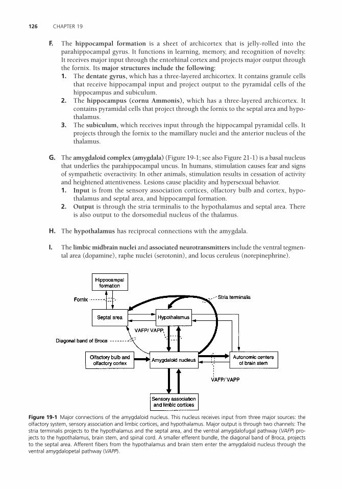
F. The hippocampal formation is a sheet of archicortex that is jelly-rolled into theparahippocampal gyrus. It functions in learning, memory, and recognition of novelty.It receives major input through the entorhinal cortex and projects major output throughthe fornix. Its major structures include the following:1. The dentate gyrus, which has a three-layered archicortex. It contains granule cells
that receive hippocampal input and project output to the pyramidal cells of thehippocampus and subiculum.
2. The hippocampus (cornu Ammonis), which has a three-layered archicortex. Itcontains pyramidal cells that project through the fornix to the septal area and hypo-thalamus.
3. The subiculum, which receives input through the hippocampal pyramidal cells. Itprojects through the fornix to the mamillary nuclei and the anterior nucleus of thethalamus.
G. The amygdaloid complex (amygdala) (Figure 19-1; see also Figure 21-1) is a basal nucleusthat underlies the parahippocampal uncus. In humans, stimulation causes fear and signsof sympathetic overactivity. In other animals, stimulation results in cessation of activityand heightened attentiveness. Lesions cause placidity and hypersexual behavior.1. Input is from the sensory association cortices, olfactory bulb and cortex, hypo-
thalamus and septal area, and hippocampal formation.2. Output is through the stria terminalis to the hypothalamus and septal area. There
is also output to the dorsomedial nucleus of the thalamus.
H. The hypothalamus has reciprocal connections with the amygdala.
I. The limbic midbrain nuclei and associated neurotransmitters include the ventral tegmen-tal area (dopamine), raphe nuclei (serotonin), and locus ceruleus (norepinephrine).
126 CHAPTER 19
Figure 19-1 Major connections of the amygdaloid nucleus. This nucleus receives input from three major sources: theolfactory system, sensory association and limbic cortices, and hypothalamus. Major output is through two channels: Thestria terminalis projects to the hypothalamus and the septal area, and the ventral amygdalofugal pathway (VAFP) pro-jects to the hypothalamus, brain stem, and spinal cord. A smaller efferent bundle, the diagonal band of Broca, projectsto the septal area. Afferent fibers from the hypothalamus and brain stem enter the amygdaloid nucleus through theventral amygdalopetal pathway (VAPP).
LWBK110-3895G-C19[125-129].qxd 7/10/08 7:58 AM Page 126 Aptara Inc.
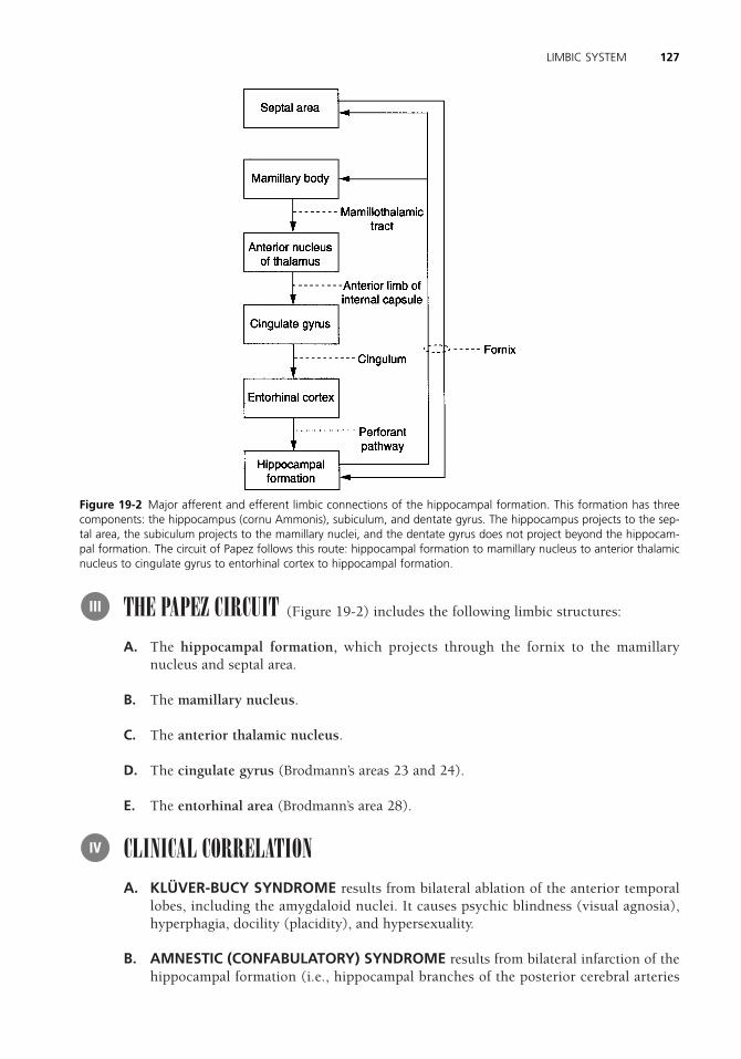
THE PAPEZ CIRCUIT (Figure 19-2) includes the following limbic structures:
A. The hippocampal formation, which projects through the fornix to the mamillarynucleus and septal area.
B. The mamillary nucleus.
C. The anterior thalamic nucleus.
D. The cingulate gyrus (Brodmann’s areas 23 and 24).
E. The entorhinal area (Brodmann’s area 28).
CLINICAL CORRELATIONA. KLÜVER-BUCY SYNDROME results from bilateral ablation of the anterior temporal
lobes, including the amygdaloid nuclei. It causes psychic blindness (visual agnosia),hyperphagia, docility (placidity), and hypersexuality.
B. AMNESTIC (CONFABULATORY) SYNDROME results from bilateral infarction of thehippocampal formation (i.e., hippocampal branches of the posterior cerebral arteries
IV
III
127LIMBIC SYSTEM
Figure 19-2 Major afferent and efferent limbic connections of the hippocampal formation. This formation has threecomponents: the hippocampus (cornu Ammonis), subiculum, and dentate gyrus. The hippocampus projects to the sep-tal area, the subiculum projects to the mamillary nuclei, and the dentate gyrus does not project beyond the hippocam-pal formation. The circuit of Papez follows this route: hippocampal formation to mamillary nucleus to anterior thalamicnucleus to cingulate gyrus to entorhinal cortex to hippocampal formation.
LWBK110-3895G-C19[125-129].qxd 7/10/08 7:58 AM Page 127 Aptara Inc.

and anterior choroidal arteries of the internal carotid arteries). It causes anterogradeamnesia (i.e., inability to learn and retain new information). Memory loss suggests hip-pocampal pathology.
C. FOSTER KENNEDY SYNDROME results from meningioma of the olfactory groove. Themeningioma compresses the olfactory tract and optic nerve. Ipsilateral anosmia and opticatrophy and contralateral papilledema occur as a result of increased intracranial pressure.
D. The hippocampus is the most epileptogenic part of the cerebrum. Lesions may causepsychomotor attacks. Sommer’s sector is very sensitive to ischemia.
E. Bilateral transection of the fornix may cause the acute amnestic syndrome (i.e., inabil-ity to consolidate short-term memory into long-term memory).
F. WERNICKE’S ENCEPHALOPATHY results from a thiamine (vitamin B1) deficiency.The clinical triad includes ocular disturbances and nystagmus, gait ataxia, and mentaldysfunction. Pathologic features include mamillary nuclei, dorsomedial nuclei of thethalamus, and periaqueductal gray and pontine tegmentum (Figure 19-3).
G. STRACHAN’S SYNDROME results from high-dose thiamine (vitamin B1) therapy. Theclinical triad includes spinal ataxia, optic atrophy, and nerve deafness.
H. Bilateral destruction or removal of the cingulate gyri causes loss of initiative and inhi-bition and dulling of the emotions. Memory is unaffected. Lesions of the anterior cin-gulate gyri cause placidity. Cingulectomy is used to treat severe anxiety and depression(Figure 19-4).
128 CHAPTER 19
A
B
C
FD
E
Figure 19-3 Midsagittal section through the brain stem and diencephalon showing the distribution of lesions inWernicke’s encephalopathy. (A) Dorsomedial nucleus of the thalamus. (B) Massa intermedia. (C) Periventricular area. (D) Mamillary nuclei. (E) Midbrain and pontine tegmentum. (F ) Inferior colliculus. Lesions in the mamillary nuclei areassociated with Wernicke’s encephalopathy and thiamine (vitamin B1) deficiency.
LWBK110-3895G-C19[125-129].qxd 7/10/08 7:58 AM Page 128 Aptara Inc.

129LIMBIC SYSTEM
Figure 19-4 Major connections of the hippocampal formation. The hippocampal formation (HF) consists of three parts:hippocampus, dentate gyrus, and subiculum. The two major hypothalamic output pathways are (1) granule cell viamossy fiber to pyramidal cell via precommissural fornix to septal nuclei and (2) subicular neuron via postcommissuralfornix to the medial mamillary nucleus. The HF plays an important role in learning and memory, and lesions of the HFresult in short-term memory defects. In Alzheimer’s disease, loss of cells in the HF and entorhinal cortex leads to loss ofmemory and cognitive function. CA, cornu ammonis. The sector CA1 is very sensitive to hypoxia (cardiac arrest or stroke).(Reprinted from JD Fix, BRS neuroanatomy, 3rd ed. Baltimore: Williams & Wilkins, 1996:332, with permission.)
Case Study A 15-year-old boy was knocked out after several rounds of boxing with friends. Computedtomography scanning showed acute subdural hematoma associated with the right cerebralhemisphere. After regaining consciousness, the boy no longer experienced normal fear andanger and demonstrated aberrant sexual behaviors and excessive oral tendencies. He alsocomplained of being very hungry all the time. What is the most likely diagnosis?
Relevant Lab Findings• Computed tomography and magnetic resonance imaging revealed lesions of the right
temporal lobe and right-dominant orbitofrontal regions, including bilateral rectal andmedial orbital gyri, and an intact left temporal lobe.
Diagnosis• Klüver-Bucy syndrome occurs when both the right and left medial temporal lobes malfunc-
tion, with frequent involvement of the amygdala. The cardinal symptom is excessive oral ten-dencies where the patient puts all types of objects into the mouth. Such patients also have anirresistible impulse to touch objects and demonstrate placidity (lack of emotional response)and a marked increase in sexual activity, without concern for social appropriateness.
LWBK110-3895G-C19[125-129].qxd 7/10/08 7:58 AM Page 129 Aptara Inc.

130
Chapter 20
Cerebellum
FUNCTION The cerebellum has three primary functions:
A. MAINTENANCE OF POSTURE AND BALANCE
B. MAINTENANCE OF MUSCLE TONE
C. COORDINATION OF VOLUNTARY MOTOR ACTIVITY
ANATOMYA. CEREBELLAR PEDUNCLES
1. The superior cerebellar peduncle contains the major output from the cerebellum,the dentatothalamic tract. This tract terminates in the ventral lateral nucleus of thethalamus. It has one major afferent pathway, the ventral spinocerebellar tract.
2. The middle cerebellar peduncle receives pontocerebellar fibers, which project tothe neocerebellum (pontocerebellum).
3. The inferior cerebellar peduncle has three major afferent tracts: the dorsal spin-ocerebellar tract, the cuneocerebellar tract, and the olivocerebellar tract from thecontralateral inferior olivary nucleus.
B. CEREBELLAR CORTEX, NEURONS, AND FIBERS1. The cerebellar cortex has three layers.
a. The molecular layer is the outer layer underlying the pia. It contains stellatecells, basket cells, and the dendritic arbor of the Purkinje cells.
b. The Purkinje cell layer lies between the molecular and the granule cell layers.c. The granule layer is the inner layer overlying the white matter. It contains
granule cells, Golgi cells, and cerebellar glomeruli. A cerebellar glomerulusconsists of a mossy fiber rosette, granule cell dendrites, and a Golgi cell axon.
2. Neurons and fibers of the cerebelluma. Purkinje cells convey the only output from the cerebellar cortex. They project
inhibitory output [i.e., �-aminobutyric acid (GABA)] to the cerebellar and
II
I
Key Concepts1) Figure 20-1 shows the most important cerebellar circuit. 2) The inhibitory �-aminobutyric acid (GABA)-ergic cerebellar output is regulated by Purkinje.3) Purkinje cells give rise to the cerebellodentatothalamic tract. 4) What are mossy and climbing fibers?
✔
LWBK110-3895G-C20[130-134].qxd 7/10/08 7:59 AM Page 130 Aptara Inc.

vestibular nuclei. These cells are excited by parallel and climbing fibers andinhibited by GABAergic basket and stellate cells.
b. Granule cells excite (by way of glutamate) Purkinje, basket, stellate, and Golgicells through parallel fibers. They are inhibited by Golgi cells and excited bymossy fibers.
c. Parallel fibers are the axons of granule cells. These fibers extend into themolecular layer.
d. Mossy fibers are the afferent excitatory fibers of the spinocerebellar, ponto-cerebellar, and vestibulocerebellar tracts. They terminate as mossy fiberrosettes on granule cell dendrites. They excite granule cells to dischargethrough their parallel fibers.
e. Climbing fibers are the afferent excitatory (by way of aspartate) fibers of the olivo-cerebellar tract. These fibers arise from the contralateral inferior olivary nucleus.They terminate on neurons of the cerebellar nuclei and dendrites of Purkinje cells.
THE MAJOR CEREBELLAR PATHWAY (Figure 20-1) consists of the followingstructures.
A. The Purkinje cells of the cerebellar cortex project to the cerebellar nuclei (e.g., den-tate, emboliform, globose, and fastigial nuclei).
III
131CEREBELLUM
Figure 20-1 The principal cerebellar connections. The major efferent pathway is the dentatothalamocortical tract. Thecerebellum receives input from the cerebral cortex through the corticopontocerebellar tract.
LWBK110-3895G-C20[130-134].qxd 7/10/08 7:59 AM Page 131 Aptara Inc.

B. The dentate nucleus is the major effector nucleus of the cerebellum. It gives rise to thedentatothalamic tract, which projects through the superior cerebellar peduncle to thecontralateral ventral lateral nucleus of the thalamus. The decussation of the superiorcerebellar peduncle is in the caudal midbrain tegmentum.
C. The ventral lateral nucleus of the thalamus receives the dentatothalamic tract. It pro-jects to the primary motor cortex of the precentral gyrus (Brodmann’s area 4).
D. The motor cortex (motor strip, or Brodmann’s area 4) receives input from the ventral lat-eral nucleus of the thalamus. It projects as the corticopontine tract to the pontine nuclei.
E. The pontine nuclei receive input from the motor cortex. Axons project as the ponto-cerebellar tract to the contralateral cerebellar cortex, where they terminate as mossyfibers, thus completing the circuit.
CEREBELLAR DYSFUNCTION includes the following triad:
A. HYPOTONIA is loss of the resistance normally offered by muscles to palpation or pas-sive manipulation. It results in a floppy, loose-jointed, rag-doll appearance with pen-dular reflexes. The patient appears inebriated.
B. DYSEQUILIBRIUM is loss of balance characterized by gait and trunk dystaxia.
C. DYSSYNERGIA is loss of coordinated muscle activity. It includes dysmetria, intentiontremor, failure to check movements, nystagmus, dysdiadochokinesia, and dys-rhythmokinesia. Cerebellar nystagmus is coarse. It is more pronounced when thepatient looks toward the side of the lesion.
CEREBELLAR SYNDROMES AND TUMORS (Figure 20-2)
A. ANTERIOR VERMIS SYNDROME involves the leg region of the anterior lobe. It resultsfrom atrophy of the rostral vermis, most commonly caused by alcohol abuse. It causesgait, trunk, and leg dystaxia.
B. POSTERIOR VERMIS SYNDROME involves the flocculonodular lobe. It is usually theresult of brain tumors in children and is most commonly caused by medulloblastomasor ependymomas. It causes truncal dystaxia.
C. HEMISPHERIC SYNDROME usually involves one cerebellar hemisphere. It is oftenthe result of a brain tumor (astrocytoma) or an abscess (secondary to otitis media ormastoiditis). It causes arm, leg, and gait dystaxia and ipsilateral cerebellar signs.
D. CEREBELLAR TUMORS. In children, 70% of brain tumors are found in the posteriorfossa. In adults, 70% of brain tumors are found in the supratentorial compartment.1. Astrocytomas constitute 30% of all brain tumors in children. They are most often
found in the cerebellar hemisphere. After surgical removal of an astrocytoma, it iscommon for the child to survive for many years.
2. Medulloblastomas are malignant and constitute 20% of all brain tumors in chil-dren. They are believed to originate from the superficial granule layer of the cere-bellar cortex. They usually obstruct the passage of cerebrospinal fluid (CSF). As aresult, hydrocephalus occurs.
V
IV
132 CHAPTER 20
LWBK110-3895G-C20[130-134].qxd 7/10/08 7:59 AM Page 132 Aptara Inc.

133CEREBELLUM
A Normal cerebellum B Cerebellar-olivary atrophy
C Diffuse paraneoplastic atrophy D Congenital atrophy of granular layer
Figure 20-2 Various forms of cerebellar atrophy. MCP, middle cerebellar peduncle; ICP, inferior cerebellar peduncle;SCP, superior cerebellar peduncle. (Reprinted from J Poirier, F Gray, R Escourolle, Manual of basic neuropathology, 3rd ed.Philadelphia: WB Saunders, 1990:155, with permission.) (continued)
LWBK110-3895G-C20[130-134].qxd 7/10/08 7:59 AM Page 133 Aptara Inc.
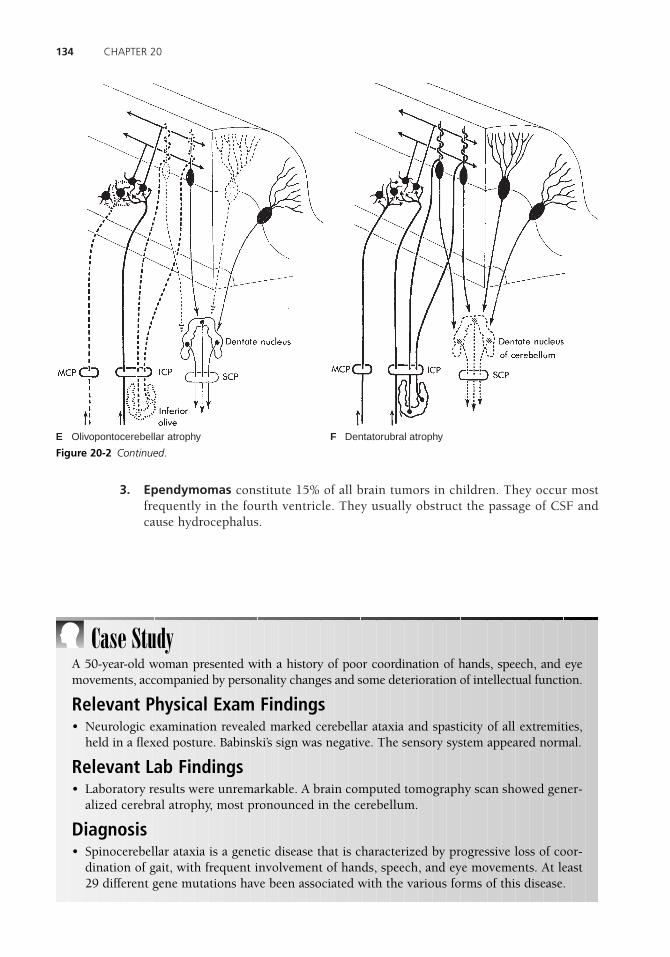
3. Ependymomas constitute 15% of all brain tumors in children. They occur mostfrequently in the fourth ventricle. They usually obstruct the passage of CSF andcause hydrocephalus.
134 CHAPTER 20
E Olivopontocerebellar atrophy F Dentatorubral atrophy
Figure 20-2 Continued.
Case StudyA 50-year-old woman presented with a history of poor coordination of hands, speech, and eyemovements, accompanied by personality changes and some deterioration of intellectual function.
Relevant Physical Exam Findings• Neurologic examination revealed marked cerebellar ataxia and spasticity of all extremities,
held in a flexed posture. Babinski’s sign was negative. The sensory system appeared normal.
Relevant Lab Findings • Laboratory results were unremarkable. A brain computed tomography scan showed gener-
alized cerebral atrophy, most pronounced in the cerebellum.
Diagnosis• Spinocerebellar ataxia is a genetic disease that is characterized by progressive loss of coor-
dination of gait, with frequent involvement of hands, speech, and eye movements. At least29 different gene mutations have been associated with the various forms of this disease.
LWBK110-3895G-C20[130-134].qxd 7/10/08 7:59 AM Page 134 Aptara Inc.

135
Chapter 21
Basal Nuclei (Ganglia) and StriatalMotor System
BASAL NUCLEI (GANGLIA) (Figure 21-1)
A. COMPONENTS1. Caudate nucleus2. Putamen 3. Globus pallidus
B. GROUPING OF THE BASAL NUCLEI (GANGLIA)1. The striatum consists of the caudate nucleus and putamen.2. The lentiform nucleus consists of the globus pallidus and putamen.3. The corpus striatum consists of the lentiform nucleus and caudate nucleus.4. The claustrum lies between the lentiform nucleus and the insular cortex. It has
reciprocal connections between the sensory cortices (i.e., visual cortex) (Figures21-2 through 21-4).
THE STRIATAL (EXTRAPYRAMIDAL) MOTOR SYSTEM (see Figure 21-1) playsa role in the initiation and execution of somatic motor activity, especially willed movement.It is also involved in automatic stereotyped postural and reflex motor activity (e.g., normalsubjects swing their arms when they walk).
A. STRUCTURE. The striatal motor system includes the following structures:1. Neocortex. 2. Striatum (caudatoputamen, or neostriatum).3. Globus pallidus. 4. Subthalamic nucleus. 5. Substantia nigra (i.e., pars compacta and pars reticularis).6. Thalamus (ventral anterior, ventral lateral, and centromedian nuclei).
II
I
Key Concepts1) Figure 21-6 shows the circuitry of the basal ganglia and their associated neurotransmitters.2) Parkinson’s disease is associated with a depopulation of neurons in the substantia nigra. 3) Huntington’s disease results in a loss of nerve cells in the caudate nucleus and putamen. 4) Hemiballism results from infarction of the contralateral subthalamic nucleus.
✔
LWBK110-3895G-C21[135-141].qxd 7/10/08 2:46 PM Page 135 Aptara Inc.
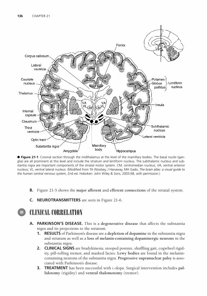
B. Figure 21-5 shows the major afferent and efferent connections of the striatal system.
C. NEUROTRANSMITTERS are seen in Figure 21-6.
CLINICAL CORRELATIONA. PARKINSON’S DISEASE. This is a degenerative disease that affects the substantia
nigra and its projections to the striatum.1. RESULTS of Parkinson’s disease are a depletion of dopamine in the substantia nigra
and striatum as well as a loss of melanin-containing dopaminergic neurons in thesubstantia nigra.
2. CLINICAL SIGNS are bradykinesia, stooped posture, shuffling gait, cogwheel rigid-ity, pill-rolling tremor, and masked facies. Lewy bodies are found in the melanin-containing neurons of the substantia nigra. Progressive supranuclear palsy is asso-ciated with Parkinson’s disease.
3. TREATMENT has been successful with L-dopa. Surgical intervention includes pal-lidotomy (rigidity) and ventral thalomotomy (tremor).
III
136 CHAPTER 21
● Figure 21-1 Coronal section through the midthalamus at the level of the mamillary bodies. The basal nuclei (gan-glia) are all prominent at this level and include the striatum and lentiform nucleus. The subthalamic nucleus and sub-stantia nigra are important components of the striatal motor system. CM, centromedian nucleus; VA, ventral anteriornucleus; VL, ventral lateral nucleus. (Modified from TA Woolsey, J Hanaway, MH Gado, The brain atlas: a visual guide tothe human central nervous system, 2nd ed. Hoboken: John Wiley & Sons, 2003:68, with permission.)
LWBK110-3895G-C21[135-141].qxd 7/10/08 2:46 PM Page 136 Aptara Inc.

B. METHYLPHENYLTETRAHYDROPYRIDINE (MPTP)-INDUCED PARKINSONISM.MPTP is an analog of meperidine (Demerol). It destroys dopaminergic neurons in thesubstantia nigra.
C. HUNTINGTON’S DISEASE (chorea). This is an inherited autosomal dominant move-ment disorder that is traced to a single gene defect on chromosome 4.1. It is associated with degeneration of the cholinergic and �-aminobutyric acid
(GABA)-ergic neurons of the striatum. It is accompanied by gyral atrophy in thefrontal and temporal lobes.
137BASAL NUCLEI (GANGLIA) AND STRIATAL MOTOR SYSTEM
● Figure 21-2 A parasagittal section through the caudate nucleus (5) and the substantia nigra (10): subthalamic nucleus(11), internal capsule (12), thalamus (13), cingulate gyrus (14). (Modified from TA Woolsey, J Hanaway, MH Gado, Thebrain atlas: a visual guide to the human central nervous system, 2nd ed. Hoboken: John Wiley & Sons, 2003:128, withpermission.)
LWBK110-3895G-C21[135-141].qxd 7/10/08 2:46 PM Page 137 Aptara Inc.

2. Glutamate (GLU) excitotoxicity results when GLU is released in the striatumand binds to its receptors on striatal neurons, culminating in an action poten-tal. GLU is removed from the extracellular space by astrocytes. In Hunting-ton’s disease, GLU is bound to the N-methyl-D-aspartate receptor, resulting inan influx of calcium ions and subsequent cell death. This cascade of events
138 CHAPTER 21
● Figure 21-3 An axial (horizontal) section through the anterior commissure (15) and the massa intermedia (16): inter-nal capsule (6), globus pallidus (7 ), insular cortex (14), putamen (17 ), claustrum (2), head of caudate nucleus (18). (Mod-ified from TA Woolsey, J Hanaway, MH Gado, The brain atlas: a visual guide to the human central nervous system, 2nded. Hoboken: John Wiley & Sons, 2003:100, with permission.)
LWBK110-3895G-C21[135-141].qxd 7/10/08 2:46 PM Page 138 Aptara Inc.

with neuronal death most likely occurs in cerebrovascular accidents (e.g.,stroke).
3. Clinical signs include choreiform movements, hypotonia, and progressive dementia.
D. OTHER CHOREIFORM DYSKINESIAS 1. Sydenham’s chorea (St. Vitus’ dance) is the most common cause of chorea over-
all. It occurs primarily in girls, typically after a bout of rheumatic fever.
139BASAL NUCLEI (GANGLIA) AND STRIATAL MOTOR SYSTEM
● Figure 21-4 A coronal section through the lentiform nucleus and the amygdaloid nucleus (1); the lentiform nucleusconsists of the putamen (8) and the globus pallidus (7 ); the amygdaloid nucleus appears as a circular profile below theuncus. (Modified from TA Woolsey, J Hanaway, MH Gado, The brain atlas: a visual guide to the human central nervoussystem, 2nd ed. Hoboken: John Wiley & Sons, 2003:60, with permission.)
LWBK110-3895G-C21[135-141].qxd 7/10/08 2:46 PM Page 139 Aptara Inc.

140 CHAPTER 21
● Figure 21-5 Major afferent and efferent connections of the striatal system. The striatum receives major input fromthree sources: the thalamus, neocortex, and substantia nigra. The striatum projects to the globus pallidus and substan-tia nigra. The globus pallidus is the effector nucleus of the striatal system; it projects to the thalamus and subthalamicnucleus. The substantia nigra also projects to the thalamus. The striatal motor system is expressed through the corti-cobulbar and corticospinal tracts. CM, centromedian nucleus; GABA, �-aminobutyric acid; VA, ventral anterior nucleus;VL, ventral lateral nucleus.
● Figure 21-6 Major neurotransmitters of the striatal motor system. Within the striatum, globus pallidus, and parsreticularis of the substantia nigra (S. nigra), �-aminobutyric acid (GABA) is the predominant neurotransmitter. GABAmay coexist in the same neuron with enkephalin (ENK ) or substance P (SP). Dopamine-containing neurons are found inthe pars compacta of the substantia nigra. Acetylcholine (ACh) is found in the local circuit neurons of the striatum. Thesubthalamic nucleus projects excitatory glutaminergic fibers to the globus pallidus. GLU, glutamate.
LWBK110-3895G-C21[135-141].qxd 7/10/08 2:46 PM Page 140 Aptara Inc.

2. Chorea gravidarum usually occurs during the second trimester of pregnancy.Many patients have a history of Sydenham’s chorea.
E. HEMIBALLISM is a movement disorder that usually results from a vascular lesion ofthe subthalamic nucleus. Clinical signs include violent contralateral flinging (ballis-tic) movements of one or both extremities.
F. HEPATOLENTICULAR DEGENERATION (WILSON’S DISEASE) is an autosomalrecessive disorder that is caused by a defect in the metabolism of copper. The genelocus is on chromosome 13.1. Clinical signs include choreiform or athetotic movements, rigidity, and wing-
beating tremor. Tremor is the most common neurologic sign.2. Lesions are found in the lentiform nucleus. Copper deposition in the limbus of
the cornea gives rise to the corneal Kayser-Fleischer ring, which is a pathogno-monic sign. Deposition of copper in the liver leads to multilobar cirrhosis.
3. Psychiatric symptoms include psychosis, personality disorders, and dementia.4. The diagnosis is based on low serum ceruloplasmin, elevated urinary excretion of
copper, and increased copper concentration in a liver biopsy specimen.5. Treatment includes penicillamine, a chelator.
G. TARDIVE DYSKINESIA is a syndrome of repetitive choreic movement that affects theface and trunk. It results from treatment with phenothiazines, butyrophenones, ormetoclopramide.
141BASAL NUCLEI (GANGLIA) AND STRIATAL MOTOR SYSTEM
Case Study A 30-year-old man presents with dysarthria, dysphagia, stiffness, and slow ataxic gait. Thereis no history of schizophrenia or depression and no family history of any neurodegenerativedisease.
Relevant Physical Exam Findings• The patient scored a 20/26 on the mini-mental status exam. The patient showed increased
tone in all extremities, with normal strength.
Relevant Lab Findings • A generalized cerebral and cerebellar atrophy and a very small caudate nucleus were
revealed on magnetic resonance imaging scans.
Diagnosis• Huntington’s disease (chorea) is caused by a trinucleotide repeat in the gene coding for the
Hungtinton protein. It is characterized by abnormal body movements and lack of coordi-nation but can also affect mental abilities.
LWBK110-3895G-C21[135-141].qxd 7/10/08 2:46 PM Page 141 Aptara Inc.
142
Chapter 22
Cerebral Cortex
INTRODUCTION The cerebral cortex, the thin, gray covering of both hemispheres ofthe brain, has two types: the neocortex (90%) and the allocortex (10%). Motor cortex isthe thickest (4.5 mm); visual cortex is the thinnest (1.5 mm).
THE SIX-LAYERED NEOCORTEX Layers II and IV of the neocortex are mainly affer-ent (i.e., receiving). Layers V and VI are mainly efferent (i.e., sending) (Figure 22-1).
A. LAYER I is the molecular layer.
B. LAYER II is the external granular layer.
C. LAYER III is the external pyramidal layer. It gives rise to association and commissuralfibers and is the major source of corticocortical fibers.
D. LAYER IV is the internal granular layer. It receives thalamocortical fibers from the thala-mic nuclei of the ventral tier (i.e., ventral posterolateral and ventral posteromedial). In thevisual cortex (Brodmann’s area 17), layer IV receives input from the lateral geniculate body.
E. LAYER V is the internal pyramidal layer. It gives rise to corticobulbar, corticospinal,and corticostriatal fibers. It contains the giant pyramidal cells of Betz, which are foundonly in the motor cortex (Brodmann’s area 4).
F. LAYER VI is the multiform layer. It is the major source of corticothalamic fibers. Itgives rise to projection, commissural, and association fibers.
FUNCTIONAL AREAS (Figure 22-2)
A. FRONTAL LOBE1. The motor cortex (Brodmann’s area 4) and premotor cortex (Brodmann’s area
6). These two cortices are somatotopically organized (Figure 22-3). Destruction
III
II
I
Key Concepts1) This chapter describes the cortical localization of functional areas of the brain. 2) How does the dominant hemisphere differ from the nondominant hemisphere? 3) Figure 22-5 shows the effects of various major hemispheric lesions. 4) What symptoms result from a lesion of the right inferior parietal lobe? 5) What is Gerstmann’s syndrome?
✔
LWBK110-3895G-C22[142-150].qxd 7/10/08 8:00 AM Page 142 Aptara Inc.

143CEREBRAL CORTEX
● Figure 22-1 Neurocortical circuits. G, granule cell; H, horizontal cell; M, Martinotti cell; P, pyramidal cell; S, stellatecell. Loops show synaptic junctions. (Reprinted from A Parent, Carpenter’s human neuroanatomy, 9th ed. Baltimore:Williams & Wilkins, 1996:868, with permission.)
of these areas of the frontal lobe causes contralateral spastic paresis. Contralat-eral pronator drift is associated with frontal lobe lesions of the corticospinaltract.
2. Frontal eye field (Brodmann’s area 8). Destruction causes deviation of the eyes tothe ipsilateral side.
3. Broca’s speech area (Brodmann’s areas 44 and 45). This is located in the poste-rior part of the inferior frontal gyrus in the dominant hemisphere (Figure 22-4).Destruction results in expressive, nonfluent aphasia (Broca’s aphasia). The patientunderstands both written and spoken language but cannot articulate speech orwrite normally. Broca’s aphasia is usually associated with contralateral facial andarm weakness because of the involvement of the motor strip.
4. Prefrontal cortex (Brodmann’s areas 9–12 and 46–47). Destruction of the ante-rior two-thirds of the frontal lobe convexity results in deficits in concentration,orientation, abstracting ability, judgment, and problem-solving ability. Otherfrontal lobe deficits include loss of initiative, inappropriate behavior, release ofsucking and grasping reflexes, gait apraxia, and sphincteric incontinence. Destruc-tion of the orbital (frontal) lobe results in inappropriate social behavior (e.g., useof obscene language, urinating in public). Perseveration is associated with frontallobe lesions.
LWBK110-3895G-C22[142-150].qxd 7/10/08 8:00 AM Page 143 Aptara Inc.
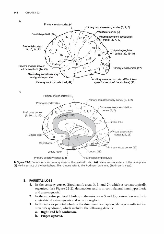
144 CHAPTER 22
Limbic lobe
Limbic lobe
Primary olfactory cortex (34) Parahippocampal gyrus
Uncus (28)
Primary visual cortex (17)
Limbic lobe
Somatosensory association cortex (5, 7)
Primary somatosensory cortex (3, 1, 2)
Visual association cortex (19, 18)
Premotor cortex (6)
Primary motor cortex (4)
Septal area
Prefrontal cortex (9, 10, 11, 12)
10
1112
1918
17
1819
38
34
37
20
28
249
8
64
5
3, 1, 2
B
Cingulate cortex
A
● Figure 22-2 Some motor and sensory areas of the cerebral cortex. (A) Lateral convex surface of the hemisphere. (B) Medial surface of the hemisphere. The numbers refer to the Brodmann brain map (Brodmann’s areas).
B. PARIETAL LOBE1. In the sensory cortex (Brodmann’s areas 3, 1, and 2), which is somatotopically
organized (see Figure 22-2), destruction results in contralateral hemihypesthesiaand astereognosis.
2. In the superior parietal lobule (Brodmann’s areas 5 and 7), destruction results incontralateral astereognosis and sensory neglect.
3. In the inferior parietal lobule of the dominant hemisphere, damage results in Ger-stmann’s syndrome, which includes the following deficits:a. Right and left confusion.b. Finger agnosia.
LWBK110-3895G-C22[142-150].qxd 7/10/08 8:00 AM Page 144 Aptara Inc.

145CEREBRAL CORTEX
● Figure 22-3 The sensory and motor homunculi. (A) Sensory representation in the postcentral gyrus. (B) Motor rep-resentation in the precentral gyrus. (Reprinted from W Penfield, T Rasmussen, The cerebral cortex of man. New York:Hafner, 1968:44, 57, with permission.)
● Figure 22-4 Cortical areas of the dominant hemisphere that play an important role in language production. Thevisual image of a word is projected from the visual cortex (Brodmann’s area 17) to the visual association cortices (Brod-mann’s areas 18 and 19) and then to the angular gyrus (Brodmann’s area 39). Further processing occurs in Wernicke’sspeech area (Brodmann’s area 22), where the auditory form of the word is recalled. Through the arcuate fasciculus, thisinformation reaches Broca’s speech area (Brodmann’s areas 44 and 45), where motor speech programs control the vocal-ization mechanisms of the precentral gyrus. Lesions of Broca’s speech area, Wernicke’s speech area, or the arcuate fas-ciculus result in dysphasia. (Reprinted from JD Fix, BRS neuroanatomy, 3rd ed. Baltimore: Lippincott Williams & Wilkins,2003:377, with permission.)
LWBK110-3895G-C22[142-150].qxd 7/10/08 8:00 AM Page 145 Aptara Inc.
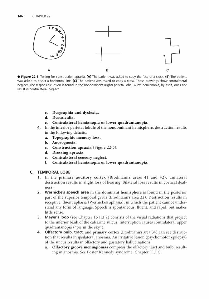
c. Dysgraphia and dyslexia.d. Dyscalculia.e. Contralateral hemianopia or lower quadrantanopia.
4. In the inferior parietal lobule of the nondominant hemisphere, destruction resultsin the following deficits:a. Topographic memory loss.b. Anosognosia.c. Construction apraxia (Figure 22-5). d. Dressing apraxia.e. Contralateral sensory neglect.f. Contralateral hemianopia or lower quadrantanopia.
C. TEMPORAL LOBE1. In the primary auditory cortex (Brodmann’s areas 41 and 42), unilateral
destruction results in slight loss of hearing. Bilateral loss results in cortical deaf-ness.
2. Wernicke’s speech area in the dominant hemisphere is found in the posteriorpart of the superior temporal gyrus (Brodmann’s area 22). Destruction results inreceptive, fluent aphasia (Wernicke’s aphasia), in which the patient cannot under-stand any form of language. Speech is spontaneous, fluent, and rapid, but makeslittle sense.
3. Meyer’s loop (see Chapter 15 II.F.2) consists of the visual radiations that projectto the inferior bank of the calcarine sulcus. Interruption causes contralateral upperquadrantanopia (“pie in the sky”).
4. Olfactory bulb, tract, and primary cortex (Brodmann’s area 34) can see destruc-tion that results in ipsilateral anosmia. An irritative lesion (psychomotor epilepsy)of the uncus results in olfactory and gustatory hallucinations.a. Olfactory groove meningiomas compress the olfactory tract and bulb, result-
ing in anosmia. See Foster Kennedy syndrome, Chapter 11.I.C.
146 CHAPTER 22
A B C
● Figure 22-5 Testing for construction apraxia. (A) The patient was asked to copy the face of a clock. (B) The patientwas asked to bisect a horizontal line. (C) The patient was asked to copy a cross. These drawings show contralateralneglect. The responsible lesion is found in the nondominant (right) parietal lobe. A left hemianopia, by itself, does notresult in contralateral neglect.
LWBK110-3895G-C22[142-150].qxd 7/10/08 8:00 AM Page 146 Aptara Inc.

147CEREBRAL CORTEX
● Figure 22-6 Focal destructive hemispheric lesions and the resulting symptoms. (A) Lateral convex surface of thedominant left hemisphere. (B) Lateral convex surface of the nondominant right hemisphere. (C) Medial surface of thenondominant hemisphere.
LWBK110-3895G-C22[142-150].qxd 7/10/08 8:00 AM Page 147 Aptara Inc.

b. Esthesioneuroblastomas (olfactory neuroblastomas) arise from bipolar sen-sory cells of the olfactory mucosa; they can extend through the cribriform plateinto the anterior cranial fossa. Presenting symptoms are similar to those of theFoster Kennedy syndrome.
5. In the hippocampal cortex (archicortex), bilateral lesions result in the inability toconsolidate short-term memory into long-term memory. Earlier memories areretrievable.
6. In the anterior temporal lobe, including the amygdaloid nucleus, bilateral dam-age results in Kläver-Bucy syndrome, which consists of psychic blindness (visualagnosia), hyperphagia, docility, and hypersexuality.
7. In the inferomedial occipitotemporal cortex, bilateral lesions result in the inabil-ity to recognize once-familiar faces (prosopagnosia).
D. OCCIPITAL LOBE. Bilateral lesions cause cortical blindness. Unilateral lesions causecontralateral hemianopia or quadrantanopia.
FOCAL DESTRUCTIVE HEMISPHERIC LESIONS AND SYMPTOMS Figure 22-6A shows the symptoms of lesions in the dominant hemisphere. Figure 22-6B showsthe symptoms of lesions in the nondominant hemisphere.
CEREBRAL DOMINANCE This dominance is determined by the Wada test. Sodiumamobarbital (Amytal) is injected into the carotid artery. If the patient becomes aphasic, theanesthetic was administered to the dominant hemisphere.
A. The dominant hemisphere is usually the left hemisphere. It is responsible for proposi-tional language (grammar, syntax, and semantics), speech, and calculation.
B. The nondominant hemisphere is usually the right hemisphere. It is responsible forthree-dimensional, or spatial, perception and nonverbal ideation. It also allows supe-rior recognition of faces.
SPLIT-BRAIN SYNDROME (Figure 22-7). This syndrome is a disconnection syndromethat results from transection of the corpus callosum.
A. The dominant hemisphere is better at vocal naming.
B. The nondominant, mute hemisphere is better at pointing to a stimulus. A person can-not name objects that are presented to the nondominant visual cortex. A blindfoldedperson cannot name objects that are presented to the nondominant sensory cortexthrough touch.
C. TEST (Figure 22-8). A subject views a composite picture of two half-faces (i.e., achimeric, or hybrid, figure). The right side shows a man; the left side shows a woman.The picture is removed, and the subject is asked to describe what he saw. He mayrespond that he saw a man, but when asked to point to what he saw, he points to thewoman.
D. In a patient who has alexia in the left visual field, the verbal symbols seen in the rightvisual cortex have no access to the language centers of the left hemisphere.
VI
V
IV
148 CHAPTER 22
LWBK110-3895G-C22[142-150].qxd 7/10/08 8:00 AM Page 148 Aptara Inc.

149CEREBRAL CORTEX
● Figure 22-7 Functions of the split brain after transection of the corpus callosum. Tactile and visual perception is pro-jected to the contralateral hemisphere, olfaction is perceived on the same side, and audition is perceived predominantlyin the opposite hemisphere. The left (L) hemisphere is dominant for language. The right (R) hemisphere is dominant forspatial construction and nonverbal ideation. (Reprinted from CR Noback, RJ Demarest, The human nervous system.Malvern, PA: Lea & Febiger, 1991:416, with permission.)
OTHER LESIONS OF THE CORPUS CALLOSUMA. ANTERIOR CORPUS CALLOSUM LESION may result in akinetic mutism or tactile anomia.
B. POSTERIOR CORPUS CALLOSUM (SPLENIUM) LESION may result in alexia with-out agraphia.
C. CALLOSOTOMY has been successfully used to treat “drop attacks” (colloid cyst ofthird ventricle).
BRAIN AND SPINAL CORD TUMORS (see Chapter 1) VIII
VII
LWBK110-3895G-C22[142-150].qxd 7/10/08 8:00 AM Page 149 Aptara Inc.

150 CHAPTER 22
● Figure 22-8 Chimeric (hybrid) figure of a face used to examine the hemispheric function of commissurotomizedpatients. The patient is instructed to fixate on the dot and is asked to describe what is seen. If the patient says that heor she sees the face of a man, the left hemisphere predominates in vocal tasks. If he or she is asked to point to the faceand points to the woman, the right hemisphere predominates in pointing tasks.
Case StudyA 50-year-old woman was referred to neurology with a year-long history of poor memoryand speech difficulties. She noted an inability to remember the names of common householdobjects and complained of forgetting her friends’ names. She complained of becoming inde-cisive, such that she had significant difficulty in making straightforward decisions and inprocessing new information.
Physical Exam Findings• On testing higher cortical function, her speech was somewhat rambling and hesitant, with
word finding difficulty. The neurologic examination was otherwise normal.
Relevant Lab Findings • Single-photon emission computed tomography cerebral perfusion disclosed markedly
reduced perfusion in the left temporal and parietal lobes and the right posterior parietalregions.
Diagnosis• Aphasia is a language disorder resulting from damage to brain regions responsible for lan-
guage (left hemisphere). Onset may occur either suddenly, owing to trauma, or slowly, asin the case of a brain tumor. Aphasia impairs expression and understanding of languageand reading and writing.
LWBK110-3895G-C22[142-150].qxd 7/10/08 8:00 AM Page 150 Aptara Inc.
151
Chapter 23
Neurotransmitters
IMPORTANT TRANSMITTERS AND THEIR PATHWAYSA. ACETYLCHOLINE is the major transmitter of the peripheral nervous system, neuro-
muscular junction, parasympathetic nervous system, preganglionic sympathetic fibers,and postganglionic sympathetic fibers that innervate sweat glands and some blood ves-sels in the skeletal muscles (Figure 23-1). Acetylcholine is found in the neurons of thesomatic and visceral motor nuclei in the brain stem and spinal cord. It is also found inthe basal nucleus of Meynert, which degenerates in Alzheimer’s disease.
B. CATECHOLAMINES. Figure 23-2 shows the biosynthetic pathway for catecholamines.Epinephrine, though a catecholamine, plays an insignificant role as a central nervoussystem neurotransmitter. In the body, epinephrine is found primarily in the adrenalmedulla. In the central nervous system, it is restricted to small neuronal clusters in thebrain stem (medulla).1. Dopamine (Figure 23-3) is depleted in patients with Parkinson’s disease and
increased in patients with schizophrenia. Dopamine is found in the arcuate nucleusof the hypothalamus. It is the prolactin-inhibiting factor. Its two major receptorsare D1 and D2. a. D1 receptors are postsynaptic. They activate adenylate cyclase and are excitatory.b. D2 receptors are both postsynaptic and presynaptic. They inhibit adenylate
cyclase and are inhibitory. Antipsychotic drugs block D2 receptors.2. Norepinephrine (Figure 23-4) is the transmitter of most postganglionic sympa-
thetic neurons. Antidepressant drugs enhance its transmission.a. Norepinephrine plays a role in anxiety states. Panic attacks are believed to
result from paroxysmal discharges from the locus ceruleus, where norepi-nephrinergic neurons are found in the highest concentration. Most postsynap-tic receptors of the locus ceruleus pathway are �1 or �2 receptors that activateadenylate cyclase and are excitatory.
I
Key Concepts1) In this chapter, the pathways of the major neurotransmitters are shown in separate
brain maps. 2) Glutamate (GLU) is the major excitatory transmitter of the brain; GABA is the major
inhibitory transmitter. Purkinje cells of the cerebellum are GABA-ergic. 3) In Alzheimer’s disease, there is a loss of acetylcholinergic neurons in the basal nucleus
of Meynert. 4) In Parkinson’s disease, there is a loss of dopaminergic neurons in the substantia nigra.
✔
LWBK110-3895G-C23[151-157].qxd 7/10/08 8:01 AM Page 151 Aptara Inc.

152 CHAPTER 23
● Figure 23-1 Distribution of acetylcholine-containing neurons and their axonal projections. The basal nucleus ofMeynert projects to the entire cortex. This nucleus degenerates in patients with Alzheimer’s disease. Striatal acetylcholinelocal circuit neurons degenerate in patients with Huntington’s disease.
● Figure 23-2 Synthesis of catecholamines from phenylalanine. Epinephrine, which is derived from norepinephrine, isfound primarily in the adrenal medulla.
b. The catecholamine hypothesis of mood disorders states that reduced norepi-nephrine activity is related to depression and that increased norepinephrineactivity is related to mania.
C. SEROTONIN [5-HYDROXYTRYPTAMINE (5-HT)] is an indolamine (Figure 23-5).Serotonin-containing neurons are found only in the raphe nuclei of the brain stem. 1. The permissive serotonin hypothesis states that when 5-HT activity is reduced,
decreased levels of catecholamines cause depression and insomnia. In addition,when 5-HT activity is increased, elevated levels of catecholamines cause mania.Dysfunction of 5-HT may underlie obsessive-compulsive disorder.
LWBK110-3895G-C23[151-157].qxd 7/10/08 8:01 AM Page 152 Aptara Inc.
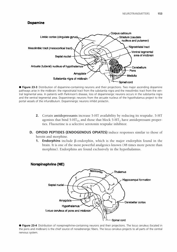
2. Certain antidepressants increase 5-HT availability by reducing its reuptake. 5-HTagonists that bind 5-HTIA and those that block 5-HT2 have antidepressant proper-ties. Fluoxetine is a selective serotonin reuptake inhibitor.
D. OPIOID PEPTIDES (ENDOGENOUS OPIATES) induce responses similar to those ofheroin and morphine.1. Endorphins include �-endorphin, which is the major endorphin found in the
brain. It is one of the most powerful analgesics known (48 times more potent thanmorphine). Endorphins are found exclusively in the hypothalamus.
153NEUROTRANSMITTERS
● Figure 23-4 Distribution of norepinephrine-containing neurons and their projections. The locus ceruleus (located inthe pons and midbrain) is the chief source of noradrenergic fibers. The locus ceruleus projects to all parts of the centralnervous system.
● Figure 23-3 Distribution of dopamine-containing neurons and their projections. Two major ascending dopaminepathways arise in the midbrain: the nigrostriatal tract from the substantia nigra and the mesolimbic tract from the ven-tral tegmental area. In patients with Parkinson’s disease, loss of dopaminergic neurons occurs in the substantia nigraand the ventral tegmental area. Dopaminergic neurons from the arcuate nucleus of the hypothalamus project to theportal vessels of the infundibulum. Dopaminergic neurons inhibit prolactin.
LWBK110-3895G-C23[151-157].qxd 7/10/08 8:01 AM Page 153 Aptara Inc.

2. Enkephalins are the most widely distributed and abundant opiate peptides. Theyare found in the highest concentration in the globus pallidus. Enkephalins coexistwith dopamine, �-aminobutyric acid (GABA), norepinephrine, and acetylcholine.They are colocalized in GABA-ergic pallidal neurons, and they play a role in painsuppression.
3. Dynorphins follow the distribution map for enkephalins.
E. NONOPIOID NEUROPEPTIDES1. Substance P plays a role in pain transmission. It is most highly concentrated in
the substantia nigra. It is also found in the dorsal root ganglion cells and substan-tia gelatinosa. It is colocalized with GABA in the striatonigral tract and plays a rolein movement disorders. Substance P levels are reduced in patients with Hunting-ton’s disease.
2. Somatostatin (somatotropin-release-inhibiting factor). Somatostatinergic neu-rons from the anterior hypothalamus project their axons to the median eminence,where somatostatin enters the hypophyseal portal system and regulates the releaseof growth hormone and thyroid-stimulating hormone. The concentration ofsomatostatin in the neocortex and hippocampus is significantly reduced in patientswith Alzheimer’s disease. Striatal somatostatin levels are increased in patients withHuntington’s disease.
F. AMINO ACID TRANSMITTERS1. Inhibitory amino acid transmitters
a. GABA (Figure 23-6) is the major inhibitory neurotransmitter of the brain.Purkinje, stellate, basket, and Golgi cells of the cerebellar cortex are GABA-ergic.
154 CHAPTER 23
● Figure 23-5 Distribution of 5-hydroxytryptamine (serotonin)-containing neurons and their projections. Serotonin-containing neurons are found in the nuclei of the raphe. They project widely to the forebrain, cerebellum, and spinalcord. The inset shows the synthetic pathway of serotonin.
LWBK110-3895G-C23[151-157].qxd 7/10/08 8:01 AM Page 154 Aptara Inc.

(1) GABA-ergic striatal neurons project to the globulus pallidus and sub-stantia nigra.
(2) GABA-ergic pallidal neurons project to the thalamus.(3) GABA-ergic nigral neurons project to the thalamus.(4) GABA receptors (GABA-A and GABA-B) are intimately associated with
benzodiazepine-binding sites. Benzodiazepines enhance GABA activity.(a) GABA-A receptors open chloride channels.(b) GABA-B receptors are found on the terminals of neurons that use
another transmitter (i.e., norepinephrine, dopamine, serotonin). Acti-vation of GABA-B receptors decreases the release of the other trans-mitter.
b. Glycine is the major inhibitory neurotransmitter of the spinal cord. It is usedby the Renshaw cells of the spinal cord.
2. Excitatory amino acid transmittersa. Glutamate (Figure 23-7). GLU is the major excitatory transmitter of the
brain. Neocortical glutamatergic neurons project to the striatum, subthalamicnucleus, and thalamus. (1) GLU is the transmitter of the cerebellar granule cells.(2) GLU is also the transmitter of nonnociceptive, large, primary afferent
fibers that enter the spinal cord and brain stem.(3) GLU is the transmitter of the corticobulbar and corticospinal tracts.
b. Aspartate. A major excitatory transmitter of the brain, aspartate is the trans-mitter of the climbing fibers of the cerebellum. Neurons of climbing fibers arefound in the inferior olivary nucleus.
c. Behavioral correlation. GLU, through its N-methyl-D-aspartate (NMDA)receptors, plays a role in long-term potentiation (a memory process) of hip-pocampal neurons. GLU plays a role in kindling and subsequent seizure activ-ity. Under certain conditions, glutamate and its analogs are neurotoxic.
d. Glutamate excitotoxicity. GLU is released in the striatum and binds to itsreceptors on striatal neurons, resulting in an action potential. GLU is removed
155NEUROTRANSMITTERS
● Figure 23-6 Distribution of �-aminobutyric acid (GABA)-containing neurons and their projections. GABA-ergic neu-rons are the major inhibitory cells of the central nervous system. GABA local circuit neurons are found in the neocortex,hippocampal formation, and cerebellar cortex (Purkinje cells). Striatal GABA-ergic neurons project to the thalamus andsubthalamic nucleus (not shown).
LWBK110-3895G-C23[151-157].qxd 7/10/08 8:01 AM Page 155 Aptara Inc.

from the extracellular space by astrocytes. In Huntington’s disease, GLU isbound to the NMDA receptor, resulting in an influx of calcium ions and sub-sequent cell death. This cascade of events with neuronal death most likelyoccurs in cerebrovascular accidents (stroke).
3. Nitric oxide is a recently discovered gaseous neurotransmitter that is producedwhen nitric oxide-synthase converts arginine to citrulline.a. It is located in the olfactory system, striatum, neocortex, hippocampal forma-
tion, supraoptic nucleus of the hypothalamus, and cerebellum.b. Nitric oxide is responsible for smooth-muscle relaxation of the corpus caver-
nosum and thus penile erection. It is also believed to play a role in memoryformation because of its long-term potentiation in the hippocampal formation.In addition, nitric oxide functions as a nitrovasodilator in the cardiovascularsystem.
FUNCTIONAL AND CLINICAL CONSIDERATIONSA. PARKINSON’S DISEASE results from degeneration of the dopaminergic neurons that
are found in the pars compacta of the substantia nigra. It causes a reduction ofdopamine in the striatum and substantia nigra (see Chapter 21, III.A).
B. HUNTINGTON’S DISEASE (CHOREA) results from a loss of acetylcholine- and GABA-containing neurons in the striatum (caudatoputamen). The effect is a loss of GABA inthe striatum and substantia nigra (see Chapter 21, III.C).
C. ALZHEIMER’S DISEASE results from the degeneration of cortical neurons and cholin-ergic neurons in the basal nucleus of Meynert. It is associated with a 60% to 90% lossof choline acetyltransferase in the cerebral cortex. Histologically, Alzheimer’s diseaseis characterized by the presence of neurofibrillary tangles, senile (neuritic) plaques,amyloid substance, granulovacuolar degeneration, and Hirano bodies.
II
156 CHAPTER 23
● Figure 23-7 Distribution of glutamate-containing neurons and their projections. Glutamate is the major excitatorytransmitter of the central nervous system. Cortical glutamatergic neurons project to the striatum. Hippocampal andsubicular glutamatergic neurons project through the fornix to the septal area and hypothalamus. The granule cells ofthe cerebellum are glutamatergic.
LWBK110-3895G-C23[151-157].qxd 7/10/08 8:01 AM Page 156 Aptara Inc.

D. MYASTHENIA GRAVIS results from autoantibodies against the nicotinic acetylcholinereceptor on skeletal muscle. These antibodies block the postganglionic acetylcholine-binding site. Thymic cells augment B-cell production of autoantibodies. The cardinalmanifestation is fatigable weakness of the skeletal muscle. The extraocular muscles,including the levator palpebrae, are usually involved. Edrophonium or neostigmineinjection is used for diagnosis.
E. LAMBERT-EATON MYASTHENIC SYNDROME is caused by a presynaptic defect ofacetylcholine release. It causes weakness in the limb muscles but not the bulbar mus-cles. Fifty percent of cases are associated with neoplasms (i.e., lung, breast, prostate).In these patients, muscle strength improves with use. In contrast, in patients with myas-thenia gravis, muscle use results in muscle fatigue, and autonomic dysfunction includesdry mouth, constipation, impotence, and urinary incontinence.
157NEUROTRANSMITTERS
Case Study A 60-year-old man presents with progressive stiffness accompanied by difficulty in walkingand going down stairs. He has an expressionless face and a vacant, staring gaze. Examina-tion revealed limited ocular motility in all directions. What is the most likely symptomatictherapy?
Relevant Physical Exam Findings• Tremor at rest, with pill-rolling motion of the hand• Rigidity observed when the patient’s relaxed wrist is flexed and extended • Bradykinesia
Diagnosis• Parkinson’s disease is a progressive neurodegenerative disorder associated with a loss of
dopaminergic nigrostriatal neurons. Often, patients are given levodopa combined withcarbidopa. Nerve cells use levodopa to make dopamine to compensate for the loss of nigro-striatal neurons, whereas carbidopa delays the conversion of levodopa into dopamine untilit reaches the brain.
LWBK110-3895G-C23[151-157].qxd 7/10/08 8:01 AM Page 157 Aptara Inc.

158
Chapter 24
Apraxia, Aphasia, and Dysprosody
APRAXIA is the inability to perform motor activities in the presence of intact motor andsensory systems and normal comprehension.
A. IDEOMOTOR APRAXIA (IDIOKENETIC APRAXIA)1. The disorder results in loss of the ability to perform intransitive or imaginary ges-
tures; the inability to perform complicated motor tasks (e.g., saluting, blowing akiss, or making the “V” for victory sign)
2. The lesion is in Wernicke’s area. 3. Bucco-facial or facial-oral apraxia is a type of idiomotor apraxia; facial apraxia is
the most common type of apraxia.
B. IDEATIONAL APRAXIA (IDEATORY APRAXIA)1. The inability to demonstrate the use of real objects (e.g., ask the patient to smoke
a pipe, a multistep complex sequence)2. A misuse of objects owing to a disturbance of identification (agnosia)3. The lesion in Wernicke’s area
C. CONSTRUCTION APRAXIA, the inability to draw or construct a geometric figure (e.g.,the face of a clock). If the patient draws only the right half of the clock, this conditionis called hemineglect, and the lesion is located in the right inferior parietal lobule.
D. GAIT APRAXIA, the inability to use the lower limbs properly. The patient has diffi-culty in lifting his feet from the floor, a frontal lobe sign seen with normal-pressurehydrocephalus (gait apraxia, dementia, incontinence).
APHASIA is impaired or absent communication by speech, writing, or signs (i.e., loss ofthe capacity for spoken language). The lesions are located in the dominant hemisphere. Asso-ciate the following symptoms and lesion sites with the appropriate aphasia (Figure 24-1).
A. BROCA’S (MOTOR) APHASIA1. Lesion in frontal lobe, in the inferior frontal gyrus (Brodmann’s areas 44 and 45)2. Good comprehension
II
I
Key Concepts1) Be able to differentiate Broca’s aphasia from Wernicke’s aphasia. 2) What is conduction aphasia?
✔
LWBK110-3895G-C24[158-161].qxd 7/10/08 8:02 AM Page 158 Aptara Inc.

3. Effortful speech4. Dysarthric speech5. Telegraphic speech6. Nonfluent speech7. Poor repetition8. Contralateral lower facial and upper limb weakness
B. WERNICKE’S (SENSORY) APHASIA1. Lesion in posterior temporal lobe, in the superior temporal gyrus (Brodmann’s
area 22)2. Poor comprehension3. Fluent speech4. Poor repetition5. Quadrantanopia6. Paraphasic errors
a. Non sequiturs (Latin, “does not follow”): statements irrelevant to the questionasked
b. Neologisms: words with no meaningc. Driveling speech
C. CONDUCTION APHASIA1. Transection of the arcuate fasciculus; the arcuate fasciculus interconnects Brod-
mann’s speech area with Wernicke’s speech area.2. Poor repetition
159APRAXIA, APHASIA, AND DYSPROSODY
Transcortical
sensory
aphasia
Transcortical
motor
aphasia
Wernicke’s
aphasiaConduction
aphasia
Fluent
speech
Nonfluent
speech
Poor r
epet
ition
Goo
d re
petition
Good
comprehension
Poor
comprehension
Broca’s
aphasiaMixed
transcortical
aphasia
● Figure 24-1 The “aphasia square” makes it easy to differentiate the six most common “national board” aphasias.Broca’s, conduction and Wernicke’s aphasias are all characterized by poor repetition. (Adapted from J Miller, N Fountain,Neurology recall. Baltimore: Williams & Wilkins, 1997:35, with permission.)
LWBK110-3895G-C24[158-161].qxd 7/10/08 8:02 AM Page 159 Aptara Inc.

3. Good comprehension4. Fluent speech
D. TRANSCORTICAL MOTOR APHASIA1. Poor comprehension2. Good repetition3. Nonfluent speech
E. TRANSCORTICAL MIXED APHASIA1. Poor comprehension2. Good repetition3. Nonfluent speech
F. TRANSCORTICAL SENSORY APHASIA1. Poor comprehension2. Good repetition3. Fluent speech
G. GLOBAL APHASIA. Resulting from a lesion of the perisylvian area, which containsBroca’s and Wernicke’s areas, global aphasia combines all of the symptoms of Broca’sand Wernicke’s aphasias.
H. THALAMIC APHASIA. A dominant thalamic syndrome, thalamic aphasia closelyresembles a thought disorder of patients with schizophrenia and chronic drug-inducedpsychosis. Symptoms include fluent paraphasic speech with normal comprehensionand repetition.
I. BASAL GANGLIA. Diseases of the basal ganglia may cause aphasia. Lesions of the ante-rior basal ganglia result in nonfluent aphasia. Lesions of the posterior basal gangliaresult in fluent aphasia.
J. WATERSHED INFARCTS. Such infarcts are areas of infarction in the boundary zonesof the anterior, middle, and posterior cerebral arteries. These areas are vulnerable tohypoperfusion and thus may separate Broca’s and Wernicke’s speech areas from the sur-rounding cortex. These infarcts cause the motor, mixed, and sensory transcorticalaphasias.
DYSPROSODY is a nondominant hemispheric language deficit that serves propositionallanguage. Emotionality, inflection, melody, emphasis, and gesturing are affected.
A. EXPRESSIVE DYSPROSODY results from a lesion that corresponds to Broca’s area butis located in the nondominant hemisphere. Patients cannot express emotion or inflec-tion in their speech.
B. RECEPTIVE DYSPROSODY results from a lesion that corresponds to Wernicke’s areabut is located in the nondominant hemisphere. Patients cannot comprehend the emo-tionality or inflection in the speech they hear.
III
160 CHAPTER 24
LWBK110-3895G-C24[158-161].qxd 7/10/08 8:02 AM Page 160 Aptara Inc.

161APRAXIA, APHASIA, AND DYSPROSODY
Case Study A 7-year-old boy presents with difficulty in articulating words, though he can hear wordsand understand their meaning. His mother noted he had difficulty in breast-feeding duringinfancy.
Relevant Physical Exam Findings• Limited ability to produce speech sounds • Groping for correct placement of the tongue, lips, and jaw during speech • Omission of words in sentences • Atypical facial expressions
Diagnosis• Apraxia results when sounds do not easily form into words. A complex coordination of
lips, tongue, and throat muscles is required to articulate words.
LWBK110-3895G-C24[158-161].qxd 7/10/08 8:02 AM Page 161 Aptara Inc.

162
Appendix I
Table of Cranial Nerves
Cranial Nerve Type Origin Function Course
I–Olfactory
II–Optic
III–OculomotorParasympathetic
Motor
IV–Trochlear
V–TrigeminalMotor
Sensory
SVA
SSA
GVE
GSE
GSE
SVE
GSA
Bipolar olfactory(olfactory epitheliumin roof of nasal cavity)
Retinal ganglion cells
Edinger-Westphalnucleus (rostral mid-brain)Oculomotor nucleus(rostral midbrain)
Trochlear nucleus(caudal midbrain)
Motor nucleus CN V(midpons)
Trigeminal ganglionand mesencephalicnucleus CN V (rostralpons and midbrain)
Smell (olfaction)
Vision
Sphincter muscle ofiris; ciliary muscle
Superior, inferior,and medial rectimuscles; inferioroblique muscle; levator palpebraemuscle
Superior obliquemuscle
Muscles of mastica-tion and tensortympani muscleTactile, pain, andthermal sensationfrom the face; theoral and nasal cavi-ties; and the supra-tentorial dura
Central axons project to theolfactory bulb via the cribriformplate of the ethmoid bone.
Central axons converge at theoptic disk and form the opticnerve, which enters the skull viathe optic canal. Optic nerveaxons terminate in the lateralgeniculate bodies.
Axons exit the midbrain in theinterpeduncular fossa, traversethe cavernous sinus, and enterthe orbit via the superior orbitalfissure.
Axons decussate in superiormedullary velum, exit dorsallyinferior to the inferior colliculi,encircle the midbrain, traversethe cavernous sinus, and enterthe orbit via the superior orbitalfissure.
Ophthalmic nerve exits via thesuperior orbital fissure; maxillarynerve exits via the foramenrotundum; mandibular nerveexits via the foramen ovale;ophthalmic and maxillary nervestraverse the cavernous sinus;GSA fibers enter the spinaltrigeminal tract of CN V.
LWBK110-3895G-AppI[162-164].qxd 7/10/08 8:08 AM Page 162 Aptara Inc.

163TABLE OF CRANIAL NERVES
Cranial Nerve Type Origin Function Course
VI–Abducent
VII–FacialParasympathetic
Motor
Sensory
Sensory
VIII–VestibulocochlearVestibular nerve
Cochlear nerve
IX–GlossopharyngealParasympathetic
Motor
Sensory
Sensory
Sensory
GSE
GVE
SVE
GSA
SVA
SSA
GVE
SVE
GSA
GVA
SVA
Abducent nucleus(caudal pons)
Superior salivatorynucleus (caudal pons)
Facial nucleus (caudalpons)
Geniculate ganglion(temporal bone)Geniculate ganglion
Vestibular ganglion(internal auditorymeatus)
Spinal ganglion(modiolus of temporalbone)
Inferior salivatorynucleus (rostralmedulla)Nucleus ambiguous(rostral medulla)Superior ganglion(jugular foramen)Inferior (petrosal) ganglion (in jugularforamen)
Inferior (petrosal) ganglion (in jugularforamen)
Lateral rectus muscle
Lacrimal gland (viasphenopalatineganglion); sub-mandibular andsublingual glands(via submandibularganglion)Muscles of facialexpression;stapedius muscleTactile sensation toskin of earTaste sensationfrom the anteriortwo-thirds oftongue (via chordatympani)
Equilibrium (inner-vates hair cells ofsemicircular ducts,saccule, and utricle)Hearing (innervateshair cells of theorgan of Corti)
Parotid gland (viathe otic ganglion)
StylopharyngeusmuscleTactile sensation toexternal earTactile sensation toposterior third oftongue, pharynx,middle ear, andauditory tube, inputfrom carotid sinusand carotid bodyTaste from posteriorthird of the tongue
Axons exit the pons from theinferior pontine sulcus, traversethe cavernous sinus, and enterthe orbit via the superior orbitalfissure.
Axons exit the pons in the cere-bellar pontine angle and enterthe internal auditory meatus;motor fibers traverse the facialcanal of the temporal bone andexit via the stylomastoid fora-men; taste fibers traverse thechorda tympani and lingualnerve; GSA fibers enter thespinal trigeminal tract of CN V;SVA fibers enter the tract.
Vestibular and cochlear nervesjoin in the internal auditorymeatus and enter the brainstem in the cerebellopontineangle; vestibular nerve projectsto the vestibular nuclei and theflocculonodular lobe of thecerebellum; cochlear nerve pro-jects to the cochlear nuclei.
Axons exit (motor) and enter(sensory) medulla from thepostolivary sulcus; axons exitand enter the skull via jugularforamen; GSA fibers enter thespinal trigeminal tract of CN V;GVA and SVA fibers enter thesolitary tract.
(continued)
LWBK110-3895G-AppI[162-164].qxd 7/10/08 8:08 AM Page 163 Aptara Inc.

Cranial Nerve Type Origin Function Course
164 APPENDIX I
X–VagalParasympathetic
Motor
Sensory
Sensory
Sensory
XI–AccessoryMotor (cranial)
Motor (spinal)
XII–Hypoglossal
GVE
SVE
GSA
GVA
SVA
Dorsal nucleus of CNX (medulla)
Nucleus ambiguus(midmedulla)Superior ganglion(jugular foramen)Inferior (nodose) ganglion (in jugularforamen)
Inferior (nodose) ganglion (in jugularforamen)
Nucleus ambiguus(medulla)
Ventral horn neuronsC1–C6
Hypoglossal nucleus(medulla)
Viscera of the tho-racic and abdomi-nal cavities to theleft colic flexure [viaterminal (mural)ganglia]Muscles of the lar-ynx and pharynxTactile sensation tothe external earMucous mem-branes of the phar-ynx, larynx, esopha-gus, trachea, andthoracic andabdominal viscerato the left colicflexureTaste from theepiglottis
Intrinsic muscles ofthe larynx (exceptthe cricothyroidmuscle) via recur-rent laryngeal nerveSternocleidomastoid and trapeziusmuscles
Intrinsic and extrin-sic muscles of thetongue (except thepalatoglossus muscle)
Axons exit (motor) and enter(sensory) medulla from the pos-tolivary sulcus; axons exit andenter the skull via the jugularforamen; GSA fibers enter thespinal trigeminal tract of CN V;GVA and SVA fibers enter thesolitary tract.
Axons from the cranial divisionexit the medulla from the pos-tolivary sulcus and join thevagal nerve; axons from spinaldivision exit the spinal cord,ascend through the foramenmagnum, and exit the skull viathe jugular foramen.
Axons exit from the preolivarysulcus of the medulla and exitthe skull via the hypoglossalcanal.
SVA, special visceral afferent; SSA, special somatic afferent; GVE, general visceral efferent; GSE, general somatic efferent; SVE, spe-cial visceral efferent; GSA, general somatic afferent; GVA, general visceral afferent; CN, cranial nerve.
LWBK110-3895G-AppI[162-164].qxd 7/10/08 8:08 AM Page 164 Aptara Inc.
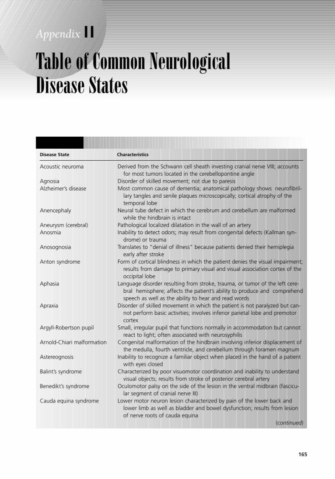
165
Appendix I I
Table of Common NeurologicalDisease States
Disease State Characteristics
(continued)
Derived from the Schwann cell sheath investing cranial nerve VIII; accountsfor most tumors located in the cerebellopontine angle
Disorder of skilled movement; not due to paresisMost common cause of dementia; anatomical pathology shows neurofibril-
lary tangles and senile plaques microscopically; cortical atrophy of thetemporal lobe
Neural tube defect in which the cerebrum and cerebellum are malformedwhile the hindbrain is intact
Pathological localized dilatation in the wall of an arteryInability to detect odors; may result from congenital defects (Kallman syn-
drome) or traumaTranslates to “denial of illness” because patients denied their hemiplegia
early after strokeForm of cortical blindness in which the patient denies the visual impairment;
results from damage to primary visual and visual association cortex of theoccipital lobe
Language disorder resulting from stroke, trauma, or tumor of the left cere-bral hemisphere; affects the patient’s ability to produce and comprehendspeech as well as the ability to hear and read words
Disorder of skilled movement in which the patient is not paralyzed but can-not perform basic activities; involves inferior parietal lobe and premotorcortex
Small, irregular pupil that functions normally in accommodation but cannotreact to light; often associated with neurosyphilis
Congenital malformation of the hindbrain involving inferior displacement ofthe medulla, fourth ventricle, and cerebellum through foramen magnum
Inability to recognize a familiar object when placed in the hand of a patientwith eyes closed
Characterized by poor visuomotor coordination and inability to understandvisual objects; results from stroke of posterior cerebral artery
Oculomotor palsy on the side of the lesion in the ventral midbrain (fascicu-lar segment of cranial nerve III)
Lower motor neuron lesion characterized by pain of the lower back andlower limb as well as bladder and bowel dysfunction; results from lesionof nerve roots of cauda equina
Acoustic neuroma
AgnosiaAlzheimer’s disease
Anencephaly
Aneurysm (cerebral)Anosmia
Anosognosia
Anton syndrome
Aphasia
Apraxia
Argyll-Robertson pupil
Arnold-Chiari malformation
Astereognosis
Balint’s syndrome
Benedikt’s syndrome
Cauda equina syndrome
#DC4E0-3895G-APPII[165-168].qxd 7/10/08 8:05 AM Page 165 Aptara Inc.
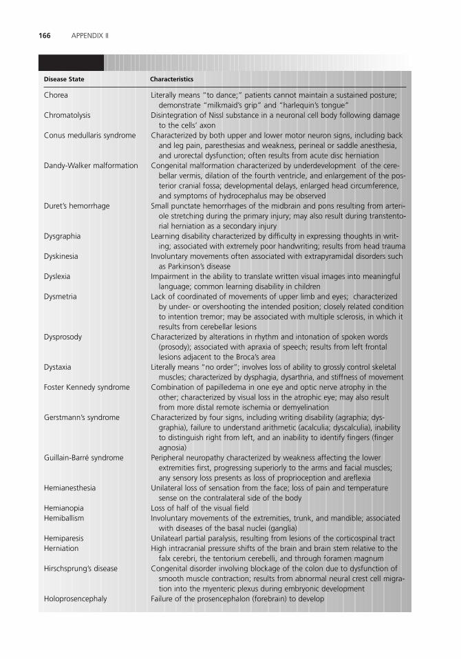
166 APPENDIX II
Disease State Characteristics
Literally means “to dance;” patients cannot maintain a sustained posture;demonstrate “milkmaid’s grip” and “harlequin’s tongue”
Disintegration of Nissl substance in a neuronal cell body following damageto the cells’ axon
Characterized by both upper and lower motor neuron signs, including backand leg pain, paresthesias and weakness, perineal or saddle anesthesia,and urorectal dysfunction; often results from acute disc herniation
Congenital malformation characterized by underdevelopment of the cere-bellar vermis, dilation of the fourth ventricle, and enlargement of the pos-terior cranial fossa; developmental delays, enlarged head circumference,and symptoms of hydrocephalus may be observed
Small punctate hemorrhages of the midbrain and pons resulting from arteri-ole stretching during the primary injury; may also result during transtento-rial herniation as a secondary injury
Learning disability characterized by difficulty in expressing thoughts in writ-ing; associated with extremely poor handwriting; results from head trauma
Involuntary movements often associated with extrapyramidal disorders suchas Parkinson’s disease
Impairment in the ability to translate written visual images into meaningfullanguage; common learning disability in children
Lack of coordinated of movements of upper limb and eyes; characterizedby under- or overshooting the intended position; closely related conditionto intention tremor; may be associated with multiple sclerosis, in which itresults from cerebellar lesions
Characterized by alterations in rhythm and intonation of spoken words(prosody); associated with apraxia of speech; results from left frontallesions adjacent to the Broca’s area
Literally means “no order”; involves loss of ability to grossly control skeletalmuscles; characterized by dysphagia, dysarthria, and stiffness of movement
Combination of papilledema in one eye and optic nerve atrophy in theother; characterized by visual loss in the atrophic eye; may also resultfrom more distal remote ischemia or demyelination
Characterized by four signs, including writing disability (agraphia; dys-graphia), failure to understand arithmetic (acalculia; dyscalculia), inabilityto distinguish right from left, and an inability to identify fingers (fingeragnosia)
Peripheral neuropathy characterized by weakness affecting the lowerextremities first, progressing superiorly to the arms and facial muscles;any sensory loss presents as loss of proprioception and areflexia
Unilateral loss of sensation from the face; loss of pain and temperaturesense on the contralateral side of the body
Loss of half of the visual fieldInvoluntary movements of the extremities, trunk, and mandible; associated
with diseases of the basal nuclei (ganglia)Unilatearl partial paralysis, resulting from lesions of the corticospinal tractHigh intracranial pressure shifts of the brain and brain stem relative to the
falx cerebri, the tentorium cerebelli, and through foramen magnum Congenital disorder involving blockage of the colon due to dysfunction of
smooth muscle contraction; results from abnormal neural crest cell migra-tion into the myenteric plexus during embryonic development
Failure of the prosencephalon (forebrain) to develop
Chorea
Chromatolysis
Conus medullaris syndrome
Dandy-Walker malformation
Duret’s hemorrhage
Dysgraphia
Dyskinesia
Dyslexia
Dysmetria
Dysprosody
Dystaxia
Foster Kennedy syndrome
Gerstmann’s syndrome
Guillain-Barré syndrome
Hemianesthesia
HemianopiaHemiballism
HemiparesisHerniation
Hirschsprung’s disease
Holoprosencephaly
#DC4E0-3895G-APPII[165-168].qxd 7/10/08 8:05 AM Page 166 Aptara Inc.

167TABLE OF COMMON NEUROLOGICAL DISEASE STATES
Disease State Characteristics
Interruption of sympathetic innervation to the eye; characterized by threeprimary signs, including ptosis, miosis, and anhidrosis
Trauma to or tumor at the jugular foramen, with resultant paralysis of cra-nial nerves IX, X, and IX; often results from a basilar skull fracture
Lesion of medial temporal lobes and amygdala; characterized by oral andtactile exploratory behavior (inappropriate licking or touching in public);hypersexuality; flattened emotions (placidity)
Disorder of presynaptic neuromuscular transmission; impaired release ofacetylcholine, resulting in proximal muscle weakness and abnormal ten-don reflexes
Also known as Wallenberg’s syndrome and posterior inferior cerebellarartery (PICA) syndrome; involves difficulty in swallowing and speaking;results from occlusion of PICA resulting in infarction of the lateral part ofthe medulla
Complete paralysis of all voluntary muscles of the body except the extraocu-lar muscles; patients fully alert but cannot move; only movements of theeyes and blinking are possible; results from stroke in the pons
Progressive degeneration of upper and lower motor neurons, resulting inhyperreflexia and spasticity (degeneration of the lateral corticospinaltracts) as well as weakness, atrophy, and fasciculations (due to muscledenervation)
Cyst containing brain tissue, cerebrospinal fluid, and the meninges, protrud-ing through a congenital defect in the skull; results from failure of theneural tube to close during gestation
Autoimmune disorder in which antibodies are created against proteins in themyelin sheath; results in inflammation of myelin and the nerves that itinvests; causes scarring (sclerosis), which slows or blocks neurotransmission
Literally means “grave muscle”; chronic autoimmune neuromuscular diseasecharacterized by weakness of the skeletal muscles of the body that wors-ens during periods of activity and improves after rest
Involuntary rhythmic shaking or wobbling of the eyes; results from a patho-logical process that damages one or more components of the vestibularsystem, including the semicircular canals, otolithic organs, and vestibulo-cerebellum
Bacterial infection of the eye, related to pinkeye, or conjunctivitis; tularemiamay infect the eye by direct or indirect entry of the bacteria into the eye
Disorder of the basal nuclei (ganglia) that results in resting tremor of a limb,slowness of movement (bradykinesia), rigidity of the limbs or trunk, andpostural instability
Vascular disease reducing blood flow to the extremities when exposed toenvironmental stimuli, including temperature changes or stress
Results of a neurologic exam used to assess the dorsal columns of the spinalcord involved in proprioception; a positive Romberg sign indicates sensoryataxia, whereas a negative Romberg sign suggests cerebellar ataxia
Occlusion of subclavian artery proximal to the origin of the vertebral artery;involves reversed blood flow in the vertebral artery; so named because itwas thought that blood was stolen by the ipsilateral vertebral artery fromthe contralateral vertebral artery
Cyst (syrinx) forms and expands within the spinal cord, destroying central
Horner’s syndrome
Jugular foramen syndrome
Kluver-Bucy syndrome
Lambert-Eaton syndrome
Lateral medullary syndrome
Locked-in syndrome
Lou Gehrig’s disease[amyotrophic lateralsclerosis (ALS)]
Meningomyelocele
Multiple sclerosis
Myasthenia gravis
Nystagmus
Parinaud’s syndrome
Parkinson’s disease
Raynaud’s disease
Romberg sign
Subclavian steal syndrome
(continued)
#DC4E0-3895G-APPII[165-168].qxd 7/10/08 8:05 AM Page 167 Aptara Inc.

168 APPENDIX II
Disease State Characteristics
Syringomelia
Tabes dorsalis
Tic douloureux
Wallerian degenerationWatershed infarct
Wilson’s disease
Weber’s syndrome
Wernicke-Korsakoffsyndrome
tissue in the cord; results in pain, weakness, and stiffness in the trunk andlimbs
Slow degeneration of the neurons in the dorsal columns of the spinal cord;results in abnormal proprioceptive signals
Also known as trigeminal neuralgia; unilateral, severe, stabbing pain of theface, particularly jaw, cheek, or lip
Process of degeneration of the axons distal to a site of transection Occurs at the junction of two nonanastomosing arterial systems; often
between branches of the anterior and middle cerebral arteriesAlso known as hepatolenticular degeneration; genetic disorder in which
copper accumulates in the liver and basal nuclei (ganglia), specifically inthe putamen and globus pallidus (lenticular nucleus)
Also known as superior alternating hemiplegia; involves oculomotor nervepalsy and contralateral hemiparesis or hemiplegia; results from midbraininfarction as a result of occlusion of the posterior cerebral artery
Neurologic disorder resulting from thiamine deficiency; usually caused bymalnutrition, common in alcoholic patients; may present with visionchanges, loss of muscle coordination, memory loss
#DC4E0-3895G-APPII[165-168].qxd 7/10/08 8:05 AM Page 168 Aptara Inc.
169
Glossary
abasia Inability to walk.
abulia Inability to perform voluntary actionsor to make decisions; seen in bilateral frontallobe disease.
accommodation Increase in thickness of thelens needed to focus a near external object onthe retina.
adenohypophysis Anterior lobe of thepituitary gland, derived from Rathke’s pouch.
adenoma sebaceum Cutaneous lesion seen intuberous sclerosis.
Adie pupil (Myotonic pupil) A tonic pupil,usually large, that constricts very slowly tolight and convergence; generally unilateraland frequently occurs in young women withabsent knee or ankle reflexes.
afferent pupil (Marcus Gunn pupil) A pupilthat reacts sluggishly to direct lightstimulation; caused by a lesion of the afferentpathway (e.g., multiple sclerosis involving theoptic nerve).
agenesis Failure of a structure to develop(e.g., agenesis of the corpus callosum).
ageusia Loss of the sensation of taste (gusta-tion).
agnosia Lack of the sensory-perceptionalability to recognize objects; visual, auditory,and tactile.
agraphesthesia Inability to recognize figures“written” on the skin.
agraphia Inability to write; seen inGerstmann’s syndrome.
akathisia (acathisia) Inability to remain in asitting position; motor restlessness; mayappear after the withdrawal of neurolepticdrugs.
akinesia Absence or loss of the power of vol-untary motion; seen in Parkinson’s disease.
akinetic mutism State in which patient canmove and speak but cannot be prompted todo so; due to bilateral occlusion of theanterior cerebral artery or midbrain lesions.
alar plate Division of the mantle zone thatgives rise to sensory neurons; receives sensoryinput from the dorsal root ganglia.
albuminocytologic dissociation Elevated cere-brospinal fluid (CSF) protein with a normalCSF cell count; seen in Guillain-Barrésyndrome.
alexia Visual aphasia; word or text blindness;loss of the ability to grasp the meaning ofwritten or printed words; seen in Gerstmann’ssyndrome.
Alzheimer’s disease Condition characterizedpathologically by the presence of senileplaques, neurofibrillary tangles, granulovacuo-lar degeneration, Hirano bodies, and amyloiddeposition; patients are demented with severememory loss.
alternating hemianesthesia Ipsilateral facialanesthesia and a contralateral body anesthesia;results from a pontine or medullary lesioninvolving the spinal trigeminal tract and thespinothalamic tract.
alternating hemiparesis Ipsilateral cranialnerve palsy and a contralateral hemiparesis(e.g., alternating abducent hemiparesis).
altitudinal hemianopia Defect in which theupper or lower half of the visual field is lost.
amaurosis fugax Transient monocular blind-ness usually related to carotid artery stenosisor, less often, to embolism of retinal arterioles.
amnesia Disturbance or loss of memory; seenwith bilateral medial temporal lobe lesions.
amusia Form of aphasia characterized by theloss of ability to express or recognize simplemusical tones.
LWBK110-3895G-GLO[169-180].qxd 7/10/08 8:04 AM Page 169 Aptara Inc.

amyotrophic lateral sclerosis (ALS) Anonhereditary motor neuron disease affectingboth upper and lower motor neurons; charac-terized by muscle weakness, fasciculations,fibrillations, and giant motor units onelectromyography. There are no sensorydeficits in ALS. It is also called Lou Gehrig’sdisease.
amyotrophy Muscle wasting or atrophy.
analgesia Insensibility to painful stimuli.
anencephaly Failure of the cerebral and cere-bellar hemispheres to develop; results fromfailure of the anterior neuropore to close.
anesthesia State characterized by the loss ofsensation.
aneurysm Circumscribed dilation of an artery(e.g., berry aneurysm).
anhidrosis Absence of sweating; found inHorner syndrome.
anisocoria Pupils that are unequal in size;found in a third-nerve palsy and Horner’s syn-drome.
anomia Anomic aphasia; the inability toname objects; may result from a lesion of theangular gyrus.
anosmia Olfactory anesthesia; loss of thesense of smell.
anosognosia Ignorance of the presence ofdisease.
Anton syndrome (visual anosognosia) Lackof awareness of being cortically blind; bilateraloccipital lesions affecting the visual associationcortex.
aphasia Impaired or absent communicationby speech, writing, or signs; loss of the capac-ity for spoken language.
aphonia Loss of the voice.
apparent enophthalmos Ptosis seen inHorner’s syndrome that makes the eye appearas if it is sunk back into the orbit.
apraxia Disorder of voluntary movement; theinability to execute purposeful movements;the inability to properly use an object (e.g., atool.)
aprosodia (aprosody) Absence of normalpitch, rhythm, and the variation of stress inspeech.
area postrema Chemoreceptor zone in themedulla that responds to circulating emeticsubstances; it has no blood–brain barrier.
areflexia Absence of reflexes.
Argyll Robertson pupil Pupil that responds toconvergence but not to light (near light disso-ciation); seen in neurosyphilis and lesions ofthe pineal region.
Arnold-Chiari malformation Characterizedby herniation of the caudal cerebellar vermis and cerebellar tonsils through the foramen magnum; associated with lumbar myelomeningocele, dysgenesis of the corpus callosum, and obstructivehydrocephalus.
arrhinencephaly Characterized by agenesis ofthe olfactory bulbs; results from malformationof the forebrain; associated with trisomy13–15 and holoprosencephaly.
ash-leaf spots Hypopigmented patches typi-cally seen in tuberous sclerosis.
astasia-abasia Inability to stand or walk.
astatognosia Position agnosia; the inability torecognize the position or disposition of anextremity or digit in space.
astereognosis (stereoanesthesia) Tactileamnesia; the inability to judge the form of anobject by touch.
asterixis Flapping tremor of the outstretchedarms seen in hepatic encephalopathy and Wil-son’s disease.
ataxia (incoordination) Inability to coordinatemuscles during the execution of voluntarymovement (e.g., cerebellar and posterior col-umn ataxia).
athetosis Slow, writhing, involuntary,purposeless movements seen in Huntington’sdisease (chorea).
atresia Absence of a normal opening(s) (e.g.,atresia of the outlet foramina of the fourthventricle, which results in Dandy-Walker syn-drome).
atrophy Muscle wasting; seen in lower motorneuron (LMN) disease.
auditory agnosia Inability to interpret thesignificance of sound; seen in Wernicke’s dysphasia/aphasia.
170 GLOSSARY
LWBK110-3895G-GLO[169-180].qxd 7/10/08 8:04 AM Page 170 Aptara Inc.

autotopagnosia (somatotopagnosia) Inabilityto recognize parts of the body; seen with pari-etal lobe lesions.
Babinski sign Extension of the great toe in response to plantar stimulation (S-I);indicates corticospinal (pyramidal) tractinvolvement.
Balint syndrome (optic ataxia) Conditioncharacterized by a failure to direct oculomo-tor function in the exploration of space; fail-ure to follow a moving object in all quadrantsof the field once the eyes are fixed on theobject.
ballism Dyskinesia resulting from damage to thesubthalamic nucleus; consists in violent flailingand flinging of the contralateral extremities.
basal plate Division of the mantle zone thatgives rise to lower motor neurons (LMNs).
Bell’s palsy Idiopathic facial nerve paralysis.
Benedikt’s syndrome Condition characterizedby a lesion of the midbrain affecting theintraaxial oculomotor fibers, medial lemniscus,and cerebellothalamic fibers.
berry aneurysm Small, saccular dilation of acerebral artery; ruptured berry aneurysms arethe most common cause of nontraumatic sub-arachnoid hemorrhage.
blepharospasm Involuntary recurrent spasmof both eyelids; effective treatment is injectionsof botulinum toxin into the orbicularis oculimuscles.
blood–brain barrier Tight junctions (zonulaeoccludentes) of the capillary endothelial cells.
blood–cerebrospinal fluid (CSF) barrier Tightjunctions (zonulae occludentes) of thechoroid plexus.
bradykinesia Extreme slowness in movement;seen in Parkinson’s disease.
Broca’s aphasia Difficulty in articulating orspeaking language; found in the dominantinferior frontal gyrus; also called expressive,anterior, motor, or nonfluent aphasia.
bulbar palsy Progressive bulbar palsy; a lowermotor neuron (LMN) paralysis affectingprimarily the motor nuclei of the medulla; theprototypic disease is amyotrophic lateral scle-rosis (ALS), characterized by dysphagia,dysarthria, and dysphonia.
caloric nystagmus Nystagmus induced byirrigating the external auditory meatus witheither cold or warm water; remember COWSmnemonic: cold, opposite, warm, same.
cauda equina Sensory and motor nerverootlets found below the vertebral level L-2;lesions of the cauda equina result in motorand sensory defects of the leg.
cerebral edema Abnormal accumulation offluid in the brain; associated with volumetricenlargement of brain tissue and ventricles;may be vasogenic, cytotoxic, or both.
cerebral palsy Defect of motor power andcoordination resulting from brain damage; themost common cause is hypoxia and asphyxiamanifested during parturition.
Charcot-Bouchard aneurysm Miliaryaneurysm; microaneurysm; rupture of thistype of aneurysm is the most common causeof intraparenchymal hemorrhage; most com-monly found in the basal ganglia.
Charcot-Marie-Tooth disease Most commonlyinherited neuropathy affecting lower motorneurons (LMNs) and dorsal root ganglioncells; also called peroneal muscular atrophy.
cherry-red spot (macula) Seen in Tay-Sachsdisease; resembles a normal-looking retina;the retinal ganglion cells surrounding thefovea are packed with lysosomes and nolonger appear red.
chorea Irregular, spasmodic, purposeless,involuntary movements of the limbs and facialmuscles; seen in Huntington’s disease.
choreiform Resembling chorea.
choreoathetosis Abnormal body movementsof combined choreic and athetoid patterns.
chromatolysis Disintegration of Nissl substancefollowing transection of an axon (axotomy).
clasp-knife spasticity Resistance that is feltinitially and then fades like the opening of apocketknife when a joint is moved briskly;seen with corticospinal lesions.
clonus Contractions and relaxations of a mus-cle (e.g., ankle or wrist clonus); seen withcorticospinal tract lesions.
cog-wheel rigidity Rigidity characteristic ofParkinson’s disease. Bending a limb results inratchetlike movements.
171GLOSSARY
LWBK110-3895G-GLO[169-180].qxd 7/10/08 8:04 AM Page 171 Aptara Inc.

conduction aphasia Aphasia in which patienthas relatively normal comprehension andspontaneous speech but difficulty with repeti-tion; results from a lesion of the arcuate fasci-culus, which interconnects the Broca andWernicke areas.
confabulation Making bizarre and incorrectresponses; seen in Wernicke-Korsakoffpsychosis.
construction apraxia Inability to draw or con-struct geometric figures; frequently seen innondominant parietal lobe lesions.
conus medullaris syndrome Condition char-acterized by paralytic bladder, fecal incon-tinence, impotence, and perianogenitalsensory loss; involves segments S3–Co.
Corti organ (spiral organ) Structure contain-ing hair cells responding to sounds thatinduce vibrations of the basilar membrane.
COWS (mnemonic) Cold, opposite, warm,same; cold water injected into the externalauditory meatus results in nystagmus to theopposite side; warm water injected into theexternal auditory meatus results in nystagmusto the ipsilateral or same side.
Creutzfeldt-Jakob disease Rapidly progressingdementia, supposedly caused by an infectiousprion; histologic picture is that of a spongiformencephalopathy; classic triad is dementia,myoclonic jerks, and typical electroencephalo-graphic (EEG) findings; similar spongiformencephalopathies are scrapie (in sheep), kuru,as well as Gerstmann-Straüssler-Scheinkerdisease, which is characterized by cerebellarataxia and dementia.
crocodile tears syndrome Lacrimation duringeating; results from a facial nerve injury proxi-mal to the geniculate ganglion; regeneratingpreganglionic salivatory fibers are misdirectedto the pterygopalatine ganglion, whichprojects to the lacrimal gland.
cupulolithiasis Dislocation of the otoliths ofthe utricular macula that causes benign posi-tional vertigo.
cycloplegia Paralysis of accommodation (CNIII) (i.e., paralysis of the ciliary muscle).
Dandy-Walker malformation Characterizedby congenital atresia of the foramina of
Luschka and Magendie, hydrocephalus, poste-rior fossa cyst, and dilatation of the fourthventricle; associated with agenesis of the cor-pus callosum.
decerebrate posture (rigidity) Posture in comatose patients in which the arms are overextended, the legs are extended, the hands are flexed, and the head isextended. The causal lesion is in the rostral midbrain.
decorticate posture (rigidity) Posture in com-atose patients in which the arms are flexedand the legs are extended. The causal lesion(anoxia) involves both hemispheres.
dementia pugilistica (punch-drunk syndrome)Condition characterized by dysarthria, parkin-sonism, and dementia. Ventricular enlargementand fenestration of the septum pellucidum arecommon; most common cause of death issubdural hematoma.
diabetes insipidus Condition characterized byexcretion of large amounts of pale urine;results from inadequate output of theantidiuretic hormone (ADH) from thehypothalamus.
diplegia Paralysis of the corresponding partson both sides of the body.
diplopia Double vision.
doll’s eyes maneuver (oculocephalic reflex)Moving the head of a comatose patient withintact brainstem; results in a deviation of theeyes to the opposite direction.
Down syndrome Condition that results froma chromosomal abnormality (trisomy 21).Alzheimer’s disease is common in Down syn-drome after the age of 40 years.
dressing apraxia Loss of the ability to dressoneself; frequently seen in nondominant pari-etal lobe lesions.
Duret’s hemorrhages Midbrain and pontinehemorrhages due to transtentorial (uncal) her-niation.
dysarthria Disturbance of articulation (e.g.,vagal nerve paralysis).
dyscalculia Difficulty in performing calcu-lations; seen in lesions of the dominantparietal lobule.
172 GLOSSARY
LWBK110-3895G-GLO[169-180].qxd 7/10/08 8:04 AM Page 172 Aptara Inc.

dysdiadochokinesia Inability to performrapid, alternating movements (e.g., supinationand pronation); seen in cerebellar disease.
dysesthesia Impairment of sensation;disagreeable sensation produced by normalstimulation.
dyskinesias Movement disorders attributed topathologic states of the striatal (extrapyrami-dal) system. Movements are generally charac-terized as insuppressible, stereotyped, andautomatic.
dysmetria Past pointing; a form of dystaxiaseen in cerebellar disease.
dysnomia Dysnomic (nominal) aphasia; diffi-culty in naming objects or persons; seen withsome degree in all aphasias.
dysphagia Difficulty in swallowing; dysagluti-tion.
dysphonia Difficulty in speaking; hoarseness.
dyspnea Difficulty in breathing.
dysprosodia (dysprosody) Difficulty of speech in producing or understanding thenormal pitch, rhythm, and variation in stress.Lesions are found in the nondominant hemi-sphere.
dyssynergia Incoordination of motor acts;seen in cerebellar disease.
dystaxia Difficulty in coordinating voluntarymuscle activity; seen in posterior column andcerebellar disease.
dystonia (torsion dystonia) Sustainedinvoluntary contractions of agonists andantagonists (e.g., torticollis); may be causedby the use of neuroleptics.
dystrophy When applied to muscle disease, itimplies abnormal development and geneticdetermination.
edrophonium (Tensilon) Diagnostic test formyasthenia gravis.
embolus Plug formed by a detachedthrombus.
emetic Agent that causes vomiting; see areapostrema.
encephalocele Result of herniation ofmeninges and brain tissue through an osseousdefect in the cranial vault.
encephalopathy Any disease of the brain.
enophthalmos Recession of the globe(eyeball) with the orbit.
epicritic sensation Discriminative sensation;posterior column–medial lemniscusmodalities.
epilepsy Chronic disorder characterized byparoxysmal brain dysfunction caused byexcessive neuronal discharge (seizure);usually associated with some alteration ofconsciousness; may be associated with areduction of �-aminobutyric acid (GABA).
epiloia Tuberous sclerosis, a neurocutaneousdisorder; characterized by dementia, seizures,and adenoma sebaceum.
epiphora Tear flow due to lower eyelid palsy(CN VII).
exencephaly Congenital condition in whichthe skull is defective with the brain exposed;seen in anencephaly.
extrapyramidal (motor) system Motor systemincluding the striatum (caudate nucleus andputamen), globus pallidus, subthalamicnucleus, and substantia nigra; also called thestriatal (motor) system.
facial apraxia Inability to perform facialmovements on command.
fasciculations Visible twitching of muscle fibersseen in lower motor neuron (LMN) disease.
festination Acceleration of a shuffling gaitseen in Parkinson’s disease.
fibrillations Nonvisible contractions of mus-cle fibers found in lower motor neuron(LMN) disease.
flaccid paralysis Complete loss of musclepower or tone resulting from lower motorneuron (LMN) disease.
folic acid deficiency Common cause of mega-loblastic anemia; may also cause fetal neuraltube defects (e.g., spina bifida).
gait apraxia Diminished capacity to walk orstand; frequently seen with bilateral frontallobe disease.
gegenhalten Paratonia; a special type ofresistance to passive stretching of muscles;seen with frontal lobe disease.
173GLOSSARY
LWBK110-3895G-GLO[169-180].qxd 7/10/08 8:04 AM Page 173 Aptara Inc.

Gerstmann’s syndrome Condition character-ized by right-left confusion, finger agnosia,dysgraphia, and dyscalculia; results from alesion of the dominant inferior parietal lobule.
glioma Tumor (neoplasm) derived from glialcells.
global aphasia Difficulty with comprehension,repetition, and speech.
graphesthesia Ability to recognize figures“written” on the skin.
hallucination False sensory perception withlocalizing value.
hematoma Localized mass of extravasatedblood; a contained hemorrhage (e.g., subduralor epidural).
hemianhidrosis Absence of sweating on halfof the body or face; seen in Horner’ssyndrome.
hemianopia Hemianopsia; loss of vision inone-half of the visual field of one or botheyes.
hemiballism Dyskinesia resulting from dam-age to the subthalamic nucleus; consists ofviolent flinging and flailing movements of thecontralateral extremities.
hemiparesis Slight paralysis affecting one sideof the body; seen in stroke involving the inter-nal capsule.
hemiplegia Paralysis of one side of the body.
herniation Pressure-induced protrusion ofbrain tissue into an adjacent compartment;may be transtentorial (uncal), subfalcine (sub-falcial), or transforaminal (tonsillar).
herpes simplex encephalitis Disorder charac-terized by headache, behavioral changes(memory), and seizures; the most commoncause of encephalitis in the central nervoussystem. The temporal lobes are preferentiallythe target of hemorrhagic necrosis.
heteronymous Referring to noncorrespondinghalves or quadrants of the visual fields (e.g.,binasal hemianopia).
hidrosis Sweating, perspiration, and diaphoresis.
Hirano bodies Eosinophilic, rodlike structures(inclusions) found in the hippocampus inAlzheimer’s disease.
holoprosencephaly Failure of the pros-encephalon to diverticulate and form twohemispheres.
homonymous Referring to correspondinghalves or quadrants of the visual fields (e.g.,left homonymous hemianopia).
Horner’s syndrome Oculosympathetic paraly-sis consisting of miosis, hemianhidrosis, mildptosis, and apparent enophthalmos.
hydranencephaly Condition in which thecerebral cortex and white matter are replacedby membranous sacs; believed to be the resultof circulatory disease.
hydrocephalus Condition marked byexcessive accumulation of cerebrospinal fluid(CSF) and dilated ventricles.
hygroma Collection of cerebrospinal fluid(CSF) in the subdural space.
hypacusis Hearing impairment.
hypalgesia Decreased sensibility to pain.
hyperacusis Abnormal acuteness of hearing;the result of a facial nerve paralysis (e.g., Bell’spalsy).
hyperphagia Gluttony; overeating, as seen inhypothalamic lesions.
hyperpyrexia High fever, as seen in hypothal-amic lesions.
hyperreflexia An exaggeration of musclestretch reflexes (MSRs) as seen with uppermotor neuron lesions (UMNs); a sign of spas-ticity.
hyperthermia Increased body temperature;seen with hypothalamic lesions.
hypertonia Increased muscle tone; seen withupper motor neuron (UMN) lesions.
hypesthesia (hypoesthesia) Diminished sensi-tivity to stimulation.
hypokinesia Diminished or slow movement;seen in Parkinson’s disease.
hypophysis Pituitary gland.
hypothermia Reduced body temperature;seen in hypothalamic lesions.
hypotonia Reduced muscle tone; seen incerebellar disease.
ideational or sensory apraxia Characterizedby the inability to formulate the ideational
174 GLOSSARY
LWBK110-3895G-GLO[169-180].qxd 7/10/08 8:04 AM Page 174 Aptara Inc.

plan for executing the several components ofa complex multistep act (e.g., patient cannotgo through the steps of lighting a cigarettewhen asked to); occurs most frequently in dif-fuse cerebral degenerating disease (e.g.,Alzheimer’s disease, multiinfarct dementia).
ideomotor or “classic” apraxia (ideokineticapraxia) Inability to button one’s clotheswhen asked; inability to comb one’s hair whenasked; inability to manipulate tools (e.g., ham-mer or screwdriver), although patient canexplain their use; and inability to pantomimeactions on request.
idiopathic Denoting a condition of an unknowncause (e.g., idiopathic Parkinson’s disease).
infarction Sudden insufficiency of blood supplycaused by vascular occlusion (e.g., emboli orthrombi), resulting in tissue necrosis (death).
intention tremor Tremor that occurs when avoluntary movement is made; a cerebellartremor.
internal ophthalmoplegia Paralysis of the irisand ciliary body caused by a lesion of the ocu-lomotor nerve.
internuclear ophthalmoplegia (INO) Medialrectus palsy on attempted conjugate lateralgaze caused by a lesion of the medial longitu-dinal fasciculus (MLF).
intraaxial Refers to structures found withinthe neuraxis; within the brain or spinal cord.
ischemia Local anemia caused by mechanicalobstruction of the blood supply.
junction scotoma Results from a lesion ofdecussating fibers from the inferior nasal reti-nal quadrant that loop into the posterior partof the contralateral optic nerve; in thecontralateral upper temporal quadrant.
Kayser-Fleischer ring Visible deposition ofcopper in Descemet’s membrane of thecorneoscleral margin; seen in Wilson’s disease(hepatolenticular degeneration).
Kernig’s sign Test method: subject lies onback with thigh flexed to a right angle, thentries to extend the leg. This movement isimpossible with meningitis.
kinesthesia Sensory perception of movement,muscle sense; mediated by the posteriorcolumn–medial lemniscus system.
Klüver-Bucy syndrome Characterized by psy-chic blindness, hyperphagia, and hypersexual-ity; results from bilateral temporal lobeablation including the amygdaloid nuclei.
labyrinthine hydrops Excess of endolymphaticfluid in the membranous labyrinth; cause ofMénière’s disease.
lacunae Small infarcts associated with hyper-tensive vascular disease.
Lambert-Eaton myasthenic syndrome Condi-tion that results from a defect in presynapticacetylcholine (ACh) release; 50% of patientshave a malignancy (e.g., lung or breasttumor).
lead-pipe rigidity Rigidity characteristic ofParkinson’s disease.
Lewy bodies Eosinophilic, intracytoplasmicinclusions found in the neurons of thesubstantia nigra in Parkinson’s disease.
Lhermitte’s sign Electriclike shocks extendingdown the spine caused by flexing the head;due to damage of the posterior columns.
lipofuscin (ceroid) Normal inclusion of manyneurons and glial cells; increases as the brainages.
Lish nodules Pigmented hamartomas of theiris seen in neurofibromatosis type 1.
lissencephaly (agyria) Results from failure ofthe germinal matrix neuroblasts to reach thecortical mantle and form the gyri. The surfaceof the brain remains smooth.
“locked-in” syndrome Results from infarctionof the base of the pons. Infarcted structuresinclude the corticobulbar and corticospinaltracts, leading to quadriplegia and paralysis ofthe lower cranial nerves; patients can commu-nicate only by blinking or moving their eyesvertically.
locus ceruleus Pigmented (neuromelanin)nucleus found in the pons and midbrain; con-tains the largest collection of norepinephriner-gic neurons in the brain.
macrographia (megalographia) Large hand-writing, seen in cerebellar disease.
magnetic gait Patient walks as if feet werestuck to the floor; seen in normal-pressurehydrocephalus (NPH).
175GLOSSARY
LWBK110-3895G-GLO[169-180].qxd 7/10/08 8:04 AM Page 175 Aptara Inc.

medial longitudinal fasciculus (MLF) Fiberbundle found in the dorsomedial tegmentumof the brain stem just under the fourth ventri-cle; carries vestibular and ocular motor axons,which mediate vestibuloocular reflexes (e.g.,nystagmus). Severance of this tract results ininternuclear ophthalmoplegia (INO).
Mees lines Transverse lines on fingernails andtoenails; due to arsenic poisoning.
megalencephaly Large brain weighing morethan 1800 g.
meningocele Protrusion of the meninges ofthe brain or spinal cord through an osseousdefect in the skull or vertebral canal.
meningoencephalocele Protrusion of themeninges and the brain through a defect inthe occipital bone.
meroanencephaly Less severe form of anen-cephaly in which the brain is present in rudi-mentary form.
microencephaly (micrencephaly) A smallbrain weighing less than 900 g. The adultbrain weighs approximately 1400 g.
micrographia Small handwriting, seen inParkinson’s disease.
microgyria (polymicrogyria) Small gyri; corti-cal lamination pattern not normal; seen in theArnold-Chiari syndrome.
Millard-Gubler syndrome Alternatingabducent and facial hemiparesis; an ipsilateralsixth and seventh nerve palsy and a contralat-eral hemiparesis.
mimetic muscles Muscles of facial expression;innervated by facial nerve (CN VII).
miosis Constriction of the pupil; seen inHorner’s syndrome.
Möbius syndrome Congenital oculofacial palsy;consists of a congenital facial diplegia (CN VII)and a convergent strabismus (CN VI).
mononeuritis multiplex Vasculitic inflamma-tion of several different nerves (e.g.,polyarteritis nodosa).
MPTP (1-methyl-4-phenyl-1,3,3,6-tetrahydropyridine) poisoning Results in thedestruction of the dopaminergic neurons inthe substantia nigra, thus resulting in parkin-sonism.
multiinfarct dementia Dementia due to thecumulative effect of repetitive infarcts; strokescharacterized by cortical sensory, pyramidal,and bulbar and cerebellar signs, which resultin permanent damage; primarily seen inhypertensive patients.
multiple sclerosis Classic myelinoclastic dis-ease in which the myelin sheath is destroyed,with the axon remaining intact; characterizedby exacerbations and remissions, with pares-thesias, double vision, ataxia, andincontinence; cerebrospinal fluid (CSF) find-ings include increased gamma globulin,increased beta globulin, presence ofoligoclonal bands, and increased myelin basicprotein.
muscular dystrophy X-linked myopathy char-acterized by progressive weakness, fibernecrosis, and loss of muscle cells; two mostcommon types are Duchenne’s and myotonicmuscular dystrophy.
mydriasis Dilation of the pupil; seen in oculo-motor paralysis.
myelopathy Disease of the spinal cord.
myeloschisis Cleft spinal cord resulting fromfailure of the neural folds to close or from fail-ure of the posterior neuropore to close.
myoclonus Clonic spasm or twitching of amuscle or group of muscles, as seen injuvenile myoclonic epilepsy; composed of sin-gle jerks.
myopathy Disease of the muscle.
myotatic reflex Monosynaptic muscle stretchreflex (MSR).
neglect syndrome Results from a unilateralparietal lobe lesion; neglect of one-half of thebody and of extracorporeal space; simultane-ous stimulation results in extinction of one ofthe stimuli; loss of optokinetic nystagmus onone side.
Negri bodies Intracytoplasmic inclusionsobserved in rabies; commonly found in thehippocampus and cerebellum.
neuraxis Unpaired part of the central nervoussystem (CNS): spinal cord, rhombencephalon,and diencephalon.
neurilemma (neurolemma) Sheath ofSchwann. Schwann cells (neurilemmal cells)
176 GLOSSARY
LWBK110-3895G-GLO[169-180].qxd 7/10/08 8:04 AM Page 176 Aptara Inc.

produce the myelin sheath in the peripheralnervous system (PNS).
neurofibrillary tangles Abnormal double heli-cal structures found in the neurons ofAlzheimer’s patients.
neurofibromatosis (von Recklinghausendisease) A neurocutaneous disorder. Neurofi-bromatosis type 1 consists predominantly ofperipheral lesions (e.g., café au lait spots, neu-rofibromas, Lish nodules, schwannomas),whereas type 2 consists primarily of intracra-nial lesions (e.g., bilateral acoustic schwanno-mas and gliomas).
neurohypophysis Posterior lobe of thepituitary gland; derived from the downwardextension of the hypothalamus, the infundibu-lum.
neuropathy Disorder of the nervous system.
Nissl bodies/substance Rough endoplasmicreticulum found in the nerve cell body anddendrites but not in the axon.
nociceptive Capable of appreciation or trans-mission of pain.
normal-pressure hydrocephalus Characterizedby normal cerebrospinal fluid (CSF) pressureand the clinical triad dementia, gait dystaxia(magnetic gait), and urinary incontinence.Shunting is effective; mnemonic is wacky,wobbly, wet.
nucleus basalis of Meynert Contains the largestcollection of cholinergic neurons in the brain;located in the forebrain between the anteriorperforated substance and the globus pallidus;neurons degenerate in Alzheimer’s disease.
nystagmus To-and-fro oscillations of the eye-balls; named after the fast component; seen investibular and cerebellar disease.
obex Caudal apex of the rhomboid fossa;marks the beginning of the “open medulla.”
Ondine curse Inability of patient to breathewhile sleeping; results from damage to the res-piratory centers of the medulla.
optokinetic nystagmus Nystagmus inducedby looking at moving stimuli (targets); alsocalled railroad nystagmus.
otitis media Infection of the middle ear,which may cause conduction deafness; mayalso cause Horner’s syndrome.
otorrhea Discharge of cerebrospinal fluid(CSF) via the ear canal.
otosclerosis New bone formation in the mid-dle ear resulting in fixation of the stapes; themost frequent cause of progressive conductiondeafness.
palsy Paralysis; often used to connote partialparalysis or paresis.
papilledema Choked disk; edema of the opticdisk; caused by increased intracranial pressure(e.g., tumor, epi- or subdural hematoma).
paracusis Impaired hearing; an auditory illu-sion or hallucination.
paralysis Loss of muscle power due to dener-vation; results from a lower motor neuron(LMN) lesion.
paraphrasia Paraphasia; a form of aphasia inwhich a person substitutes one word foranother, resulting in unintelligible speech.
paraplegia Paralysis of both lowerextremities.
paresis Partial or incomplete paralysis.
paresthesia Abnormal sensation such as tin-gling, pricking, or numbness; seen with poste-rior column disease (e.g., tabes dorsalis).
Parinaud’s syndrome Lesion of the midbraintegmentum resulting from pressure of agerminoma, a tumor of the pineal region. Thepatient has a paralysis of upward gaze.
Pick’s disease Dementia affecting primarily thefrontal lobes; always spares the posterior one-third of the superior temporal gyrus; clinicallyindistinguishable from Alzheimer’s disease.
pill-rolling tremor Tremor at rest; seen inParkinson’s disease.
planum temporale Auditory association cor-tex found posterior to the transverse gyri ofHeschl on the inferior bank of the lateral sul-cus; a part of Wernicke’s area; larger on theleft side in males.
poikilothermia Inability to thermoregulate;seen with lesions of the posteriorhypothalamus.
polydipsia Frequent drinking; seen in lesionsof the hypothalamus (diabetes insipidus).
polyuria Frequent micturition; seen withhypothalamic lesions (diabetes insipidus).
177GLOSSARY
LWBK110-3895G-GLO[169-180].qxd 7/10/08 8:04 AM Page 177 Aptara Inc.

porencephaly Cerebral cavitation caused bylocalized agenesis of the cortical mantle; thecyst is lined with ependyma.
presbycusis (presbyacusia) Inability toperceive or discriminate sounds as part of theaging process; due to atrophy of the organ ofCorti.
progressive supranuclear palsy Characterizedby supranuclear ophthalmoplegia, primarily adowngaze paresis followed by paresis of othereye movements. As the disease progresses, theremainder of the motor cranial nervesbecomes involved.
proprioception Reception of stimuli originat-ing from muscles, tendons, and other internaltissues. Conscious proprioception is mediatedby the dorsal column–medial lemniscussystem.
prosopagnosia Difficulty in recognizingfamiliar faces.
protopathic sensation Pain, temperature, andlight (crude) touch sensation; the modalitiesmediated by the spinothalamic tracts.
pseudobulbar palsy (pseudobulbar supranu-clear palsy) Upper motor neuron (UMN) syn-drome resulting from bilateral lesions thatinterrupt the corticobulbar tracts. Symptomsinclude difficulties with articulation, mastica-tion, and deglutition; results from repeatedbilateral vascular lesions.
psychic blindness Type of visual agnosia seenin the Klüver-Bucy syndrome.
psychosis Severe mental thought disorder.
ptosis Drooping of the upper eyelid; seen inHorner’s syndrome and oculomotor nerveparalysis (CN III).
pyramidal (motor) system Voluntary motorsystem consisting of upper motor neurons(UMNs) in the corticobulbar and corticospinaltracts.
quadrantanopia Loss of vision in onequadrant of the visual field in one or both eyes.
quadriplegia Tetraplegia; paralysis of all fourlimbs.
rachischisis Spondyloschisis; failure of thevertebral arches to develop and fuse and formthe neural tube.
raphe nuclei Paramedian nuclei of the brain stem that contain serotoninergic (5-hydroxytryptamine) neurons.
Rathke’s pouch Ectodermal outpocketing ofthe stomodeum; gives rise to theadenohypophysis (anterior lobe of thepituitary gland).
retrobulbar neuritis Optic neuritis frequentlycaused by the demyelinating disease multiplesclerosis.
rhinorrhea Leakage of cerebrospinal fluid(CSF) via the nose.
rigidity Increased muscle tone in both exten-sors and flexors; seen in Parkinson’s disease;cog-wheel rigidity and lead-pipe rigidity.
Romberg’s sign On standing with feettogether and closing eyes, subject losesbalance; a sign of dorsal column ataxia.
saccadic movement Quick jump of the eyesfrom one fixation point to another. Impairedsaccades are seen in Huntington’s disease.
scanning speech (scanning dysarthria) Break-ing up of words into syllables; typical of cere-bellar disease and multiple sclerosis (e.g., IDID not GIVE any TOYS TO my son FORCHRISTmas).
schizophrenia Psychosis characterized by adisorder in the thinking processes (e.g., delu-sions and hallucinations); associated withdopaminergic hyperactivity.
scotoma Blind spot in the visual field.
senile (neuritic) plaques Swollen dendritesand axons, neurofibrillary tangles, and a coreof amyloid; found in Alzheimer’s disease.
shagreen spots Cutaneous lesions found intuberous sclerosis.
shaken baby syndrome Three major physicalfindings: retinal hemorrhages, large head cir-cumference, and bulging fontanelle. Eightypercent of the subdural hemorrhages are bilat-eral.
sialorrhea (ptyalism) Excess of saliva (e.g.,drooling), seen in Parkinson’s disease.
simultanagnosia Inability to understand themeaning of an entire picture even thoughsome parts may be recognized; the inability toperceive more than one stimulus at a time.
178 GLOSSARY
LWBK110-3895G-GLO[169-180].qxd 7/10/08 8:04 AM Page 178 Aptara Inc.

singultus Hiccups; frequently seen in the pos-terior inferior cerebellar artery (PICA)syndrome.
somatesthesia (somesthesia) Bodilysensations that include touch, pain, and tem-perature.
spasticity Increased muscle tone (hypertonia)and hyperreflexia [exaggerated muscle stretchreflexes (MSRs)]; seen in upper motor neuron(UMN) lesions.
spastic paresis Partial paralysis withhyperreflexia resulting from transection of thecorticospinal tract.
spina bifida Neural tube defect with variants:spina bifida occulta, spina bifida withmeningocele, spina bifida with meningomye-locele, and rachischisis; results from failure ofthe vertebral laminae to close in the midline.
status marmoratus Hypermyelination in theputamen and thalamus; results from perinatalasphyxia; clinically presents as double athetosis.
stereoanesthesia (astereognosis) Inability tojudge the form of an object by touch.
“stiff-man syndrome” A myopathy character-ized by progressive and permanent stiffness ofthe muscles of the back and neck and spread-ing to involve the proximal muscles of theextremities. The syndrome is caused by a dis-turbance of the inhibitory action of Renshawcells in the spinal cord.
strabismus Lack of parallelism of the visualaxes of the eyes; squint; heterotropia.
striae medullares (of the rhombencephalon)Fiber bundles that divide the rhomboid fossainto a rostral pontine part and a caudalmedullary half.
stria medullaris (of the thalamus) Fiber bun-dle extending from the septal area to the habe-nular nuclei.
stria terminalis Semicircular fiber bundleextending from the amygdala to the hypothal-amus and septal area.
Sturge-Weber syndrome Neurocutaneouscongenital disorder including a port-winestain (venous angioma) and calcifiedleptomeningeal angiomatoses (railroad trackimages seen on plain film); seizures occur inup to 90% of patients.
subclavian steal syndrome Occlusion of thesubclavian artery, proximal to the vertebralartery, resulting in a shunting of blooddown the vertebral and into the ipsilateralsubclavian artery. Physical activity of theipsilateral arm may cause signs ofvertebrobasilar insufficiency (dizziness orvertigo).
sulcus limitans Groove separating the sensoryalar plate from the motor basal plate; extendsfrom the spinal cord to the mesencephalon.
sunset sign Downward look by eyes, inwhich the sclerae are above the irides and theupper eyelids are retracted; seen in congenitalhydrocephalus and in progressivesupranuclear palsy.
swinging flashlight sign Test to diagnose arelevant afferent pupil. Light shone into theafferent pupil results in a small change inpupil size bilaterally, and light shone into thenormal pupil results in a decrease in pupilsize in both eyes.
sympathetic apraxia Motor apraxia in the lefthand; seen in lesions of the dominant frontallobe.
syringomyelia Cavitation of the cervicalspinal cord results in bilateral loss of pain andtemperature sensation and wasting of theintrinsic muscles of the hands. Syringes maybe found in the medulla (syringobulbia) andpons (syringopontia) and in Arnold-Chiarimalformation.
tabes dorsalis Locomotor ataxia; progressivedemyelination and sclerosis of the dorsalcolumns and roots; seen in neurosyphilis.
tactile agnosia Inability to recognize objectsby touch.
tardive dyskinesia Syndrome of repetitive,choreoathetoid movements frequently affect-ing the face; results from treatment withantipsychotic agents.
Tay-Sachs disease (GM2 gangliosidosis) Best-known inherited metabolic disease of the cen-tral nervous system (CNS); characterized bymotor seizures, dementia, and blindness; acherry-red spot (macula) occurs in 90% ofcases; caused by a deficiency ofhexosaminidase A; affects Ashkenazi Jews.
179GLOSSARY
LWBK110-3895G-GLO[169-180].qxd 7/10/08 8:04 AM Page 179 Aptara Inc.

tethered cord syndrome (filum terminale syn-drome) Characterized by numbness of thelegs and feet, foot drop, loss of bladdercontrol, and impotence.
thrombus Clot in an artery that is formedfrom blood constituents; gives rise to anembolus.
tic douloureux Trigeminal neuralgia.
tinnitus Ringing in the ear(s); seen with irri-tative lesions of the cochlear nerve (e.g.,acoustic neuroma).
titubation A head tremor in the anteroposte-rior direction, often accompanying midlinecerebellar lesions; also a staggering gait.
tremor Involuntary, rhythmic, oscillatorymovement.
tuberous sclerosis (Bourneville’s disease)Neurocutaneous disorder characterized by thetrilogy of mental retardation, seizures, andadenoma sebaceum. Cutaneous lesionsinclude periungual fibromas, shagreenpatches, and ash-leaf spots.
uncinate fit Form of psychomotor epilepsy,including hallucinations of smell and taste;results from lesions of the parahippocampalgyrus (uncus).
upper motor neurons (UMNs) Corticalneurons that give rise to the corticospinal andcorticobulbar tracts. Destruction of UMNs ortheir axons results in a spastic paresis. Someauthorities include brain stem neurons thatsynapse on lower motor neurons (LMNs) (i.e.,neurons from the red nucleus).
vertigo Sensation of whirling motion due tovestibular disease.
visual agnosia Inability to recognize objectsby sight.
von Hippel-Lindau disease Disordercharacterized by lesions of the retina and cere-bellum; retinal and cerebellarhemangioblastomas. Non–central nervous sys-tem (CNS) lesions may include renal, epididy-mal, and pancreatic cysts, as well as renal car-cinoma.
Wallenberg’s syndrome Condition character-ized by hoarseness, cerebellar ataxia, anesthe-sia of the ipsilateral face and contralateralbody, and cranial nerve signs of dysarthria,dysphagia, dysphonia, vertigo, and nystagmus;results from infarction of the lateral medulladue to occlusion of the vertebral artery or itsmajor branch, the posterior inferior cerebellarartery (PICA); Horner’s syndrome isfrequently found on the ipsilateral side.
Wallerian degeneration Anterograde degener-ation of an axon and its myelin sheath afteraxonal transection.
Weber’s syndrome Lesion of the midbrainbasis pedunculi involving the root fibers ofthe oculomotor nerve and the corticobulbarand cortospinal tracts.
Werdnig-Hoffman syndrome (spinal muscularatrophy) Early childhood disease of the ante-rior horn cells [lower motor neuron (LMN)disease].
Wernicke’s aphasia Difficulty in comprehend-ing spoken language; also called receptive, pos-terior, sensory, or fluent aphasia.
witzelsucht Inappropriate facetiousness andsilly joking; seen with frontal lobe lesions.
180 GLOSSARY
LWBK110-3895G-GLO[169-180].qxd 7/10/08 8:04 AM Page 180 Aptara Inc.

181
Index
AAbducent nerve, 79, 163Accessory nerve. See Spinal accessory nerveAcetylcholine, 56, 151, 152fAcoustic neuroma, 82, 112–113, 113f, 165Adenohypophysis, 13Adie’s pupil, 108AFP. See Alpha-fetoproteinAgnosia, 165AICA. See Anterior inferior cerebellar arteryAlar plate, 9, 10fAlpha-fetoprotein (AFP), 9ALS. See Amyotrophic lateral sclerosisAlzheimer’s disease, 156, 165Amino acid transmitters, 154–156, 155f–156f
excitatory, 155–156, 156finhibitory, 154–155, 155fnitric oxide, 57, 156
Amnestic syndrome, 127–128Amygdaloid complex, 126, 126fAmyotrophic lateral sclerosis (ALS), 65, 67f, 167Anencephaly, 13, 165Aneurysms, 165
oculomotor nerve and, 76Angiography, 41–43, 42f–43fAnosmia, 165Anosognosia, 165ANS. See Autonomic nervous systemAnterior cerebral artery, 38, 39fAnterior choroidal artery, 38, 118Anterior inferior cerebellar artery (AICA), 41, 42fAnterior nucleus, of thalamus, 120, 125Anterior spinal artery, 38, 39f, 40
syndrome See Medial medullary syndromeAnterior temporal lobe, 148Anterior vermis syndrome, 132Anton syndrome, 165Aorticopulmonary septum, 9Aphasia, 158–160, 159f, 165Apraxia, 158, 165Arachnoid mater, 30, 31fArcuate nucleus, 120, 121fArgyll Robertson pupil, 105f, 108, 165Arnold-Chiari, 14, 15f, 165Aspartate, 155Astereognosis, 165Astrocytes, 3Astrocytomas, 6f, 132Auditory pathway, 93–95, 94fAuditory system, 93–96Auditory tests, 95–96, 95tAutonomic nervous system (ANS), 53–57
clinical correlation of, 57communicating rami, 53, 56cranial nerves, 53divisions of, 53, 54f–55f, 56tneurotransmitters of, 56–57
Axonal transport, 1–2
BBacterial meningitis, 31–32BAEPS. See Brain stem auditory evoked potentialsBalint’s syndrome, 165Basal ganglia, aphasia of, 160Basal nuclei, 135, 136fBasal plate, 9, 10fBasilar artery, 41, 42fBell’s palsy, 81Benedikt’s syndrome, 165. See also Paramedian midbrain syndromeBilateral facial nerve palsies, 81Bipolar neurons, 1, 2fBlood supply, 38–48
angiography, 41–43, 42f–43finternal capsule supply, 41internal carotid system, 38–40, 39f–40fmiddle meningeal artery, 43, 47fof spinal cord and lower brain stem, 38, 39fof thalamus, 118–119veins of brain, 41venous dural sinuses, 41, 42fvertebrobasilar system, 39f, 40–41
Blood-brain barrier, 5Blood-CSF barrier, 5Botulism, 57Brain
abscesses of, 6fatlas of, 17, 24f–29fcross-sectional anatomy of, 17–29veins of, 41
Brain stem, 70–73atlas of, 17, 24f–29fblood supply of, 38, 39fcorticonuclear fibers, 73, 78flesions of, 109–115medulla, 70, 72fmidbrain, 72–73, 73fpons, 70–72, 72fsurface anatomy of, 70, 71f
Brain stem auditory evoked potentials (BAEPS), 96Brain tumors, 5–7, 6f–7fBrain vesicles, 9, 11fBroca’s and Wernicke’s speech areas, 39f, 40Broca’s aphasia, 158–159, 159fBroca’s speech area, 143, 145fBrodmann’s areas, 144fBrown-Séquard syndrome. See Spinal cord, hemisection
CCallosotomy, 149Caloric nystagmus, 99–100, 99fCarbamazepine, for trigeminal neuralgia, 91Carotid angiography, 41, 42f, 44fCatecholamines, 151–152, 152f–153fCauda equina, 50
syndrome, 68–69, 165
Page numbers followed by “f ” indicate figure; those followed by “t” indicate table.
LWBK110-3895G-INDX[181-196].qxd 8/14/08 8:06 PM Page 181 KSC

Caudate nucleus, 40, 40f, 135, 136f–139fCavernous sinus, 41, 42f, 92, 92fCentral gustatory pathway, 79f, 81Central nervous system (CNS)
congenital malformations of, 13–16, 15fdevelopment of, 9, 10f–11fmyelination of, 12, 12fregeneration in, 3tumors of, 5–7, 6f–7f
Central pontine myelinolysis, 114Centromedian nucleus, 116Cerebellar artery, superior, 41, 42fCerebellar astrocytomas, 7fCerebellar cortex, 130Cerebellar neurons, 130–131Cerebellar peduncles, 130Cerebellar tumors, 132–134Cerebellar veins, superior, 41Cerebellopontine angle, 115Cerebellum
anatomy of, 130–131cerebellar pathway, 131–132, 131fdysfunction of, 132function of, 130syndromes and tumors of, 132–134, 133f–134f
Cerebral aqueduct, 32Cerebral artery
anterior, 38, 39fmiddle, 39f, 40posterior, 39f–40f, 41, 42f, 118
Cerebral cortex, 142–150corpus callosum lesions, 149dominance in, 148functional areas of, 142–148, 144f–146flayers of, 142, 143flesions and symptoms of, 147f, 148split-brain syndrome, 148, 149f–150f
Cerebral veins, 43, 44fgreat, 41, 43, 44finternal, 43, 44fsuperior, 43, 44f
Cerebrospinal fluid (CSF), 3, 33–34, 33tChiasma, 13Choked disk. See PapilledemaChorda tympani, 81Chorea, 166Chorea gravidarum, 141Choroid fissure, 13Choroid plexus, 32–33Choroid plexus papillomas, 7fChromaffin cells, 9Chromatolysis, 3, 166Ciliary ganglion, 53, 105Ciliospinal center of Budge, 49, 51f, 106Cingulectomy, 128, 129fClaustrum, 135, 136f–139fClimbing fibers, 131Clostridium botulinum, 57CNS. See Central nervous systemCochlear nerve, 82, 93, 94f
acoustic neuroma, 112Cochlear nuclei, 93, 94fColloid cysts, 6fColoboma iridis, 13Communicating rami, of autonomic nervous system, 53, 56Conduction aphasia, 159–160, 159fConduction deafness, 95Congenital aganglionic megacolon. See MegacolonConstruction apraxia, 158Contralateral hemianopia, 103Conus medullaris
syndrome, 69, 166termination of, 49
Convergent strabismus, 79Corneal reflex, 90, 91t
loss of, 78, 78f, 81
Corpus callosum, lesions of, 149Corpus striatum, 135, 136f–139fCortical centers for ocular motility, 106–107,
107fCorticonuclear fibers, 73, 78fCorticospinal tracts, diseases of, 65, 66f–67fCowdry type A inclusion bodies, 5Cranial nerves, 74–87
abducent nerve, 79, 163accessory nerve, 85–87, 85f, 113, 164facial nerve, 79–81, 79f–80f, 163glossopharyngeal nerve, 82–84, 83f, 163hypoglossal nerve, 86f–87f, 87, 164oculomotor nerve, 75–76, 162olfactory, 74, 75f, 162optic nerve, 13, 74, 101, 103f, 162with parasympathetic components, 53table of, 162–164trigeminal nerve, 76–78, 77f–78f, 162trochlear nerve, 76, 77f, 104f, 162vagal nerve, 83f, 84–85, 164vestibulocochlear nerve, 82, 94f, 98f, 163
Craniopharyngiomas, 7f, 13, 124Cranium bifidum, 14Crocodile tears syndrome, 81Crus cerebri, 73CSF. See Cerebrospinal fluidCuneate fasciculus, 58Cuneus, 103Cutaneous receptors, 7, 8f
DDandy-Walker malformation, 14, 15f, 166Dendrites, 1, 2fDentate nucleus, 131f, 132Diabetes insipidus, 124Diabetes mellitus, oculomotor nerve and, 76Digital subtraction angiography, 43, 45f–46fDisequilibrium, 82Dopamine, 57, 151, 152f–153fDorsal column disease, 65, 67fDorsal midbrain syndrome, 111, 112fDorsomedial nucleus, 120Dura mater, 30, 31fDuret’s hemorrhage, 166Dynorphins, 154Dysarthria, 85Dysequilibrium, 132Dysgraphia, 166Dyskinesia, 166
tardive, 141Dyslexia, 166Dysmetria, 166Dysphagia, 85Dysphonia, 85Dyspnea, 85Dysprosody, 160, 166Dyssynergia, 132Dystaxia, 166
EEmissary veins, 41Encephalopathy, Wernicke’s, 128, 128fEndorphins, 153Enkephalins, 154Ependymal cells, 3Ependymomas, 7, 7f, 134Epidural hematomas, 31, 36f–37f, 43, 47fEpidural space, 30Esthesioneuroblastomas, 148Excitatory amino acid transmitters, 155–156, 156fExpressive dysprosody, 160
182 INDEX
LWBK110-3895G-INDX[181-196].qxd 8/14/08 8:06 PM Page 182 KSC

FFace, trigeminal system, 88–92Facial colliculus syndrome, 111Facial nerve, 79–81, 79f–80f, 163
acoustic neuroma, 113Familial dysautonomia, 57Fast anterograde axonal transport, 2Fast retrograde axonal transport, 2Fetal alcohol syndrome, 14Flaccid paralysis, 78, 81Food intake regulation, 123, 123fForamen magnum, transforaminal herniation, 34, 34f–37fForamen spinosum, 43, 47fFoster Kennedy syndrome, 74, 128, 166Fourth-nerve palsy, 105fFriedreich’s ataxia, 68Frontal eye field, 143Frontal lobe, 142–143, 144f–145f
GGABA, 154–155, 155fGag reflex, 84Gait apraxia, 158General somatic afferent (GSA)
facial nerve, 79–81, 79f–80fglossopharyngeal nerve, 82–84, 83ftrigeminal nerve, 76–78, 77f–78fvagal nerve, 83f, 84–85
General somatic efferent (GSE)abducent nerve, 79, 163oculomotor nerve, 75trochlear nerve, 76, 77f, 104f
General visceral afferent (GVA)facial nerve, 79–81, 79f–80fglossopharyngeal nerve, 82–84, 83fvagal nerve, 83f, 84–85
General visceral efferent (GVE)facial nerve, 79–81, 79f–80fglossopharyngeal nerve, 82–84, 83foculomotor nerve, 75vagal nerve, 83f, 84–85
Geniculocalcarine tract, 102, 104fGerminomas, 6fGerstmann’s syndrome, 144, 166Glial cells, 3Glioblastoma multiforme, 6f, 7Global aphasia, 159f, 160Globus pallidus, 40, 40f, 135, 136f–139f, 140fGlomus jugulare tumor, 113Glossopharyngeal nerve, 82–84,
83f, 163jugular foramen syndrome, 113
Glossopharyngeal neuralgia, 84Glucose levels, of CSF, 33Glutamate, 155, 156f
excitotoxicity, 138–139, 155–156Glycine, 155Gracile fasciculus, 58Granule cells, 131Gray rami communicans, 49, 50f, 56Great cerebral vein, 41, 43, 44fGSA. See General somatic afferentGSE. See General somatic efferentGuillain-Barré syndrome, 68, 166GVA. See General visceral afferentGVE. See General visceral efferent
HHair cells of organ of Corti, 93, 94fHead tilting, trochlear nerve and, 76Hearing defects, 95
Hearing loss, 82Hemangioblastomas, 7fHemianesthesia, 78, 166Hemianopia, 166Hemiballism, 141, 166Hemiparesis, 166Hemispheric syndrome, 132Hepatolenticular degeneration, 141Herniation, 166
cerebral, 34, 34f–37fintervertebral disk, 68
Hippocampal cortex, 148Hippocampal formation, 126Hirano bodies, 5Hirschsprung’s disease, 166. See also MegacolonHoloprosencephaly, 14, 166Horizontal diplopia, 79Horner’s syndrome, 57, 64, 105, 105f, 108, 1675-HT. See SerotoninHuntington’s disease, 137–139, 156Hydranencephaly, 14Hydrocephalus, 14, 32–335-Hydroxytryptamine. See SerotoninHyperacusis, 81Hypoglossal nerve, 86f–87f, 87, 164Hypophysis, 13, 13f–14fHypothalamospinal tract, 63–64, 64fHypothalamus, 53, 120–124, 126
functions of, 122–123, 123fmajor fiber systems of, 122nuclei and functions, 120–122, 121f
Hypotonia, 132
IIdeational apraxia, 158Ideomotor apraxia, 158IHCs. See Inner hair cellsInferior colliculus nucleus, 94, 94fInferior parietal lobule, 144–145Inferomedial occipitotemporal cortex, 148Inhibitory amino acid transmitters, 154–155, 155fInner hair cells (IHCs), 93, 94fIntermediolateral nucleus, 49, 51fInternal capsule, 40, 40f, 118–119, 118f
axial image through, 17, 21f–22fInternal carotid system, 38–40, 39f–40fInternal cerebral vein, 43, 44fInternuclear ophthalmoplegia, 105f, 107Interventricular foramina, 32Intervertebral disk herniation, 68Intraparenchymal hemorrhage, 36fIpsilateral anosmia, 74
JJaw jerk reflex, 90, 91f, 91tJugular foramen syndrome, 113, 167
KKinetic labyrinth, 97Klüver-Bucy syndrome, 127, 148, 167
LLabyrinth, 97Labyrinthine artery, 41Lacrimal pathway, 79f, 81Lambert-Eaton myasthenic syndrome, 57, 157, 167Laryngeal paralysis, 85Larynx paralysis, 87Lateral corticospinal tract, 62f, 63, 66f
183INDEX
LWBK110-3895G-INDX[181-196].qxd 8/14/08 8:06 PM Page 183 KSC

Lateral geniculate body, 102Lateral hypothalamic nucleus, 122Lateral inferior pontine syndrome, 110, 111fLateral lemniscus, 94, 94fLateral medullary syndrome, 109, 167Lateral spinothalamic tract, 59, 61f, 66fLateral striate arteries, 40–41, 40fLentiform nucleus, 135, 136f–139fLeptomeninges, 9Lewy bodies, 5Limbic lobe, 125Limbic midbrain nuclei, 126Limbic system, 125–129
major components and connections of, 125–126, 126fPapez circuit of, 127, 127f
Lingual gyrus, 103Lingual nerve, 81Lipofuscin granules, 5LMN. See Lower motor neuron“Locked-in” syndrome, 114, 167Locus ceruleus, 126Lou Gehrig’s disease. See Amyotrophic lateral sclerosisLower motor neuron (LMN), lesions of, 65, 66f–67f
MMacroglia, 3Major dural sinuses, 43, 44fMamillary bodies
axial image through, 17, 22f–24fcoronal section through, 17, 20f–21f
Mamillary nucleus, 122Mandibular nerve, 88, 89fMarcus Gunn. See Relative afferent pupilMastication, 88Maxillary artery, 43, 47fMaxillary nerve, 88, 89fMedial geniculate body, 94, 94fMedial inferior pontine syndrome, 110Medial longitudinal fasciculus (MLF) syndrome, 111, 111fMedial medullary syndrome, 109Medial midbrain syndrome, 112, 112fMedial preoptic nucleus, 120, 121fMedial striate arteries, 39f, 40Mediodorsal nucleus, 125Medulla, 70, 72f
lesions of, 109, 110fMedulloblastomas, 7, 7f, 132Megacolon, 57Melanin, 5Memory loss, 128Meningeal spaces, 30, 31fMeningeal tumors, 30–31Meninges, 30–32, 31fMeningiomas, 6f, 7, 30
olfactory groove, 146Meningitis, 31–32Meningomyelocele, 167Metathalamus, 118Methylphenyltetrahydropyridine-induced Parkinsonism, 137Meyer’s loop, 146Microglia, 3, 11Midbrain
axial image through, 17, 22f–24fcross section through, 72–73, 73flesions of, 111–112, 112fpretectal nucleus of, 105
Middle cerebral artery, 39f, 40Middle meningeal artery, 43, 47fMidsagittal section, of brain, 17, 18f–19fMitral cells, 74MLF syndrome. See Medial longitudinal fasciculus syndromeMöbius’ syndrome, 81Mossy fibers, 131Motor cortex, 131f, 132, 142–143, 144f–145f
Motor neurons, 9, 10fdiseases of, 65, 66f–67fof spinal cord, 49–50, 51f
MSR. See Muscle stretch reflexMultiple sclerosis, 67f, 68, 167Multipolar neurons, 1, 2f, 9Muscle stretch reflex (MSR), 50, 50tMyasthenia gravis, 157Myelination, 11–12, 12fMyotatic reflex, 50, 50f, 50t
NNegri bodies, 5Neocortex, 135, 140fNerve deafness, 95Nerve fibers, classification of, 5, 6tNervous system, development of, 9–16Neural crest, 9, 10fNeural tube, 9, 10f–11fNeuralgia
glossopharyngeal, 84trigeminal, 78, 91
Neurofibrillary tangles, 5Neurofibromatosis type 2, 113Neurohistology, 1–8Neurohypophysis, 13Neuromelanin, 5Neurons
regeneration of, 3, 4ftypes of, 1, 2f
Neuropore, 11, 11fNeurotransmitters, 136, 140f, 151–157
of autonomic nervous system, 56–57Nissl substance, 1Nitric oxide (NO), 57, 156NO. See Nitric oxideNodes of Ranvier, 3Nonopioid neuropeptides, 154Norepinephrine, 56, 151–152, 152f–153fNuclei. See Specific nucleiNucleus dorsalis of Clark, 49, 51fNystagmus, 82, 167
OOccipital eye fields, 106Occipital lobe, 148Oculocardiac reflex, 91, 91tOculomotor nerve, 75–76, 162Oculomotor nucleus, 105Oculomotor paralysis, 75–76Odontoblasts, 9OHCs. See Outer hair cellsOlfactory bulb, 146Olfactory groove meningiomas, 146Olfactory nerve, 74, 75f, 162Oligodendrocytes, 3, 12Oligodendrogliomas, 6fOne-and-a-half syndrome, 107Ophthalmic artery, 38Ophthalmic nerve, 88, 89fOphthalmic vein, superior, 43, 44fOpioid peptides, 153–154Optic chiasm, 101
coronal section through, 17, 19f–20fOptic nerve, 13, 74, 101, 103f, 162Optic tract, 101
axial image through, 17, 22f–24fOral cavity, trigeminal system, 88–92Orbitofrontal cortex, 125Otic ganglion, 53Otic vesicle, 93, 97Outer hair cells (OHCs), 93, 94f
184 INDEX
LWBK110-3895G-INDX[181-196].qxd 8/14/08 8:06 PM Page 184 KSC

PPalsy. See Oculomotor paralysisPapez circuit, 127, 127fPapilledema, 108Parafollicular cells, 9Parallel fibers, 131Paramedian midbrain syndrome, 111–112, 112fParaventricular nucleus, 120, 121fParietal lobe, 144–146, 144f, 146fParietal lobule, superior, 144Parinaud’s syndrome, 105f, 167.
See also Dorsal midbrain syndromeParkinson’s disease, 136, 156, 167Peptic ulcer disease, 57Perikaryon, 1, 2fPeripheral nervous system (PNS)
development of, 9, 10flesions of, 68myelination of, 12, 12fregeneration in, 3, 4ftumors of, 5–7, 6f–7f
Phrenic nucleus, 50, 51fPia mater, 30, 31fPICA. See Posterior inferior cerebellar arteryPigment cells, 9Pituitary adenomas, 7f, 124Pituitary gland. See HypophysisPNS. See Peripheral nervous systemPons, 70–72, 72f
lesions of, 110–111, 111fPontine arteries, 41Pontine nuclei, 131f, 132Posterior cerebral artery, 39f–40f, 41, 42f, 118Posterior column-medial lemniscus pathway, 58–59, 60f, 66fPosterior communicating artery, 38, 118Posterior hypothalamic nucleus, 122Posterior inferior cerebellar artery (PICA), 40, 42f. See also Lateral
medullary syndromePosterior vermis syndrome, 132Postrotatory nystagmus, 99Prefrontal cortex, 143Premotor cortex, 142–143, 144f–145fPretectal nucleus, of midbrain, 105Primary auditory cortex, 146Projection neurons, 53Protein levels, in CSF, 34Pseudounipolar neurons, 1, 2f, 9Pterygopalatine ganglion, 53Ptosis, 75Pulvinar, 116Pupillary dilation pathway, 105–106, 105fPupillary light reflex pathway, 104f, 105Purkinje cells, 130–131, 131fPutamen, 40, 40f, 135, 136f–139f
RRaphe nuclei, 126Rathke’s pouch, 13Raynaud’s disease, 57, 167Receptive dysprosody, 160Red blood cells, in CSF, 33Reflex. See Specific reflexRegeneration, 3, 4fRelative afferent pupil, 105f, 108Retina, 101, 102f–104f, 105Riley-Day syndrome. See Familial dysautonomiaRomberg sign, 167
SSacral parasympathetic nucleus, 49, 51fSagittal sinus, superior, 41
Salivation, 82Schwann cells, 3, 9, 12
acoustic neuroma, 112–113, 113fSchwannoma, 7, 7f. See also Acoustic neuromaSDH. See Subdural hematomasSensory cortex, 144, 144fSensory neurons, 9, 10f
of spinal cord, 49–50, 51fSensory pathway lesions, 65, 67fSeptal area, 125Serotonin (5-HT), 152–153, 154fShy-Drager syndrome, 57Sixth-nerve palsy, 105fSkull fracture, 37fSlow anterograde axonal transport, 2Somatostatin, 154Special somatic afferent (SSA)
optic nerve, 74vestibulocochlear nerve, 82
Special visceral afferent (SVA)facial nerve, 79–81, 79f–80fglossopharyngeal nerve, 82–84, 83fvagal nerve, 83f, 84–85
Special visceral efferent (SVE)facial nerve, 79–81, 79f–80fglossopharyngeal nerve, 82–84, 83fspinal accessory nerve, 85–87, 85ftrigeminal nerve, 76–78, 77f–78fvagal nerve, 83f, 84–85
Spina bifida, 11, 11f, 13Spinal accessory nerve, 85–87, 85f, 164
jugular foramen syndrome, 113Spinal cord, 49–52
blood supply of, 38, 39fcauda equina, 50conus medullaris termination, 49gray and white rami communicans, 49, 50fhemisection, 65–66, 67flesions of, 65–69major motor and sensory nuclei of, 49–50, 51fmyotatic reflex, 50, 50f, 50tpositional changes in development of, 13tracts of, 58–64, 59f
Split-brain syndrome, 148, 149f–150fSSA. See Special somatic afferentSt. Vitus’ dance. See Sydenham’s choreaStatic labyrinth, 97Sternocleidomastoid muscle paralysis, 87Strachan’s syndrome, 128Striatal motor system, 135–136, 136f, 140fStriatum, 135, 136f–139f, 140fSubacute combined degeneration, 67, 67fSubarachnoid space, 30, 31fSubclavian steal syndrome, 114f, 115, 167Subcortical centers for ocular motility, 106–107, 107fSubdural hematomas (SDH), 31, 41, 43, 47fSubdural space, 30Subfalcine herniation, 34, 34f–37fSubmandibular ganglion, 53Submandibular pathway, 79f, 81Substance P, 154Substantia nigra, 135, 140fSubthalamic nucleus, 135, 140fSuperior cerebellar artery, 41, 42fSuperior cerebellar veins, 41Superior cerebral vein, 43, 44fSuperior cervical ganglion, 106Superior colliculi, 72Superior olivary nucleus, 93, 94fSuperior ophthalmic vein, 43, 44fSuperior parietal lobule, 144Superior sagittal sinus, 41Suprachiasmatic nucleus, 120Supranuclear facial palsy, 81Supraoptic nucleus, 120Supratentorial dura, trigeminal system, 88–92
185INDEX
LWBK110-3895G-INDX[181-196].qxd 8/14/08 8:06 PM Page 185 KSC

SVA. See Special visceral afferentSVE. See Special visceral efferentSwallowing, 82Sydenham’s chorea, 139Syncope, 84Syringomyelia, 67f, 68, 167
TTabes dorsalis, 167Tanycytes, 3Tardive dyskinesia, 141Taste, 81–82, 84Tearing reflex, 91, 91tTegmentum, 72Temperature regulation, 122Temporal lobe, 146–148Tensor tympani paralysis, 78Tentorial incisure, transtentorial herniation, 34, 34f–37fTerminal ganglion, 53Thalamic aphasia, 159f, 160Thalamus, 116–119, 135, 140f
axial image through, 17, 21f–22fblood supply of, 118–119internal capsule, 118–119, 118fnuclei and connections of, 116–118, 117f
Third-nerve palsy, 38, 105fTic douloureux, 167Tinnitus, 82Tongue, 86f–87f, 87“Top of the basilar” syndrome, 115Tracts
corticospinal, 65, 66f–67fgeniculocalcarine, 102, 104fhypothalamospinal, 63–64, 64flateral corticospinal, 62f, 63, 66flateral spinothalamic, 59, 61f, 66fposterior column-medial lemniscus pathway, 58–59, 60f, 66f
Transcortical mixed aphasia, 159f, 160Transcortical motor aphasia, 159f, 160Transcortical sensory aphasia, 159f, 160Transforaminal herniation, 34, 34f–37fTranstentorial herniation, 34, 34f–37f, 76, 108Transverse temporal gyri of Heschl, 94f, 95Trapezius muscle paralysis, 87Trapezoid body, 93, 94fTrigeminal ganglion, 88, 89fTrigeminal nerve, 76–78, 77f–78f, 162Trigeminal neuralgia, 78, 91Trigeminal reflexes, 90–91, 91f, 91tTrigeminal system, 88–92Trigeminothalamic pathways, 88–90, 90fTrochlear nerve, 76, 77f, 104f, 162Tumors
cerebellar, 132–134of CNS and PNS, 5–7, 6f–7fglomus jugulare, 113meningeal, 30–31
Tuning fork tests, 95, 95t
UUMN. See Upper motor neuronUncal. See Transtentorial herniationUpper motor neuron (UMN), lesions of, 65, 66f–67f
VVagal nerve, 83f, 84–85, 164
jugular foramen syndrome, 113Vasoactive intestinal polypeptide (VIP), 57Veins, of brain, 41Venous dural sinuses, 41, 42fVentral spinal artery occlusion, 66, 67fVentral tegmental area, 126Ventricular system, 32–33Ventromedial nucleus, 120Vertebral angiography, 41, 42f–44fVertebral artery, 40, 42fVertebrobasilar system, 39f, 40–41Vertical diplopia, trochlear nerve and, 76Vertigo, 82Vestibular nerve, 82, 98f
acoustic neuroma, 112Vestibular nuclei, 70, 72f, 99Vestibular nystagmus, 99Vestibular pathways, 97–99, 98fVestibular system, 97–100Vestibulocochlear nerve, 82, 94f, 98f, 163
acoustic neuroma, 112–113, 113fVestibuloocular reflexes, 99–100, 99fVIP. See Vasoactive intestinal polypeptideViral meningitis, 31–32Visual cortex, 103Visual pathway, 101–103, 102f–104fVisual system, 101–108
cortical and subcortical centers for ocular motility, 106–107,107f
near reflex and accommodation pathway, 106pupillary dilation pathway, 105–106, 105fpupillary light reflex pathway, 104f, 105visual pathway, 101–103, 102f–104f
WWada test, 148Wallenberg. See Lateral medullary syndromeWallerian degeneration, 2, 167Water balance regulation, 122Watershed infarcts, 160, 167Weber’s syndrome, 167. See also Medial midbrain syndromeWernicke-Korsakoff syndrome, 116, 167Wernicke’s aphasia, 159, 159fWernicke’s encephalopathy, 128, 128fWernicke’s speech area, 146White rami communicans, 49, 50f, 53Wilson’s disease, 167. See also Hepatolenticular degeneration
186 INDEX
LWBK110-3895G-INDX[181-196].qxd 8/14/08 8:06 PM Page 186 KSC