high throughput identification of monoclonal antibodies to
TRANSCRIPT

High Throughput Identification of MonoclonalAntibodies to Membrane Bound and Secreted ProteinsUsing Yeast and Phage DisplayLequn Zhao1, Liang Qu1, Jing Zhou1, Zhengda Sun1, Hao Zou1, Yunn-Yi Chen2, James D. Marks1*,
Yu Zhou1*
1 Department of Anesthesia and Perioperative Care, University of California San Francisco, San Francisco General Hospital, San Francisco, California, United States of
America, 2 Departments of Pathology & Laboratory Medicine, University of California San Francisco, San Francisco, California, United States of America
Abstract
Antibodies are ubiquitous and essential reagents for biomedical research. Uses of antibodies include quantifying proteins,identifying the temporal and spatial pattern of expression in cells and tissue, and determining how proteins function undernormal or pathological conditions. Specific antibodies are only available for a small portion of the proteome, limiting studyof those proteins for which antibodies do not exist. The technologies to generate target-specific antibodies need to beimproved to obtain high quality antibodies to the proteome at reasonable cost. Here we show that renewable, validated,and standardized monoclonal antibodies can be generated at high throughput, without the need for antigen production oranimal immunizations. In this study, 60 protein domains from 24 selected secreted proteins were expressed on the surfaceof yeast and used for selection of phage antibodies, over 400 monoclonal antibodies were identified within 3 weeks. Asubset of these antibodies was validated for binding to cancer cells that overexpress the target protein by flow cytometry orimmunohistochemistry. This approach will be applicable to many of the membrane-bound and the secreted proteins, 20–40% of the proteome, accelerating the timeline for Ab generation while reducing the cost.
Citation: Zhao L, Qu L, Zhou J, Sun Z, Zou H, et al. (2014) High Throughput Identification of Monoclonal Antibodies to Membrane Bound and Secreted ProteinsUsing Yeast and Phage Display. PLoS ONE 9(10): e111339. doi:10.1371/journal.pone.0111339
Editor: Dimiter S. Dimitrov, CIP, NCI-Frederick, NIH, United States of America
Received July 10, 2014; Accepted September 23, 2014; Published October 29, 2014
Copyright: � 2014 Zhao et al. This is an open-access article distributed under the terms of the Creative Commons Attribution License, which permitsunrestricted use, distribution, and reproduction in any medium, provided the original author and source are credited.
Data Availability: The authors confirm that all data underlying the findings are fully available without restriction. All relevant data are within the paper.
Funding: Research reported in this publication was supported by NCI of the National Institutes of Health under award number P50 CA58207. The content issolely the responsibility of the authors and does not necessarily represent the official views of the National Institutes of Health. The funder had no role in studydesign, data collection and analysis, decision to publish, or preparation of the manuscript.
Competing Interests: The authors have declared that no competing interests exist.
* Email: [email protected] (YZ); [email protected] (JDM)
Introduction
Availability of antibodies (Abs) strongly determines which
proteins of the proteome are studied [1]. Over half the human
proteome is not annotated, and functional Abs are not reliably
available for these proteins. Even when monoclonal or polyclonal
Abs are commercially available, a high proportion of these Abs
show either poor specificity or fail to recognize their targets [2–6].
For example, a recent editorial by Michel et al. highlighted the
lack of target specificity for 49 Abs against 19 subtypes of GPCRs
[7]. An additional problem is lot-to-lot variability in Ab specificity,
including monoclonal Abs (mAbs) made via hybridoma technol-
ogy, resulting in inconsistent assay results [5].
Among the proteome, the secretome includes membrane-bound
and extracellular proteins that are processed through the secretory
pathway [8]. Secreted proteins are involved in a myriad of normal
functions [8–11], as well as in disease processes [12,13]. This class
of proteins is extensively studied for their roles in the pathogenesis
of disease, as diagnostic and prognostic biomarkers and as targets
of therapeutics [12,14]. As of May 2014, 39 of the 40 FDA
approved Abs target proteins in a subset of the human secretome
[15–17]. This is also true of the majority of the more than 338
therapeutic Abs under clinical development. Secreted proteins are
ideal candidates for a high throughput recombinant Ab (rAb)
generation platform because they are frequently implicated in
disease pathogenesis, and because expression and purification of
these types of proteins for use in Ab generation is challenging.
Secreted proteins generally do not fold properly in the bacterial
cytosol, necessitating use of the bacterial secretion system for
expression. The presence of multiple disulfide bonds in the
extracellular proteins and in the extracellular domains of type 1
and type 2 membrane proteins is typical, and their large size
makes expression yields in bacteria frequently too low to be useful
[18]. This can be partially overcome by expressing isolated protein
domains. Although expression in either insect or mammalian cells
is often required but these are difficult systems to automate and
expression yields are variable [19]. Multi-pass transmembrane
proteins are even more difficult to express and purify. Because of
the large hydrophobic transmembrane domains, they must be
harvested from membrane fractions and purified in the presence of
detergents [20,21]. It is not uncommon for them to denature
during purification making recognition of the native conformation
unlikely. Furthermore, many of these proteins are evolutionarily
conserved, limiting the robustness of the immune response when
the protein is used as an immunogen [22].
PLOS ONE | www.plosone.org 1 October 2014 | Volume 9 | Issue 10 | e111339

Yeast display is an attractive platform for generating antigens
for phage Ab selections as no antigen purification is required [23].
Yeast display is a robust system for displaying a variety of different
proteins in their properly folded states on the yeast surface. The
antigen of interest is fused to either the N- or C-terminus of the
yeast Aga2 protein which disulfide bonds to the Aga1 membrane
protein. A flexible linker between the antigen of interest and Aga2
ensures accessibility of the antigen to the Abs. Domains of human
EGFR, T-cell receptor, NY-Eso-1, breast cancer antigens, and
botulinum neurotoxin have been functionally displayed on the
yeast surface and used to map Ab epitopes [24–27] [28].
Here we report a high throughput scalable approach to
generate widely available, renewable, validated and standardized
sets of recombinant Ab reagents (rAbs) to plasma membrane and
extracellular proteins. We demonstrate the generality of our
approach by generating Abs to a variety of protein classes. A key
benefit of the technology is that the expensive, time-consuming
and tedious task of antigen generation and purification is bypassed
by displaying the antigen at high levels on the surface of yeast.
Antigens expressed on yeast were used for selection of phage Abs
as well as for validation and characterization. The use of phage
display bypasses the low throughput, time-consuming, and
expensive immunization of animals to generate polyclonal Abs
or the use of hybridoma technology to generate mAbs. Moreover,
the Ab genes are cloned, and the rAbs are forever renewable and
can easily be formatted for expression as Ab fragments or
traditional mAbs with any Fc.
Results
Display of membrane and secreted proteins on thesurface of yeast cells
Sixty different cDNA from 17 single pass, 1 multi-pass
transmembrane, 2 GPI-anchored, and 4 secreted proteins were
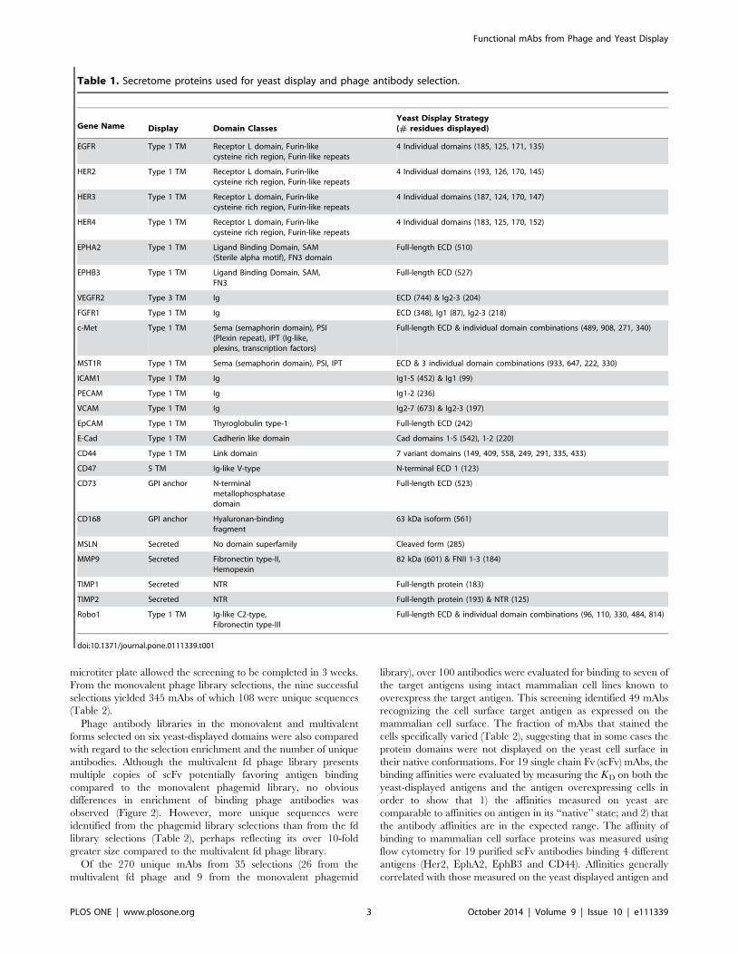
displayed on the surface of yeast (Table 1). These included 14
different single domain classes, multiple domains, and full-length
extracellular domains (ECD). Domains were identified based on
experimentally determined or homology-modeled structures, and
cloned into the yeast display vector pYD2. Display was induced
and quantitated using the SV5 tag at the C-terminus of the
displayed protein with 86% of the proteins showing detectable
display above the background (Fig. 1a). The display level was
weakly inversely correlated with the protein size (Fig. 1a). Some
protein domains were poorly expressed, such as the semaphorin
domain of c-Met.
Selection of phage single chain (scFv) antibody library onyeast displayed proteins
To determine whether phage antibodies could be successfully
selected on yeast-displayed antigens, a naı̈ve human scFv
multivalent phage display library [29,30] was selected on each of
the 60 yeast-displayed protein domains. After three rounds of
selection, the polyclonal phage outputs were analyzed for binding
to the target protein domains displayed on yeast cells by flow
cytometry and a percentage of antigen binding phage above 10%
used as an indicator of successful antibody selection. Of 60
selections, 49 specifically bound the yeast displayed target antigen
compared to the un-induced yeast cells (Fig. 1b, Fig. 2a). On
average, 47% of the polyclonal phage from the selections bound
the yeast-displayed antigens, with a range of 11% to 77%. Of the
failed selections, 9 were from proteins that were poorly displayed.
Since most proteins were represented by more than one domain,
there was at least one domain from each of the 24 target antigens
that displayed and resulted in antigen binding phage antibodies.
The exception was the antigen NRP1, which was only displayed as
full-length ECD. The correlation between protein density on the
yeast surface and the percentage of target binding phage suggested
that display levels greater than 50,000 copies per yeast cell are
required for successful phage selections. However, good display
was insufficient by itself, since some of the better-displayed protein
domains failed to generate Abs, such as FGFR1 Ig domain 1,
NRP1, and VEGFR2 Ig domain 2&3 (Fig. 1b).
A naı̈ve scFv phage display library in the monovalent format
(scFv) was also selected on 9 of the yeast-displayed protein
domains, including ErbB2 domain1, EphA2 ECD, PECAM
domain 1-2, MMP9 FN domain 1-3, and 5 CD44 variants (link,
H, v6, v67, and v47) (Fig. 2b). All nine selections yielded an
antigen binding polyclonal phage population.
Characterization of phage antibodiesFrom the multivalent phage library selections, the initial
screening of 96 random clones from each of the 26 polyclonal
phage outputs yielded 1152 mAbs that bound the yeast-displayed
antigens. DNA sequencing revealed 162 unique sequences for Abs
binding the 26 yeast-displayed domains (Table 2). Flow cytometry
analysis and RCA amplification for DNA sequencing using 96-well
Figure 1. Yeast surface display of proteins and results of phage antibody library selections. A) The density of each protein domain on theinduced yeast surface was quantitated and plotted against the domain size; B) The percentage of the polyclonal phage output binding the targetantigen is shown as a function of antigen receptor density on the yeast surface.doi:10.1371/journal.pone.0111339.g001
Functional mAbs from Phage and Yeast Display
PLOS ONE | www.plosone.org 2 October 2014 | Volume 9 | Issue 10 | e111339
microtiter plate allowed the screening to be completed in 3 weeks.
From the monovalent phage library selections, the nine successful
selections yielded 345 mAbs of which 108 were unique sequences
(Table 2).
Phage antibody libraries in the monovalent and multivalent
forms selected on six yeast-displayed domains were also compared
with regard to the selection enrichment and the number of unique
antibodies. Although the multivalent fd phage library presents
multiple copies of scFv potentially favoring antigen binding
compared to the monovalent phagemid library, no obvious
differences in enrichment of binding phage antibodies was
observed (Figure 2). However, more unique sequences were
identified from the phagemid library selections than from the fd
library selections (Table 2), perhaps reflecting its over 10-fold
greater size compared to the multivalent fd phage library.
Of the 270 unique mAbs from 35 selections (26 from the
multivalent fd phage and 9 from the monovalent phagemid
library), over 100 antibodies were evaluated for binding to seven of
the target antigens using intact mammalian cell lines known to
overexpress the target antigen. This screening identified 49 mAbs
recognizing the cell surface target antigen as expressed on the
mammalian cell surface. The fraction of mAbs that stained the
cells specifically varied (Table 2), suggesting that in some cases the
protein domains were not displayed on the yeast cell surface in
their native conformations. For 19 single chain Fv (scFv) mAbs, the
binding affinities were evaluated by measuring the KD on both the
yeast-displayed antigens and the antigen overexpressing cells in
order to show that 1) the affinities measured on yeast are
comparable to affinities on antigen in its ‘‘native’’ state; and 2) that
the antibody affinities are in the expected range. The affinity of
binding to mammalian cell surface proteins was measured using
flow cytometry for 19 purified scFv antibodies binding 4 different
antigens (Her2, EphA2, EphB3 and CD44). Affinities generally
correlated with those measured on the yeast displayed antigen and
Table 1. Secretome proteins used for yeast display and phage antibody selection.
Gene Name Display Domain ClassesYeast Display Strategy(# residues displayed)
EGFR Type 1 TM Receptor L domain, Furin-likecysteine rich region, Furin-like repeats
4 Individual domains (185, 125, 171, 135)
HER2 Type 1 TM Receptor L domain, Furin-likecysteine rich region, Furin-like repeats
4 Individual domains (193, 126, 170, 145)
HER3 Type 1 TM Receptor L domain, Furin-likecysteine rich region, Furin-like repeats
4 Individual domains (187, 124, 170, 147)
HER4 Type 1 TM Receptor L domain, Furin-likecysteine rich region, Furin-like repeats
4 Individual domains (183, 125, 170, 152)
EPHA2 Type 1 TM Ligand Binding Domain, SAM(Sterile alpha motif), FN3 domain
Full-length ECD (510)
EPHB3 Type 1 TM Ligand Binding Domain, SAM,FN3
Full-length ECD (527)
VEGFR2 Type 3 TM Ig ECD (744) & Ig2-3 (204)
FGFR1 Type 1 TM Ig ECD (348), Ig1 (87), Ig2-3 (218)
c-Met Type 1 TM Sema (semaphorin domain), PSI(Plexin repeat), IPT (Ig-like,plexins, transcription factors)
Full-length ECD & individual domain combinations (489, 908, 271, 340)
MST1R Type 1 TM Sema (semaphorin domain), PSI, IPT ECD & 3 individual domain combinations (933, 647, 222, 330)
ICAM1 Type 1 TM Ig Ig1-5 (452) & Ig1 (99)
PECAM Type 1 TM Ig Ig1-2 (236)
VCAM Type 1 TM Ig Ig2-7 (673) & Ig2-3 (197)
EpCAM Type 1 TM Thyroglobulin type-1 Full-length ECD (242)
E-Cad Type 1 TM Cadherin like domain Cad domains 1-5 (542), 1-2 (220)
CD44 Type 1 TM Link domain 7 variant domains (149, 409, 558, 249, 291, 335, 433)
CD47 5 TM Ig-like V-type N-terminal ECD 1 (123)
CD73 GPI anchor N-terminalmetallophosphatasedomain
Full-length ECD (523)
CD168 GPI anchor Hyaluronan-bindingfragment
63 kDa isoform (561)
MSLN Secreted No domain superfamily Cleaved form (285)
MMP9 Secreted Fibronectin type-II,Hemopexin
82 kDa (601) & FNII 1-3 (184)
TIMP1 Secreted NTR Full-length protein (183)
TIMP2 Secreted NTR Full-length protein (193) & NTR (125)
Robo1 Type 1 TM Ig-like C2-type,Fibronectin type-III
Full-length ECD & individual domain combinations (96, 110, 330, 484, 814)
doi:10.1371/journal.pone.0111339.t001
Functional mAbs from Phage and Yeast Display
PLOS ONE | www.plosone.org 3 October 2014 | Volume 9 | Issue 10 | e111339
Figure 2. Selection enrichment demonstrated by flow cytometry analysis of polyclonal phage binding to target antigen expressingyeast cells. Binding of fifteen polyclonal phage Ab outputs (A) and nine phagemid Ab outputs (B) from the first, second and third round of selectioncompared to binding to an irrelevant yeast displayed antigen. The antigen used for selection is shown above each dot-plot.doi:10.1371/journal.pone.0111339.g002
Functional mAbs from Phage and Yeast Display
PLOS ONE | www.plosone.org 4 October 2014 | Volume 9 | Issue 10 | e111339

12 of them were less than 30 nM (Fig. 3). The correlation
coefficient for these two measurements was 0.47. Variations in
some KD values may reflect the conformational differences
between the epitopes displayed on yeast surface and that on the
cancer cells (Fig. 3).
Validation of antibodiesOf the unique mAbs generated, we examined those binding
CD44 in detail due to the importance of this antigen in tumor
biology. Expression of multiple isoforms of CD44 has been
identified in tumors [31–38]. To distinguish mAbs that are
selective for different CD44 isoforms, we displayed 5 splice
variants (CD44 V6, CD44 V7, CD44 V67, CD44 V4-7, CD44
V3-10), and 2 standard forms (CD44 link domain and CD44 H)
on the yeast surface (Figure 4a). These yeast displayed CD44
forms were used for Ab library selection, mAb screening, as well as
evaluation of Ab binding specificities and affinities. MAbs binding
specifically to the V6, V7, or the constant domains with affinities
ranging from 7 to 482 nM were identified from the phagemid
library (Figure 4b). The CD44 domain binding profile suggested
that antibody V6-2C5, V47-2B8, V47-2B12 bound CD44 v6,
Table 2. Results of phage antibody selections.
Multivalent phagelibrary Domain
Binding Abs(per 95 screened)
Unique mAbsidentified Cell binding (cell line and number of mAbs)
EGFR III 62 1 A431 (1)
IV 54 2 A431 (2)
ErbB2 I 90 2 AU565 (2)
II 15 1 AU565 (1)
III 11 1 AU565 (1)
IV 5 6 AU565 (1)
ErbbB3 IV 59 12 N.D.
ErbbB4 IV 42 10 N.D.
EphA2 ECD 50 3 MDAMB231 (3)
EphB3 ECD 53 10 MDAMB453 (5)
VEGFR2 ECD 10 4 N.D.
FGFR1 Ig2-3 78 4 N.D.
ECD 57 9 N.D.
c-Met PSI-IPTs 16 4 MDAMB231 (2)
NRP1 ECD 20 2 N.D.
ICAM1 Ig1 76 14 SUM159PT (5)
Ig1-5 30 6 SUM159PT (3)
PECAM 1 Ig1-2 21 3 N.D.
EpCAM ECD 67 13 N.D.
E-Cad Cad1-2 2 2 N.D.
CD44 Link 60 6 MDAMB231 (1)
H 76 2 N.D.
CD47 ECD1 60 16 N.D.
MMP9 FN1-3 51 16 N.D.
ECD 22 N.D.
hFc CH2-CH3 65 13 N.D.
Monovalent phage library
ErbB2 I 63 8 SKBR3 (5)
EphA2 ECD 57 18 MDAMB231 (10)
CD44 Link 16 2 MDAMB231 (2)
H 40 6 MDAMB231 (1)
V6 11 5 JIMT (1)
V67 34 13 JIMT (1)
V47 48 29 JIMT (4)
MMP9 FN1-3 50 17 N.D.
PECAM 1 Ig1-2 26 10 N.D.
N.D. This test was not done.Antigen and antigen domain used for selection are indicated along with the number of binding mAbs, the number of unique mAbs, and the number of mAbs bindingmammalian cells expressing the target antigen. The specific cell line used is also indicated.doi:10.1371/journal.pone.0111339.t002
Functional mAbs from Phage and Yeast Display
PLOS ONE | www.plosone.org 5 October 2014 | Volume 9 | Issue 10 | e111339

V47-2A6 bound CD44 v7, and V47-2G10 bound the CD44
standard form at the membrane proximal region.
The anti-CD44 phage antibodies with the highest affinities were
converted to full length IgG which were used to stain breast cancer
cell lines (Figure 5), and to detect specific variant in the breast
tumor sections (Figure 6). Both MDA-MB-231 and JIMT-1 [39]
are known to express CD44 at high level, and MDA-MB-453 cells
express lower levels of CD44 [40]. The V6 specific mAb 2B12
showed membrane binding on JIMT-1 cells but not MDA-MB-
231 or MDA-MB-453 cells. The link domain specific mAb F2-1A6
showed positive binding on both JIMT-1 and MDA-MB-231, but
not MDA-MB-453 cells, consistent with prior flow cytometry
analysis [23]. The CD44-H specific mAb showed the strongest
signal, while the V7 specific mAb showed a weaker signal, and
both antibodies showed some intracellular signal in MDA-MB-453
cells.
Breast tumor sections stained with three antibodies, 2A6, 2B12,
and 2G10, also demonstrated unique CD44 staining patterns. The
V7 and H specific Abs showed diffusing staining with epithelial
cells including the normal mammary duct, the V6 specific 2B12
antibody stained a subset of the tumor epithelial cells with a
distinctive membrane pattern (Figure 6). The biological relevance
of V6 within the tumor tissue is unknown.
Other cell binding antibodies were also validated for cell
staining and internalization. Ab internalization was evaluated
using the chelated ligand-mediated internalization assay (CLIA).
Specifically, the histidine-tagged scFv coupled with the Ni-NTA
coated fluorescent liposome bound the target receptor on the
intact mammalian cell surface at 4uC compared to that
internalized into the cells at 37uC. The surface bound liposomes
were removed by imidazole wash showing no fluorescence signal
left for the 4uC incubation group, while the internalized liposomes
remained for the 37uC incubation group (Figure 7). The ratio of
fluorescence between the imidazole and the PBS wash reflected
the fraction of internalized Abs. Both monoclonal scFv antibodies
to ErbB2 domain 1 and EphA2 ECD were internalized into
SKOV3 and MDA-MB-231 cells, respectively (Figure 7). Some
mAbs showed comparable internalization to mAbs F5 and D2-
1A7, which bound ErbB2 and EphA2, respectively, but were
isolated from selection for internalization into intact cells [23,41].
Breast cancer cell lines known to express the target antigens
ErbB2, EphA2, EphB3, and CD44 with high and low levels were
stained with the identified phage mAbs and evaluated by
fluorescence microscopy. Consistent with the gene expression
data [40], EphB3 antibodies stained MDA-MB-453 cells, CD44
and EphA2 antibodies were positive on MDA-MB-231, and the
ErbB2 domain 1 binders showed intensive intracellular staining of
ErbB2 positive AU565 cells (Figure 8).
Discussion
We have demonstrated that antigen yeast display combined
with Ab phage display is a rapid method to generate highly specific
mAbs to secretome proteins that recognize native proteins.
Previously, we showed that the extracellular domains of EphA2
and CD44 can be displayed on the yeast cell surface and used to
identify mAbs from a phage antibody library pre-selected on
breast cancer cells overexpressing EphA2 and CD44 [23]. Here,
we demonstrate the generality of this approach using un-selected
naı̈ve phage and phagemid antibody libraries. The approach
presented here could be amenable to automation allowing high
throughput antibody selection.
Yeast antigen display proved to be a robust system to display
protein domains for antibody selection and screening based on the
range of proteins we tested. The advantages over other antigen
production system include 1) easy and fast cloning of antigen using
gap repair, 2) simultaneous display of multiple domains, 3) high
density of homogeneous antigens on cell surface, and 4) relatively
clean background for antibody selection. Potential limitations of
this method are the non-natural epitope space caused by
mannosylation [42,43] and inability to express multi-pass trans-
membrane proteins and GPI-linked membrane proteins.
The expression level of antigen domains was weakly correlated
with the antigen size and more strongly correlated with the protein
fold and the structure. Generally, the Ig fold and the Fn3 fold were
better displayed in the yeast expression system, while the sema
domain was difficult to display. Expression could potentially be
Figure 3. Binding affinities of scFv antibodies. The KD of purified scFv antibody was measured on yeast displayed antigen domains andcompared with the KD for binding to antigen (Ag) expressing mammalian cells by flow cytometry. scFv mAbs were incubated with the yeast cellstransformed and induced for antigen expression, or with the antigen overexpressing cancer cells; SKOV3 cells for HER2 mAbs, MDA-MB-231 for EphA2and CD44 mAbs, Colo205 for EphB3 mAbs.doi:10.1371/journal.pone.0111339.g003
Functional mAbs from Phage and Yeast Display
PLOS ONE | www.plosone.org 6 October 2014 | Volume 9 | Issue 10 | e111339
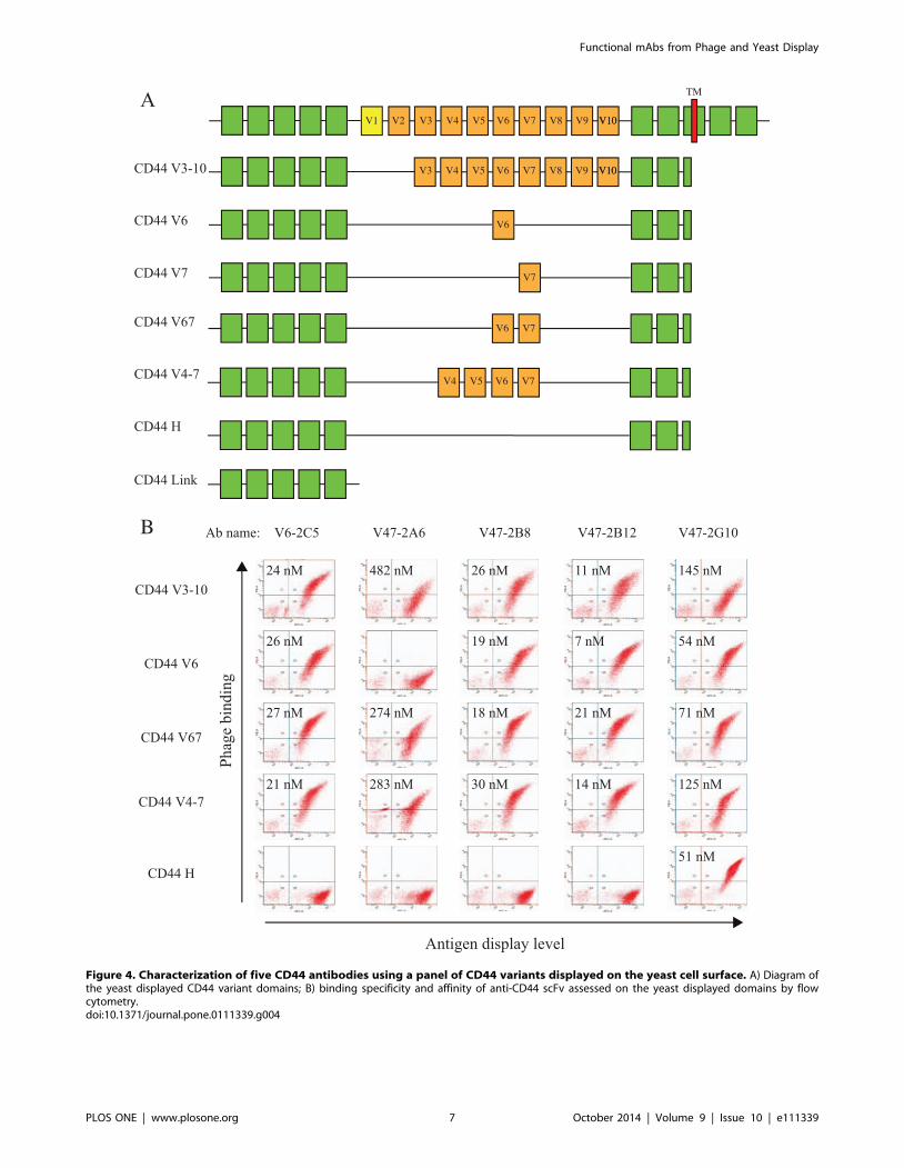
Figure 4. Characterization of five CD44 antibodies using a panel of CD44 variants displayed on the yeast cell surface. A) Diagram ofthe yeast displayed CD44 variant domains; B) binding specificity and affinity of anti-CD44 scFv assessed on the yeast displayed domains by flowcytometry.doi:10.1371/journal.pone.0111339.g004
Functional mAbs from Phage and Yeast Display
PLOS ONE | www.plosone.org 7 October 2014 | Volume 9 | Issue 10 | e111339

improved with codon and domain optimization, changing the
orientation of Aga2, or by including yeast chaperones [44].
The number of phage antibodies generated was directly
correlated with the display level of the antigen, but not associated
with the antigen size. A higher number of unique mAbs seemed to
be generated from the phagemid library compared to the phage
Ab library, suggesting that some Ab sequences were lost during the
construction of the smaller phage library by sub-cloning from the
phagemid library [29,30].
A number of the mAbs we generated were able to recognize the
native antigen as presented on the cell surface. Interestingly, we
identified identical mAbs that we previously identified using direct
selection on intact cells, namely F5 and D2-1A7, which binds
HER2 and EphA2, respectively [23,41]. This observation
confirms the native structure of the antigen as it is presented on
yeast and suggests similar selection pressures.
The yeast display of antigen domains allowed flexible, rapid,
and easy expression of multiple antigen domains as well as direct
affinity assessment by flow cytometry [28], which proved
extremely useful for CD44 splice variants where the soluble
proteins are not available. We displayed seven different variant
forms of CD44 on the yeast surface, and completed the phage
antibody library selection and screening within a month. The
resulting mAbs have affinities ranging from 7 nM to 482 nM,
were differentially specific to different domains, and could stain
breast tumor sections.
MAb internalization via receptor-mediated endocytosis was
evaluated for HER2 and EphA2 mAbs; the majority of mAbs to
the HER2 domain 1 Abs and EphA2 Abs demonstrated
intracellular staining as determined by CLIA [45] and fluorescent
microscopy. The results indicate that the functionality of mAbs are
associated with the methods of Ab selection, and can also be
approached by using the correct epitope domain of the antigens,
such as the HER2 domain 1.
The described approach may be challenging in the absence of
knowledge of a target’s domain structure. However, the yeast
system allowed us to rapidly display multiple domains with
different starting and ending amino acids to search for the optimal
domains. We also noticed that some antigen epitopes are difficult
to recapitulate on the yeast cells as exemplified by the failure of
Figure 5. Immunohistochemistry staining of breast cancer cell buttons using CD44 mAbs. Breast cancer cell lines with known CD44expression levels were processed to make paraffin embedded cell buttons, and used to evaluate anti-CD44 mAb staining patterns. Abs studiedincluded V7 specific mAb 2A6, V6 specific mAb 2B12, CD44 standard form binding mAb 2G10, and CD44 link domain specific mAb F2-1A6.doi:10.1371/journal.pone.0111339.g005
Figure 6. Breast tumor tissue array staining by immunohistochemistry with CD44 mAbs. A tissue array containing samples from 8different patients (numbered 1–8) was evaluated after staining with different CD44 mAbs. A) V7 specific mAb 2A6, B) V6 specific mAb 2B12, C) CD44standard from binding mAb 2G10, D) an enlarged view of tissue #3 stained with 2B12 showing distinct membrane bound CD44v6 in the center of asubset of cancerous cells. Magnification is 400x, and the scale bar is 50 mm.doi:10.1371/journal.pone.0111339.g006
Functional mAbs from Phage and Yeast Display
PLOS ONE | www.plosone.org 8 October 2014 | Volume 9 | Issue 10 | e111339

mAbs isolated by using recombinant antigens to bind the yeast
displayed domains. Such epitopes will definitely be missed using
this approach.
We conclude that the yeast display of antigen domains and
domain combinations are useful for isolating recombinant
antibodies from naı̈ve antibody libraries in a short time period
starting from the antigen sequences. This platform will allow Ab
discovery for a large function of the antigen epitope space, but not
all the epitopes, which will need other approaches to complete Ab
discovery.
Materials and Methods
Cell lines, media, antibodies and full-length cDNA clonesBreast cancer cell lines A431, AU565, Colo205, JIMT-1, MDA-
MB-231, MDA-MB-453, MDA-MB-468, SKBR3, SKOV3,
SUM159PT and HEK293A cells were obtained from the
American Type Culture Collection (ATCC). The cell lines were
cultured using conditions described previously [46]. The yeast
strain EBY100 was grown in YPD medium (Current Protocols inMolecular Biology, John Wiley and Sons, Chapter 13.1.2).
EBY100 was transfected with expression vector pYD2 [47] and
was selected on SD-CAA medium [48] (Current Protocols,Chapter 13). The Aga2p antigen fusion was expressed on the
yeast surface by induction in SG-CAA medium (identical to SD-
CAA medium except the glucose is replaced by galactose) at 18uC
for 24–48 hr as described previously [49]. E. coli strains DH5aand TG1 were used for the preparation of plasmid DNA and the
expression of soluble scFv antibodies respectively. SV5 antibody
was purified from hybridoma supernatant using Protein G and
directly labeled with Alexa-488 or Alexa-647 using a kit provided
by the manufacturer (Invitrogen; Carlsbad, CA). Biotin conjugated
rabbit anti-fd bacteriophage (Sigma), biotin conjugated anti-rabbit
(Vector Labs), Streptavidin Phycoerythrin (PE) (Biosource/Invi-
trogen), streptavidin Texas Red (GE Healthcare), streptavidin
HRP conjugate were used to detect antibodies. The full-length
cDNAs were obtained from the ATCC and Open Biosystems.
Cloning of protein domain coding genes into the yeastdisplay vector
For antigen display, the cDNA encoding the full length antigen
or antigen domain was amplified by PCR using primers that
anneal to the 59 and 39 ends of the antigen cDNA and with 25
nucleotide overlaps with the yeast display vector pYD2 digested
with the restriction enzymes NcoI and NotI. The antigen DNA
fragment was then cloned into NcoI-NotI-digested pYD2 vector
by using gap repair [50,51]. Gap repair allows cloning of virtually
all cDNA fragments including those with internal digestion sites.
Primers for cloning the target antigen gene by gap repair:
N Ag-Gap5 primer 59-GTTGTTCTGCTAGCGGGGC-CATGG---39
Figure 7. Internalization of mAbs into cancer cells as determined by chelated ligand-mediated internalization assay (CLIA). Ni-NTAliposomes loaded with dye were internalized into cells mediated by hexa-histidine tagged scFv mAbs prebound to the cell surface at 37uC comparedto 4uC. Imidazole buffer was used to remove the surface bound dye-loading liposomes by disrupting the Ni-NTA and hexa-Histidine interaction. A)Internalization of ErbB2 mAbs into ErbB2 expressing SKBR3 cells; B) internalization of EphA2 mAbs into EphA2 expressing MDA-MB-231 cells.doi:10.1371/journal.pone.0111339.g007
Figure 8. Binding of mAbs to cancer cell lines that overexpress the target antigens as determined by fluorescent microscopy. A)EphB3 mAbs showed positive staining on EphB3 overexpressing cell MDA-MB-453, B) CD44 and EphA2 mAbs showed positive staining on MDA-MB-231 cells, C) ErbB2 domain 1 binding mAbs showed positive staining on the ErbB2 overexpressing cell AU565.doi:10.1371/journal.pone.0111339.g008
Functional mAbs from Phage and Yeast Display
PLOS ONE | www.plosone.org 9 October 2014 | Volume 9 | Issue 10 | e111339

N Ag-Gap3 primer 59-TTCGAAGGGCCCGCCTGCGGC-CGC---39
The 59 sequence indicated anneals to Nco1-Not1 digested
pYD2 DNA for cloning by gap repair, the bolded sequences are
the Nco1 and Not1 sites. The dashed sequence should include
approximately 24 nucleotides that anneal to the 59 and 39 ends of
the target antigen DNA.
Display of protein domains on the surface of yeast cellsMultiple target genes were amplified by PCR in 96-well plates
using high-fidelity DNA polymerase, and used to transform LiAc
treated EBY100 cells together with NcoI/NotI digested vector
pYD2 using the TRAFO method with gap repair in 1 ml 96-well
plate [50,51]. Specifically, TRAFO mix including the vector with
large enough quantity for 96 transformation was prepared and
used to resuspend the EBY100 yeast cells followed by aliquoting
359 ml of TRAFO-treated cells into each well that contains 1 ml of
the target gene PCR fragment. After incubation for 30 min at
30uC followed by 45 min at 42uC, the cells were pelleted by
centrifugation at 3000 rpm for 2 min, the transformation mix
removed, cells washed once in 1 ml of sterile water, 1/10 of the
cells transferred to a new 1 ml deep 96-well plate containing
600 ml of SD-CAA media, and cultured for 24 hrs at 30uC.
Dilution of untransformed yeast cells was repeated by growing 1/
10 of 24 hr culture in a new 1 ml 96-well plate containing 600 ml
of SD-CAA media followed by induction culture in SG-CAA
media for 24 hrs at 18uC.
Quantitation of protein density on yeast surface by flowcytometry
A fraction of the induced yeast cells were transferred into a V-
bottom 96-well plate, washed once by resuspending in 100 ml of
FACS buffer, followed by centrifugation, aspiration of the
supernatant, resuspension in 50 ml of FACS buffer, and stained
by incubation with 50 ml of 1 mg/mL Alexa-647 labeled anti-SV5
IgG diluted in FACS buffer for 1 hr at 4uC. After washing the
stained yeast cells once in 200 ml of FACS buffer, the cell
fluorescence was measured in a FACS LSRII flow cytometer using
QuantumTM Alexa Fluor 647 bead (Bangs Laboratories Inc.) as
reference to calculate the protein density on each cell.
Selection of phage antibody library against yeastdisplayed protein domains
A phage display naı̈ve human single chain library was used for
target specific antibody selections [29,30]. The induced yeast cells
displaying an human Fc domain were used to deplete the non-
specific binders by incubating 161013 phage particles with 109
yeast cells for 2 hr at 4uC. The filtered supernatant containing the
depleted phage library was then aliquoted into each well of 1 ml
96-well plate containing about 16107 yeast cells displaying
individual target protein domains for 1 h at 4uC. Yeast cells were
washed ten times with cold PBS and pelleted by centrifugation.
The bound phage antibodies were eluted by incubating yeast cells
with 100 ml of 100 mM triethylamine, neutralized with 50 ml of
1 M Tris-HCl (pH 7.4), and used to infect exponentially growing
E. coli TG1 as described previously [52]. Specifically, half of the
phage eluent was transferred to a new 1 ml 96-well plate,
incubated with 1 ml of E. coli TG1 cells for 30 min at 37uC,
and 1/10 of the infected TG1 transferred to a new 1 ml 96-well
plate containing 2YT/Amp/2% glucose, and cultured for 16 hrs
at 30uC. A small fraction of the overnight culture was transferred
to another 1 ml 96-well plate containing 150 ml of 2YT/Amp/
0.1% glucose using 96-pin transfer device followed by culture for
2.5 hr at 37uC, infection with VCSM13 for 30 min at 37uC, and
culture in additional of 450 ml of 2YT/Amp for 16 hrs at 30uC. In
the case of fd phage library, the TG1 infected phage eluent was
cultured in 2YT/Tet for 16 hrs at 30uC.
The resulting phage output of each target was harvested,
precipitated using 20% PEG6000/1.5 M NaCl, resuspended in
100 ml of cold PBS, and subjected to another round of selection to
the corresponding target protein domain.
For the second round of selection, the 100 ml of output phage
from the round 1 selection was depleted in 1 ml 96-well plate by
incubating with 108 yeast cells displaying an human Fc domain for
2 hr at 4uC followed by centrifugation to pellet the yeast cells. The
supernatant of each well was transferred to a new 1 ml 96-well
plate, and mixed with 16107 of yeast cells displaying the
corresponding target protein domains for 1 h at 4uC. The rest
of the selection procedure was the same as the first round. If
needed, the third round of selection can be performed following
the same procedure as the second round of selection. When the
selection was completed, each selection output can be plated for
monoclonal phage analysis.
Evaluation of phage antibody binding to yeast-displayedprotein domains
Polyclonal phage antibodies from each round of selections were
evaluated by flow cytometry for binding to the corresponding
target protein domains displayed on yeast cells. In detail, the
supernatant of phage culture in 96-well plate was transferred to V-
bottom 96-well plate and incubated with yeast cells displaying the
corresponding target antigen for 2 hrs at 4uC. The bound phage
antibodies were detected by incubating cells with biotin conjugat-
ed anti-fd antibody (1 mg/ml) for 30 min at 4uC and streptavidin-
PE followed by flow cytometry analysis. The binding of phage
antibodies to the yeast displayed target antigens were represented
by the percentile of yeast population in the Q2 quadrant of the dot
plot.
Characterization of phage antibodiesAfter two or three rounds of selection, 96 individual colonies
from each target specific selection were isolated and the phage
prepared by growing the single colonies in 96-well microtiter
plates as described [53]. Binding of each phage antibody to yeast
displayed antigen was determined by incubation of 105 yeast cells
with 100 ml phage supernatant diluted in FACS buffer (PBS with
1 mM MgCl2, 0.1 mM CaCl2 and 0.3% BSA) for 2 h at 4uC in
conical 96-well microtiter plates, followed by incubation with
biotinylated anti-fd antibody and streptavidin-phycoerythrin
conjugate, and analyzed using a FACS LSRII (Becton Dickinson).
The number of unique phage antibodies was determined by 96-
well RCA reaction followed by DNA sequencing.
Expression of soluble antibodies as scFv and IgGThe scFv coding genes from the identified phage antibodies
were subcloned from the phage-display pHEN1 [52] via NcoI-
NotI into expression vector pSyn1 for scFv expression [54], or
AbVec with rabbit Fc for IgG expression [55]. scFvs were
produced in the periplasm of E. coli strain TG1, and purified by
osmotic shock and immobilized metal affinity chromatography, as
reported previously [54]. IgG proteins were secreted in the media
by transfected HEK293A cells as described elsewhere [55], and
purified by Protein A affinity chromatography (GE Healthcare).
The homogeneity and purity of the protein preparations were
verified by SDS-PAGE stained with coomassie blue; protein
Functional mAbs from Phage and Yeast Display
PLOS ONE | www.plosone.org 10 October 2014 | Volume 9 | Issue 10 | e111339

concentrations were measured by micro-bicinchoninic acid assay
(Pierce, Rockford, IL).
Binding affinity assay by flow cytometryEach scFv was incubated with 16105 yeast cells induced to
express the target protein domain for 1 hr-16 hrs at the indicated
concentration, or the target antigen overexpressing cancer cells
[28,56]. Cell binding was performed at 4uC in PBS containing
0.3% BSA in volume sufficient to maintain constant antibody
concentration for equilibrium conditions. After two washes with
200 ml of PBS, bound scFv was detected by the addition of 100 ml
(1 mg/ml) of biotinylated His probe (Santa Cruz Biotech.) and
streptavidin-PE (Biosource/Invitrogen), and the yeast displayed Ag
co-stained with Alexa 647 labeled anti-SV5. After incubating
30 minutes at 4uC, the cells were washed twice and resuspended in
PBS containing 4% paraformaldehyde. Fluorescence was mea-
sured by flow cytometry in a FACS LSRII (Becton Dickinson), and
median fluorescence intensity (MFI) was calculated using Cellquest
software (Becton Dickinson). Equilibrium constants were deter-
mined as described [57], except that values were fitted to the
equation MFI = MFImin + MFImax* [Ab]/(KD+[Ab]) using
Kaleidagraph (Synergy Software).
Antibody internalization assayThe anti-ErbB2 and anti-EphA2 scFv antibodies were evaluated
for the ability to internalize into human breast cancer cells SKBR3
and MDA-MB-231 cells, respectively, by chelated ligand-mediated
internalization assay (CLIA) as described previously [45]. Briefly,
the cells were grown to 80–90% confluence in the media
recommended by ATCC supplemented with 10% fetal calf serum
(FCS) and harvested by trypsinization, seeded at 10,000/well in
96-well plates, and incubated overnight at 37uC. The next day, Ni-
NTA liposomes (0–1 mM total phospholipid) were incubated for
4 hrs with the cells along with the (His) 6-containing scFv (20 mg/
mL unless otherwise indicated) in 100 mL tissue culture media
supplemented with 10% FCS under four different conditions. The
conditions are 37uC followed by PBS wash, 37uC with 250 mM
phosphate buffered imidazole wash, 4uC with PBS wash, and 4uCwith 250 mM phosphate buffered imidazole wash. Cells were then
trypsinized, washed, and the fluorescence measured using a FACS
LSRII (Becton Dickinson).
ImmunohistochemistryThree breast cancer cell lines, MDA-MB-231, MDA-MB-453,
and JIMT-1, were used to make paraffin embedded cell button at
UCSF tissue core, and stained with CD44 antibodies following
standard IHC protocol.
De-identified human breast tumor tissue samples were obtained
from University of California, San Francisco, Department of
Pathology archives. Samples were collected with consent under
UCSF IRB# 11-06720 ‘‘Retrospective Study of Adjunctive
Pathologic Diagnostic Features, Biomarkers Expression, and
Molecular Alterations in Early Breast Neoplasms and in Morpho-
logic Mimics’’. The UCSF Mount Zion Panel Institutional Review
Board approved the use of this archived patient tissue samples in
research and waived the need of consent.
Breast tumor tissue samples were arrayed, and cut to five-
micrometer sections. After antigen retrieval, the samples were
stained with three different anti-CD44 IgG antibodies with rabbit
Fc, 2A6, 2B12, and 2G10, respectively, followed by staining with
biotin-conjugated anti-rabbit secondary antibody (Vector Labs
Co.), ABC complex (Vector Labs Co.), and standard DAB color
development (Vector Labs Co.) sequentially. A pathologist (YYC)
evaluated the staining result.
ImmunofluorescenceMDA-MB-231, MDA-MB-453, MDA-MB-468, and AU565
cells were grown on coverslips to 70% of confluence in 12 well-
plates and incubated with 1011 phage antibodies for three hours at
37uC. The coverslips were washed once with PBS, twice with PEM
(80 mM Potassium PIPES (pH 6.8), 5 mM EGTA (pH 7), 2 mM
MgCl2), and fixed with PEM containing 4% (W/V) paraformal-
dehyde for 30 min on ice. Cells were quenched with 0.1 M
NH4Cl, permeabilized with 0.5% Triton X-100, and blocked with
5% non-fat dry milk in TBS-T buffer overnight at 4uC. After
blocking endogenous biotin with Avidin-Biotin Kit (Lab Vision),
intracellular phages were detected with biotinylated anti-fd
polyclonal antibody (Sigma) and streptavidin Texas Red (GE
healthcare). Coverslips were inverted on a slide on mounting
medium and microscopic images were taken with a Zeiss
fluorescence microscope (Zeiss, Germany).
Acknowledgments
Research reported in this publication was supported by NCI of the
National Institutes of Health under award number P50 CA58207.
Author Contributions
Conceived and designed the experiments: YZ JDM. Performed the
experiments: ZS HZ LQ LZ JZ YZ YYC. Analyzed the data: YZ JDM
YYC. Contributed reagents/materials/analysis tools: YYC. Contributed to
the writing of the manuscript: YZ JDM.
References
1. Isserlin R, Emili A (2007) Nine steps to proteomic wisdom: A practical guide to
using protein-protein interaction networks and molecular pathways as a
framework for interpreting disease proteomic profiles. Proteomics Clin Appl 1:
1156–1168.
2. Grimsey NL, Goodfellow CE, Scotter EL, Dowie MJ, Glass M, et al. (2008)
Specific detection of CB1 receptors; cannabinoid CB1 receptor antibodies are
not all created equal! J Neurosci Methods 171: 78–86.
3. Jensen BC, Swigart PM, Simpson PC (2009) Ten commercial antibodies for
alpha-1-adrenergic receptor subtypes are nonspecific. Naunyn Schmiedebergs
Arch Pharmacol 379: 409–412.
4. Jositsch G, Papadakis T, Haberberger RV, Wolff M, Wess J, et al. (2009)
Suitability of muscarinic acetylcholine receptor antibodies for immunohisto-
chemistry evaluated on tissue sections of receptor gene-deficient mice. Naunyn
Schmiedebergs Arch Pharmacol 379: 389–395.
5. Pozner-Moulis S, Cregger M, Camp RL, Rimm DL (2007) Antibody validation
by quantitative analysis of protein expression using expression of Met in breast
cancer as a model. Lab Invest 87: 251–260.
6. Saper CB (2005) An open letter to our readers on the use of antibodies. J Comp
Neurol 493: 477–478.
7. Michel MC, Wieland T, Tsujimoto G (2009) How reliable are G-protein-
coupled receptor antibodies? Naunyn Schmiedebergs Arch Pharmacol 379:
385–388.
8. Klee EW (2008) The zebrafish secretome. Zebrafish 5: 131–138.
9. Caneparo L, Huang YL, Staudt N, Tada M, Ahrendt R, et al. (2007) Dickkopf-1
regulates gastrulation movements by coordinated modulation of Wnt/beta
catenin and Wnt/PCP activities, through interaction with the Dally-like
homolog Knypek. Genes Dev 21: 465–480.
10. Merritt WM, Sood AK (2007) Markers of angiogenesis in ovarian cancer. Dis
Markers 23: 419–431.
11. Pickart MA, Klee EW, Nielsen AL, Sivasubbu S, Mendenhall EM, et al. (2006)
Genome-wide reverse genetics framework to identify novel functions of the
vertebrate secretome. PLoS One 1: e104.
12. Karagiannis GS, Pavlou MP, Diamandis EP (2010) Cancer secretomics reveal
pathophysiological pathways in cancer molecular oncology. Mol Oncol 4: 496–
510.
Functional mAbs from Phage and Yeast Display
PLOS ONE | www.plosone.org 11 October 2014 | Volume 9 | Issue 10 | e111339

13. Baggetta R, De Andrea M, Gariano GR, Mondini M, Ritta M, et al. (2010) The
interferon-inducible gene IFI16 secretome of endothelial cells drives the earlysteps of the inflammatory response. Eur J Immunol 40: 2182–2189.
14. Xue H, Lu B, Lai M (2008) The cancer secretome: a reservoir of biomarkers.
J Transl Med 6: 52.15. Nelson AL, Dhimolea E, Reichert JM (2010) Development trends for human
monoclonal antibody therapeutics. Nat Rev Drug Discov 9: 767–774.16. Reichert JM (2009) Probabilities of success for antibody therapeutics. MAbs 1:
387–389.
17. Dimitrov DS, Marks JD (2009) Therapeutic antibodies: current state and futuretrends–is a paradigm change coming soon? Methods Mol Biol 525: 1–27, xiii.
18. Wagner S, Bader ML, Drew D, de Gier JW (2006) Rationalizing membraneprotein overexpression. Trends Biotechnol 24: 364–371.
19. Tate CG, Haase J, Baker C, Boorsma M, Magnani F, et al. (2003) Comparisonof seven different heterologous protein expression systems for the production of
the serotonin transporter. Biochim Biophys Acta 1610: 141–153.
20. Ren H, Yu D, Ge B, Cook B, Xu Z, et al. (2009) High-level production,solubilization and purification of synthetic human GPCR chemokine receptors
CCR5, CCR3, CXCR4 and CX3CR1. PLoS One 4: e4509.21. Sarramegna V, Talmont F, Demange P, Milon A (2003) Heterologous
expression of G-protein-coupled receptors: comparison of expression systems
from the standpoint of large-scale production and purification. Cell Mol Life Sci60: 1529–1546.
22. Goding J (1983) Monoclonal Antibodies: principles and practice.23. Zhou Y, Zou H, Zhang S, Marks JD (2010) Internalizing cancer antibodies from
phage libraries selected on tumor cells and yeast-displayed tumor antigens. J MolBiol 404: 88–99.
24. Cochran JR, Kim YS, Olsen MJ, Bhandari R, Wittrup KD (2004) Domain-level
antibody epitope mapping through yeast surface display of epidermal growthfactor receptor fragments. J Immunol Methods 287: 147–158.
25. Johns TG, Adams TE, Cochran JR, Hall NE, Hoyne PA, et al. (2004)Identification of the epitope for the epidermal growth factor receptor-specific
monoclonal antibody 806 reveals that it preferentially recognizes an untethered
form of the receptor. J Biol Chem 279: 30375–30384.26. Piatesi A, Howland SW, Rakestraw JA, Renner C, Robson N, et al. (2006)
Directed evolution for improved secretion of cancer-testis antigen NY-ESO-1from yeast. Protein Expr Purif 48: 232–242.
27. Aggen DH, Chervin AS, Insaidoo FK, Piepenbrink KH, Baker BM, et al. (2010)Identification and engineering of human variable regions that allow expression
of stable single-chain T cell receptors. Protein Eng Des Sel.
28. Levy R, Forsyth CM, LaPorte SL, Geren IN, Smith LA, et al. (2007) Fine anddomain-level epitope mapping of botulinum neurotoxin type A neutralizing
antibodies by yeast surface display. J Mol Biol 365: 196–210.29. Sheets MD, Amersdorfer P, Finnern R, Sargent P, Lindquist E, et al. (1998)
Efficient construction of a large nonimmune phage antibody library: the
production of high-affinity human single-chain antibodies to protein antigens.Proc Natl Acad Sci U S A 95: 6157–6162.
30. Huie MA, Cheung MC, Muench MO, Becerril B, Kan YW, et al. (2001)Antibodies to human fetal erythroid cells from a nonimmune phage antibody
library. Proc Natl Acad Sci USA 98: 2682–2687.31. Khan SA, Cook AC, Kappil M, Gunthert U, Chambers AF, et al. (2005)
Enhanced cell surface CD44 variant (v6, v9) expression by osteopontin in breast
cancer epithelial cells facilitates tumor cell migration: novel post-transcriptional,post-translational regulation. Clin Exp Metastasis 22: 663–673.
32. Al-Hajj M, Wicha MS, Benito-Hernandez A, Morrison SJ, Clarke MF (2003)Prospective identification of tumorigenic breast cancer cells. Proc Natl Acad
Sci U S A 100: 3983–3988.
33. Berner HS, Nesland JM (2001) Expression of CD44 isoforms in infiltratinglobular carcinoma of the breast. Breast Cancer Res Treat 65: 23–29.
34. Yang Q, Liu Y, Huang Y, Huang D, Li Y, et al. (2013) Expression of COX-2,CD44v6 and CD147 and relationship with invasion and lymph node metastasis
in hypopharyngeal squamous cell carcinoma. PLoS One 8: e71048.
35. Shiozaki M, Ishiguro H, Kuwabara Y, Kimura M, Mitsui A, et al. (2011)Expression of CD44v6 is an independent prognostic factor for poor survival in
patients with esophageal squamous cell carcinoma. Oncol Lett 2: 429–434.
36. Zavrides HN, Zizi-Sermpetzoglou A, Panousopoulos D, Athanasas G,
Elemenoglou I, et al. (2005) Prognostic evaluation of CD44 expression in
correlation with bcl-2 and p53 in colorectal cancer. Folia Histochem Cytobiol
43: 31–36.
37. Watanabe O, Kinoshita J, Shimizu T, Imamura H, Hirano A, et al. (2005)
Expression of a CD44 variant and VEGF-C and the implications for lymphatic
metastasis and long-term prognosis of human breast cancer. J Exp Clin Cancer
Res 24: 75–82.
38. Cho EY, Choi Y, Chae SW, Sohn JH, Ahn GH (2006) Immunohistochemical
study of the expression of adhesion molecules in ovarian serous neoplasms.
Pathol Int 56: 62–70.
39. Olsson E, Honeth G, Bendahl PO, Saal LH, Gruvberger-Saal S, et al. (2011)
CD44 isoforms are heterogeneously expressed in breast cancer and correlate
with tumor subtypes and cancer stem cell markers. BMC Cancer 11: 418.
40. Spellman PT (2006) Transcription profiling of 51 human breast cancer cell lines.
41. Poul M-A, Becerril B, Nielsen UB, Morrison P, Marks JD (2000) Selection of
internalizing human antibodies from phage libraries. J Mol Biol 301: 1149–
1161.
42. Gerngross TU (2004) Advances in the production of human therapeutic proteins
in yeasts and filamentous fungi. Nat Biotechnol 22: 1409–1414.
43. Hamilton SR, Bobrowicz P, Bobrowicz B, Davidson RC, Li H, et al. (2003)
Production of complex human glycoproteins in yeast. Science 301: 1244–1246.
44. Robinson AS, Hines V, Wittrup KD (1994) Protein disulfide isomerase
overexpression increases secretion of foreign proteins in Saccharomyces
cerevisiae. Biotechnology (N Y) 12: 381–384.
45. Nielsen UB, Kirpotin DB, Pickering EM, Drummond DC, Marks JD (2006) A
novel assay for monitoring internalization of nanocarrier coupled antibodies.
BMC Immunol 7: 24.
46. Neve RM, Chin K, Fridlyand J, Yeh J, Baehner FL, et al. (2006) A collection of
breast cancer cell lines for the study of functionally distinct cancer subtypes.
Cancer Cell 10: 515–527.
47. Razai A, Garcia-Rodriguez C, Lou J, Geren IN, Forsyth CM, et al. (2005)
Molecular evolution of antibody affinity for sensitive detection of botulinum
neurotoxin type A. J Mol Biol 351: 158–169.
48. (2008) Current Protocols in Molecular Biology: Wiley Interscience.
49. Feldhaus MJ, Siegel RW, Opresko LK, Coleman JR, Feldhaus JM, et al. (2003)
Flow-cytometric isolation of human antibodies from a nonimmune Saccharo-
myces cerevisiae surface display library. Nat Biotechnol 21: 163–170.
50. Gietz RD, Schiestl RH (1991) Applications of high efficiency lithium acetate
transformation of intact yeast cells using single-stranded nucleic acids as carrier.
Yeast 7: 253–263.
51. Orr-Weaver TL, Szostak JW (1983) Yeast recombination: the association
between double-strand gap repair and crossing-over. Proc Natl Acad Sci U S A
80: 4417–4421.
52. Marks JD, Hoogenboom HR, Bonnert TP, McCafferty J, Griffiths AD, et al.
(1991) By-passing immunization. Human antibodies from V-gene libraries
displayed on phage. J Mol Biol 222: 581–597.
53. O’Connell D, Becerril B, Roy-Burman A, Daws M, Marks JD (2002) Phage
versus phagemid libraries for generation of human monoclonal antibodies. J Mol
Biol 321: 49–56.
54. Schier R, Bye J, Apell G, McCall A, Adams GP, et al. (1996) Isolation of high-
affinity monomeric human anti-c-erbB-2 single chain Fv using affinity-driven
selection. J Mol Biol 255: 28–43.
55. Wrammert J, Smith K, Miller J, Langley WA, Kokko K, et al. (2008) Rapid
cloning of high-affinity human monoclonal antibodies against influenza virus.
Nature 453: 667–671.
56. Zhou Y, Goenaga AL, Harms BD, Zou H, Lou J, et al. (2012) Impact of intrinsic
affinity on functional binding and biological activity of EGFR antibodies. Mol
Cancer Ther 11: 1467–1476.
57. Benedict CA, MacKrell AJ, Anderson WF (1997) Determination of the binding
affinity of an anti-CD34 single-chain antibody using a novel, flow cytometry
based assay. J Immunol Methods 201: 223–231.
Functional mAbs from Phage and Yeast Display
PLOS ONE | www.plosone.org 12 October 2014 | Volume 9 | Issue 10 | e111339