high hydrostatic pressure processing: a method having high success potential in pollen protein...
TRANSCRIPT
This article was downloaded by: [University of Oklahoma Libraries]On: 24 April 2013, At: 06:24Publisher: Taylor & FrancisInforma Ltd Registered in England and Wales Registered Number: 1072954 Registeredoffice: Mortimer House, 37-41 Mortimer Street, London W1T 3JH, UK
High Pressure Research: AnInternational JournalPublication details, including instructions for authors andsubscription information:http://www.tandfonline.com/loi/ghpr20
High hydrostatic pressure processing: amethod having high success potential inpollen protein extractionErgin Murat Altuner a , Talip Çeter a & Hami Alpas ba Department of Biology, Kastamonu University, Kastamonu, Turkeyb Department of Food Engineering, Middle East TechnicalUniversity, Ankara, TurkeyVersion of record first published: 12 Apr 2012.
To cite this article: Ergin Murat Altuner , Talip Çeter & Hami Alpas (2012): High hydrostatic pressureprocessing: a method having high success potential in pollen protein extraction, High PressureResearch: An International Journal, 32:2, 291-298
To link to this article: http://dx.doi.org/10.1080/08957959.2012.678341
PLEASE SCROLL DOWN FOR ARTICLE
Full terms and conditions of use: http://www.tandfonline.com/page/terms-and-conditions
This article may be used for research, teaching, and private study purposes. Anysubstantial or systematic reproduction, redistribution, reselling, loan, sub-licensing,systematic supply, or distribution in any form to anyone is expressly forbidden.
The publisher does not give any warranty express or implied or make any representationthat the contents will be complete or accurate or up to date. The accuracy of anyinstructions, formulae, and drug doses should be independently verified with primarysources. The publisher shall not be liable for any loss, actions, claims, proceedings,demand, or costs or damages whatsoever or howsoever caused arising directly orindirectly in connection with or arising out of the use of this material.
High Pressure ResearchVol. 32, No. 2, June 2012, 291–298
High hydrostatic pressure processing: a method having highsuccess potential in pollen protein extraction
Ergin Murat Altunera*, Talip Çetera and Hami Alpasb
aDepartment of Biology, Kastamonu University, Kastamonu, Turkey; bDepartment of Food Engineering,Middle East Technical University, Ankara, Turkey
(Received 09 August 2011; final version received 19 March 2012)
Even a single peptide that is present in the pollen wall and cytoplasm could cause pollen allergy. To produceskin-prick test kits, the first step is the extraction of these molecules. In this study, Cedrus atlantica pollenswere subjected to 220 and 330 MPa for 10 and 30 min in order to extract these molecules. After highhydrostatic pressure processing (HHPP), the total amounts of proteins (TAPs) are measured and comparedwith the results of the conventional extraction method (CEM). As a result, the TAPs extracted by HHPPis 18.0210 μg/mL at 220 MPa for 10 min, 22.5770 μg/mL at 220 MPa for 30 min, 23.3810 μg/mL at330 MPa for 10 min and 25.9270 μg/mL at 330 MPa for 30 min, while this is 1.9460 μg/mL in 24 h by theCEM. In addition to these results, visual pollen deformation and eruption, pollen wall and surface damagehave also been observed.
Keywords: Cedrus atlantica; atlas cedar; high hydrostatic pressure processing; pollen protein extraction
1. Introduction
Cedrus (Cedar) is a genus of coniferous trees in the plant family Pinaceae [1]. They are tallevergreen trees. Their long shoots bear scattered leaves, while the short shoots bear leaves inwhorls. Leaves are needle-like and persistent. They have erect and cylindrical male cones. Ripefemale cones are large and erect. Their scales are woody, overlapping, fan-shaped and fall whenripe. Two seeds are located in each scale, with a broad, membranous apical wing [2]. Floweringtime is from September to November. Cedrus atlantica is very commonly cultivated especiallyin temperate climates [3].
C. atlantica pollens are of a bisaccate nature (Figure 1). They have elongated elliptical centralbody, with two lateral air bladders (sacchi). The size of the pollen is 75.2 (69.2–79.1) μm (longestdiameter, measured with sacchi). Sacchi may or may not insignificantly protrude in shape fromthe central body, forming a half spherical shape globe or less. The corpus is oblate, suboblateor oblate spheroidal. Under the light microscope, the sacci are reticulate. The wall of the cap isthick. The wall of the corpus is thick and sinuate in the dorsal roof of sacci. Under the scanningelectron microscope, the ornamentation of sacci is scabrate with puncta. The ornamentation ofcap of C. atlantica pollen is scabrate with puncta [4,5].
*Corresponding author. Email: [email protected]
ISSN 0895-7959 print/ISSN 1477-2299 online© 2012 Taylor & Francishttp://dx.doi.org/10.1080/08957959.2012.678341http://www.tandfonline.com
Dow
nloa
ded
by [
Uni
vers
ity o
f O
klah
oma
Lib
rari
es]
at 0
6:24
24
Apr
il 20
13
292 E.M. Altuner et al.
Proteins, glycoproteins or even a single peptide which is present in the pollen wall and cytoplasmcould cause pollen allergy [6]. In order to produce skin-prick test kits for pollen allergy, the firststep is the extraction of these molecules. There are several methods to extract these molecules,but high hydrostatic pressure processing (HHPP) method has greater potential to be used for thispurpose.
HHPP is a non-thermal food-processing method in which the food is subjected to elevatedpressure, which is mostly between 100 and 800 MPa or even more [7]. HHPP is a cold isostaticsuper-high hydraulic pressure that is not only used in food engineering, but also in other applicationareas, such as the extraction of active ingredients from natural biomaterials [8].
HHHP does not require any kind of heating process, it can be operated at room temperature [9].According to the compression, a slight rise in temperature is also expected [8]. However, sometypes of HHPP equipment can get rid of this rise with their cooling systems.
During HHPP, the solubility increases, as the pressure increases according to the phasebehaviour theory [8,10,11]. It is also known that pressurized cells show increased permeabil-ity due to the mass transfer theory [8,12]. This means the higher the hydrostatic pressure, themore the solvent that can enter into the cell. As they enter, more compounds can permeate thecell membrane, which leads to higher yield of extraction. A rapid permeation is observed underHHPP due to the large differential pressure between the cell interior and the exterior of the cellmembranes [8].
In this study, HHPP and conventional extraction method (CEM) are used to extract proteinsfrom C. atlantica pollens and the main aim of this study is to compare the total amounts of proteinsextracted by HHPP and CEM.
2. Materials and methods
2.1. Pollen samples
Pollen samples were collected in October from a C. atlantica (Endl.) Carriere tree located inKastamonu, Turkey. Male cones of closed branches were collected in zip lock plastic bags andtransferred to the lab in a thermal-insulated flask. Collected male cones were air-dried at roomtemperature in wide flat plates for 2 days. One hundred and fifty grams of pollen sample wascollected after sieving the dried male cones. Pollen samples were checked for their 99% purityusing a microscope. Pollen samples were stored at −80◦C until extraction.
2.2. Conventional extraction method
Proteins were extracted from C. atlantica pollens 1:5 (w/v) in 0.2 M phosphate buffer saline(PBS) (pH 7.5) at 4◦C by shaking for 24 h. The extract was clarified by 15,000g centrifugationfor 30 min at 4◦C [13].
The supernatant was used for total protein determination and the pellet was used for microscopyand electron microscopy.
2.3. High hydrostatic pressure processing
A C. atlantica pollen solution 1:5 (w/v) was prepared by using 0.2 M PBS buffer at pH 7.5.High hydrostatic pressure was applied with an industrial high pressure system (SITEC CH-
8124, Zürich, Switzerland). Samples were pressurized at 220 and 330 MPa at room temperaturefor 10 and 30 min. Water was used as the pressure transmitting medium.A built-in heating-cooling
Dow
nloa
ded
by [
Uni
vers
ity o
f O
klah
oma
Lib
rari
es]
at 0
6:24
24
Apr
il 20
13

High Pressure Research 293
system (Huber Circulation Thermostat, Offenburg, Germany) was used to maintain and controlthe required temperature, which was measured by a thermocouple. The pressurization rates were400 MPa/min for 220 MPa and 300 MPa/min for 330 MPa. The decompression time was lessthan 20 s.
After applying pressure, the samples were centrifuged at +4◦C, 15,000g for 30 min. The super-natant was used for total protein determination and the pellet was used for microscopy and electronmicroscopy.
2.4. Establishing standard curve
A Bradford assay reagent was prepared by dissolving 100 mg of Coomassie Brilliant Blue G250(Sigma-Aldrich) in 50 mL of ethanol (anhydrous) (Sigma-Aldrich). The solution was then mixedwith 100 mL of 85% phosphoric acid (Sigma-Aldrich) and made up to 1 L with distilled water. Thereagent was filtered through Whatman no. 1 filter paper and then stored in an amber bottle [14].
In order to establish a standard curve, a protein stock solution of 1 mg/mL was prepared byusing bovine serum albumin (BSA) (Sigma-Aldrich). Then 10, 20, 40, 60, 80, 100 μL of BSAstock solutions were taken into separate tubes and completed to 100 μL by adding distilled H2O.Five millilitres of the Bradford reagent was added to each tube and incubated for 5 min. Theabsorbance of each tube was observed at λ595 by using a spectrophotometer. Results were usedto draw a calibration curve [15].
2.5. Measuring the extracted total proteins
Extracted proteins by both CEM and HHPP were dyed with the Bradford reagent as described byBradford [15] and the absorbance of the samples was observed at λ595. Results were comparedagainst the calibration curve and the total amount of proteins in each sample was identified.
2.6. Light microscopy
To observe pollen morphology by a light microscope, glycerin-jelly with safranin was preparedas described by Zafar et al. [16].
Pollen grains were stained with glycerin-jelly with safranin, any bubbles formed were carefullyremoved by slightly heating the slide and then a cover slip was placed on it. The prepared slideswere studied under the light microscope. Their photographs were taken with the Leica DM3000digital photomicrograph system (Germany).
2.7. Electron microscopy
For scanning electron microscopy (SEM) studies, dried pollen grains were transferred on alu-minum stubs and coated with gold. The JEOL JSM-6060 scanning electron microscope was usedfor SEM microscopy at Gazi University, Department of Biology, SEM Laboratory.
2.8. Statistical analysis
The data were determined from the mean of three parallel studies. All values given here are meanvalues of these three parallel studies.
The statistical analysis was performed using a non-parametric method Kruskal–Wallis one-wayanalysis of variance. A value of P < 0.05 was considered statistically significant.
Dow
nloa
ded
by [
Uni
vers
ity o
f O
klah
oma
Lib
rari
es]
at 0
6:24
24
Apr
il 20
13
294 E.M. Altuner et al.
3. Results
In this study, CEM and HHPP were compared in terms of their efficiency to extract proteinsfrom C. atlantica pollens. The amounts of proteins extracted by using both methods are given inTable 1.
Figures 1–6 present light and electron microscopy images of different conditions.
Table 1. The amount of proteins extracted by the methods used.
Samples Absorbance (λ595) Protein concentration (μg/μL)
Conventional extraction method 0.079 1.946220 MPa, 10 min 0.199 18.021220 MPa, 30 min 0.233 22.577330 MPa, 10 min 0.239 23.381330 MPa, 30 min 0.258 25.927
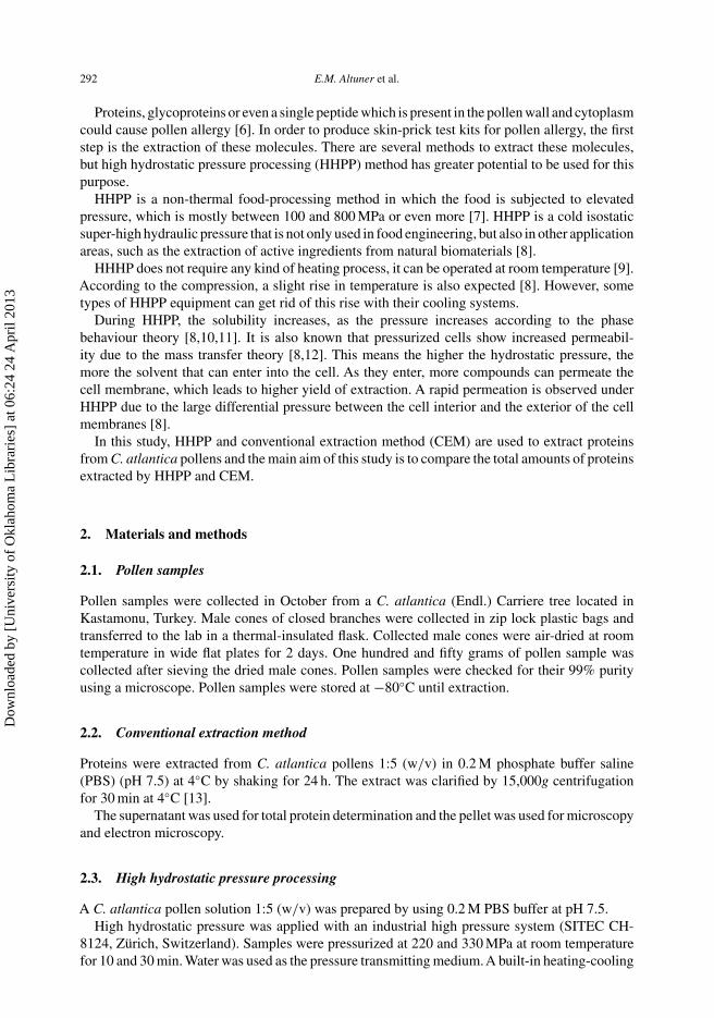
Figure 1. Untreated C. atlantica pollens. (a) light microscopy, (b) electron microscopy (top view), (c) electronmicroscopy (side view), and (d–f) surface ornamentation.
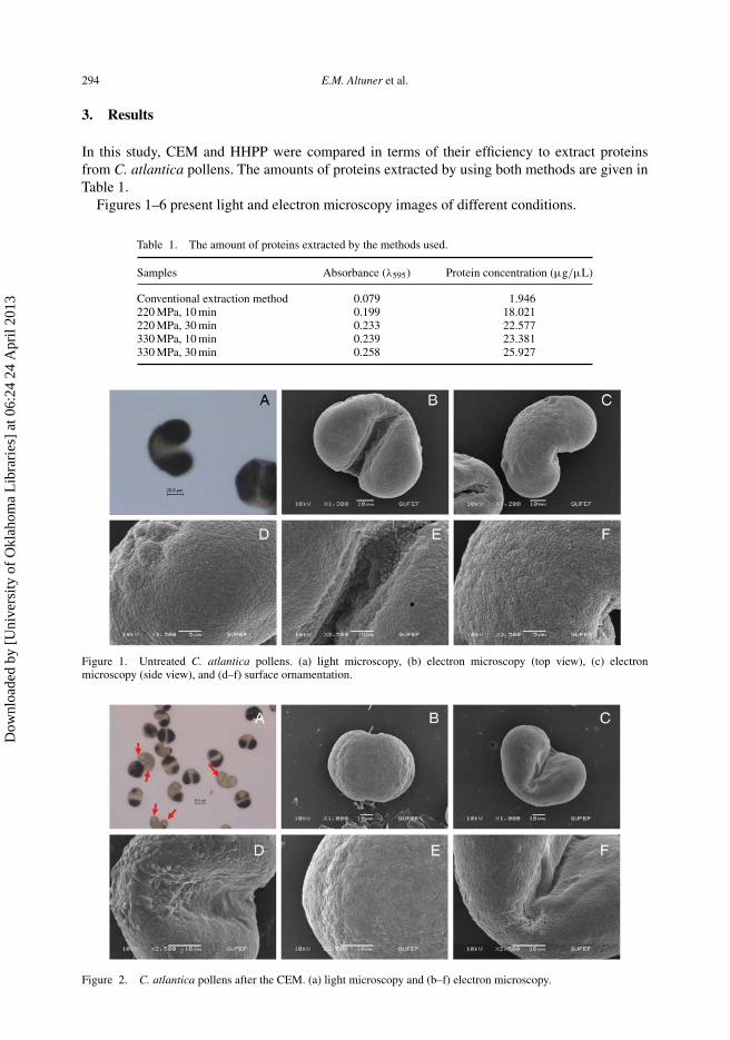
Figure 2. C. atlantica pollens after the CEM. (a) light microscopy and (b–f) electron microscopy.
Dow
nloa
ded
by [
Uni
vers
ity o
f O
klah
oma
Lib
rari
es]
at 0
6:24
24
Apr
il 20
13
High Pressure Research 295
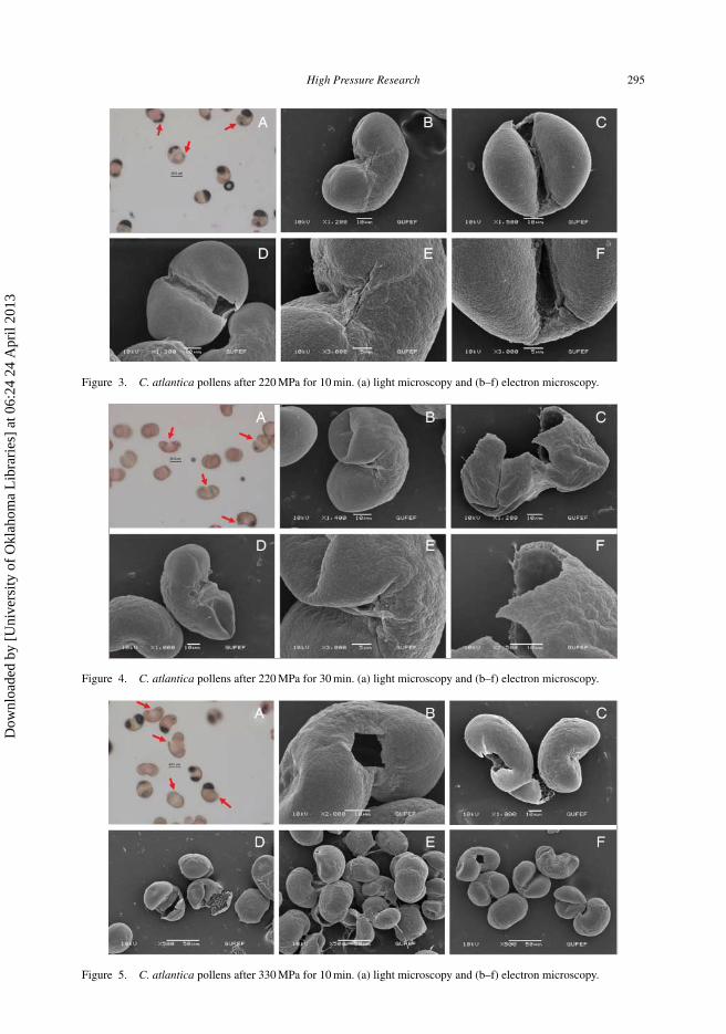
Figure 3. C. atlantica pollens after 220 MPa for 10 min. (a) light microscopy and (b–f) electron microscopy.
Figure 4. C. atlantica pollens after 220 MPa for 30 min. (a) light microscopy and (b–f) electron microscopy.
Figure 5. C. atlantica pollens after 330 MPa for 10 min. (a) light microscopy and (b–f) electron microscopy.
Dow
nloa
ded
by [
Uni
vers
ity o
f O
klah
oma
Lib
rari
es]
at 0
6:24
24
Apr
il 20
13
296 E.M. Altuner et al.
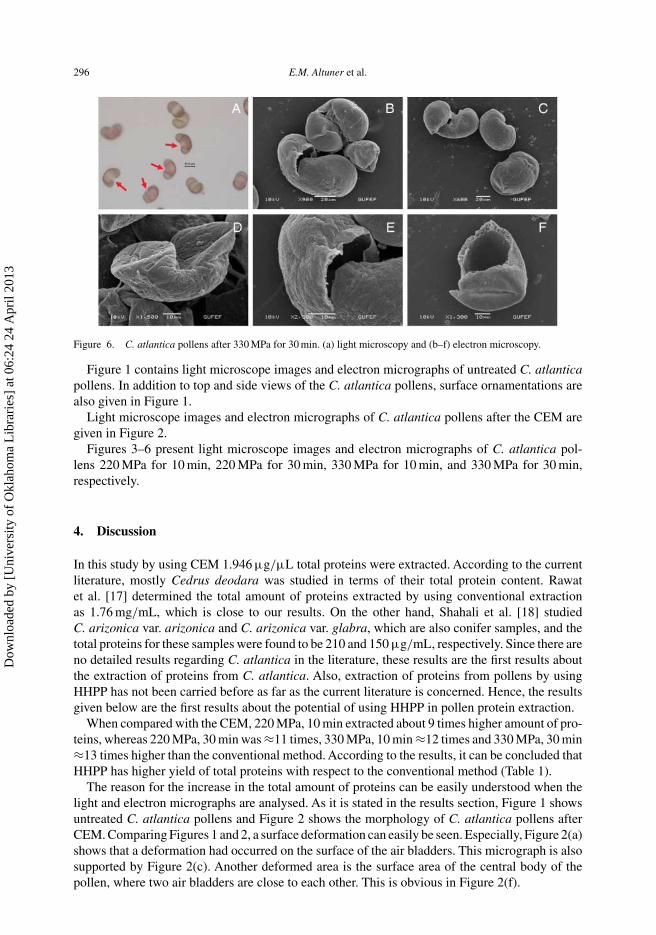
Figure 6. C. atlantica pollens after 330 MPa for 30 min. (a) light microscopy and (b–f) electron microscopy.
Figure 1 contains light microscope images and electron micrographs of untreated C. atlanticapollens. In addition to top and side views of the C. atlantica pollens, surface ornamentations arealso given in Figure 1.
Light microscope images and electron micrographs of C. atlantica pollens after the CEM aregiven in Figure 2.
Figures 3–6 present light microscope images and electron micrographs of C. atlantica pol-lens 220 MPa for 10 min, 220 MPa for 30 min, 330 MPa for 10 min, and 330 MPa for 30 min,respectively.
4. Discussion
In this study by using CEM 1.946 μg/μL total proteins were extracted. According to the currentliterature, mostly Cedrus deodara was studied in terms of their total protein content. Rawatet al. [17] determined the total amount of proteins extracted by using conventional extractionas 1.76 mg/mL, which is close to our results. On the other hand, Shahali et al. [18] studiedC. arizonica var. arizonica and C. arizonica var. glabra, which are also conifer samples, and thetotal proteins for these samples were found to be 210 and 150 μg/mL, respectively. Since there areno detailed results regarding C. atlantica in the literature, these results are the first results aboutthe extraction of proteins from C. atlantica. Also, extraction of proteins from pollens by usingHHPP has not been carried before as far as the current literature is concerned. Hence, the resultsgiven below are the first results about the potential of using HHPP in pollen protein extraction.
When compared with the CEM, 220 MPa, 10 min extracted about 9 times higher amount of pro-teins, whereas 220 MPa, 30 min was ≈11 times, 330 MPa, 10 min ≈12 times and 330 MPa, 30 min≈13 times higher than the conventional method. According to the results, it can be concluded thatHHPP has higher yield of total proteins with respect to the conventional method (Table 1).
The reason for the increase in the total amount of proteins can be easily understood when thelight and electron micrographs are analysed. As it is stated in the results section, Figure 1 showsuntreated C. atlantica pollens and Figure 2 shows the morphology of C. atlantica pollens afterCEM. Comparing Figures 1 and 2, a surface deformation can easily be seen. Especially, Figure 2(a)shows that a deformation had occurred on the surface of the air bladders. This micrograph is alsosupported by Figure 2(c). Another deformed area is the surface area of the central body of thepollen, where two air bladders are close to each other. This is obvious in Figure 2(f).
Dow
nloa
ded
by [
Uni
vers
ity o
f O
klah
oma
Lib
rari
es]
at 0
6:24
24
Apr
il 20
13
High Pressure Research 297
As previously mentioned in the results section, Figure 3 shows the pollen morphology ofC. atlantica after 220 MPa for 10 min and Figure 4 presents the pollen morphology of C. atlanticaafter 220 MPa for 30 min.
Figure 3(a) shows a deformation around the surface of the air bladders. Although it is not veryclear in the micrograph taken using the light microscope, a very clear pollen wall deformationcan be seen in Figure 3(c) and (d), which had occurred between two air bladders. ComparingFigures 2 and 3, especially Figure 3(c) and (d) explains why nine times higher amount of proteinswere extracted compared with the CEM. Due to the pollen wall deformation, it is possible thatproteins inside the pollens were released into the buffer.
According to the micrographs taken using the light microscope given in Figure 4(a) and theelectron micrographs presented in Figure 4(c) and (f), eruptions in C. atlantica pollens startedwhen 220 MPa of pressure was applied for a longer period of time. Figure 4(b), (d) and (e) showsthat air bladders had collapsed. This is also obvious in Figure 4(a), where the arrow is pointing atthe bottom right.
As stated in the results section, Figure 5 shows the pollen morphology of C. atlantica after330 MPa for 10 min and Figure 6 shows the pollen morphology of C. atlantica after 330 MPafor 30 min. HHPP of 330 MPa for 10 min caused a similar effect as 220 MPa for 30 min. At thiscondition, one can see that the air bladders have torn and are without a central body (Figure 5(f)).
When Figure 6 is analysed in detail, it is observed that at this pressure, the number of deformedand damaged pollens was increased.
In general, if we compare all the figures, the effect of 220 MPa for 30 min, 330 MPa for 10 minand 330 MPa for 30 min is quite close to each other. This may be the reason why total proteinsextracted at these conditions are very close to each other as given in Table 1.
As a conclusion, according to the results, it is possible to say that due to high pressure, a visualpollen deformation and eruption, pollen wall and surface damage were observed. Since HHPPcaused some morphological changes such as deformation and membrane damage, the total proteinextracted by this method was dramatically increased.
HHPP has a lesser cost of operation, is efficient, takes less time and is an energy saver. Therefore,it has a great opportunity to be used to extract proteins from pollens. However, it is known thatHHPP could cause protein denaturation. For this reason, further studies should be conducted toobserve whether a denaturation had occurred or not.
Acknowledgements
The authors thank Gülçin Karakaya and Emre Filiz for their help during the study and Gazi University, Department ofBiology for their help during SEM studies. We also thank Dr Kerim Güney for the identification of C. atlantica by meansof botanical criteria.
References
[1] A. Farjon, Pinaceae. Drawings and Descriptions of the Genera: Abies, Cedrus, Pseudolarix, Keteleeria, Nothotsuga,Tsuga, Cathaya, Pseudotsuga, Larix and Picea, Koeltz Scientific Books, Koenigstein, Germany, 1990.
[2] P.H. Davis, Flora of Turkey and the East Aegean Islands, Vol. 1, Edinburgh University Press, Edinburgh, 1965.[3] S.M. Walters, A. Brady, C.D. Brickell, J. Cullen, P.S. Green, J. Lewis, V.A. Matthews, D.A. Webb, P.F. Yeo, and
J.C.M. Alexander, The European Garden Flora, Vol. 1, Cambridge University Press, Cambridge, 2001.[4] F. Toshiyuki, I. Yasushi, and Y. Yoshinori, Pollen morphology of Cedrus (Pinaceae), Jap. J. Palynol. 49 (2003),
pp. 21–24.[5] F. Toshiyuki, I. Yasushi, and Y. Yoshinori, Pollen morphology of Cedrus (Pinaceae), Jap. J. Palynol. 49 (2003),
pp. 21–24.[6] S. Chanda, Pollen grains as aeroallergens: morphological, biological and chemical Approach. in Recent Trends in
Aerobiology, Allergy and Immunology, S.N. Agashe (ed.), Oxford & IBH Publishing Company Pvt ltd, New Delhi,1994, pp. 85–92.
Dow
nloa
ded
by [
Uni
vers
ity o
f O
klah
oma
Lib
rari
es]
at 0
6:24
24
Apr
il 20
13
298 E.M. Altuner et al.
[7] E.M. Altuner, C. Islek, T. Çeter, and H. Alpas, High hydrostatic pressure extraction of phenolic compounds fromMaclura pomifera fruits, Afr. J. Biotechnol. 11 (2012), pp. 930–937.
[8] S. Zhang, J. Xi, and C. Wang, High hydrostatic pressure extraction of flavonoids from propolis, J. Chem. Technol.Biotechnol. 80 (2005), pp. 50–54.
[9] E.M. Altuner, H. Alpas, Y.K. Erdem, and F. Bozoglu, Effect of high hydrostatic pressure on physicochemical andbiochemical properties of milk, Eur. Food Res. Technol. 222 (2006), pp. 392–396.
[10] J.S. Richard, High Pressure Phase Behaviour of Multicomponent Fluid Mixtures, Elsevier, Amsterdam, 1992.[11] W.J. Le Noble, Organic High Pressure Chemistry, Elsevier, Amsterdam, 1988.[12] H. Yan, Separation Engineering, China Petrochemical Press, Beijing, 2002.[13] S. Patriarca, S. Voltolini, R. Navone, S. Martini, C. Montanari, A. Negrini, and E. Cosulich, Biochemical and
immunochemical characterization of hop-hornbeam (Ostrya Carpinifolia Scop.) pollen A new allergenic pollenfrom the Corylaceae family, Aerobiologia 16 (2000), pp. 255–260.
[14] H.J. Chial, H.B. Thompson, and A.G. Splittgerber, A spectral study of the charge forms of Coomassie Blue G, Anal.Biochem. 209 (1993), pp. 258–266.
[15] M.M. Bradford, A rapid and sensitive method for the quantitation of microgram quantities of protein utilizing theprinciple of protein-dye binding. Anal. Biochem. 72 (1976), pp. 248–254.
[16] M. Zafar, M. Ahmed, and M.A. Khan, Palynological studies of Verbenaceae from Margalla Hills, Islamabad,Pakistan, Pak. J. Plant Sci. 12 (2006), pp. 21–25.
[17] A. Rawat, A. Singh, A.B. Singh, S.N. Gaur, L. Kumar, I. Roy, and P. Ravindrun, Clinical and immunologic evaluationof Cedrus deodara pollen: a new allergen from India, Allergy 55 (2000), pp. 620–626.
[18] Y. Shahali, A. Majd, Z. Pourpak, G. Tajadod, M. Haftlang, and M. Moin, Comparative study of the pollen proteincontents in two major varieties of Cupressus Arizonica planted in Tehran, Iran J. Allergy Asthma Immunol. 6 (2007),pp. 123–127.
Dow
nloa
ded
by [
Uni
vers
ity o
f O
klah
oma
Lib
rari
es]
at 0
6:24
24
Apr
il 20
13