hidden dynamic allostery in a pdz · pdf filehidden dynamic allostery in a pdz domain chad m....
TRANSCRIPT

Hidden dynamic allostery in a PDZ domainChad M. Petita, Jun Zhangb, Paul J. Sapienzaa, Ernesto J. Fuentesc, and Andrew L. Leea,b,1
aDivision of Medicinal Chemistry and Natural Products, Eshelman School of Pharmacy and bDepartment of Biochemistry and Biophysics, School of Medicine,University of North Carolina, Chapel Hill, NC 27599; and cDepartment of Biochemistry, University of Iowa, Iowa City, IA 52242
Edited by Susan S. Taylor, University of California San Diego, La Jolla, CA, and approved September 4, 2009 (received for review April 28, 2009)
Structure–function relationships in proteins are predicated on thespatial proximity of noncovalently interacting groups of atoms. Thus,structural elements located away from a protein’s active site aretypically presumed to serve a stabilizing or scaffolding role for thelarger structure. Here we report a functional role for a distal structuralelement in a PDZ domain, even though it is not required to maintainPDZ structure. The third PDZ domain from PSD-95/SAP90 (PDZ3) hasan unusual additional third alpha helix (�3) that packs in contiguousfashion against the globular domain. Although �3 lies outside theactive site and does not make direct contact with C-terminal peptideligand, removal of �3 reduces ligand affinity by 21-fold. Furtherinvestigation revealed that the difference in binding free energiesbetween the full-length and truncated constructs is predominantlyentropic in nature and that without �3, picosecond-nanosecondside-chain dynamics are enhanced throughout the domain, as deter-mined by 2H methyl NMR relaxation. Thus, the distal modulation ofbinding function appears to occur via a delocalized conformationalentropy mechanism. Without removal of �3 and characterization ofside-chain dynamics, this dynamic allostery would have gone unno-ticed. Moreover, what appeared at first to be an artificial modificationof PDZ3 has been corroborated by experimentally verified phosphor-ylation of �3, revealing a tangible biological mechanism for this novelregulatory scheme. This hidden dynamic allostery raises the possibil-ity of as-yet unidentified or untapped allosteric regulation in this PDZdomain and is a very clear example of function arising from dynamicsrather than from structure.
NMR � PSD-95 � spin relaxation � entropy
Proteins owe their functionality to the 3-dimensional arrange-ment of atoms. A typical protein’s structure stabilizes its
active site, allowing for specific interactions with substrate orligand. These basic structure–function relationships are wellunderstood for countless types of proteins. Because most activesites are relatively small, it has been presumed that the remainingbulk of the globular structure provides a scaffolding role. Thus,even though similar domains belonging to the same family mayhave substrate specificity preferences, the folds of those domainsare composed of invariant structural elements (1). Nonetheless,variations in tertiary fold composition, such as additional ele-ments of secondary structure, are not uncommon. An exampleof this can be seen within the PDZ domain family of proteins.From this, the question of whether there is a specific role for suchauxiliary structural elements remains open. In other words, howmight these additional elements influence the core domain?
PDZ domains (eg, PSD-95, Discs Large, Zo-1) are small, �90-aamodular structures that typically bind C-terminal tails (�4–6residues) of target proteins (2). They are frequently found inmultiple copies in proteins with diverse functions, especially thoseinvolved in signal transduction complexes and in the anchoring ofreceptors at membrane specializations (2–4). Thus, one commonfunction of PDZ domains is to provide scaffolding. Although thePDZ family has a highly conserved fold consisting of a 5- or6-stranded half �-barrel and 2 �-helices (5), some members exhibitadditional secondary structural elements or differences in lengthsof helices, �-strands, or loops (6–9). For instance, the third PDZdomain from the PSD-95/SAP90 protein (referred to herein asPDZ3) contains an additional �-helix at its carboxyl terminus (�3)that was first reported in its homologue Dlg1 (5, 6). It is ironic that
this atypical helix is present in PDZ3, because PDZ3 is consideredthe archetype of PDZ domains, being the first PDZ domainstructure to be solved (2). Naively, �3 is not expected to impactfunction, because it lies on a different surface from the PDZligand-binding site (Fig. 1). However, studies on various proteinshave shown that intramolecular communication can occur betweensites that would not be expected to be energetically linked based onprotein structure alone (10–14). This notion was exemplified earlyon in the PDZ domain family using coevolutionary analysis, fromwhich an evolutionarily conserved communication pathway wasproposed (15). Subsequently, individual PDZ domains were foundto exhibit allosteric behavior (8, 16–19). For these reasons, weinvestigated the role of �3 in PDZ3 function, structure, anddynamics.
Here we show that the �3 appendage to PDZ3 modulatesC-terminal peptide binding function through a novel, dynamics-based mechanism. Removal of �3 has a minimal effect on globalPDZ3 structure, but results in a �21-fold reduction in bindingaffinity that is entirely entropic in origin, within experimental error.Backbone (15N) and side-chain (2H methyl) NMR relaxation ex-periments indicate that removal of �3 results in increased side-chaindynamics throughout the structure on time scales faster than �6 ns.These enhanced motions are quenched on peptide binding, pro-viding the entropic driving force for the reduced binding affinity.This example of dynamically regulated binding combines features ofdynamic allostery observed at the backbone level (20) with thecontrol of ligand-binding affinity through the tuning of side-chainconformational entropy (21). These findings, along with the recentreport that Tyr-397 in �3 is phosphorylated (22), provide supportfor the intriguing possibility that in vivo, PDZ3 function may beregulated by an entropically controlled dynamic mechanism. Thatthis particular PDZ domain is not known to be allosteric or toundergo conformational change (5, 23) suggests that similar mech-anisms might be found in other PDZ domains, such as PDZ fromharmonin (24), or even in a wide variety of domain types.
ResultsThe third PDZ domain of the neuronal signaling protein PSD-95/SAP90 (PDZ3, residues 303–402) contains an uncharacter-istic, third �-helix (�3, residues 394–399) at its C-terminus thatpacks up against the core domain (the widely conserved 5 or 6�-strands and 2 �-helices) in a region distinct from the peptideligand-binding pocket (Fig. 1) (5). The closest end-to-end ap-proach between �3 and peptide is made at the �3 position (Gln)of peptide [using the numbering scheme of Doyle et al. (5)],whose side-chain nitrogen is located �6 Å from the hydroxyloxygen of Tyr-397 (Fig. 1). There are no bridging waters, and theremaining residues of �3 and peptide ligand extend away fromeach other; all C�–C� distances are �10 Å. Thus, �3 does notdirectly contact the peptide. To investigate the role of �3 inPDZ3 function and behavior, a C-terminal truncation mutant(�7ct) was constructed with residues 396–402 removed. This
Author contributions: C.M.P. and A.L.L. designed research; C.M.P., J.Z., and E.J.F. performedresearch; C.M.P., J.Z., P.J.S., and E.J.F. analyzed data; and C.M.P. and A.L.L. wrote the paper.
The authors declare no conflict of interest.
This article is a PNAS Direct Submission.
1To whom correspondence should be addressed. E-mail: [email protected].
www.pnas.org�cgi�doi�10.1073�pnas.0904492106 PNAS � October 27, 2009 � vol. 106 � no. 43 � 18249–18254
BIO
PHYS
ICS
AN
DCO
MPU
TATI
ON
AL
BIO
LOG
Y

truncation was engineered to disrupt the helix while leaving thecore PDZ domain intact.
Consistent with previous studies (25), �7ct retains its native PDZstructure. The 1H-15N heteronuclear single quantum coherence(HSQC) spectrum shows a well-dispersed single set of peaks,indicative of a well-folded protein (Fig. 2). Aside from �3, allsecondary structures are intact, as determined from 13C� chemicalshifts (26). A more detailed analysis of potential structural effectsis provided below. These data show that �7ct is stable with a typicalPDZ fold. Having verified no gross structural changes in �7ct, weset out to study the thermodynamics, structure, and dynamics of thissystem and found that �3 has a significant effect on both ligand-binding affinity and the internal dynamics of PDZ3.
Binding Thermodynamics of �7ct-PDZ3. Isothermal titration calorim-etry (ITC) was used to evaluate the thermodynamic characteristicsof �7ct and the full-length PDZ3 (i.e., PDZ3303–402) binding to apeptide derived from the carboxyl terminus of CRIPT (Ac-TKNYKQTSV-COOH), a naturally occurring binding partner forPDZ3 (27). For PDZ3303–402, binding occurs with relatively highaffinity (Kd � 1.2 � 0.1 �M), whereas truncation lowers the affinity(Kd � 25.7 � 3.6 �M), as shown in Fig. 3 and summarized in Table1. This demonstrates an important role for �3 in PDZ3’s bindingfunction. Interestingly, the enthalpic contribution to the free energyof binding is nearly identical for both constructs, whereas -T�Schanges from 1.7 kcal/mol for PDZ3303–402 to 4.0 kcal/mol for �7ctat 25 °C (Table 1). Thus, the difference in binding is entropic inorigin. Because binding takes place in a separate location from �3,and there is no change in the enthalpic contribution to binding
between the 2 constructs (which might reflect structural changes),differences in desolvation on binding are an unlikely source of theentropic change. It is possible that the core PDZ domain undergoessome structural adjustment such that individual changes in enthalpycancel out, resulting in differences in desolvation on binding. Thereis no direct evidence of this, however. The remaining possibleentropy-based explanation for the reduced affinity of �7ct forpeptide is that significant changes in the conformational entropy ofthe system occur after removal of �3.
�3 Modulates Picosecond-Nanosecond Dynamics Throughout PDZ3.To investigate the source of any differences in conformationalentropy on binding, we used backbone (28) and side-chain (29)NMR spin relaxation experiments to monitor the picosecond-nanosecond (ps-ns) dynamics of both the free and bound forms ofPDZ3303–402 and �7ct. For the backbone, 15N T1, T2, and {1H}-15NNOE relaxation experiments were acquired at 2 field strengths (500and 600 MHz) to achieve high-quality fits of the spectral densityfunction. The relaxation analysis used statistical model selection toyield rigorous model-free order parameters for N-H bond vectors,S2. The order parameter is a measure of a given bond’s angularrigidity (on a scale of 0–1) on timescales faster than the overallrotational tumbling time (30), which in this case is on the order of6 ns. For side-chain groups, methyl 2H relaxation data sets (IzCz,IzCzDz, and IzCzDy) at 2 field strengths (500 and 600 MHz) werefitted to obtain the order parameter corresponding to the methylC–CH3 bond, denoted by S2
axis (31).Order parameters were first compared between PDZ3303–402 and
�7ct constructs in the absence of CRIPT peptide. Backbone N-Hbond vectors showed limited changes in S2 after removal of �3 (Fig.4A). The largest observed changes were increased flexibility for a
Fig. 1. Structure of PSD-95/SAP90 PDZ3 complexed with CRIPT peptide. The �3helix is shown in blue. The van der Waals surface of the CRIPT peptide is in purple,and the nearest approaching residues (Tyr-397 and Phe-400) of �3 are in cyan.
Fig. 2. 1H-15N HSQC spectrum of PDZ3303–402 (black) overlain with the 1H-15NHSQC spectrum of �7ct (red). Resonances with 1H-15N assignments for deletedresidues 396–402 are indicated by arrows.
Fig. 3. The �3 helix affects CRIPT peptide binding. ITC data for binding ofPSD-95/SAP90 PDZ3303–402 (A) and �7ct (B) to C-terminal 9 residues of CRIPT(Ac-TKNYKQTSV-COOH) at 25 °C. Thermograms and integrated titrationcurves are shown in the top and bottom panels, respectively. Heats of dilutionwere obtained from independent buffer–buffer titration experiments (seeMaterials and Methods).
Table 1. Thermodynamic parameters for the PDZ3–CRIPT peptideinteraction at 25 °C
Protein Kd (�M) �H (kcal/mol) �T�S (kcal/mol)
PDZ3303–402 1.2 (0.1) �9.70 (0.12) 1.66 (0.06)�7ct 25.7 (3.6) �10.27 (0.68) 4.00 (0.59)
Values are the average of 2 independent experiments. The fitted stoichi-ometry (n) ranged from 0.95 to 1.06.
18250 � www.pnas.org�cgi�doi�10.1073�pnas.0904492106 Petit et al.
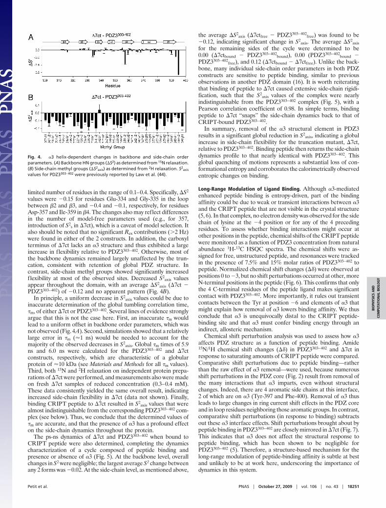
limited number of residues in the range of 0.1–0.4. Specifically, �S2
values were �0.15 for residues Glu-334 and Gly-335 in the loopbetween �2 and �3, and �0.4 and �0.1, respectively, for residuesAsp-357 and Ile-359 in �4. The changes also may reflect differencesin the number of model-free parameters used (e.g., for 357,introduction of S2
s in �7ct), which is a caveat of model selection. Italso should be noted that no significant Rex contributions (�2 Hz)were found in either of the 2 constructs. In addition, the carboxylterminus of �7ct lacks an �3 structure and thus exhibited a largeincrease in flexibility relative to PDZ3303–402. Otherwise, most ofthe backbone dynamics remained largely unaffected by the trun-cation, consistent with retention of global PDZ structure. Incontrast, side-chain methyl groups showed significantly increasedflexibility at most of the observed sites. Decreased S2
axis valuesappear throughout the domain, with an average �S2
axis (�7ct �PDZ3303–402) of �0.12 and no apparent pattern (Fig. 4B).
In principle, a uniform decrease in S2axis values could be due to
inaccurate determination of the global tumbling correlation time,�m, of either �7ct or PDZ3303–402. Several lines of evidence stronglyargue that this is not the case here. First, an inaccurate �m wouldlead to a uniform offset in backbone order parameters, which wasnot observed (Fig. 4A). Second, simulations showed that a relativelylarge error in �m (�1 ns) would be needed to account for themajority of the observed decreases in S2
axis. Global �m times of 5.9ns and 6.0 ns were calculated for the PDZ3303–402 and �7ctconstructs, respectively, which are characteristic of a globularprotein of �10 kDa (see Materials and Methods for all �m values).Third, both 15N and 2H relaxation on independent protein prepa-rations of �7ct were performed, and measurements also were madeon fresh �7ct samples of reduced concentration (0.3–0.4 mM).These data consistently yielded the same overall result, indicatingincreased side-chain flexibility in �7ct (data not shown). Finally,binding CRIPT peptide to �7ct resulted in S2
axis values that werealmost indistinguishable from the corresponding PDZ3303–402 com-plex (see below). Thus, we conclude that the determined values of�m are accurate, and that the presence of �3 has a profound effecton the side-chain dynamics throughout the protein.
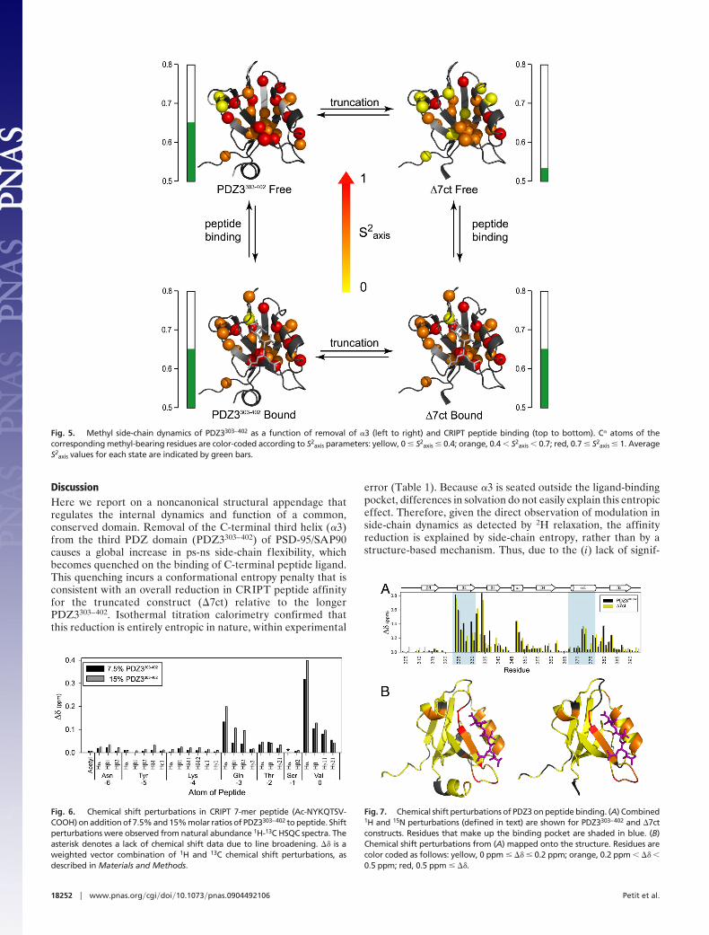
The ps-ns dynamics of �7ct and PDZ3303–402 when bound toCRIPT peptide were also determined, completing the dynamicscharacterization of a cycle composed of peptide binding andpresence or absence of �3 (Fig. 5). At the backbone level, overallchanges in S2 were negligible; the largest average S2 change betweenany 2 forms was �0.02. At the side-chain level, as mentioned above,
the average �S2axis (�7ctfree � PDZ3303–402
free) was found to be�0.12, indicating significant change in S2
axis. The average �S2axis
for the remaining sides of the cycle were determined to be0.00 (�7ctbound � PDZ3303–402
bound), 0.00 (PDZ3303–402bound �
PDZ3303–402free), and 0.12 (�7ctbound � �7ctfree). Unlike the back-
bone, many individual side-chain order parameters in both PDZconstructs are sensitive to peptide binding, similar to previousobservations in another PDZ domain (16). It is worth reiteratingthat binding of peptide to �7ct caused extensive side-chain rigidi-fication, such that the S2
axis values of the complex were nearlyindistinguishable from the PDZ3303–402 complex (Fig. 5), with aPearson correlation coefficient of 0.98. In simple terms, bindingpeptide to �7ct ‘‘snaps’’ the side-chain dynamics back to that ofCRIPT-bound PDZ3303–402.
In summary, removal of the �3 structural element in PDZ3results in a significant global reduction in S2
axis, indicating a globalincrease in side-chain flexibility for the truncation mutant, �7ct,relative to PDZ3303–402. Binding peptide then returns the side-chaindynamics profile to that nearly identical with PDZ3303–402. Thisglobal quenching of motions represents a substantial loss of con-formational entropy and corroborates the calorimetrically observedentropic changes on binding.
Long-Range Modulation of Ligand Binding. Although �3-mediatedenhanced peptide binding is entropy-driven, part of the bindingaffinity could be due to weak or transient interactions between �3and the CRIPT peptide that are not visible in the crystal structure(5, 6). In that complex, no electron density was observed for the sidechain of lysine at the �4 position or for any of the 4 precedingresidues. To assess whether binding interactions might occur atother positions in the peptide, chemical shifts of the CRIPT peptidewere monitored as a function of PDZ3 concentration from naturalabundance 1H-13C HSQC spectra. The chemical shifts were as-signed for free, unstructured peptide, and resonances were trackedin the presence of 7.5% and 15% molar ratios of PDZ3303–402 topeptide. Normalized chemical shift changes (��) were observed atpositions 0 to �3, but no shift perturbations occurred at other, moreN-terminal positions in the peptide (Fig. 6). This confirms that onlythe 4 C-terminal residues of the peptide ligand makes significantcontact with PDZ3303–402. More importantly, it rules out transientcontacts between the Tyr at position �6 and elements of �3 thatmight explain how removal of �3 lowers binding affinity. We thusconclude that �3 is unequivocally distal to the CRIPT peptide-binding site and that �3 must confer binding energy through anindirect, allosteric mechanism.
Chemical shift perturbation analysis was used to assess how �3affects PDZ structure as a function of peptide binding. Amide15N/1H chemical shift changes (��) in PDZ3303–402 and �7ct inresponse to saturating amounts of CRIPT peptide were compared.Comparative shift perturbations due to peptide binding—ratherthan the raw effect of �3 removal—were used, because numerousshift perturbations in the PDZ core (Fig. 2) result from removal ofthe many interactions that �3 imparts, even without structuralchanges. Indeed, there are 4 aromatic side chains at this interface,2 of which are on �3 (Tyr-397 and Phe-400). Removal of �3 thusleads to large changes in ring current shift effects in the PDZ coreand in loop residues neighboring those aromatic groups. In contrast,comparative shift perturbations (in response to binding) subtractsout these �3 interface effects. Shift perturbations brought about bypeptide binding in PDZ3303–402 are closely mirrored in �7ct (Fig. 7).This indicates that �3 does not affect the structural response topeptide binding, which has been shown to be negligible forPDZ3303–402 (5). Therefore, a structure-based mechanism for thelong-range modulation of peptide-binding affinity is subtle at bestand unlikely to be at work here, underscoring the importance ofdynamics in this system.
Fig. 4. �3 helix-dependent changes in backbone and side-chain orderparameters. (A) Backbone HN groups (�S2) as determined from 15N relaxation.(B) Side-chain methyl groups (�S2
axis) as determined from 2H relaxation. S2axis
values for PDZ3303–402 were previously reported by Law et al. (44).
Petit et al. PNAS � October 27, 2009 � vol. 106 � no. 43 � 18251
BIO
PHYS
ICS
AN
DCO
MPU
TATI
ON
AL
BIO
LOG
Y
DiscussionHere we report on a noncanonical structural appendage thatregulates the internal dynamics and function of a common,conserved domain. Removal of the C-terminal third helix (�3)from the third PDZ domain (PDZ3303–402) of PSD-95/SAP90causes a global increase in ps-ns side-chain flexibility, whichbecomes quenched on the binding of C-terminal peptide ligand.This quenching incurs a conformational entropy penalty that isconsistent with an overall reduction in CRIPT peptide affinityfor the truncated construct (�7ct) relative to the longerPDZ3303–402. Isothermal titration calorimetry confirmed thatthis reduction is entirely entropic in nature, within experimental
error (Table 1). Because �3 is seated outside the ligand-bindingpocket, differences in solvation do not easily explain this entropiceffect. Therefore, given the direct observation of modulation inside-chain dynamics as detected by 2H relaxation, the affinityreduction is explained by side-chain entropy, rather than by astructure-based mechanism. Thus, due to the (i) lack of signif-
Fig. 5. Methyl side-chain dynamics of PDZ3303–402 as a function of removal of �3 (left to right) and CRIPT peptide binding (top to bottom). C� atoms of thecorresponding methyl-bearing residues are color-coded according to S2
axis parameters: yellow, 0 � S2axis � 0.4; orange, 0.4 � S2
axis � 0.7; red, 0.7 � S2axis � 1. Average
S2axis values for each state are indicated by green bars.
Fig. 6. Chemical shift perturbations in CRIPT 7-mer peptide (Ac-NYKQTSV-COOH) on addition of 7.5% and 15% molar ratios of PDZ3303–402 to peptide. Shiftperturbations were observed from natural abundance 1H-13C HSQC spectra. Theasterisk denotes a lack of chemical shift data due to line broadening. �� is aweighted vector combination of 1H and 13C chemical shift perturbations, asdescribed in Materials and Methods.
Fig. 7. Chemical shift perturbations of PDZ3 on peptide binding. (A) Combined1H and 15N perturbations (defined in text) are shown for PDZ3303–402 and �7ctconstructs. Residues that make up the binding pocket are shaded in blue. (B)Chemical shift perturbations from (A) mapped onto the structure. Residues arecolor coded as follows: yellow, 0 ppm � �� � 0.2 ppm; orange, 0.2 ppm � �� �0.5 ppm; red, 0.5 ppm � ��.
18252 � www.pnas.org�cgi�doi�10.1073�pnas.0904492106 Petit et al.

icant structural changes, (ii) lack of direct interactions between�3 and CRIPT peptide residues, and (iii) participation of ps-nsside-chain dynamics throughout the domain, this �3/PDZ3/peptide ligand system is a clear example of allostery mediated byfast side-chain dynamics. It is allosteric in that �3 and the ligandare distal to one another, and it is also allosteric in the sense thatit is driven by dynamic changes outside the ligand-binding site.We also note that several backbone sites, such as residuesGlu-334 and Gly-335 in the glycine-rich �2-�3 loop, appear tobehave like the side chains and contribute to the overall effect.Importantly, because this is observed in a small protein domainnot prone to structural changes and considered ‘‘rigid’’ (23), itsuggests that any folded protein could in principle use dynamicallostery as a regulatory mechanism, although recognition of thismechanism likely would require characterization of side-chaindynamics.
The finding that the distal �3 element can regulate PDZ3function raises an interesting question as to whether this dynamicallostery has a defined biological role. In support of this, PSD-95 hasbeen shown to be phosphorylated at multiple positions (32, 33),including Tyr-397 (22) located in the �3 helix. Because the Tyrphenol ring packs against the core PDZ domain, phosphorylationwould alter �3 packing or lead to loss of �3 structure, either ofwhich could affect ligand affinity. Although Tyr-397 is partiallyburied in the crystal structure, its accessibility to kinases should besignificant, based on the high amide exchange rates within �3 (25)(data not shown for PDZ3303–402). A particularly relevant findingcomes from the Drosophila homologue Discs Large (Dlg) protein,which, along with PSD-95, belongs to the MAGUK (membrane-associated guanylate kinase) family of proteins (34). It was shownthat deletion of a portion of �3 (residues corresponding to PSD-95396–401) disrupts the interdomain communication between PDZ3and the SH3-GK module located 25 residues downstream; in turn,this communication was shown to modulate Dlg binding to thelocalization protein GukHolder (35). This suggests that the highlyconserved linker region between PDZ3 and the SH3 domains isessential for modulating both intramolecular and intermolecularinteractions (35). Furthermore, it seems reasonable that the phos-phorylation state and conformation of �3 could play an importantrole in this regulation. Thus, in a broader context, the dynamicallostery observed here with PDZ3 arises out of a larger, morecomplex interdomain regulatory scheme. Such regulatory mod-ules within the context of larger interdomain interactions havepreviously been reported in the case of the GTPase exchangefactor Vav1 (36).
Dynamics-based modulation of binding affinity has previouslybeen reported in 2 notable cases. First, negative cooperative bindingof cAMP to the dimeric catabolite activator protein was found tobe driven by ps-ns backbone dynamics (20). Specifically, binding ofthe first cAMP occurs with little change in backbone flexibility,whereas binding the second cAMP triggers rigidification through-out both subunits and thus has a substantial conformational en-tropic cost (20). In the second example, changes in side-chainconformational entropy in calmodulin were shown to correlateremarkably well with calorimetric entropy changes for a series ofcalmodulin-binding peptides binding to calmodulin (21). In relationto these studies, the critical difference in the present study is that thelong-range dynamic mechanism becomes apparent only after re-moval of the �3 helix. Furthermore, the driving force occursthrough side-chain equilibrium dynamics, rather than effects fromthe backbone. In this sense, the data presented here bridge theside-chain entropy findings of Frederick et al. (21) with the dynam-ically driven allosteric effect reported by Popovych et al. (20). Ourresults provide further support for the general notion of entropyvis-a-vis dynamics as a bona fide mechanism for allosteric commu-nication without conformational change (10, 37–39).
Materials and MethodsCloning, Expression, and Purification of PSD-95/SAP90 PDZ3. The third PDZdomain of PSD-95/SAP90 from Rattus norvegicus was subcloned into a T7 expres-sion vector (pET28a; Novagen) using primers designed to amplify residues 303–402 [using the numbering scheme of Doyle et al. (5)] of the full-length plasmid ofPSD95/SAP90 (a gift from Ken Prehoda). The PSD-95 PDZ3 plasmid was trans-formed into DE3 Star Eschericha coli cells and grown in minimal media supple-mented with the appropriate isotope for the desired labeling scheme, as de-scribed previously (16). A 50-mL starter culture was grown for 18 h and used toinoculate 950 mL of culture media. Protein expression was induced by addingisopropyl �-D-1-thiogalactopyranoside to a final concentration of 1 mM once thebacteria reached an OD600 of �0.6–0.8. The culture was allowed to grow for 4 hat 37 °C. Bacteria were harvested by centrifugation and frozen until further use.Frozen, pelleted cells were resuspended in lysis buffer [50 mM Tris, 150 mM NaCl,10 mM EDTA (pH 6.8)] and lysed using 2 freeze–thaw steps followed by ultra-sonication. The PDZ3303–402 protein was purified using Source-Q (GE Biosciences)chromatography media, followed by Fast-Flow Q-Sepharose chromatography(GE Biosciences). Pooled fractions containing PDZ were subjected to size-exclusion chromatography using a G50-Sephadex column (GE Biosciences) equil-ibrated in NMR buffer [20 mM NaPO4, 50 mM NaCl, 1 mM EDTA (pH 6.8)]. Typicalyields were 30 mg/L with �95% purity, as verified by SDS/PAGE analysis.
Generation of �7ct. The �7ct truncation mutant (PSD-95/SAP90 numbering303–395) was generated by PCR using oligonucleotides designed to amplify DNAwithin the �3 helix. This amplified DNA was subcloned into a T7 expression vector(pET28a; Novagen). Purification of truncated PDZ3 (�7ct) was achieved in thesame manner as for PDZ3303–402.
Peptide Synthesis. The C-terminal peptide from CRIPT (Ac-TKNYKQTSV-COOH orAc-NYKQTSV-COOH) was manually synthesized using standard Fmoc solid-phasepeptide chemistry. Peptide cleavage from the Wang resin and side-chain depro-tection were achieved simultaneously using TFA:triisopropyl silane:anisole:water(90:5:3:2) for 2 h under nitrogen. The volume of filtrate was reduced under astream of nitrogen, followed by peptide precipitation using 60 mL of cold diethylether. The mixture was extracted 3 times with 0.1% TFA in water (20 mL each).The aqueous phase was then combined and lyophilized. The crude powder wasdissolved in water and then purified by reverse-phase (C18) HPLC using a lineargradient from 0 to 20% (95% acetonitrile, 4.9% water, 0.1% TFA) over 10 min,followed by a gradient from 20% to 35% over 15 min. Pure CRIPT was eluted at20–22 min. The final peptide product was verified by mass spectrometry.
Isothermal Titration Calorimetry. ITC was carried out on a Microcal VP-ITCmicrocalorimeter at 25 °C. All measurements were collected by titration of CRIPTpeptide into the protein solution. Both protein and peptide ITC buffer solutionswere composed of 20 mM NaPO4, 50 mM NaCl, and 1 mM EDTA (pH 6.8). The pHof the peptide and protein solutions were determined to be identical to within0.02 pH units at room temperature. All solutions were degassed for �5–10 minwithout stirring and kept on ice before the ITC experiments.
For all ITC experiments, PDZ3 (100 �M) was placed in the sample chamber (ca.2 mL), while the peptide solution (1 mM) was transferred into a 250-�L injectionsyringe. Each titration experiment comprised a single 2-�L injection, followed by7-�L injections at 180-s intervals. The c values for PDZ3303–402 and �7ct were �90and �5, respectively. To account for the heat of dilution, an identical calorimetryexperiment was performed in the absence of protein. The data from eachexperiment were collected and analyzed using ORIGIN version 5.0 (Microcal).Thermodynamic parameters were determined using nonlinear least squaresfitting, assuming a single-site–binding model. The parameters reported are theresults of 2 independent measurements.
NMR Spectroscopy. NMR experiments were carried out at 25 °C (calibrated usingamethanol standard)usingVarian Inovaspectrometersequippedwith 1H/15N/13Cprobes and z-axis pulsed-field gradients operating at 500- and 600-MHz 1Hfrequencies. For both PDZ3303–402 and �7ct in free and bound forms, backboneand side-chain methyl groups were assigned using standard triple-resonanceassignment experiments. Stereospecific assignments were obtained from a 10%13C-labeled PDZ3 sample (40). All NMR experiments were processed usingNMRPipe and analyzed using NMRView compiled for Linux workstations (41,42). Chemical shift perturbation analysis was performed using a weightedvector combination of shifts, calculated by applying the formulas [��H
2 �(0.25*��C) 2]0.5 for 1H-13C shifts and [��H
2 � (0.1*��N) 2]0.5 for 1H-15N shifts.
15N Backbone Relaxation. 15N T1, T2, and {1H}-15N NOE values were obtainedthrough standard experiments (28) at 500 and 600 MHz for PDZ3303–402 (�1.0mM) and �7ct (�1.0 mM), both bound and free forms. For T1 and T2 experiments,
Petit et al. PNAS � October 27, 2009 � vol. 106 � no. 43 � 18253
BIO
PHYS
ICS
AN
DCO
MPU
TATI
ON
AL
BIO
LOG
Y

9 relaxation time points along with 3 duplicates were typically collected. For the{1H}-15N NOE, a recycle delay of 4.5 s was used. Relaxation decays were best fit tosingle exponentials using in-house programs. For Lipari-Szabo analysis, global �m
correlation times were found to be 5.89 ns for PDZ3303–402free, 6.23 ns for
PDZ3303–402bound, 5.98 ns for �7ctfree, and 6.34 ns for �7ctbound. Anisotropic rota-
tional models were tested, but it was concluded that the isotropic rotationalmodel was the best for both PDZ3303–402 and �7ct (data not shown). To assesspotential aggregation effects on global �m, 15N relaxation sets were indepen-dently collected and analyzed at 2 different concentrations (1.0 and 0.4 mM) forbothconstructsusing independentlypreparedprotein samples (datanot shown).For each residue, the selection of 1 of 5 standard model-free models (S2; S2 and �e;S2 and Rex; S2, �e, and Rex; or S2
s, S2f, and �s) were carried out using RVI software,
a front-end interface for relxn2.2 that analyzes 15N relaxation using simplemodel-free formalism(30)andtheAkaike informationcriterion (43),asdescribedpreviously (14).
2H-Methyl Relaxation. Side-chain 2H-methyl relaxation experiments were col-lected for samples containing randomly fractionally incorporated 2H (29). Multi-coherence relaxation experiments (IzCzDz, IzCzDy, and IzCz) were collected at 500and 600 MHz (29). For each experiment, 9 relaxation time points along with 3duplicate points were collected for PDZ3303–402 (�1.0 mM) and �7ct (�1.0 mM) inboth free and bound forms. All decays were best fit to single exponentials.Side-chain order parameters (S2
axis) were derived from fits of S2 and �e to the datausing the appropriate isotropic �m values determined for each construct.
ACKNOWLEDGMENTS. We thank Karl Koshlap (UNC Eshelman School of Phar-macy, NMR Facility), Greg Young (UNC Biomolecular NMR Laboratory), AshutoshTripathy (UNC Macromolecular Interactions Facility), Dan Cline (UNC EshelmanSchool of Pharmacy), and Brenda Temple (UNC Structural Bioinformatics Facility)for their technical assistance. We also thank Ken Prehoda for providing DNA forthe third PDZ domain of PSD-95. This work was funded by NSF grant 0344354(to A.L.L.).
1. BatemanA,etal. (2002)ThePfamproteinfamiliesdatabase. NucleicAcidsRes30:276–280.2. Zhang MJ, Wang WN (2003) Organization of signaling complexes by PDZ-domain scaffold
proteins. Acc Chem Res 36:530–538.3. Garner CC, Nash J, Huganir RL (2000) PDZ domains in synapse assembly and signalling.
Trends Cell Biol 10:274–280.4. Kim E, Sheng M (2004) PDZ domain proteins of synapses. Nat Rev Neurosci 5:771–781.5. DoyleDA,etal. (1996)Crystal structuresofacomplexedandpeptide-freemembraneprotein-
binding domain: Molecular basis of peptide recognition by PDZ. Cell 85:1067–1076.6. Cabral JHM, et al. (1996) Crystal structure of a PDZ domain. Nature 382:649–652.7. Birrane G, Chung J, Ladias JA (2003) Novel mode of ligand recognition by the Erbin PDZ
domain. J Biol Chem 278:1399–1402.8. Peterson FC, Penkert RR, Volkman BF, Prehoda KE (2004) Cdc42 regulates the Par-6 PDZ
domain through an allosteric CRIB–PDZ transition. Mol Cell 13:665–676.9. Mishra P, et al. (2007) Dynamic scaffolding in a G protein–coupled signaling system. Cell
131:80–92.10. Pan H, Lee JC, Hilser VJ (2000) Binding sites in Escherichia coli dihydrofolate reductase
communicate by modulating the conformational ensemble. Proc Natl Acad Sci USA97:12020–12025.
11. Luque I, Leavitt SA, Freire E (2002) The linkage between protein folding and functionalcooperativity: Two sides of the same coin? Annu Rev Biophys Biomol Struct 31:235–256.
12. Clarkson MW, Lee AL (2004) Long-range dynamic effects of point mutations propagatethrough side chains in the serine protease inhibitor eglin c. Biochemistry 43:12448–12458.
13. Gunasekaran K, Ma BY, Nussinov R (2004) Is allostery an intrinsic property of all dynamicproteins? Proteins 57:433–443.
14. Clarkson MW, Gilmore SA, Edgell MH, Lee AL (2006) Dynamic coupling and allostericbehavior in a nonallosteric protein. Biochemistry 45:7693–7699.
15. Lockless SW, Ranganathan R (1999) Evolutionarily conserved pathways of energetic con-nectivity in protein families. Science 286:295–299.
16. Fuentes EJ, Der CJ, Lee AL (2004) Ligand-dependent dynamics and intramolecular signal-ing in a PDZ domain. J Mol Biol 335:1105–1115.
17. Ota N, Agard DA (2005) Intramolecular signaling pathways revealed by modeling aniso-tropic thermal diffusion. J Mol Biol 351:345–354.
18. Gianni S, et al. (2006) Demonstration of long-range interactions in a PDZ domain by NMR,kinetics, and protein engineering. Structure 14:1801–1809.
19. van den Berk LC, et al. (2007) An allosteric intramolecular PDZ–PDZ interaction modulatesPTP-BL PDZ2-binding specificity. Biochemistry 46:13629–13637.
20. Popovych N, Sun S, Ebright RH, Kalodimos CG (2006) Dynamically driven protein allostery.Nat Struct Mol Biol 13:831–838.
21. Frederick KK, Marlow MS, Valentine KG, Wand AJ (2007) Conformational entropy inmolecular recognition by proteins. Nature 448:325–329.
22. Ballif BA, Carey GR, Sunyaev SR, Gygi SP (2008) Large-scale identification and evolutionindexing of tyrosine phosphorylation sites from murine brain. J Proteome Res 7:311–318.
23. ChiCN,etal. (2008)Reassessingasparseenergeticnetworkwithinasingleproteindomain.Proc Natl Acad Sci USA 105:4679–4684.
24. Pan L, Yan J, Wu L, Zhang M (2009) Assembling stable hair cell tip link complex viamultidentate interactions between harmonin and cadherin 23. Proc Natl Acad Sci USA106:5575–5580.
25. Feng H, Vu ND, Bai Y (2005) Detection of a hidden folding intermediate of the thirddomain of PDZ. J Mol Biol 346:345–353.
26. Wishart DS, Sykes BD (1994) The 13C chemical-shift index: A simple method for theidentification of protein secondary structure using 13C chemical-shift data. J Biomol NMR4:171–180.
27. NiethammerM,etal. (1998)CRIPT,anovelpostsynapticprotein thatbinds to thethirdPDZdomain of PSD-95/SAP90. Neuron 20:693–707.
28. Farrow NA, et al. (1994) Backbone dynamics of a free and a phosphopeptide-complexed Src homology-2 domain studied by N-15 NMR relaxation. Biochemistry33:5984–6003.
29. Muhandiram DR, Yamazaki T, Sykes BD, Kay LE (1995) Measurement of H-2 T-1 and T-1prelaxation times in uniformly C-13–labeled and fractionally H-2–labeled proteins in solu-tion. J Am Chem Soc 117:11536–11544.
30. Lipari G, Szabo A (1982) Model-free approach to the interpretation of nuclear magneticresonance relaxation in macromolecules, 1: Theory and range of validity. J Am Chem Soc104:4546–4559.
31. Lipari G, Szabo A (1982) Model-free approach to the interpretation of nuclear magnetic-resonance relaxation in macromolecules, 2: Analysis of experimental results. J Am ChemSoc 104:4559–4570.
32. Morabito MA, Sheng M, Tsai LH (2004) Cyclin-dependent kinase 5 phosphorylates theN-terminal domain of the postsynaptic density protein PSD-95 in neurons. J Neurosci24:865–876.
33. DuCP,etal. (2009) IncreasedtyrosinephosphorylationofPSD-95bySrc familykinasesafterbrain ischaemia. Biochem J 417:277–285.
34. Fujita A, Kurachi Y (2000) SAP family proteins. Biochem Biophys Res Commun 269:1–6.35. Qian Y, Prehoda KE (2006) Interdomain interactions in the tumor suppressor discs large
regulate binding to the synaptic protein GukHolder. J Biol Chem 281:35757–35763.36. Li P, Martins IR, Amarasinghe GK, Rosen MK (2008) Internal dynamics control activation
and activity of the autoinhibited Vav DH domain. Nat Struct Mol Biol 15:613–618.37. Cooper A, Dryden DTF (1984) Allostery without conformational change: A plausible
model. Eur Biophys J 11:103–109.38. Wand AJ (2001) Dynamic activation of protein function: A view emerging from NMR
spectroscopy. Nat Struct Biol 8:926–931.39. Tsai CJ, del Sol A, Nussinov R (2008) Allostery: Absence of a change in shape does not imply
that allostery is not at play. J Mol Biol 378:1–11.40. Neri D, et al. (1989) Stereospecific nuclear magnetic resonance assignments of the
methyl groups of valine and leucine in the DNA-binding domain of the 434 repressorby biosynthetically directed fractional C-13 labeling. Biochemistry 28:7510–7516.
41. Johnson BA, Blevins RA (1994) NMR View: A computer program for the visualization andanalysis of NMR data. J Biomol NMR 4:603–614.
42. Delaglio F, et al. (1995) NMRPIPE: A multidimensional spectral processing system based onUnix pipes. J Biomol NMR 6:277–293.
43. d’Auvergne EJ, Gooley PR (2003) The use of model selection in the model-free analysis ofprotein dynamics. J Biomol NMR 25:25–39.
44. Law AB, Fuentes EJ, Lee AL (2009) Conservation of side-chain dynamics within a proteinfamily. J Am Chem Soc 131:6322–6323.
18254 � www.pnas.org�cgi�doi�10.1073�pnas.0904492106 Petit et al.