herpetic whitlow
TRANSCRIPT

Clinical Microbiology Newsletter Vol. 7, No. 1 January l, 1985
Herpetic Whitlow Cheryl A. Smith, M.D. Infectious Diseases and Epidemiology St. Francis Hospital and Medical Center Hartford, Connecticut 06105
Herpetic whitlow is a herpes sim- plex virus (HSV) infection of the distal phalanx. Adamson (I) reported the first cases in 1909, but the term "her- petic whitlow" was not used until 1959 wflen Stem et al. (18) applied it to a series of HSV digital infections observed in nurses. The presenting clinical appearance suggested a whit- low, a painful pus-producing infection of a finger's distal phalanx. The term whitlow is actually a misnomer be- cause incision of the apparently pus- filled vesicles yields only clear sero- sanguineous fluid. Herpetic whitlow is now a well-described entity, but un- fortunately, not a well-recognized one by many physicians.
E p i d e m i o l o g y Herpetic whitlow results from direct
contact between macroscopic or micro- scopic breaks in the skin and HSV- containing material (usually oral secre- tions). Herpes simplex virus has been isolated from the oral secretions of 1- 20% of asymptomatic children and 0.75-9.6% of normal adults (3, I0, 13). In hospitalized patients, HSV has been isolated from tracheal aspirates of 6.5% of tracheostomized neurosurgical patients and from 21% of critical care unit patients' oral secretions (18). Most of these patients shed HSV without obvious herpetic lesions (18, Smith, C. et al. 1984. Frequency of
CMNEEJ 7(1)1-8, 1985
herpes simplex virus isolation from critical care unit patients' oral secre- tions. Abstr. Intersci. Conf. Antimi- crob. Agents Chemother. Abstr. No. 356, p. 154).
Herpetic whitlow has been described in both children and adults following exogenous or autogenous HSV-1 or HSV-2 infection (I, 4, 5, 7). Medical personnel such as nurses, dentists, re- spiratory therapists, anesthesiologists, and physicians having contact with pa- tients' oropharyngeal secretions com- prise the majority of reported herpetic whitlow cases (5, 6, 9, 12, 17, 18). Additionally, initial HSV digital infec- tion may follow primary HSV gingi- vostomatitis in children or herpes pro- genitalis in nonmedically employed adults. Herpetic whitlow may follow primary or secondary HSV exposure. Preexisting HSV antibody does not prevent herpetic whitlow acquisition, but may lessen the severity of the clin- ical manifestations (11, 14-16).
The average incubation period of hepetie whitlow is 6 days with a range of 2-20 days (8, 12, 14, 17). In some cases lesions have remained HSV-culture positive for 2 to 3 weeks (6, 11).
Clinical Manifes tat ions Typically, the onset of signs and
symptoms is abrupt. Some patients re- call prior local trauma to the affected digit. After an average 3 to 7-day in- cubation period, digital pain and tin- gling or burning occur. Next, ery- thema, swelling, tenderness, and one or more vesicles appear at the portal of
Elsevier
entry into the skin. Characteristically, the vesicles are distributed in the pa- ronychia and volar digital skin where they coalesce resulting in a honey- combed appearance. Lesions may re- main localized or spread as crops of fresh vesicles. Skin surrounding the vesicles may become necrotic and sub- cuticular blisters may form and extend under the nail, wholly or partly sepa- rating it from the nail bed.
Severe throbbing, usually constant digital pain is the patient's chief com- plaint. Fever, chills, lymphangitis, and lymphadenopathy may be present during the acute phase, suggesting a primary infection, but frequently sys- temic manifestations are absent.
Classically, herpetic whitlow in- volves either the thumb or index finger, but multiple digits are involved in 10% of the cases (18). Examina- tion reveals an exquisitely tender fin- gertip, usually lacking the tense pulp associated with pyogenic infections.
In T h i s I s sue
Herpetic Whitlow . . . . . . . . . . . . . . . . 1 A hazard for health-care workers
Qu.ality Control . . . . . . . . . . . . . . . . . . 3 Which QC procedures are cost effective?
Escherichia vulneris Isolation . . . . 5 An unusual Escherichia species
Letter to the Editor . . . . . . . . . . . . . . 7 Workshops and Meetings . . . . . . . . 7
I I l l
01~-4399/8.~I~0.00 + 02.20
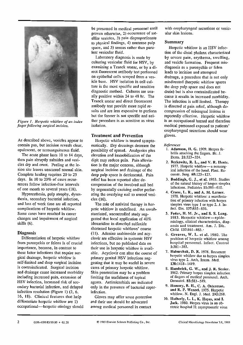
Figure 1. Herpetic whitlow of an index finger following surgical incision.
As described above, vesicles appear to contain pus, but incision reveals clear, opalescent, or serosanguineous fluid.
The acute phase lasts 10 to 14 days, then pain abruptly subsides and vesi- cles dry and crust. Peeling at the le- sion site leaves unscarred normal skin. Complete healing requires 20 to 25 days. In 10 to 20% of cases recur- rences follow infection-free intervals of one month to several years (18).
Hyperesthesia, pulp atrophy, dyses- thesia, secondary bacterial infection, and loss of work time are all reported complications of herpetic whitlow. Some cases have resulted in career changes and impairment of surgical skills (6).
Diagnos is Differentiation of herpetic whitlow
from paronychia or felons is of crucial importance, because, in contrast to these latter infections that require sur- gical drainage, herpetic whitlow is self-limited and deep surgical incision is contraindicated. Surgical incision and drainage cause increased morbidity including increased pain, extension of HSV infection, increased risk of sec- ondary bacterial infection, and delayed infection resolution (Figure 1) (2, 6, 16, 18). Clinical features that help differentiate herpetic whitlow are 1) occupational--herpetic etiology should
be presumed in medical personnel until proven otherwise, 2) occurrence of sat- ellite vesicles, 3) pain disproportionate to physical findings, 4) nontense pulp space, and 5) serous rather than puru- lent vesicular fluid.
Laboratory diagnosis is made by culturing vesicular fluid for HSV, by examining a Tzanck smear, or by a di- rect fluorescent antibody test performed on epithelial cells scraped from a ves- icle base. HSV isolation in cell cul- ture is the most specific and sensitive diagnostic method. Cultures are usu- ally positive within 24 to 48 hr. The Tzanck smear and direct fluorescent antibody test provide more rapid re- sults and are less expensive to perform but the former is not specific and nei- ther procedure is as sensitive as virus isolation.
Treatment and Prevent ion Herpetic whitlow is treated sympto-
matically. Dry dressings decrease the possibility of spread. Analgesics plus elevation and immobilization of the digit may reduce pain. Pain allevia- tion is the major concern, although surgical incision and drainage of the deep pulp space is detrimental. Pain relief has been reported after de- compression of the involved nail bed by segmentally excising and/or perfor- ating the overlying nail to unroof vesi- cles (16).
The role of antiviral therapy in her- petic whitlow is undefined. An unsub- stantiated, uncontrolled study sug- gested that local application of 40% idoxuridine in dimethyl sulfoxide shortened herpetic whitlows' course (1 I). Adenine arabinoside and acy- clovir are effective in systemic HSV infections, but no published data on their use in herpetic whitlow is avail- able. Acyclovir can alter the course of primary genital HSV infections sug- gesting that it may be useful in severe cases of primary herpetic whitlow. Skin penetration may be a problem limiting the usefulness of topical agents. Antimicrobials are indicated only in the presence of bacterial super- infection.
Gloves may offer some protection and their use should be advocated among medical personnel in contact
with oropharyngeal secretions or vesic- ular skin lesions.
S u m m a r y
Herpetic whitlow is an HSV infec- tion of the distal phalanx characterized by severe pain, erythema, swelling, and vesicle formation. Frequent mis- diagnosis as a paronychia or felon leads to incision and attempted drainage, a procedure that is not only misdirected (herpetic whitlow spares the deep pulp space and does not drain) but is also contraindicated be- cause it results in increased morbidity. The infection is self-limited. Therapy is directed at pain relief, although de- compression of subungual lesions is reportedly effective. Herpetic whitlow is an occupational hazard and therefore medical personnel exposed to patients' oropharyngeal secretions should wear gloves.
References 1. Adamson, H. G. 1909. Herpes fe-
brilis attacking the fingers. Br. J. Derm. 21:323-324.
2. Berkowitz, R. L., and V. R. Hentz. 1977. Herpetic whitlow--a nonsurg- ical infection of the hand. Plast. Re- eonstr. Surg. 60:125-127.
3. Buddingh, G. J., et al. 1953. Studies of the natural history of herpes simplex infections. Pediatrics 11:595-610.
4. Crane, L. R., and A. M. Lerner. 1978. Herpetic whitlow: a manifesta- tion of primary infection with herpes simplex virus type 1 or type 2. J. In- fect. Dis. 137:855-856.
5. Feder, H. M. Jr. , and S. S. Long. 1983. Herpetic whitlow--epide- miology, clinical characteristics, diag- nosis and treatment. Am. J. Dis. Child. 137:861-863.,
6. Greaves, W. L. et al. 1980. The problem of herpetic whitlow among hospital personnel. Infect. Control 1:381-385.
7. Haburchak, D. R. 1978. Recurrent herpetic whitlow due to herpes simplex virus type 2. Arch. Intem. Med. 138:1418-1419.
8. ltambrick, G. W., and J. R. Senior. 1962.-Primary herpes simplex infection of fingers of medical personnel. Arch. Dermatol. 85:583-589.
9. ltamory, B. H., C. A. Osterman, and R. P. Wenzel. 1975. Herpetic whitlow. N. Engl. J. Med. 292:268.
10. Hatherly, L. I., K. Hayes, and I. Jack. 1980. Herpes virus in an ob- stetric hospital II: asymptomatic virus
2 0196-4399/85/$0.00 + 02.20 © 1985 Elsevier Science Publishing Co.. Inc. Clinical Microbiology Newsletter 7:1, 1985

excretion in staff members. Med. J. Aust. 2:273-275.
11. Juel-Jensen, B. E. 1971. Herpetic whitlows: a medical risk. Br. Med. J. 4:681.
12. LaRossa, D., and R. Hamilton. 1971. Herpes simplex infections of the digits. Arch. Surg. 102:600-603.
13. Lindgren, K. M,, R. G. Douglas, Jr. , and R. B. Couch. 1968. Signifi- cance of herpes virus hominis in respi-
ratory secretions of man. N. Engl. J. Med. 278:517-523.
14. Nahmias, A. J., and B. Roizman. 1973. Infection with herpes simplex virus 1 and 2. N. Engl. J. Med. 289:781-789.
15. Nahmias, A., and W. E. Josey. 1983. Herpes simplex virus 1 and 2, pp. 351-372, In A. S. Evans (ed.), Viral infections of humans. 2rid ed. Plenum, New York.
16. Polayes, I. M., and M. S. Arons. 1980. The treatment of herpetic whi- tlow--a new surgical concept. Plast. Reconstr. Surg. 65:811-817.
17. Rosato, F. E., E. F. Rosato, and S. A. Plotkin. 1970. Herpetic parony- chia--an occupational hazard of med- ical personnel. N. Engl. J. Med. 283:804-805.
18. Stern, H. et al. 1959. Herpetic whit- low, a form of cross-infection in hos- pitals. Lancet 2:871-874.
E d i t o r i a l
Cost Effective Quality Control in Microbiology
Raymond C. Bartlett, M.D. Director, Division of Microbiology Hartford Hospital Hartford, Connecticut 06115
Historical Appra i sa l A wave of public concern, created
by the reporting and documentation of slipshod practices in some clinical lab- oratories, resulted in the Clinical Lab- oratory Improvement Act in 1967 (6). During the 1960s new medical tech- nology was introduced with the as- sumption that any added cost would produce a justifiable increase in health care quality. Now, however, new technology is being scrutinized for its cost effectiveness before implementa- tion and existing practices of question- able value are discontinued. As a re- suit, attention has been focused on the health care value of quality control testing. As laboratorians began to im- plement mandated quality control prac- tices, they found that many materials, procedures, and equipment rarely (and some never) displayed deficiencies. This caused many laboratorians to dis- continue such monitoring procedures. In other laboratories reluctant microbi- ologists refused to implement moni- toring practices of questionable useful- ness. Through inspection and accred- itation efforts regulators found that ob- taining complete compliance with the regulations as originally published was difficult. Progressive liberalization of the requirements occurred and opera-
tional guidelines were developed that often had inconsistencies between dif- ferent inspecting agencies and even geographic inconsistencies among in- spection teams from the same agency.
In 1964 our microbiology laboratory began conducting an extensive quality control program. In keeping with the attitude of the times, our program was originally quite extensive. After sub- stantial data had been accumulated, however, we began to decrease the frequency of various procedures and eliminate others. This practice did not jeopardize our own accreditation be- cause inspecting agencies accepted our extensive documentation and did not view us as noncompliant. I suspect that other laboratories could satisfy in- spectors by providing much objective evidence for curtailing or discontinuing quality control activity. Laboratorians must understand that the inspectors are caught in the middle and are torn be- tween accepting what seems rational and what they are required to expect and thus inconsistencies in their reac- tions should not be surprising. A compilation of data from numerous laboratories is needed for an organized approach to prioritizing quality control requirements based on observed cost effectiveness. This might lead to a ra- tional revision in the operational guidelines of inspecting agencies even if the regulations that were published in the Federal Register are not revised (7). In 1979 we reported an extensive study of the cost effectiveness of quality control in the bacteriology section of our microbiology labora- tory (5).
What is the health care benefit of spending $10, $100, $1,000, or $10,000 to pre~,ent one laboratory error? We must ask ourselves two questions. 1) What is the possibility that failure to detect and prevent that error would create an equivalent cost in the care of the patient affected? 2) How often will a laboratory error cause an error in diagnosis or treat- ment by the physician? The cost could express itself as prolonged hos- pitalization, performance of additional unnecessary laboratory tests or other diagnostic procedures, unnecessary treatment procedures, or administration of the wrong medications. More diffi- cult is the task of estimating the mor- bidity or mortality that might result from a laboratory error. Although nu- merical values are occasionally as- cribed to human life, attempts to ana- lyze cost effectiveness on this basis can easily appear to justify any cost. I believe that attempting to evaluate the usefulness of quality control practices on the basis of the health care benefit is premature.
Hartford Hospi ta l Exper ience We reviewed 62 surveillance proce-
dures within our quality control pro- gram that had been applied to different tests, materials, equipment, or per- sonttel functions. Some of the surveil- lance procedures had been conducted for up to 15 years and others for as few as 1 to 2 years. In all, 4421 sur- veillance procedures had been con- ducted over the 15 years that our quality control program had been in operation. The average number of
Clinical Microbiology Newsletter 7:1, 1985 © 1985 Elsevier Science Publishing Co.. Inc. 0196-4399/85/50.00 + 02.20 3