heparan sulfate chain valency controls syndecan-4 function in
TRANSCRIPT
Heparan Sulfate Chain Valency Controls Syndecan-4 Functionin Cell Adhesion*□S
Received for publication, August 18, 2009, and in revised form, February 5, 2010 Published, JBC Papers in Press, February 12, 2010, DOI 10.1074/jbc.M109.056945
Sandeep Gopal‡1,2, Adam Bober§1,3, James R. Whiteford§4, Hinke A. B. Multhaupt‡, Atsuko Yoneda‡,and John R. Couchman‡5
From the ‡Department of Biomedical Sciences, University of Copenhagen, 2200 Copenhagen N, Denmark and the §NationalHeart & Lung Institute, Faculty of Medicine, Imperial College London, London SW7 2AZ, United Kingdom
Fibroblasts null for the transmembrane proteoglycan, synde-can-4, have an altered actin cytoskeleton, compared withmatching wild-type cells. They do not organize �-smooth mus-cle actin into bundles, but will do so when full-length synde-can-4 is re-expressed. This requires the central V region of thecore protein cytoplasmic domain, though not interactions withPDZ proteins. A second key requirement is multiple heparansulfate chains. Mutant syndecan-4 with no chains, or only onechain, failed to restore the wild-type phenotype, whereasthose expressing two or three were competent. However,clustering of one-chain syndecan-4 forms with antibodiesovercame the block, indicating that valency of interactionswith ligands is a key component of syndecan-4 function.Measurements of focal contact/adhesion size and focal adhe-sion kinase phosphorylation correlated with syndecan-4 sta-tus and �-smooth muscle actin organization, being reducedwhere syndecan-4 function was compromised by a lack ofmultiple heparan sulfate chains.
Syndecans are an ancient group of transmembrane hepa-ran sulfate proteoglycans, with four members in every mam-mal examined to date (1–3). Syndecans associate with theactin cytoskeleton, and syndecan-4 in particular, is known asa focal adhesion component (3). In fibroblast adhesion stud-ies (4), it has been shown that the HepII domain of fibronec-tin will interact with the heparan sulfate chains of synde-can-4 to promote a signaling response that includes bindingand activation of protein kinase C� and downstream to theRho family of G proteins (4–6). Together with integrins,
syndecan-4 then promotes assembly of focal adhesions(3–7). Of the syndecan family, much focus has been on syn-decan-4 because of its role in adhesion, and also because keyelements of its structure and signaling have been elucidated.The cytoplasmic domain of syndecan-4 forms twisted clampdimers, stabilized by the presence of phosphatidylinositol4,5-bisphosphate (PtdIns4,5P2), which in turn bind to pro-tein kinase C�, becoming persistently activated (8, 9). It islikely that the dimer is the basal state of all syndecans,because the transmembrane domains of all syndecans formSDS-resistant homodimers by virtue of a highly conservedGXXXG motif (10, 11).One report suggests that heparan sulfate chains are not
required for focal adhesion promotion (12), in this casewhere syndecan-4 cDNA was transfected into a CHO cellmutant incapable of glycosaminoglycan synthesis. However,syndecan-4 does also possess a site in its external core protein,including an NXIP motif, which triggers integrin-mediated celladhesion (13). A similar principle has been demonstrated forsyndecan-1, where �3 or �5 integrins are influenced directlyby a portion of the ectodomain, now known as synstatin (14).However, most studies have indicated a need for heparan sulfatesubstitutionon syndecan-4, and some fine structure requirementsof the chains for interaction with fibronectin, have been eluci-dated. These include N-sulfation (but not 2-O-sulfation) and sul-fated subdomains of �12 sugar residues (15).
The syndecan-4 knock-out mouse has vascular repair andcell migration defects (16). It also has defects in skeletal mus-cle regeneration (17). Cell migration defects are not cor-rected by selected growth factors, suggesting that there arealtered cell adhesion properties (16). This is consistent with invitro studies showing a role for the proteoglycan in directionallypersistentmigration (18) and zebrafish studies demonstrating asimilar role in neural crest migration (19). One report suggeststhat only under limited circumstances is focal adhesion assem-bly compromised in null fibroblasts (20), but a second reportindicates that a difference in �-smooth muscle actin (�SMA)6organization is observable in knock-out cells (21). Whereaswild-type mouse embryo fibroblasts have a high proportionwith this actin isoform incorporated into stress fibers, synde-can-4-null cells do not, though this can be corrected by exoge-nous transforming growth factor-� (21). This difference in
* This work was supported initially by Wellcome Trust Programme Grant065940 (to J. R. C.), then by the Danish National Research Foundation,Haensch Foundation, Wilhelm Pedersen Fonden through the Novo Nor-disk Fonden, and the Department of Biomedical Sciences at the Universityof Copenhagen (to J. R. C.).Author’s Choice—Final version full access.
□S The on-line version of this article (available at http://www.jbc.org) containssupplemental Fig. S1 and Table S1.
1 Both authors contributed equally to this work.2 Supported by the Molecular Medicine Ph.D. program at the University of
Copenhagen.3 Supported by a Medical Research Council UK (MRC) studentship.4 Present address: Centre for Microvascular Research, William Harvey
Research Institute, Barts and the London School of Medicine & Dentistry,Queen Mary University of London, UK.
5 To whom correspondence should be addressed: Dept. of Biomedical Sci-ences, University of Copenhagen, Biocenter, Ole Maaløes Vej 5, 2200Copenhagen N, Denmark. Tel.: 45-353-25670; Fax: 45-353-25669; E-mail:[email protected].
6 The abbreviations used are: �SMA, �-smooth muscle actin; FAK, focal adhe-sion kinase; HA, hemagglutinin; PBS, phosphate-buffered saline; MEF,murine embryonic fibroblast; GFP, green fluorescent protein.
THE JOURNAL OF BIOLOGICAL CHEMISTRY VOL. 285, NO. 19, pp. 14247–14258, May 7, 2010Author’s Choice © 2010 by The American Society for Biochemistry and Molecular Biology, Inc. Printed in the U.S.A.
MAY 7, 2010 • VOLUME 285 • NUMBER 19 JOURNAL OF BIOLOGICAL CHEMISTRY 14247
by guest on January 7, 2019http://w
ww
.jbc.org/D
ownloaded from

cytoskeletal organization is used here as an assay for synde-can-4 functionality.A continuing mystery is how ligand binding to heparan sul-
fate on the cell surface triggers cytoplasmic signaling events insyndecan-4. Because this proteogly-can becomes clustered into focaladhesions, an obvious possibility isthat lateral association of synde-can-4 dimers is sufficient to drivethe process. At the same time, allsyndecans are potentially multiva-lent, given that each core proteinhas at least three Ser-Gly motifssuitable for substitution with glyco-saminoglycans (3, 6, 8).Where stud-ied, it appears that glycanation ismaximal, i.e. if three sites are avail-able, each tends to be substitutedwith a chain (22). If this applies tosyndecan-4 on the cell surface, thenup to six heparan sulfate chainsper core protein dimer may beavailable. The question arises, how-ever, whether multiple chains are infact required to promote a signalingresponse. To address this, we pre-pared all possible variants of synde-can-4, ranging from the wild typewith three Ser-Gly dipeptides, totwo, one, and no Ser-Gly dipeptides,eight variants in total. In mutantforms, key serine residues weremutated to alanine. These cDNAconstructs were introduced intosyndecan-4-null cells and assessedfor their ability to restore a wild-type actin cytoskeleton. The datasuggest that a functional synde-can-4 dimer requires a minimumof four heparan sulfate chains, un-derlining the importance of va-lency, and suggesting that undernormal circumstances, two synde-can-4 dimers must associate in thepresence of ligand, to form a sig-naling unit.
EXPERIMENTAL PROCEDURES
Cell Culture and Transfection—Syndecan-4-null (S4KO) and match-ing wild-type murine embryonicfibroblasts (MEFs) (20) were grownand maintained in �MEM (Lonza)supplemented with 10% v/v fetalbovine serum (FBS). COS7 cellswere grown in Dulbecco’s modifiedEagle’s medium (DMEM, GIBCO)containing 10% FBS and 2% glu-
tamax. Syndecan-4-null fibroblast and COS7 transfectionswere performed using Lipofectamine (Invitrogen) or Lipo-fectamine LTX (Invitrogen) respectively, according to theman-ufacturers’ instructions.
Syndecan-4 and the Cytoskeleton
14248 JOURNAL OF BIOLOGICAL CHEMISTRY VOLUME 285 • NUMBER 19 • MAY 7, 2010
by guest on January 7, 2019http://w
ww
.jbc.org/D
ownloaded from

Antibodies—Antibodies used included monoclonal anti-�-smoothmuscle actin antibody (Clone 1A4; Sigma), anti-paxillinantibody (clone Z035; Zymed Laboratories Inc.), monoclonalanti-HA (Clone HA.11; Covance), polyclonal goat anti-�-acti-nin (C-20; Santa Cruz Biotechnology), polyclonal rabbit anti-bodies against focal adhesion kinase (FAK; BD Pharmingen),and tyrosine 397-phosphorylated FAK (Cell Signaling), mono-clonal mouse anti-Rac (Clone 23A8;Millipore), polyclonal rab-bit anti-RhoA (119; Santa Cruz Biotechnology), polyclonal goatanti-ezrin (C-19; Santa Cruz Biotechnology), polyclonal rabbitanti-HA (SG. 77; Zymed Laboratories Inc.), and monoclo-nal anti-�-tubulin (Clone TUB 2.1; Sigma). Syndecan-4 anti-body was raised in chickens against a synthetic peptide corre-sponding to the N terminus of the syndecan-4 core protein asdescribed (15).Preparation of Fibronectin 110-kDa (III3–III11 Repeats) Frag-
ment and HepII Domain—Fibronectin was purified from freshhuman plasma by adapting the protocol of Miekka et al. (23).The enzymatic cleavage and purification of the central, inte-grin-binding 110-kDa fibronectin fragment was as previously(15). Recombinant His-tagged HepII (FN repeats III12–15)domain was expressed in Escherichia coli BL21 cells and puri-fied with His select� cobalt affinity gel (Sigma) according to themanufacturer’s instructions. The purity of the protein was con-firmed by SDS-PAGE with Coomassie Blue staining. The con-struct in pQE-30 was a kind gift from Dr. Jean Schwarzbauer(Princeton University).GAG Chain Mutagenesis—The three serine residues that
serve as syndecan-4 glycosaminoglycan attachment siteswere mutated using a two step overlap PCR approach asfollows. During the first step 5� and 3� PCR products weregenerated from full-length rat syndecan-4 cDNA usingprimers complementary to the 5� and 3� non-coding regionsof syndecan-4 and reverse and forward primers containingpoint mutations so that one serine residue (44, 65, or 67) wasmutated to alanine (supplemental Table S1). The resultanttwo PCR products were combined and used as template ina second reaction using S4for and S4rev primers to gener-ate full-length syndecan-4 cDNA. EcoRI and BamHI restric-tion sites were incorporated into these primer sequences,and PCR products were digested with these enzymes andligated into the corresponding sites of pIRES2-EGFP (Clon-tech) using standard procedures. Using the primers de-scribed in supplemental Table S1 it was not only possible togenerate single Ser3Alamutations at each position but alsoto generate all combinations of the three mutations (Fig. 2).
A similar approach was used to fuse the HA epitope se-quence between isoleucine 32 and aspartate 33 in the synde-can-4 constructs. Primers S4for and S4rev were used in con-junction with primers containing the HA coding sequence togenerate 5� and 3� PCR products from each of the 6 mutatedsyndecan-4 cDNAs and the wild-type sequence. Full-lengthproducts were obtained for each cDNA, and these weredigested and ligated into pIRES2-EGFP as described. Theexpression of each cDNA was analyzed in COS7 cells byWestern blots with anti-HA antibody followed by horserad-ish peroxidase-conjugated goat anti-mouse antibody (Dako).In control experiments, cell layers were treated with chon-droitinase ABC (Sigma Aldrich) before performing theSDS-PAGE to remove chondroitin and dermatan sulfatechains from syndecan-4 core protein. Syndecan-4 mutantcDNAs encoding truncated cytoplasmic domains were pre-pared as previously (24), and the HA coding sequence wasinserted as described above. To create single amino acid sub-stitution of either Tyr-184 or Tyr-192 with phenylalanine insyndecan-4 cytoplasmic domain (25), syndecan-4 cDNAsencoding those sequences in pIRES2-EGFP were con-structed using overlap PCR extension methods. The cell sur-face expression of recombinant syndecan-4 was confirmedin COS7 by flow cytometry. Cells expressing wild-type andrecombinant syndecan-4 were harvested using cell dissocia-tion buffer (Invitrogen), resuspended in 1% bovine serumalbumin (BSA) in PBS; then the live cells were stained usinganti-HA antibody in 1% BSA and rabbit anti-mouse Alexa-fluor 647 IgG (Molecular Probes). Dead cells were identified bystaining with 20 �g/ml propidium iodide solution (BD Bio-science). Controls were prepared by staining with Alexafluor647-conjugated anti-mouse IgG without primary antibodies.Samples were analyzed using a FACS Calibur (Becton Dickin-son) and analyzed with CellQuest Pro software.Immunofluorescence Microscopy and Western Blotting—
Transfected syndecan-4-null mouse fibroblasts were seededonto coverslips in growth medium for 24 h prior to serumstarvation for a further 24 h. Cells were fixed in 4% paraform-aldehyde in PBS and permeabilized with 0.1% Triton X-100in PBS for 10 min; then stained using conventional proce-dures using a primary antibody to �SMA and Alexafluor568-conjugated goat-anti mouse IgG (Molecular Probes).Samples were analyzed on a Zeiss Axioplan-2 microscope(�40 objective), and images were processed using Meta-morph and Adobe Photoshop. For focal adhesion area mea-surement, the structures were stained with paxillin antibody,
FIGURE 1. Syndecan-4 regulates �SMA incorporation into fibroblast stress fibers. A, rat syndecan-4 protein sequence showing the signal peptide in normaltext and the mature protein in bold. Serine residues 44, 65, 67 in the serine-glycine consensus sequences (red text) are the putative sites of glycosaminoglycanchain substitution. Sites of cytoplasmic domain truncation �199E (terminating at Glu-199) and �191I (terminating at Ile-191) are marked by an asterisk. TheY192F and Y184F mutations are marked with an arrow. The site of HA tag insertion is shown, as are the subdomains of the cytoplasmic domain, C1, V, and C2.B, wild-type and syndecan-4-null murine fibroblast showed differences in the stress fiber and focal adhesion phenotype when stained with phalloidin andanti-paxillin antibodies. C, similarly, immunostaining shows incorporation of �SMA into stress fibers of wild type, but not syndecan-4-null fibroblasts. Thewild-type �SMA phenotype of stress fibers in syndecan-4-null MEF is restored by the reintroduction of rat syndecan-4 (SDC4) but not transfection of theequivalent empty vector (pIRES2-EGFP). A series of mutant syndecan-4 cDNA transfections shows that the C2 region is dispensable, but the V region andtyrosine residues 184 and 192 are essential for restoration of a wild-type �SMA phenotype. For all transfections except SDC4-�199E and SDC4-�191I, thetransfected cells were identified by expression of GFP encoded in the pIRES-EGFP plasmid. For SDC4-�199E and SDC4-�191I the transfected cells are distin-guished from untransfected cells by immunostaining for syndecan-4 expressed on cell surface. D, for each transfection represented in C, �100 cells werecounted, and the data are means � S.E. from triplicate experiments. E, Western blots show that protein levels of �SMA are equivalent in wild-type andsyndecan-4-null fibroblasts. The scale bar is marked in each panel.
Syndecan-4 and the Cytoskeleton
MAY 7, 2010 • VOLUME 285 • NUMBER 19 JOURNAL OF BIOLOGICAL CHEMISTRY 14249
by guest on January 7, 2019http://w
ww
.jbc.org/D
ownloaded from
followed by Alexafluor 568-conju-gated goat anti-mouse IgG. Thearea of each focal adhesion wasmeasured and calibrated usingMetamorph and ImageJ imagingsoftware. For each transfection aminimum of 100 cells was ana-lyzed for focal adhesion area.Transfected cells were identifiedby the expression of GFP from thebicistronic vector or by immuno-logical detection of the HA insertin the syndecan-4. For fibronectindomain experiments, transfectedsyndecan-4-null fibroblasts wereseeded onto coverslips coated withfibronectin (1 �g/coverslip) or FN110 (1 �g/coverslip) in growthmedium, followed by incubationfor 3–4 h and HepII (1–10 �g/ml)solution was added to themedium.Cells were stained for focal adhe-sion components, and the per-centage of cells containing theseorganelles was assessed, with aminimum of 100 cells per condi-tion. For all experiments trans-fected cells were identified by theexpression of GFP or by immuno-logical detection of the HA tag inthe syndecan-4 core protein. Tocompare the expression of differ-ent focal adhesion proteins inwild-type and syndecan-4-nullmurine fibroblasts, Western blotswere performed with antibodiesagainst paxillin, �-actinin, Rac 1,Rho A, and ezrin.Analysis of Focal Adhesion Ki-
nase—Syndecan-4-null MEFs weretransfected with cDNAs correspond-ing towild type, andglycosaminogly-can chain substitution mutants ofsyndecan-4 and allowed to expressthe protein for 36 h. The expressionof FAK and the level of its phosphor-ylation was analyzed by Westernblotting with anti-FAK and anti-phospho-FAK (tyrosine 397) anti-bodies followed by horseradish per-oxidase-conjugated goat anti-rabbitantibody. Western blots were cali-brated by quantitation of �-tubulinas a loading control.Syndecan-4 Clustering Experi-
ments—Transfected syndecan-4-nullfibroblasts were seeded on glasscoverslips in the presence of serum
Syndecan-4 and the Cytoskeleton
14250 JOURNAL OF BIOLOGICAL CHEMISTRY VOLUME 285 • NUMBER 19 • MAY 7, 2010
by guest on January 7, 2019http://w
ww
.jbc.org/D
ownloaded from

for 24 h prior to serum starvation for a further 24 h. Syndecan-4clustering was achieved by first incubating cells with 10 �g/mlof anti-syndecan-4 polyclonal antibody in serum-free medium(15) for 15 min at 37 °C prior to washing twice with PBS. Cellswere then treated with Alexaflour 568-conjugated goat anti-chicken IgY (Invitrogen; 1 �g/ml in serum-free medium) for 15min at 37 °C prior to fixation. The cells were then convention-ally stained for �SMA as described as above.Syndecan-4 Glycosaminoglycan Analysis—COS7 cells ex-
pressing wild-type and recombinant syndecan-4 mutantswere washed with PBS and DMEM was supplied withoutFBS. Cultures were metabolically labeled with 35S (Na2SO4;specific activity 43Ci/mg sulfur; MP Biomedicals) at a con-centration of 140–150 �Ci/ml. The cells were incubated for48 h at 37 °C in a CO2 incubator. Syndecan-4 was purified byimmunoprecipitation with a polyclonal anti-HA antibody(Zymed Laboratories Inc.) using protein-A beads. Radiola-beled glycosaminoglycan chains were cleaved from synde-can-4 core protein by alkaline elimination (0.1 M NaBH4 in0.1 M NaOH) for 24–30 h at 4 °C, followed by neutralizationwith 4% HCl. For gel filtration chromatography, the heparansulfate chains (�8000 cpm) were applied to a Superdex 20010/300GL gel filtration column (GE Healthcare) pre-equili-brated in 50mMNaH2PO4 (pH 7.0) containing 150mMNaCl.The heparan sulfate chains were eluted in the same buffer ata flow rate of 0.4 ml/min. The column was calibrated withblue dextran (2000 kDa), ovalbumin (42 kDa), and vitaminB12 (1.35 kDa). Eluted fractions were collected and analyzedfor radiolabel content by scintillation spectroscopy. Foranion-exchange chromatography, metabolically labeledheparan sulfate chains were applied to a MonoQ10/100GLcolumn (GE Healthcare). The column was pre-equilibratedwith 20 mM Tris-HCl containing 0.5 M NaCl. The unboundradioactivity was removed by washing columnwith 5 columnvolumes of 20 mM Tris-HCl containing 0.5 M NaCl. Boundradioactivity was eluted in a linear gradient of NaCl (0.5–2.0M) in 20 mM Tris-HCl buffer (10 column volumes). Theeluted fractions were collected and analyzed for radiolabelcontent.Low pH (1.5) nitrous acid treatment of heparan sulfate
chains was performed according to Shively and Conrad (26),to cleave the heparan sulfate chains at N-sulfated glucos-amine residues. Immunoprecipitated syndecan-4 was alsotreated with 5 milliunits of chondroitinase ABC (Sigma, EC4.2.2.4) in 50 mM Tris pH 8.0, 60 mM sodium acetate at 37 °Cfor 16h. Heparinase III treatments were as described (27).Briefly, 35S-labeled heparan sulfate chains from purified syn-decan-4 were mixed with unlabeled heparan sulfate (0.01mg/ml final) and incubated with 1.5 milliunits of heparinaseIII (heparinase EC 4.2.2.8, Seikagaku America) for 6 h at37 °C in 0.1 M sodium acetate, pH 7.0 containing 0.1 mM
calcium acetate. Another 1.2-milliunit aliquot of the enzymewas added after 6 h, and the mixture was incubated for 16 h.The products from nitrous acid, chondroitinase ABC andheparinase III treatments were applied to Superdex 75 col-umn (GEHealthcare) pre-equilibrated with 50mMNaH2PO4(pH 7.0) containing 150 mM NaCl. The column was cali-brated with commercially available protein standards anddisaccharide, �UA-GlcNS (462 Da; Iduron UK).
RESULTS
Syndecan-4-null Cells Have an Altered Actin Cytoskeleton—Two independent strains of newborn and embryonic synde-can-4-null fibroblasts exhibit the same defect in organiza-tion of their actin cytoskeleton. On fibronectin substrates,these cells have �-actin containing microfilament bundles,often oriented circumferentially, and small focal contacts(Fig. 1B). These focal contacts contained several expectedmarker proteins including paxillin (Fig. 1B), vinculin, talin,and phosphotyrosine. In contrast, wild-type cells have moresubstantial stress fibers, frequently running from end-to-end, and larger focal adhesions (Fig. 1B). A key differencehowever, is the organization of �SMA. This is a stress fibercomponent of normal mouse (Fig. 1C), and rat fibroblastsbut is not a component of the finer microfilament structuresof the null cells (Fig. 1C). Quantitative data are shown in Fig.1D. This difference is not a result of decreased �SMA proteinlevels in the null cells (Fig. 1E) but its organization. More-over it can be overcome by transfection of null cells withcDNA constructs encoding wild type syndecan-4 (Fig. 1C).These patterns of �SMA organization are the phenotypicmarker for subsequent experiments where mutated forms ofsyndecan-4 are expressed in null cells to determine theparameters necessary for reconstitution of the wild-typephenotype. The role of the syndecan-4 cytoplasmic domainwas confirmed by transfection of a series of mutant con-structs, shown in Fig. 1A. Deletion of the three C-terminalamino acids, which prevents PDZ proteins from binding, hadno effect on syndecan-4 ability to restore a normal actincytoskeleton (Fig. 1C). However, a truncation to the centerof the V region (terminating at Ile-191) prevented a restora-tion of actin cytoskeleton. Similarly, mutation of either oftwo tyrosine residues at proximal and distal ends of the Vregion rendered the syndecan-4 non-functional in theseassays (Fig. 1C). Quantitation of these experiments is shownin Fig. 1D. These data suggest that signaling through thecytoplasmic domain, and/or interactions with actin-associ-ated proteins, is critical to the �-smooth muscle phenotypepromoted by syndecan-4. FACS data confirmed that all con-structs were expressed on the cell surface of the fibroblasts(supplemental Fig. S1) to broadly equivalent levels.
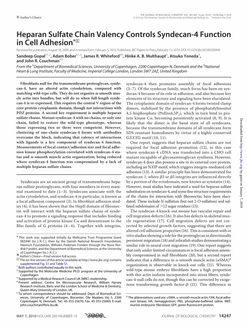
FIGURE 2. A minimum of two heparan sulfate chains on syndecan-4 core protein are required to restore a normal actin cytoskeleton in syndecan-4-nullfibroblasts. A, syndecan-4 variants, where the serine residues 44, 65, and 67 were mutated to alanine either individually or in combination, are showndiagrammatically. Each HA-tagged mutant was expressed in COS7 cells and lysates analyzed by Western blot with an HA tag-specific antibody. The hetero-geneous products are consistent with GAG substitution. In the AAA mutant with no glycosaminoglycan chains, dimeric core protein was detectable as a 40-kDapolypeptide. B, immunostaining of the syndecan-4-null fibroblasts transfected with wild-type and glycosaminoglycan chain mutant syndecan-4 constructsshowed that syndecan-4 can mediate signaling to incorporate �SMA into stress fibers if at least two of three serine residues are substituted by heparan sulfatechains. This is quantified in C. The scale bar is 10 �m.
Syndecan-4 and the Cytoskeleton
MAY 7, 2010 • VOLUME 285 • NUMBER 19 JOURNAL OF BIOLOGICAL CHEMISTRY 14251
by guest on January 7, 2019http://w
ww
.jbc.org/D
ownloaded from
Multiple Heparan Sulfate ChainsAre Required for Syndecan-4 Func-tion in Cell Adhesion—Null cellstransfected with wild-type synde-can-4 cDNA become essentiallyidentical to wild-type cells within48 h of transfection, when examin-ing the distribution of �SMA (Fig.2B). Correspondingly, focal adhe-sions were much enhanced in sizeand number (Fig. 3). In contrast, anempty vector control had no influ-ence on cell morphology or cytoskel-eton (Figs. 2B and 3). To directlyascertain how many glycosamino-glycan chains were required for thephenotype restoration and whethertheir position on the core proteinwas important, all possible combi-nations of mutations in the threeserine residues of the N-terminalregion of the core protein were pro-duced (Fig. 2A). ComplementaryDNAs for all six mutants weretransfected into COS7 and nullmouse embryo fibroblasts and thecytoskeleton examined 48 h later.Glycanation of each syndecan-4
variant was examined by Westernblotting of COS7 cell lysates, intowhich HA-tagged forms for synde-can-4 cDNA had been transfected(Fig. 2A). Where the AAA mutantwas expressed, a single prominentpolypeptide of �40 kDa was de-tected, consistent with an SDS-re-sistant dimeric core protein (9, 10).Where a single serine was present(SAA, ASA, and AAS), glycanationwas apparent, with heterodispersepopulations of syndecan-4 visible.This confirmed the presence of gly-cosaminoglycan in all three variantsof syndecan-4. Similar heterogene-ous populations of syndecan-4 weredetected by Western blotting ofSSA, SAS, ASS, and SDC4 (wild-type) forms of syndecan-4, thoughall extended into higher massregions of the gel than the “singlechain” forms. The SDC4 materialhad the highest mass range of all,and the data are consistentwith syn-decan-4 being substituted on eachavailable serine residue, as notedpreviously (22).When the syndecan-4 cDNA
constructs were transfected into
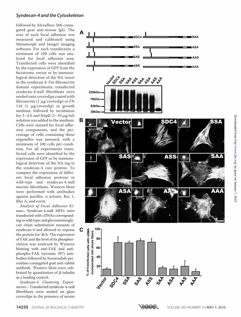
FIGURE 3. Syndecan-4 with multiple heparan sulfate chains promotes larger focal adhesions withincreased FAK activity. A, wild-type, syndecan-4-null, and syndecan-4 re-expressing-null fibroblasts werestained for the focal adhesion component paxillin. In each case focal contacts or focal adhesions were formed,but those of null cells, or those expressing syndecan-4 with one or no heparan sulfate chains were smallerin area than wild-type cells or those expressing syndecan-4 with two or three heparan sulfate chains.Quantification of 75–100 adhesions, from �50 cells per condition is shown in B. C, Western blot of focaladhesion proteins and cytoskeletal regulators in both wild-type and syndecan-4-null fibroblasts revealedthat protein expression levels were mostly comparable, although the latter contain less FAK protein.D, FAK activity was assessed from the extent of tyrosine 397 phosphorylation by Western blotting with aspecific antibody. Null cells and those expressing syndecan-4 with one chain have lower FAK activity thannull cells expressing two or three chains. FAK protein and tyrosine 397 phosphorylation are calibrated to�-tubulin levels. In C and D, quantitation is the mean of three independent experiments. Scale bar is50 �m.
Syndecan-4 and the Cytoskeleton
14252 JOURNAL OF BIOLOGICAL CHEMISTRY VOLUME 285 • NUMBER 19 • MAY 7, 2010
by guest on January 7, 2019http://w
ww
.jbc.org/D
ownloaded from

null fibroblasts, the results showed that at least two of threeserine residues were required in order that �SMA becameincorporated into microfilament bundles. Quantitative dataare shown in Fig. 2C. However, and importantly, it did notmatter which two of three serine residues were present(Figs. 2B and C). In all cases though, where only one serineresidue was present, allowing only one heparan sulfate chain
substitution on the core protein,no restoration of normal cytoskel-etal phenotype was seen. The datatherefore suggest that two or moreheparan sulfate chains are re-quired to interact with the matrixsubstrate for productive synde-can-4 function. In each case, fo-cal adhesion characteristics fol-lowed that of the microfilamentarchitecture, being larger andmore numerous where �SMA wasincorporated into microfilamentbundles (Fig. 3). Quantitativeanalysis showed that focal adhe-sions were �5 �m2 in null cells,and where single chain variants ofsyndecan-4 were expressed. Incontrast, focal adhesions were onaverage �10 �m2 in wild-typecells and those null cells express-ing syndecan-4 with two or threeheparan sulfate chains (Fig. 3B). Incontrol experiments, cell surfaceexpression of each syndecan-4variant in terms of heparan sulfatechain composition was confirmedby FACS analysis (supplementalFig. S1). The difference in focaladhesion size between null andwild-type cells was not related tothe expression level of proteincomponents such as �-actinin,paxillin, Rac1, RhoA, or ezrin pro-teins that were comparable in syn-decan-4-null and wild-type cells(Fig. 3C). This suggests that orga-nization, rather than proteinexpression levels, is relevant tosyndecan-4 promotion of focaladhesions and large microfilamentbundles containing �SMA. How-ever, null cells reproduciblyexpressed �30% less FAK proteinthan wild-type cells (Fig. 3C), andthis was further studied by exami-nation of FAK activity.Tyrosine 397 phosphorylation
of FAK is an index of kinase ac-tivity (28). Syndecan-4-null fibro-blasts together with transfectants
encoding one, two, or all three (wild type) were examinedby Western blotting with a tyrosine 397 FAK phosphospe-cific antibody (Fig. 3D). The results were consistent withfocal adhesion size; lower levels of phosphorylation weredetected in null cells and those expressing syndecan-4 withonly one glycosaminoglycan chain. Higher levels were pres-ent in cells expressing wild-type syndecan-4 and a mutant
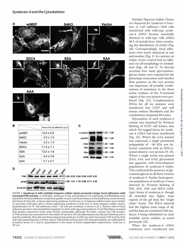
FIGURE 4. Clustering of syndecan-4 bearing one or no heparan sulfate chains, with antibodies triggersincorporation of �SMA into stress fibers. A–L, syndecan-4-null mouse fibroblasts were transfected with thesyndecan-4 cDNAs encoding none or one GAG attachment site. Transfected cells were grown in the presenceof serum for 24 h prior to serum starvation for 24 h. Syndecan-4 clustering was achieved with a polyclonalanti-syndecan-4 antibody raised against an N-terminal peptide sequence, followed by an Alexafluor-568 con-jugated anti-chicken IgY. Transfected cells were identified by GFP from the bicistronic vector, cell surfacesyndecan-4 visualized directly, and the �SMA detected by an Alexafluor 405 antibody (blue). M, percentage oftransfected cells with �SMA incorporated into stress fibers was calculated in syndecan-4-expressing cells thathad been clustered as above (black bars) or remained untreated (light gray bars). n � 110 –130 cells from eachcondition. The scale bar is 10 �m.
Syndecan-4 and the Cytoskeleton
MAY 7, 2010 • VOLUME 285 • NUMBER 19 JOURNAL OF BIOLOGICAL CHEMISTRY 14253
by guest on January 7, 2019http://w
ww
.jbc.org/D
ownloaded from
expressing two chains, these having larger focal adhesions(Fig. 3).As a further test of heparan sulfate chain number and valency
in cell-matrix interactions, mouse fibroblasts expressing onlyone chain per syndecan-4 core pro-tein (i.e. SAA, ASA, and AAS forms)were clustered with a core protein-specific polyclonal antibody, di-rected to the N-terminal region,before fixation and staining. In nocase was a wild-type phenotypereconstituted.Moreover, the synde-can-4 remained diffusely present onthe cell surface with no evidence ofclustering (not shown). In contrast,where both a primary anti-synde-can-4 antibody and a secondaryantibody were sequentially added tocells expressing single serine synde-can-4 constructs, a restoration ofcytoskeletal phenotype was seen inmany cells (Fig. 4,A–I andM). Here,syndecan-4 was clustered by thedouble layer of antibody. Onceagain, it did not matter in whichposition the serine residuewas pres-ent, �SMA incorporation into mi-crofilament bundles was inducedby the clustering treatments. Be-cause it has been reported that clus-tering of syndecan-4 expressed in aCHO mutant lacking heparan sul-fate synthesis could lead to micro-filament bundle and focal contactformation (12), a similar experi-ment was carried out with a AAAmutant form of syndecan-4 trans-fected into null cells. Whereas theeffects were weaker, nevertheless,some focal adhesion and �SMA-positive microfilaments were ob-served (Fig. 4, J–M).
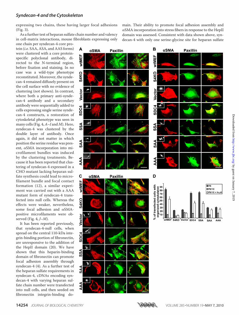
It has been reported previously,that syndecan-4-null cells, whenspread on the central 110-kDa inte-grin-binding portion of fibronectin,are unresponsive to the addition ofthe HepII domain (20). We haveshown that this heparin-bindingdomain of fibronectin can promotefocal adhesion assembly throughsyndecan-4 (4). As a further test ofthe heparan sulfate requirements insyndecan-4, cDNAs encoding syn-decan-4 with varying heparan sul-fate chain number were transfectedinto null cells, and then seeded onfibronectin integrin-binding do-
main. Their ability to promote focal adhesion assembly and�SMA incorporation into stress fibers in response to the HepIIdomain was assessed. Consistent with data shown above, syn-decan-4 with only one serine-glycine site for heparan sulfate
Syndecan-4 and the Cytoskeleton
14254 JOURNAL OF BIOLOGICAL CHEMISTRY VOLUME 285 • NUMBER 19 • MAY 7, 2010
by guest on January 7, 2019http://w
ww
.jbc.org/D
ownloaded from

substitution was not able to restore a wild-type phenotype innull cells (Fig. 5), while wild-type syndecan-4 or mutants lack-ing only one substitution site, were responsive to the HepIIdomain.Heparan Sulfate Chain Length Is Unaltered Where Chain
Number Is Reduced, but Sulfation Density Can Vary—Animportant factor where glycosaminoglycan chain number wasmanipulated, was to ascertain heparan sulfate chain size,because thismight affectmigration on SDS-PAGE aswell as thebiological responses of cells expressing the chains. This wasdetermined for each mutant and wild-type syndecan-4expressed in COS7 cells by immunoprecipitationwithHA-spe-cific antibodies, then gel chromatography of the glycosamino-glycan chains released by alkaline elimination (Fig. 6). Regard-less of whether the chains were derived from wild-type ormutant forms of syndecan-4 with reduced chain number, chainsize was consistent and within the range of 36–42 kDa. Thiscorresponds well with previous data from cell surface proteo-glycan analyses (29).The properties of the heparan sulfate chains from each of
the expressed syndecan-4 molecules were also analyzed bygradient anion exchange chromatography. Here differencesin elution profile were noted, indicating variation in sulfa-tion density that depended on chain number and their ori-gin. Chains from wild-type syndecan-4 transfectants hadthe highest charge density, equaled by chains from the AASmutant. Those with the lowest charge density originatedfrom the SAA mutant form of syndecan-4, with others beingintermediate. This interesting variation in chain character-istics appeared to have two underlying themes. Generally,chain number was proportional to charge density. For exam-ple SAS-derived chains had higher charge density than thosefrom SAA, while SDC4 had the highest density. The otherconsideration is position of the chain on the core protein.Chains closer to the N terminus, particularly serine 44,had lower charge density than more membrane-proximalchains, e.g. on serine 67. Charge density was in the orderSAA�ASA�AAS. However, it was also the case that chainsfrom SAS, for example, were intermediate in charge densitybetween SAA and SDC4 (or AAS), but only present as asingle population, not two populations of lower and highercharge density.In further experiments, the glycosaminoglycans from SAA,
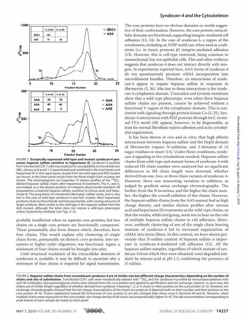
ASA, AAS, ASS, and wild-type syndecan-4 from 35sulfate-la-beled cultures were treated with chondroitinase ABC, hepa-rinize III, or nitrous acid (pH 1.5). The results showed thatchains from AAS and wild-type syndecan-4 were close to 100%
heparan sulfate, but the others had varying chondroitin/derma-tan sulfate content (Fig. 7). All populations contained heparansulfate cleaved by nitrous acid, generatingmostly disaccharidesand tetrasaccharides, therefore indicating the presence ofN-sulfate. The heparinase III degradation profiles were broadlysimilar for chains from each form of syndecan-4. However, tet-rasaccharides and larger saccharides were not as abundant inchains derived from the SAA and ASA mutants of syndecan-4comparedwith heparan sulfate fromothermutants orwild type(Fig. 7).This has implications for the regulation of heparan sulfate
synthesis, but does not, in fact, bear on syndecan-4 functionin regulation of the cytoskeleton. This is because two-chainforms of syndecan-4 were functional, even though the sulfa-tion was less than in the wild type. Similarly, AAS forms ofsyndecan-4 were not functional, unless clustered by antibod-ies, yet had similar charge density and heparan sulfate char-acteristics to chains derived from the SDC4 (wild type).
DISCUSSION
Syndecan-4 is unique among the four mammalian familymembers in being a focal adhesion component and promot-ing their assembly in response to heparin-binding domainsof fibronectin (3, 4). All syndecans have been reported toassociate with the actin cytoskeleton, and interactions with�-actinin, cortactin and ezrin have been reported (3, 6, 8).Moreover, signaling to second messengers has been demon-strated, most notably in syndecan-4 where in the presence ofPtdIns4,5P2 and protein kinase C�, downstream signaling toRhoGTPases occurs (3, 5, 6, 8). Evidence from in vivo and invitro studies suggests a role for syndecan-4 in cell adhesionand particularly, cell migration (18, 19, 24). This is coupledto the fact, shown before and here, that there is a distinctionin actin cytoskeleton organization when wild-type and syn-decan-4-null cells are compared (21). The incorporation of�SMA into stress fibers appears to be syndecan-4 depen-dent, and may indicate a decreased ability of null cells toexert tension on extracellular matrix substrates (8, 31). Fur-thermore, previous data (32), and that presented here, sug-gest that syndecan-4 can influence FAK activity. Wild-typesyndecan-4 when expressed in null cells had higher levels ofFAK tyrosine 397 phosphorylation than the parent null cells,or those cells expressing syndecan-4 with only one glycos-aminoglycan chain. FAK activity data were commensuratewith the extent of focal adhesion assembly. How syndecan-4regulates FAK activity is not known, but may be indirect,
FIGURE 5. Syndecan-4 with multiple heparan sulfate chains can signal focal adhesion assembly in response to the HepII domain of fibronectin.Wild-type and untransfected or transfected syndecan-4-null mouse fibroblasts were seeded on coverslips coated with whole FN (A) or its integrin-binding110-kDa fragment (FN110; B) incubated for 3– 4 h and stained for paxillin and �SMA. On fibronectin, the wild-type mouse fibroblasts (wtMEF) and syndecan-4knock-out MEFs transfected with multiple heparan sulfate chain forms of syndecan-4 (SDC4 and SSA) had well developed focal adhesions with �SMAincorporated into stress fibers. The syndecan-4-null cells (S4KO) and those expressing syndecan-4 with a single (SAA) or no (AAA) heparan sulfate chains hadreduced �SMA organization. B, none of the fibroblasts plated on FN110 alone were able to assemble a well organized actin cytoskeleton. C, same fibroblastpopulations were seeded on coverslips coated with FN110 fragment but after 3– 4 h, fibronectin HepII domain was added for a further 1–2 h. Cytoskeletalorganization was analyzed by staining for paxillin and �SMA. The addition of HepII enabled the wild-type fibroblasts (wtMEF) and the null cells expressingsyndecan-4 with two (SSA) or three heparan sulfate chains (SDC4) to organize the actin cytoskeleton and substantial focal adhesions. Syndecan-4-null cells andthose expressing syndecan-4 with one or no heparan sulfate chains responded weakly or not at all. For all experiments a control transfection was performedusing empty vector. Where applicable, transfected cells are shown as insets by their GFP expression from the bicistronic vector. Quantitative analysis is shownin D where �100 cells from each condition were counted, � S.E. from triplicate experiments. The scale bar is 50 �m.
Syndecan-4 and the Cytoskeleton
MAY 7, 2010 • VOLUME 285 • NUMBER 19 JOURNAL OF BIOLOGICAL CHEMISTRY 14255
by guest on January 7, 2019http://w
ww
.jbc.org/D
ownloaded from

because no direct interactionbetween these two molecules hasbeen recorded.Taken as a whole, the data here
support a hypothesis that valency isan important consideration in hepa-ran sulfate-ligand interactions. Formsof syndecan-4 that possess onlyone chain were unable to reconsti-tute a wild-type cytoskeletal phe-notype when expressed in nullcells, unlike wild-type syndecan-4or forms lacking a single serine forheparan sulfate substitution. Thelack of response, could, however, beovercome by antibody-mediatedclustering of single chain forms.This suggests that receptor cluster-ing is an important facet of synde-can-4 biology. Whether binding ofHepII domains of fibronectin toheparan sulfate is positively co-op-erative as has been shown foranother heparan sulfate-bindingprotein, FGF-2 (33), is not knownbut of interest. Previous affinity co-electrophoresis showed that singleheparan sulfate chains have a�2-fold lower affinity for fibronec-tin, than do whole proteoglycansfrom the same rat fibroblast synde-cans (19).In addition, the native state of
syndecan-4 core protein impactsour understanding of the process.Our previous NMR spectroscopydata revealed that the cytoplasmicdomain of syndecan-4 forms twistedclampdimers (34, 35). In addition, thetransmembrane domains of all syn-decans form homodimers, due inpart to the GXXXG motif in theouter half of the putative trans-membrane domains (10). As aresult, all syndecans when pre-pared as core proteins resolve asdimers on SDS-PAGE. This verystrong tendency to dimer formationprobably means that all syndecansare homodimeric in their nativestate (3, 11). Furthermore, singlechain forms of syndecan-4 were notcompetent to restore the wild-typephenotype, even though they wouldalso form dimers, given that thetransmembrane and cytoplasmicdomains were unaltered (i.e. wild-type). Therefore two chains are
Syndecan-4 and the Cytoskeleton
14256 JOURNAL OF BIOLOGICAL CHEMISTRY VOLUME 285 • NUMBER 19 • MAY 7, 2010
by guest on January 7, 2019http://w
ww
.jbc.org/D
ownloaded from
probably insufficient when on separate core proteins, but twochains on a single core protein are functionally competent.These presumably also form dimers which, therefore, havefour chains. This would explain why clustering of singlechain forms, presumably on dimeric core proteins, into tet-ramers or higher order oligomers, was functional. Again, aminimum of four chains would be brought into play.Until structural resolution of the extracellular domains of
syndecans is available, it may be difficult to ascertain why aminimum of four chains is required for signal transmission.
The core proteins have no obvious domains or motifs sugges-tive of their conformation. However, the core protein extracel-lular domains are functional, supporting integrin-mediated celladhesion (13, 14). In the case of syndecan-4, a region of theectodomain, including an NXIP motif can, when used as a sub-strate (i.e. in trans), promote �1 integrin-mediated adhesion(13). However, this is cell-type restricted, being common tomesenchymal, but not epithelial cells. This and other evidencesuggests that syndecan-4 does not interact directly with inte-grin. In experiments reported here, AAA forms of syndecan-4do not spontaneously promote �SMA incorporation intomicrofilament bundles. Therefore, cis interactions of synde-can-4 appear to require heparan sulfate in responses tofibronectin (3, 36). Also key to these interactions is the synde-can-4 cytoplasmic domain. Truncation and tyrosine mutationsshow that a wild-type phenotype, even when three heparansulfate chains are present, cannot be achieved without afunctional V region of the cytoplasmic domain. This is con-sistent with signaling through protein kinase C� (5, 25). Syn-decan-4 interactions with PDZ proteins through its C-termi-nal FYA motif (30) appear, however, to be dispensable, atleast for normal fibroblast-matrix adhesion and actin cytoskel-etal organization.It has been shown in vivo and in vitro, that high affinity
interactions between heparan sulfate and the HepII domainof fibronectin require N-sulfation, and S-domains of 12sugar residues or more (15). Under these conditions, synde-can-4 signaling to the cytoskeleton resulted. Heparan sulfatechains from wild-type and mutant forms of syndecan-4 withreduced HS chain number were analyzed but no significantdifferences in HS chain length were detected, whetherderived from one, two, or three chain variants of syndecan-4.However, there was interesting variation in sulfation, asjudged by gradient anion exchange chromatography. Thefurther from the N terminus, and the higher the chain num-ber, the higher the overall sulfation of the chains. However,the heparan sulfate chains from the AAS mutant had as highcharge density, and similar elution profiles after nitrousacid and heparinase III treatments as the wild-type chains, sothat the results, while intriguing, seem not to bear on the roleof multiple heparan sulfate chains in cell adhesion. More-over, antibody clustering of any of the single chain bearingmutants of syndecan-4 led to increased organization of�SMA into stress fibers. In this context, we have shown pre-viously that N-sulfate content of heparan sulfate is impor-tant in syndecan-4-mediated cell adhesion (15). All theheparan sulfate samples, regardless of which mutant of syn-decan-4 from which they were obtained, were degraded sim-ilarly by nitrous acid at pH 1.5, confirming the presence ofN-sulfate.
FIGURE 6. Heparan sulfate chains from recombinant syndecan-4 are of similar size but different charge characteristics depending on the number ofchains and site of substitution. Transfected COS7 cells were metabolically labeled with 35SO4 and the syndecan-4 purified by immunoprecipitation withanti-HA antibodies. Glycosaminoglycan chains were cleaved from the core protein and applied to gel filtration and ion exchange columns. In each case, thechains are of similar length regardless of whether derived from syndecan-4 bearing 1, 2, or 3 chains or their position on the core protein (A–G). However, ionexchange chromatography showed that the net charge characteristics of the chains on syndecan-4 depends both on their number and their position on thecore protein (H–N). Chains substituted toward the N-terminal of core protein (L) are less charged than those more C-terminal (M and N). However, whenmultiple chains were expressed on the core protein, the charge density of all chains was proportionally higher (H–K). The salt concentrations corresponding topeak elution of each sample are noted on each panel.
FIGURE 7. Ectopically expressed wild-type and mutant syndecan-4 pos-sesses heparan sulfate sensitive to heparinase III. Syndecan-4 purifiedfrom transfected COS-7 cells was analyzed for susceptibility to chondroitinaseABC, nitrous acid at pH 1.5 (quantitated and combined in the inset boxes) andheparinase III. In the upper panel, results from the wild-type and ASS mutantare shown, in the lower panel results from the three single chain mutants areshown. The chromatograms are Superdex 75 elution profiles of the 35SO4-labeled heparan sulfate chains after heparinase III treatment. The Vo and Vtare marked, as is the elution position of a heparin disaccharide standard. Allpreparations contained heparan sulfate, sensitive to nitrous acid, and hepa-rinase III. The proportion of chondroitin/dermatan sulfate varies, and is verylow in the case of wild-type syndecan-4 and AAS mutant. Most heparinaseproducts elute as disaccharide and tetrasaccharide, with varying amounts oflarger products. Most similar to the wild type is the heparan sulfate from theAAS mutant, although the latter does not restore a wild-type phenotypeunless clustered by antibody (see Figs. 2– 6).
Syndecan-4 and the Cytoskeleton
MAY 7, 2010 • VOLUME 285 • NUMBER 19 JOURNAL OF BIOLOGICAL CHEMISTRY 14257
by guest on January 7, 2019http://w
ww
.jbc.org/D
ownloaded from

In summary, syndecan-4 has a role in focal adhesion andstress fiber assembly by virtue of at least two important prop-erties, multiple heparan sulfate chains, and a functional cyto-plasmic domain. This may regulate migration, but also thestrength of cell adhesion, because the loss of �SMA from stressfibers accompanies the knock-out of this proteoglycan.
REFERENCES1. Bishop, J. R., Schuksz, M., and Esko, J. D. (2007) Nature 446, 1030–10372. Tkachenko, E., Rhodes, J. M., and Simons, M. (2005) Circ. Res. 96,
488–5003. Couchman, J. R. (2003) Nat. Rev. Mol. Cell Biol. 4, 926–9374. Woods, A., Longley, R. L., Tumova, S., and Couchman, J. R. (2000) Arch.
Biochem. Biophys. 374, 66–725. Dovas, A., Yoneda, A., and Couchman, J. R. (2006) J. Cell Sci. 119,
2837–28466. Morgan, M. R., Humphries, M. J., and Bass, M. D. (2007) Nat. Rev. Mol.
Cell Biol. 8, 957–9697. Mostafavi-Pour, Z., Askari, J. A., Parkinson, S. J., Parker, P. J., Ng, T. T., and
Humphries, M. J. (2003) J. Cell Biol. 161, 155–1678. Okina, E., Manon-Jensen, T., Whiteford, J. R., and Couchman, J. R. (2009)
Scand. J. Med. Sci. Sports 19, 479–4899. Keum, E., Kim, Y., Kim, J., Kwon, S., Lim, Y., Han, I. O., andOh, E. S. (2004)
Biochem. J. 378, 1007–101410. Dews, I. C., and Mackenzie, K. R. (2007) Proc. Natl. Acad. Sci. U.S.A. 104,
20782–2078711. Choi, S., Lee, E., Kwon, S., Park, H., Yi, J. Y., Kim, S., Han, I. O., Yun, Y., and
Oh, E. S. (2005) J. Biol. Chem. 280, 42573–4257912. Echtermeyer, F., Baciu, P. C., Saoncella, S., Ge, Y., and Goetinck, P. F.
(1999) J. Cell Sci. 112, 3433–344113. Whiteford, J. R., and Couchman, J. R. (2006) J. Biol. Chem. 281,
32156–3216314. Beauvais, D. M., Ell, B. J., McWhorter, A. R., and Rapraeger, A. C. (2009) J.
Exp. Med. 206, 691–70515. Mahalingam, Y., Gallagher, J. T., and Couchman, J. R. (2007) J. Biol. Chem.
282, 3221–323016. Echtermeyer, F., Streit, M., Wilcox-Adelman, S., Saoncella, S., Denhez, F.,
Detmar, M., and Goetinck, P. (2001) J. Clin. Invest. 107, R9–R1417. Cornelison, D. D.W.,Wilcox-Adelman, S. A., Goetinck, P. F., Rauvala, H.,
Rapraeger, A. C., and Olwin, B. R. (2004) Genes Dev. 18, 2231–223618. Bass, M. D., Roach, K. A., Morgan, M. R., Mostafavi-Pour, Z., Schoen, T.,
Muramatsu, T., Mayer, U., Ballestrem, C., Spatz, J. P., and Humphries,M. J. (2007) J. Cell Biol. 177, 527–538
19. Matthews, H. K., Marchant, L., Carmona-Fontaine, C., Kuriyama, S., Lar-raín, J., Holt, M. R., Parsons, M., and Mayor, R. (2008) Development 135,1771–1780
20. Ishiguro, K., Kadomatsu, K., Kojima, T., Muramatsu, H., Tsuzuki, S., Na-kamura, E., Kusugami, K., Saito, H., and Muramatsu, T. (2000) J. Biol.Chem. 275, 5249–5252
21. Chen Y., Shi-Wen, X., van Beek, J., Kennedy, L., McLeod, M., Renzoni,E. A., Bou-Gharios, G., Wilcox-Adelman, S., Goetinck, P. F., Eastwood,M., Black, C. M., Abraham, D. J., and Leask, A. (2005) Am. J. Pathol. 167,1699–1711
22. Shworak, N. W., Shirakawa, M., Mulligan, R. C., and Rosenberg, R. D.(1994) J. Biol. Chem. 269, 21204–21214
23. Miekka, S. I., Ingham, K. C., and Menache, D. (1982) Thromb. Res. 27,1–14
24. Longley, R. L., Woods, A., Fleetwood, A., Cowling, G. J., Gallagher, J. T.,and Couchman, J. R. (1999) J. Cell Sci. 112, 3421–3431
25. Lim, S. T., Longley, R. L., Couchman, J. R., and Woods, A. (2003) J. Biol.Chem. 278, 13795–13802
26. Shively, J. E., and Conrad, H. E. (1976) Biochemistry 15, 3932–394227. Tumova, S., Woods, A., and Couchman, J. R. (2000) J. Biol. Chem. 275,
9410–941728. Mitra, S. K., Hanson, D. A., and Schlaepfer D. D. (2005)Nat. Rev.Mol. Cell
Biol. 6, 56–6829. Yamada, S., Morimoto, H., Fujisawa, T., and Sugahara, K. (2007) Glycobi-
ology 17, 886–89430. Zimmermann, P., Tomatis, D., Rosas, M., Grootjans, J., Leenaets, I., De-
geest, G., Reekmans, G., Coomans, C., and David, G. (2001)Mol. Biol. Cell12, 339–350
31. Goffin, J. M., Pittet, P., Csucs, G., Lussi, J. W., Meister, J. J., and Hinz, B.(2006) J. Cell Biol. 172, 259–268
32. Wilcox-Adelman, S. A., Denhez, F., and Goetinck, P. F. (2002) J. Biol.Chem. 277, 32970–32977
33. Robinson, C. J., Harmer, N. J., Goodger, S. J., Blundell, T. L., andGallagher,J. T. (2005) J. Biol. Chem. 280, 42274–42282
34. Shin, J., Lee, W., Lee, D., Koo, B. K., Han, I., Lim, Y., Woods, A., Couch-man, J. R., and Oh, E. S. (2001) Biochemistry 40, 8471–8478
35. Whiteford, J. R., Ko, S., Lee, W., and Couchman, J. R. (2008) J. Biol. Chem.283, 29322–29330
36. Whiteford, J. R., Behrends, V., Kirby, H., Kusche-Gullberg, M., Mura-matsu, T., and Couchman, J. R. (2007) Exp. Cell Res. 313, 3902–3913
Syndecan-4 and the Cytoskeleton
14258 JOURNAL OF BIOLOGICAL CHEMISTRY VOLUME 285 • NUMBER 19 • MAY 7, 2010
by guest on January 7, 2019http://w
ww
.jbc.org/D
ownloaded from

Yoneda and John R. CouchmanSandeep Gopal, Adam Bober, James R. Whiteford, Hinke A. B. Multhaupt, Atsuko
Heparan Sulfate Chain Valency Controls Syndecan-4 Function in Cell Adhesion
doi: 10.1074/jbc.M109.056945 originally published online February 12, 20102010, 285:14247-14258.J. Biol. Chem.
10.1074/jbc.M109.056945Access the most updated version of this article at doi:
Alerts:
When a correction for this article is posted•
When this article is cited•
to choose from all of JBC's e-mail alertsClick here
Supplemental material:
http://www.jbc.org/content/suppl/2010/02/12/M109.056945.DC1
http://www.jbc.org/content/285/19/14247.full.html#ref-list-1
This article cites 36 references, 22 of which can be accessed free at
by guest on January 7, 2019http://w
ww
.jbc.org/D
ownloaded from