hemoglobin as the red pigment of microsomes
TRANSCRIPT
VOI.. 19 (I956) BIOCHIMICA ET BIOPHYSICA ACTA 297
H E M O G L O B I N AS T H E R E D P I G M E N T O F M I C R O S O M E S
by
KENNETH PAIGEN*
The Virus Laboratory, University o/California, Berkeley, Calil. (U.S.A.)
INTRODUCTION
The characteris t ic reddish color of microsomes was originally ascribed by BENSLEY to a lipid-like materiaP. In a later repor t 2 BENSLEY suggested tha t the color of micro-
somes was due to the presence of blood pigments. STRITT.'~IATTER AND BALL 3 however,
have demons t ra ted by spect rophotometr ic methods tha t the principal visible p igment
is a hemochromogen. Results obtained in this laboratory indicate tha t the red color
of microsomes is in fact due to the presence of hemoglobin, which, however, is present
in too low concentra t ion relat ive to the brownish hemochromogen to affect signifi-
cant ly the absorpt ion spectrum in the visible region.
IVfATERIALS AND METHODS
Microsomes were isolated from the livers of mice which were given no food for 18 hours prior to sacrifice. The procedure of SCHNEIDER AND HOGEBOOM 4 employing o.25 M sucrose was followed throughout. A solution of authentic mouse hemoglobin was obtained from heparinized mouse blood. The erythrocytes were centrifuged off, washed three times with isotonic saline, lysed by the addition of 3 volumes of distilled water, and the ghosts centrifuged down.
Zone electrophoresis was carried out in the cold employing O.lO3¢ glycylglycine buffer in o.25M sucrose, pH 7.6, with starch as the supporting medium. A description of the apparatus used is published elsewhere 5. Sedimentation measurements were made in a Spinco model E ultra- centrifuge at concentrations of o.oo9 % protein, by using absorption optics 6. For this purpose essentially monochromatic light of wavelength 415 m# was employed. This was obtained with an interference filter having a half band width of 2 m/,. Absorption spectra were obtained with a Cary recording spectrophotometer.
RESULTS
When a suspension of microsomes was subjected to zone electrophorises at p H 7.6
in the cold, a pink band appeared which moved rapidly along the starch column tt ward the cathode. The microsome zone, remained at the origin, and became brown in
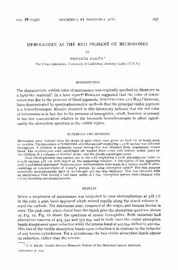
color. The pink zone, when eluted from the starch geve the absorption spec t rum shown
on Fig. Ia. Fig. Ib shows the spect rum of mouse hemoglobin. Both mater ia ls had absorpt ion m a x i m a at 415, 54 ° and 575 m/x, and in both cases the visible absorpt ion bands disappeared upon reduct ion while the intense band at 415 m/x shifted to 42o mr*. This loss of the visible absorpt ion bands upon reduct ion is in contrast to the behavior of any known cytochrome. For a cy tochrome the two visible absorpt ion bands appear on reduction, ra ther than the reverse.
* U.S. Public Health Service Research Fellow of the National Cancer Institute.
lCe/erences p. 299.
298 K.P.\IGEN VOL. 19 (19561
0 6
04 z
u_ 0 2
o
I , I i i I ~ t J t I
4 oo 500 600 mpu
Fig. I (a)
0.6 >- v-
~,lz04
° 0 , 2 C:- I:L 0
i
400 500 600 ml~
t;ig, ~ (b)
Fig. I . (a) m i c r o s o m e p i g m e n t . (b) M ous e h e m o g l o b i n . S p e c t r a w e r e o b t a i n e d in 0.04 M g l y c y l - g lyc ine buffer , p H 7.6, c o n t a i n i n g o . 2 5 M sucrose . - - o x id i zed ; . . . . . , r e d u c e d wi th a t r a c e
of s o d i u m hyd ros u l f i t e .
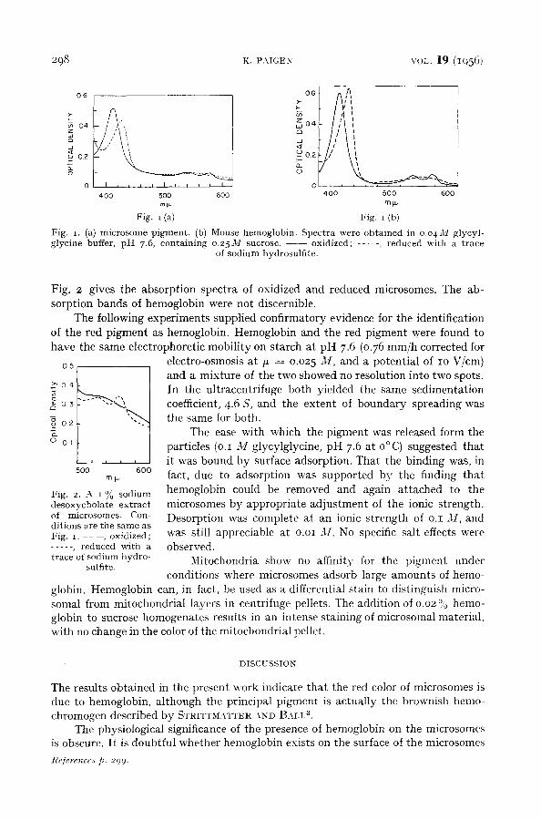
Fig. 2 gives the absorption spectra of oxidized and reduced microsomes. The ab- sorption bands of hemoglobin were not discernible.
The following experiments supplied confirmatory evidence for the identification of the red pigment as hemoglobin. Hemoglobin and the red pigment were found to have the same electrophoretic mobility on starch at pH 7.6 (0.76 mm/h corrected for
05 electro-osmosis at /x = 0.025 M, and a potential of IO V/cm) and a mixture of the two showed no resolution into two spots.
o4 ------.--,C-~ ~ In the ultracentrifuge both yielded the same sedimentation ~g o 3 - -- , coefficient, 4.6 S, and the extent of boundary spreading was
02 ~ the same for both. The ease with which the pigment was released form the
o Ot particles (o.I M glycylglycine, pH 7.6 at o ° C) suggested that , , , it was bound by surface adsorption. That the binding was, in
500 1600 mF fact, due to adsorption was supported by the finding that
Fig. 2. A 1% sodium hemoglobin could be removed and again attached to the desoxycholate extract microsomes by appropriate adjustment of the ionic strength. of microsomes. Con- Desorption was complete at an ionic strength of o.I M, and di t ions a re t h e s a m e as Fig. i . - - - , oxidized; was still appreciable at o.oi M. No specific salt effects were . . . . . , r e d u c e d w i t h a observed. trace of sodium hydro- Mitochondria show no affinity for the pigment under
sulfite. conditions where microsomes adsorb large amounts of hemo-
globin. Hemoglobin can, in fact, be used as a differential stain to distinguish micro- somal from mitochondrial layers in centrifuge pellets. The addition of o 02 % hemo- globin to sucrose homogenates results in an intense staining of microsomal material, with no change in the color of the mitochondrial pellet.
DISCUSSION
The results obtained in the present work indicate that the red color of microsomes is due to hemoglobin, although the principal pigment is actually the brownish hemo- chromogen described by STRITT~.~TTER AND BALP.
The physiological significance of the presence of hemoglobin on the microsomes is obscure. It is doubtful whether hemoglobin exists on the surface of the microsomes
References p. 299.
VOL. 1 9 ( 1 9 5 6 ) HEMOGLOBIN AS THE RED PIGMENT OF MICROSOMES 2 9 9
intracellularly. The most likely source of the hemoglobin is an occasional red cell which is ruptured during the original preparation of the homogenate.
The existence of hemoglobin binding by microsomes does serve to demonstrate a fundamental difference in the surface properties of the two types of particles.
The ability of microsomes to adsorb protein is not limited to hemoglobin. The adsorption of eytochrome c 7 and nucleases a has been reported previously, and adsorp- tion of lysozyme has also been observed in this laboratory. I t is possible that the ad- sorption of basic proteins by microsomes, as contrasted with the failure of these proteins to adsorb to mitochondria, is related to the high nucleic acid content of the microsomes.
ACKNOWLEDGEMENTS
The author wishes to thank Professor W. M. STANLEY for making the facilities of the Virus Laboratory available throughout this work, and Dr. H. K. SCHACHMAN and Dr. V. SCHUMAKER for their generous assistance with the physical measurements.
SUMMARY
The red color of isolated microsomes results from the presence of adsorbed hemoglobin. Adsorption occurs only at very low ionic s t rengths. In contras t to microsomes, mitochondria do not adsorb hemoglobin. Hemoglobin added to tissue homogenates is useful as a specific stain to differentiate microsomal from mitochondrial material in mixed centrifuge pellets.
RI~SUME
La coloration rouge des microsomes isol6s r6sulte de la pr6sence d'h6moglobine adsorbde. L 'ad- sorpt ion ne se produi t qu'/t des forces ioniques trbs basses. A l ' inverse des microsomes, les mito- chondries n ' adsorben t pas l 'h6moglobine. De l 'h6moglobine, ajout6e /~ des tissus homog6n6is6s, peut servir de colorant sp6cifique pour diff6rencier les microsomes des mitochondries dans des culots de centrifugations nlixtes.
ZUSAMMENFASSUNG
Adsorbiertes Hemoglobin verursacht die rote Farbe von isolierten Mikrosomen. Adsorption kommt nur bei sehr geringen Ionenstgrken vor. I m Gegensatz zu Mikrosomen adsorbieren Mito- chondrien kein Hemoglobin. Zu Gewebshonlogenaten hinzugeffigtes Hemoglobin kann als spezifischer Farbstoff beniitzt werden, um in gemischten Zentrifugenrfickst/tnden Mikrosomen von Mitochondrien zu unterscheiden.
R E F E R E N C E S
1 R. R. BENSLEY, Anat. Record, 98 (1947) 609. 2 R. R. BENSLEY, J. Hislochem. Cytochem., I (19.53) 179. a C. F. STRITTMATTER AND E. G. BALL, Proc. Natl. Acad. Sci. U.S., 38 (1952) t9- 4 VV. C. SCHNEIDER AND G. t-I. HOGEBOOM, .]. Biol. Chem., 183 (195o) t23.
K. PAIGEN, .4hal. Chem., (in press). K. PAIGEN AND H. K. SCHACHMAN, unpubl ished data.
7 H. BEINERT, J. Biol. Chem., 19o (1951) 287. s \V. C. SCHNEIDER AND G. H. HOGEBOOM, J. Biol. Chem., 198 (1952) 155.
Received June 27th, 1955