hematology, morphology and ultrastructure of blood cells ... for...
TRANSCRIPT

Kasetsart J. (Nat. Sci.) 36 : 35 - 43 (2002)
1 Faculty of Veterinary Medicine, Kasetsart University, Nakorn Pathom 73140, Thailand.
2 Queen Saovabha Memorial Institute, Thai Red Cross Society, Bangkok 10330, Thailand.
Hematology, Morphology and Ultrastructure of Blood Cells andBlood Parasites from Puff-faced Watersnakes (Homalopsis buccata)
Chaleow Salakij1, Jarernsak Salakij1, Piyawan Suthunmapinunta1 and Lawan Chanhome2
ABSTRACT
Blood samples of 45 puff-faced watersnakes (Homalopsis buccata) in the Queen Saovabha
Memorial Institute were collected from ventral caudal vein for both basic hematology and light microscopic,
scanning and transmission electron microscopic features of blood cells. Seventeen samples (37.8%) were
positive for hematozoa infections. The single infection of Hepatozoon sp., trypanosome and Haemogregarina
sp. was found in 4, 4 and 5 snakes respectively. The other four snakes were infected by both trypanosome
and Hepatozoon sp. There were no significant differences of all hematological value between the
hematozoa-negative and the hematozoa-positive snakes except fibrinogen concentration which was found
higher in the negative group. Lymphocytes were the most commonly observed leukocytes and average 6-
8 µm in diameter. Azurophils were the second most commonly observed leukocytes, average 10-17 µm in
diameter and might play a major role in eliminating the trypanosome. Heterophils were the largest
leukocytes, average 16-19 µm in diameter and the third commonly observed leukocytes. Eosinophils
usually were medium-sized cells, average 10-14 µm in diameter but in some occasion the very large cells
were also detected. Basophils were smaller than heterophils and eosinophils. Scanning electron microscopy
revealed the membrane surfaces of normal and abnormal erythrocytes, Hepatozoon sp. infected erythrocytes,
thrombocytes, eosinophil and trypanosomes. Transmission electron microscopy revealed the organelles
within azurophil, eosinophil, heterophil and trypanosome.
Key words: Electronmicroscopy, Haemogregarina, Hematology, Hepatozoon, Homalopsis buccata,
puff-faced watersnakes, trypanosome
INTRODUCTION
Puff-faced or mask-faced watersnake
(Homalopsis buccata) is immediately identified by
the large broad head and a white, mask-like pattern
on the top of the head. It consumes fish and frogs.
Its habitat is commonly found in most of Southeast
Asia (Cox et al., 1998). The Queen Saovabha
Memorial Institute (QSMI) had initiated a captive
breeding program since 1994 to supply healthy
snakes for venom and antivenom production. These
venomous snakes prey on mice and occasionally on
puff-faced watersnakes. The venomous snakes were
highly infected with Hepatozoon sp. (Salakij et al.,
2001). Reptile blood cell morphologic
characteristics are heterogeneous. Variations in cell
characteristics and cell populations were existed
between species within the order Squamata
(Alleman et al., 1999).
The purpose of this study was to obtain the
hematological data, characterization of blood cells

36 Kasetsart J. (Nat. Sci.) 36 (1)
and compare if the hematozoas in the puff-faced
water snakes were the same as those found in the
venomous snakes.
MATERIALS AND METHODS
Blood samples of 45 puff-faced watersnakes
were collected from ventral caudal vein during
January to March 2000. Blood smears were prepared
immediately and air-dried. They were stained with
Wright’s and Wright-Giemsa stained for white
blood cell differentiation and hemoparasite
examination. The collected blood samples were
kept immediately in EDTA, immediately stored at
4°C and processed within 2 hours.
The packed cell volume (PCV) were
determined by microcapillary technique. The total
solids were measured using a AtagoSPR-N
refractometer (Japan). Fibrinogen was calculated
as the difference between the total solids before and
after incubation for 3 minutes at 56°C and
recentrifugation (Jain, 1986). The total red blood
cell count (RBC), were determined manually with
the improved Neubauer counting chamber after the
blood was diluted 200 times with the Natt and
Herrick’s solution (Natt and Herrick, 1952). The
total number of the RBC was counted in the five red
blood cell squares of the center large square of the
chamber in duplicate. The duplicates were averaged
for agreement within 15% difference and multiplied
by 10,000 to calculated the number per microlitre
(Campbell, 1986). The leukocyte count (WBC)
was determined in the same counting chamber as
the RBC count except the leukocytes were counted
in duplicate from four large squares of the chamber
at 40X magnification. The leukocyte neuclei were
stained blue whereas the thrombocyte neuclei were
unstained or very pale blue. The duplicates were
averaged for agreement within 15% difference and
multiplied by 500 to calculate the number per
microlitre. The hemoglobin concentration (Hb) was
determined by the cyanomethemoglobin method
which free RBC neuclei were removed by the
centrifugation before reading the absorbance
(Campbell, 1986).
Blood smears were fixed in methanol and
stained with Wright-Giemsa (WG) stain (Benjamin,
1978) for determination of differential leukocyte
count, identification of hematozoa infection and
morphological evaluation of all blood cells. Grading
of Hepatozoon sp. and Haemogregarina sp. was
quantitated by the number of infected erythrocytes
as described elsewhere (Salakij et al., 2001). A
minimum of 200 leukocytes were counted for
differential leukocyte determination. For
comparison, blood smear from 5 puff-faced
watersnakes were stained with one step Wright’s
staining method that did not required methanol
fixation prior to staining stain (Benjamin, 1978).
Blood samples were also prepared for
reticulocyte count by staining with new methylene
blue using wet preparation (Benjamin, 1978). The
percentage of reticulocytes presented in 1,000
erythrocytes was determined. The reticulocytes that
contained distinct aggregated reticulum were
described as aggregate reticulocytes whereas
punctate reticulocytes contained a few small dots
(Jain, 1986).
For each parameter obtained, data from
hematozoa-negative and positive were calculated
for means, variances and standard error using
SPSS for window (Norusis, 1993). Significant
difference between means were determined using
an independent sample T-test model.
For scanning electron microscopy (SEM), a
drop of blood were fixed using 1.5% glutaraldehyde
(GA) in 0.1 M phosphate buffer (PB) at 4°C for 24
hr. Specimens were dehydrated through a graded
acetone series. Gold-coated smears were examined
under Jeol JSM-35CF scanning electron
microscopy.
For transmission electron microscopy
(TEM), buffy coats were fixed in 2.5% GA (PB) for
24 hr and postfixed in 1% osmium tetroxide.
Specimens were dehydrated through a graded
acetone series and embedded in Spurr’s epoxy

Kasetsart J. (Nat. Sci.) 36 (1) 37
resin. Ultrathin sections, stained with uranyl acetate
and lead citrate, were examined using Jeol 1200Ex
TEM.
RESULTS
Seventeen samples (37.8%) were positive
for hematozoa infections. The single infection was
found in thirteen snakes including: Hepatozoon sp.,
trypanosome and Haemogregarina sp. The other
four snakes (8.9%) were mixed infection between
trypanosome and Hepatozoon sp. (Table 1). There
were no significant differences of all hematological
values between the hematozoa-negative and the
positive groups except fibrinogen concentration
which was higher in the negative group (Table 2).
Erythrocytes were homogeneous in color
but moderately anisocytosis (Figure 1, 2, 3).
Cytoplasmic holes were detected in less than 1%
erythrocytes (Figure 3c). The other shapes of
abnormal erythrocytes were seldom detected (Figure
3b, 3c). Hepatozoon– and Haemogregarina-infected
erythrocytes were larger than those non-infected
ones (Figure 2b, 2d, 3e). By SEM, erythrocytes
were ellipsoidal lacking the doming appearance in
the site of the nucleus (Figure 3a, 3b, 3c).
Thrombocytes were elongate and
approximately half the size of mature erythrocytes.
Under the Wright’s stain, cytoplasm was slightly
basophilic (Figure 1i) compared with the unstained
cytoplasm with azurophil granules in Wright-
Giemsa stain (Figure 1a). Thrombocytes were easily
differentiated from lymphocytes by the
characteristic perinuclear and cytoplasmic
vacuolation (Figure 1a, 1i). By SEM, their
membranes were more irregular than those of
erythrocytes (Figure 3a).
Leukocytes of puff-faced water snakes were
categorized into 6 groups; azurophil, heterophil,
eosinophil, basophil, lymphocyte and monocyte.
For comparison, the blood smears stained with one
step Wright’s stain provided staining quality for
identification of all blood cell type but in Wright’s
stain the erythrocytes stained more basophilic
(Figure 1h, 1i).
Lymphocytes in puff-faced watersnakes
were the most prevalent circulating cells (Table 2).
They were small, well differentiated and averaged
6-8 µm in diameter (Figure 1a).
Azurophils were the second most commonly
observed leukocytes, which contained fine indistinct
azurophilic granules. They were round and 10-17
µm in diameter. The nuclei were round to irregular
with clump chromatin and located centrally to
eccentric (Figure 1b, 1d). Ultrastucturally, they
contained numerous membrane-bound granules,
some mitochondria and rough endoplasmic
reticulum (Figure 4b). Phagocytosis of trypanosome
by azurophils was detected only by TEM (Figure
4b). The number of monocytes is very rare and their
characters were similar to mammalian monocytes
(Figure 1d).
Heterophils were the largest of the leukocytes
and average 16-19 µm in diameter. They contained
large numbers of irregular shape, dull eosinophilic
granules (Figure 1c). By Wright’s stain, heterophil
granules were easily seen by reddish-orange bright
granules (Figure 1h). Ultrastructurally, heterophils
contained pleomorphic population of large granules
with variable electron density (Figure 4d).
Eosinophils contained numerous round and
light blue granules that often occluded visualization
of the nucleus (Figure 1e, 1h). They usually were
medium-sized cells (10-14 µm in diameter) but in
some cases very large cells were also detected
(Figure 1f, 1i). By Wright’s stain, eosinophil
granules were stained dark blue when compared to
the heterophils (Figure 1h). By SEM, their granule
contour was bulging showing the custard apple-
liked appearance (Figure 3d). Ultrastructurally,
eosinophils contained homogeneous electron
density granules with some cytoplasmic projections
(Figure 4c). The eosinophil number were very high
both in the negative and positive groups (Table 2).
Basophils were very low, average 9-12 µm
in diameter and were slightly smaller than

38 Kasetsart J. (Nat. Sci.) 36 (1)
Table 2 Comparative hematology (mean ± SE) between hematozoa-negative and positive puff-faced
watersnakes.
Hematozoa-negative Hematozoa-positive
Number 28 17
PCV (%) 19.6 ± 1.2 22.3 ± 1.6
Hb (g/dl) 6.34 ± 0.38 6.99 ± 0.46
RBC (x106/µl) 0.530 ± 0.046 0.618 ± 0.068
WBC (x103/µl) 12.11 ± 1.00 11.95 ± 1.78
Azurophils (x103/µl) 4.22 ± 0.48 3.96 ± 0.82
Heterophils (x103/µl) 1.91 ± 0.26 1.31 ± 0.17
Basophils (x103/µl) 0.005 ± 0.003 0.02 ± 0.01
Eosinophils (x103/µl) 0.69 ± 0.10 0.70 ± 0.14
Lymphocytes (x103/µl) 5.16 ± 0.70 5.93 ± 1.26
Monocytes (x103/µl) 0.08 ± 0.03 0.03 ± 0.02
Azurophils (%) 35.8 ± 3.0 32.5 ± 3.9
Heterophils (%) 15.3 ± 1.6 13.1 ± 2.1
Basophils (%) 0.07 ± 0.05 0.2 ± 0.09
Eosinophils (%) 6.5 ± 0.9 6.2 ± 0.9
Lymphocytes (%) 41.3 ± 2.8 47.5 ± 4.3
Monocytes (%) 0.8 ± 0.4 0.4 ± 0.2
PP (g/dl) 5.7 ± 0.30 5.6 ± 0.40
Fibrinogen (mg/dl) 142.8 ± 20.8* 64.7 ± 17.0*
Agg. Reticulocytes (%) 0.74 ± 0.30 3.27 ± 1.68
Punct. Reticulocytes (%) 4.11 ± 1.32 9.61 ± 2.83
* Significant difference at p<0.05.
Table 1 Number and percentage of hematozoa-negative and positive puff-faced watersnakes which was
subgrouped according to sex.
Male Female Total %
Negative 15 13 28 62.2
Hepatozoon sp. 2 2 4 8.9
Trypanosoma sp. 4 0 4 8.9
Trypanosoma sp. and Hepatozoon sp. 3 1 4 8.9
Haemogregarina sp. 5 0 5 11.1
Total 29 16 45 100
% 64.4 35.6 100

Kasetsart J. (Nat. Sci.) 36 (1) 39
Figure 1 Blood cells in puff-faced watersnakes (a) a lymphocyte (lower cell) and a thrombocyte (b) an
azurophil, (c) a heterophil, (d) a monocyte (left cell) and an azurophil, (e) an eosinophil, (f) a large
eosinophil, (g) a basophil, Wright-Giemsa stain, (h) a 17 µm heterophil and an 14 µm eosinophil.
Wright’s stain, (i) a large eosinophil, Wright’s stain.
eosinophils (Figure 1g). They contained small dark
purple staining metachromatic granules that obscure
the lobed nucleus.
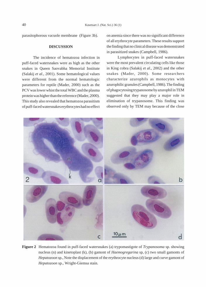
Trypomastigote form of trypanosomes were
large, broad body width (Figure 2a). By SEM, they
were often in a cluster (Figure 3f). Ultrastructurally,
they contained nucleus, very large mitochondria,
abundant ribosomes (Figure 4b), some dense and
multivesicular bodies (Figure 4a). The gamonts of
Haemogregarina sp. were easily defined from those
of the Hepaozoon sp. by their very large-size and
more granularity (Figure 2b). There were two kinds
of Hepaozoon gamonts; the small (Figure 2c) and
the large gamonts (Figure 2d). Both Hepaozoon
and Haemogregarina gamonts were resided in the
cytoplasm of enlarged erythrocytes (Figure 3e) and
displaced the nucleus (Figure 2b, 2c). Some gamonts
were free from erythrocytes within their
40 Kasetsart J. (Nat. Sci.) 36 (1)
parasitophorous vacuole membrane (Figure 3b).
DISCUSSION
The incidence of hematozoa infection in
puff-faced watersnakes were as high as the other
snakes in Queen Saovabha Memorial Institute
(Salakij et al., 2001). Some hematological values
were different from the normal hematologic
parameters for reptile (Mader, 2000) such as the
PCV was lower whist the total WBC and the plasma
protein was higher than the reference (Mader, 2000).
This study also revealed that hematozoa parasitism
of puff-faced watersnakes erythrocytes had no effect
on anemia since there was no significant difference
of all erythrocyte parameters. These results support
the finding that no clinical disease was demonstrated
in parasitized snakes (Campbell, 1986).
Lymphocytes in puff-faced watersnakes
were the most prevalent circulating cells like those
in King cobra (Salakij et al., 2002) and the other
snakes (Mader, 2000). Some researchers
characterize azurophils as monocytes with
azurophilic granules (Campbell, 1986). The finding
of phagocytosing trypanosome by azurophil in TEM
suggested that they may play a major role in
elimination of trypanosome. This finding was
observed only by TEM may because of the close
Figure 2 Hematozoa found in puff-faced watersnakes (a) trypomastigote of Trypanosoma sp. showing
nucleus (n) and kinetoplast (k), (b) gamont of Haemogregarina sp, (c) two small gamonts of
Hepatozoon sp., Note the displacement of the erythrocyte nucleus (d) large and curve gamont of
Hepatozoon sp., Wright-Giemsa stain.
Kasetsart J. (Nat. Sci.) 36 (1) 41
contact of the azurophils and the trypanosomes in
the buffy coat before they were fixed with
glutaraldehyde.
It is difficult to differentiate eosinophils
from basophils in WG stained smears because of
the bluish coloration of their granules. They were
identifed more easily on Wright’s stained
preparation. The eosinophil granule characteristic
Figure 3 Scanning electron photomicrographs of (a) two normal erythrocytes and a thrombocyte, (b)
euglenoil-shaped erythrocyte and parasitophorous vacuole membrane containing gamont of
Hapatozoon sp. (star), (c) abnormal erythrocytes showing double appendages and cytoplasmic
hole (arrow), (d) an eosinophil showing custard apple-like appearance of granule contour, (e) an
enlarged erythrocyte containing Hepatozoon gamont, (f) a cluster of trypanosomes.
in puff-faced watersnakes was similar to those of
iguanas and psittacines (Hawkey and Dannett, 1989)
which they contained dark purple staining
metachromatic granules that obscure the unlobed
nucleus. The large-sized eosinophils found in puff-
faced watersnakes should be the characteristic of
eosinophil in snakes which were larger than those
of the other reptiles (Mader, 2000).

42 Kasetsart J. (Nat. Sci.) 36 (1)
The high number of eosinophils in the
negative and positive groups (Table 2) may be
influenced by parasitic stimuli or other stimuli
(Mader, 2000). The finding of eosinophils in puff-
faced watersnake confirm the existence of these
leukocytes in snakes eventhough they were not
identified in eastern diamondback rattlesnakes
(Alleman et al., 1999).
Trypomastigote form of trypanosomes in
puff-faced watersnakes was different from
Trypanosoma hydrae in broad-band watersnake
from Louisiana (Chia and Miller, 1984).
Trypanosomes were detected only in puff-faced
and rainbow watersnakes of the QSMI (Salakij et
al., 2001).
Figure 4 Transmission electron photomicrographs of (a) cross-section of a trypanosome containing
nucleus, mitochondria, multivesicular bodies (m) and dense granular granule (arrow), (b) an
azurophil (A) pseudopodia is surrounding a trypanosome (T), (c) an eosinophil with homogene-
ous granules and cytoplasmic process, (d) a heterophil with vacuoles and heterogeneous electron
density granules.

Kasetsart J. (Nat. Sci.) 36 (1) 43
The small gamonts of Hepatozoon sp. were
similar to those found in the banded krait (Bangarus
fasciatus) of the QSMI (Salakij et al., 2001). The
large gamonts found in puff-faced watersnakes
were referred as medium-sized gamonts when
compared with the larger gamonts found in
mangrove snakes and mangrove pit vipers of the
QSMI (Salakij et al., 2001). Haemogregarina sp.
was found not only in watersnakes but also in
Burmese python, mangrove snakes and rainbow
watersnakes of the QSMI (Salakij et al., 2001).
These three kinds of snakes were not fed on puff-
faced watersnakes so they were not transmitted by
eating.
This study provides more information on
the hematology, morphology and ultrastructure of
blood cells in puff-faced watersnakes. This may be
benificial for further study and related research.
ACKNOWLEDGMENTS
Financial support was received from
Kasetsart University Research and Development
Institute.
LITERATURE CITED
Alleman, A. R., E. R. Jacobson, and R. E. Raskin.
1999. Morphologic, cytochemical staining and
ultrstructural characteristics of blood cells from
eastern diamonback rattlesnakes (Crotalus
adamanteus). Am. J. Vet. Res. 60 : 507-514.
Benjamin, M. M. 1978. Outline of Veterinary
Clinical Pathology. 3rd ed. The Iowa State
University Press, Ames. 351p.
Campbell T. W. 1986. Clinical Pathology, pp. 248-
257. In D. R. Mader (ed.). Reptile Medicine
and Surgery. WB Saunders, Philadelphia.
Chia, N. M. and J. H. Miller. 1984. Morphological
and Developmental Studies of the Snake
Trypanosome Trypanosoma hydrae Ayala,
Atkinson, Vakalis, 1983 in Experimentally
Infected Hosts and in Culture. J. Protozool. 31
: 352-356.
Cox, M. J., P. P. van Dijk, J. Nabhitabhata and K.
Thirakhupt. 1998. A Photographic Guide to
Snakes and Other Reptiles of Thailand and
Southeast Asia. ASIA BOOKS. Bangkok, 144
p.
Hawkey, C. M. and T. B. Dannett. 1989. A Color
Atlas of Comparative Veterinary Haematology.
Wolfe Publishing Limited. Ipswick. 192 p.
Jain, N. C. 1986. Schalm’s Veterinary Hematology.
4th ed. Lea and Febiger. Philadelphia, 1221p.
Mader, D. R. 2000. Normal Hematology of Reptiles,
pp. 1126-1132. In B. E., Feldman, J. G. Zinkl
and N. C. Jain (eds.) Schalm’s Veterinary
Hematology. 5th ed. Lippincott Williams and
Wilkins, Philadelphia.
Natt. M. P. and C. A. Herrick. 1952. A new blood
diluent for counting erythrocytes and leukocytes
of the chicken. Poult. Sci. 31 : 735-738.
Norusis, M. J. 1993. SPSS for Window Base
System User’s Guide Release 6.0.SPSS inc.,
Chicago, Ill. 828 p.
Salakij, J., C. Salakij, N. Narkkong, L. Chanhome,
N. Rochanapat, and P. Suthunmapinunta. 2001.
Hematozoa of snakes in Queen Saovabha
Memorial Institute. Kasetsart J (Nat Sci) 35 :
149-156.
Salakij, C., J. Salakij, S. Apibal, N. Narkkong, L.
Chanhome, N. Rochanapat, and P. Suthun-
mapinunta. 2002. Hematology, morphology,
cytochemical staining and ultrastructural
characteristics of blood cells in King cobras
(Ophiophagus hannah). Vet. Clin. Path. 31 :
116-126.
Received date : 3/01/02Accepted date : 29/03/02