heart failure - - rn.org® · heart failure ® reviewed september 2017, expires september 2019...
TRANSCRIPT

Heart Failure WWW.RN.ORG®
Reviewed September 2017, Expires September 2019 Provider Information and Specifics available on our Website
Unauthorized Distribution Prohibited
©2017 RN.ORG®, S.A., RN.ORG®, LLC By Wanda Lockwood, RN, BA, MA
Purpose The purpose of this course is to describe the causes of chronic heart failure, the different types and classification systems, the symptoms of right ventricular failure and left ventricular failure, compensatory mechanisms, treatment and medications as well as symptoms and treatment of acute heart failure.
Goals Upon completion of this course, the healthcare practitioner should be able to:
• Describe the normal cardiac circulation.
• Provide definitions for cardiac terminology.
• List and describe at least 6 causes of heart failure.
• Discuss at least 5 diagnostic procedures.
• Differentiate between systolic and diastolic heart failure.
• Describe at least 7 symptoms associated with left ventricular failure.
• Describe 4 primary symptoms associated with right ventricular failure.
• Differentiate between the New York Heart Association and the American College of Cardiology-American Heart Association classification systems.
• Describe at least 5 compensatory mechanisms and the adverse effects related to these mechanisms.
• Describe at least 6 treatment approaches to chronic heart failure.
• Discuss at least 5 medications used to treat chronic heart failure.
• Explain how acute heart failure relates to chronic heart failure.
• Describe emergency treatment options for acute heart failure.
Introduction About 5.8 million people in the United States have heart failure, with 670,000 newly diagnosed each year. Because diagnosis is often delayed, 20% die within a year of diagnosis, so early diagnosis and proper medical management are critical to survival. In a normal heart, blood flows into the right atrium from the superior and inferior vena cava. As the right ventricle relaxes after contraction, blood is pulled from the right atrium through the tricuspid valve into the right
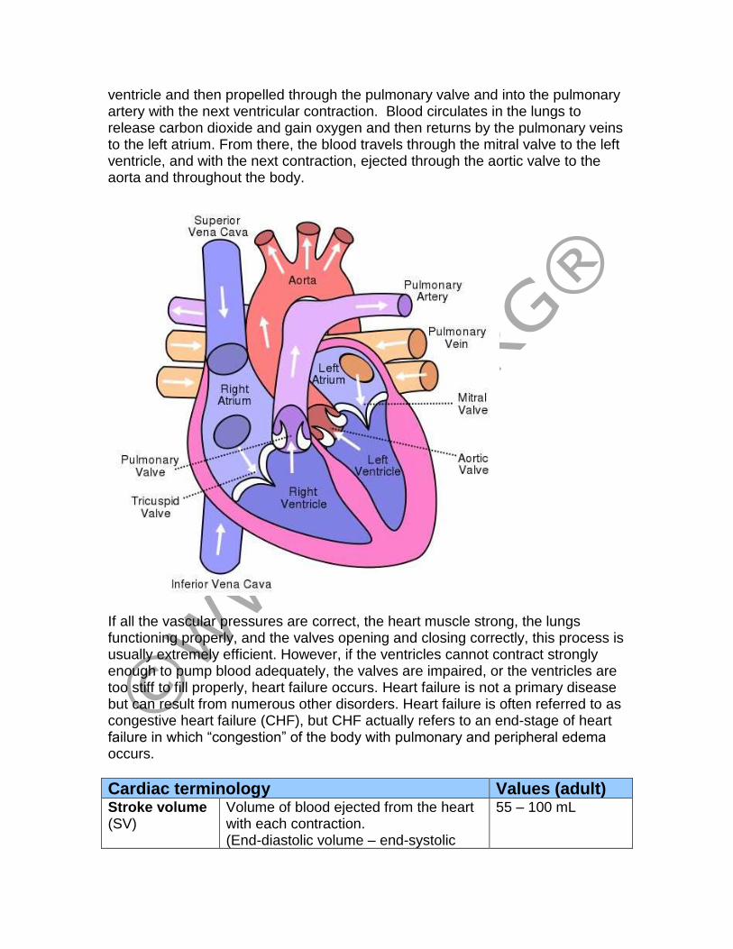
ventricle and then propelled through the pulmonary valve and into the pulmonary artery with the next ventricular contraction. Blood circulates in the lungs to release carbon dioxide and gain oxygen and then returns by the pulmonary veins to the left atrium. From there, the blood travels through the mitral valve to the left ventricle, and with the next contraction, ejected through the aortic valve to the aorta and throughout the body.
If all the vascular pressures are correct, the heart muscle strong, the lungs functioning properly, and the valves opening and closing correctly, this process is usually extremely efficient. However, if the ventricles cannot contract strongly enough to pump blood adequately, the valves are impaired, or the ventricles are too stiff to fill properly, heart failure occurs. Heart failure is not a primary disease but can result from numerous other disorders. Heart failure is often referred to as congestive heart failure (CHF), but CHF actually refers to an end-stage of heart failure in which “congestion” of the body with pulmonary and peripheral edema occurs.
Cardiac terminology Values (adult) Stroke volume (SV)
Volume of blood ejected from the heart with each contraction. (End-diastolic volume – end-systolic
55 – 100 mL

volume = SV)
Cardiac output (CO)
Total volume of blood ejected from the heart in on minute. (Heart rate (HR) X SV =CO)
4.9 L /min
End-diastolic volume (EDV)
Volume of blood in filled ventricles. 120 mL
End-systolic volume (ESV)
Volume of blood left in ventricles after contraction.
50 mL
Ejection fraction (Ef)
Fraction (expressed as percentage) of blood pumped from ventricles during contraction (usually refers to left ventricle). (Ef = SV/EDV
55 – 75%
Heart rate Number of heart beats per minute 60 – 80 BPM
Preload (Volume)
Degree of elasticity (stretch) in the myocardium with end-diastolic volume. Increased stretch (and volume) correlates with increased contraction.
(Estimated with EDV—cannot be measured in vivo).
Afterload (Pressure/ Resistance)
Degree of systemic vascular resistance to ventricular contractions (left ventricular ejection of blood through the aorta and systemic circulation and right ventricular ejection of blood to the pulmonary vascular system).
Systemic vascular resistance (SVR)
Resistance in the peripheral vascular system to left ventricular ejection. Vascular resistance is determined by tone in small and pre-capillary arterioles and by viscosity of blood.
What causes heart failure? The primary risk factors for heart failure are coronary artery disease and advancing age, but a number of different disorders may bring about heart failure. If disorders associated with increased risk are diagnosed early and treated, heart failure may be averted in some cases.
Common causes of heart failure Coronary artery disease (60%)
Decreases oxygenation of the heart muscle, interfering with the conduction system that stimulates the heart to contract and with the ability of the ventricles to properly contract.
Myocardial infarction
May cause structural damage that interferes with function of heart muscle or may interfere with conduction system. Remodeling of left ventricle is common after MI.
Cardiomyopathy • Hypertrophic: Causes thickened ventricular walls with inability to effectively pump blood throughout the body
and may cause arrhythmias that further impair contractions.
• Dilated: Causes damage to heart muscle (from coronary artery disease, MI, myocarditis), which weakens and dilates ventricles, interfering with ability to contract.
• Restrictive: Causes stiffened heart muscles that cannot contract adequately.
Congenital heart defects
• May interfere with flow of blood through the heart, oxygenation, and/or ability of the heart to pump.
Valvular disease • Stenosis: Increases the work of the heart muscle to circulate blood, causing hypertrophy as the heart tries to overcome the restriction.
• Insufficiency (regurgitation): Increases the work of the heart muscle, causing enlargement (dilation) and decreased pumping ability.
Diabetes mellitus
Associated with hyperlipemia and atherosclerosis, increasing work of the heart. Hyperglycemia may impair cardiac muscle.
Hypertension Increases systemic vascular resistance to blood flow, increasing work of heart.
Arrhythmias Interfere with the ability of the heart to effectively circulate blood, so heart works harder but less effectively. May decrease cardiac output and increase workload and oxygen demands of the myocardium.
Chronic kidney disease
Resulting hypertension increase systemic vascular resistance and retention of sodium and water increases workload on the heart.
Obesity Left ventricular hypertrophy common as the heart works harder to supply the body with adequate amounts of oxygenated blood.
In addition to diseases and disorders that can cause heart failure, a number of precipitating factors may lead to heart failure:
• Anemia stimulates the heart to increase cardiac output to compensate for decreased oxygen-carrying capacity.
• Infection increases need for tissue oxygenation, stimulating increased cardiac output.
• Malnutrition may impair cardiac function by decreasing myocardial muscle mass and ability to contract.
• Hypervolemia increases preload and causes volume load on right ventricle.
• Pulmonary embolism increases pulmonary pressure, exerting increased pressure on the right ventricle and causing right ventricular hypertrophy and failure.
Diagnosis
Because heart failure results from another disease process, the first step in diagnosis is to determine the underlying cause with a complete history and physical examination.
• Blood gases, chemistries, and liver function tests: Help identify underlying cause and damage related to HF.
• Chest X-ray: Identifies an enlarged heart or indications of pulmonary edema.
• Echocardiogram: Shows valve function, flow of blood through heart, and ejection fraction (to help differentiate between diastolic and systolic heart failure). Most common test for EF.
• Electrocardiogram: Identifies cardiac damage, arrhythmias.
• Exercise stress testing: Shows functional limitations.
• Plasma natriuretic peptide levels: Correlate with degree of LV dysfunction.
• Nuclear imaging studies, MRI, cardiac catheterization: May be indicated in some patients.
How is heart failure described? Heart failure may be described in relation to the type of impairment—pumping (systole) or filling (diastole)—or by the primary site of impairment—right or left ventricular. In most cases, biventricular failure occurs as heart failure progresses, but heart failure most commonly first begins with left ventricular /systolic failure, which leads to right ventricular failure although signs of heart failure may become more evident with right ventricular failure.
Systolic failure (Pumping) Most heart failure derives from systolic failure, or the inability of the heart to adequately pump blood because of a defect in the ventricles. Typically, the left ventricles cannot generate adequate pressure to eject blood through the aorta. The most common causes of systolic failure are those that interfere with contractility, such as myocardial infarction, hypertension, cardiomyopathy, and valvular heart disease. Evaluation of the ejection fraction (EF) differentiates systolic from diastolic failure. Systolic failure is associated with a decrease in EF <40% because the heart is not able to pump an adequate amount of blood. EF is usually normal with diastolic failure.
Diastolic failure (Filling) With diastolic failure, the ventricles are able to contract adequately, but stroke volume decreases because the ventricles can’t fill properly. With diastolic failure, backpressure builds in the circulatory system, resulting in venous engorgement in the pulmonary and systemic vascular systems. Pulmonary hypertension is present with a normal ejection fraction. Diastolic failure is most common in older adults who have developed left ventricular hypertrophy from hypertension, aortic stenosis, or hypertrophic cardiomyopathy.
Mixed diastolic and systolic failure
Some people, such as those with dilated cardiomyopathy, will exhibit signs of both systolic and diastolic failure and often exhibit both increased pulmonary pressure and low ejection fractions with signs of failures of both right and left ventricles.
Left ventricular (LV) failure LV failure is sometimes referred to as backward failure. The left ventricle cannot pump the blood out of the ventricle to the body, so LV end-diastolic blood volume increases, and this increases LV end-diastolic pressure, causing blood to back up into the left atrium and pulmonary veins, increasing pulmonary pressure and causing pulmonary edema as fluid is forced from the pulmonary capillaries into the interstitial tissues and alveoli. A common cause of LV failure is increased systemic vascular resistance (as from atherosclerosis and hypertension), which increases the force of LV contractions, leading to increased demand for oxygen to the LV myocardium and LV hypoxia, which in turn decreases the force of contractions. As the cardiac output decreases, this results in decreased perfusion of all organs and tissues and stimulation of the sympathetic nervous system. The sympathetic nervous system protects the heart from extrinsic damage, such as trauma and blood loss, by increasing heart rate and blood pressure and constricting vessels, and in the early stages of LV failure, this helps to compensate, but over time these changes worsen heart failure.
Symptoms of LV failure Pulmonary edema/ Dyspnea
Dyspnea of exertion (DOE): Maybe caused by minimal or moderate exertion, depending on extent of LV failure. Orthopnea: Dyspnea intensifies when lying flat. Patient may need 2-3 pillows or may sit up to sleep. Paroxysmal nocturnal dyspnea: Acute attack of dyspnea from pulmonary edema, occurring 1-2 hours after going to sleep and associated with severe HF.
Cough Initial: Usually dry hacking non-productive cough. Later: Moist cough with copious amounts of frothy (sometime pink-tinged) sputum.
Decreased oxygen saturation level
Normal values: 95-100%. Oxygen saturation levels may begin to fall to with slight increase in carbon dioxide levels because of poor oxygen exchange.
Extra heart sound S3 and S4 heart sounds may be evident on auscultation.
Pulmonary changes
Initial: Crackles heard in bases of both lungs. Later: Crackles throughout lung fields. Pleural effusion: Increasing pressure in pleural capillaries causes fluid to move into pleural space.
Renal changes Oliguria: Results from decreased renal perfusion.
Nocturia: Output increases because workload of heart decreases during sleep, resulting in better renal perfusion.
GI abnormalities Decreased perfusion may cause altered digestion, nausea, vomiting, and anorexia.
CNS abnormalities Decreased cerebral perfusion results in dizziness, light-headedness, confusion, anxiety, and restlessness
Tachycardia Decreased cardiac output causes increased heart rate with weak, thready pulse.
Skin changes Constriction of peripheral vascular system causes skin to be pale, cool, and clammy.
Fatigue Cardiac output is not equal to demands for energy, so the person has chronic fatigue.
Left ventricular thrombus
Enlargement of the LV and decreased cardiac output increase incidence of LV thrombus formation, further decreasing contractility and perfusion and increasing risk of stroke, heart attack, or pulmonary embolism.
Right ventricular (RV) failure Right ventricular (RV) failure primarily results from severe left ventricular failure, so for most people, the symptoms associated with RV failure will add to those of LV failure. When the right ventricle fails, the right side of the heart is unable to adequately eject blood to the lungs or to accommodate blood entering the right atrium from the superior and inferior vena cava, increasing venous pressure and causing blood to back up and fluid to enter the tissues and cause edema. The picture presented by RV failure is that typically associated with congestive heart failure: Weakness, dyspnea, fatigue, and central and peripheral edema.
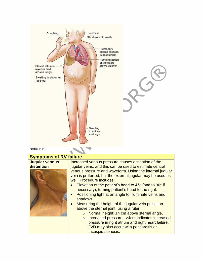
NHIBI, NIH
Symptoms of RV failure Jugular venous distention
Increased venous pressure causes distention of the jugular veins, and this can be used to estimate central venous pressure and waveform. Using the internal jugular vein is preferred, but the external jugular may be used as well. Procedure includes:
• Elevation of the patient’s head to 45 (and to 90 if necessary), turning patient’s head to the right.
• Positioning light at an angle to illuminate veins and shadows.
• Measuring the height of the jugular vein pulsation above the sternal joint, using a ruler.
o Normal height: 4 cm above sternal angle. o Increased pressure: >4cm indicates increased
pressure in right atrium and right heart failure. JVD may also occur with pericarditis or tricuspid stenosis.
Edema Peripheral: Edema of the feet and ankles increases with dependency and decreases with elevation. Over time, the edema may envelope the entire leg, external genitalia, and lower trunk. Ascites: Some people may only exhibit abdominal edema, evidenced by increasing girth measurement, or it may be in addition to peripheral edema. Ascites usually develops as the result of increased portal hypertension from hepatic dysfunction. Sacral: Those on bed rest may develop edema of the sacral area because it’s dependent. Anasarca: Severe, generalized edema. Monitoring edema requires daily weight, girth measurement, and inspection. Edema is classified on a 1-4 point scale, but pitting is usually not evident until at least 10 lbs/4.5 L of fluid have been retained:
• 1+ = Slight pitting to about 2 mm (persists 10-15 seconds).
• 2+ = Moderate pitting to about 4 mm (persists 10-15 seconds).
• 3+ = Moderate-severe pitting to about 6 mm (persists >1 minute).
• 4+ = Severe pitting to 8 mm or more (persists 2-5 minutes).
Hepatomegaly • As the liver becomes congested, the person may experience pain in the right upper quadrant and increased liver size on palpation.
• The engorgement results in hepatic dysfunction and increased portal hypertension, which can cause ascites.
• Pressure on the diaphragm may increase dyspnea and on the intestines may increase GI upset (anorexia, bloating).
• As liver cells are destroyed, fibrosis and cirrhosis may occur.
Weakness The combination of reduced cardiac output, impaired circulation, and inadequate removal of waste products cause pronounced weakness.
Classification systems
Two different classification systems are used to describe heart failure. The New York Heart Association (1994) developed a classification system that focuses on function.
New York Heart Association Classification of Heart Failure
Class I (Early HF)
The patient is essentially asymptomatic during normal activities with no pulmonary congestion or peripheral hypotension. There is no restriction on activities, and prognosis is good.
Class II (Early HF)
Mild symptoms occur with physical exertion but usually absent at rest, resulting in some limitations of ADLs. Slight pulmonary edema may be evident by basilar rales. Prognosis is good.
Class III (Advanced HF)
Obvious limitations of ADLs and discomfort on any exertion, even walking 20-100 yards. Comfortable only at rest. Prognosis is fair.
Class IV (Advanced HF)
Symptoms at rest, often resulting in being bedbound. Prognosis is poor.
The American College of Cardiology and the American Heart Association (2001) developed a newer system that includes structural disorders and symptoms. The NYHA and ACC-AHA classification systems do not completely correspond.
ACC-AHA Classification of Heart Failure
A Patients are at high risk because of conditions associated with HF but have no identified structural or functional abnormalities of the heart and no signs or symptoms. (No corresponding NYHA class.)
B
Patients have structural heart disease strongly associated with HF but have no signs or symptoms. (Corresponds to NYHA class I.)
C Patients have current or prior symptoms of HF associated with underlying structural heart disease. (Corresponds to NYHA class II and III.)
D Patients have advanced structural heart disease and marked symptoms of HF at rest despite medical therapy and interventions. (Corresponds to NYHA class IV.)
How does the heart compensate for heart failure? As heart function declines, the body compensates in order to maintain adequate cardiac output, but the compensatory measures themselves eventually contribute to and worsen heart failure.
• Cardiac compensation occurs when the compensatory mechanisms are able to maintain adequate CO and tissue perfusion.
• Cardiac decompensation occurs when the compensatory mechanisms are no longer able to maintain adequate CO or tissue perfusion.
Compensatory mechanisms
Mechanism Initial response Adverse effects
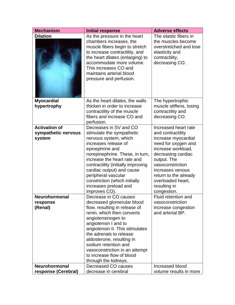
Dilation
As the pressure in the heart chambers increases, the muscle fibers begin to stretch to increase contractility, and the heart dilates (enlarging) to accommodate more volume. This increases CO and maintains arterial blood pressure and perfusion.
The elastic fibers in the muscles become overstretched and lose elasticity and contractility, decreasing CO.
Myocardial hypertrophy
As the heart dilates, the walls thicken in order to increase contractility of the muscle fibers and increase CO and perfusion.
The hypertrophic muscle stiffens, losing contractility and decreasing CO.
Activation of sympathetic nervous system
Decreases in SV and CO stimulate the sympathetic nervous system, which increases release of epinephrine and norepinephrine. These, in turn, increase the heart rate and contractility (initially improving cardiac output) and cause peripheral vascular constriction (which initially increases preload and improves CO).
Increased heart rate and contractility increase myocardial need for oxygen and increase workload, decreasing cardiac output. The vasoconstriction increases venous return to the already overloaded heart, resulting in congestion.
Neurohormonal response (Renal)
Decrease in CO causes decreased glomerular blood flow, resulting in release of renin, which then converts angiotensinogen to angiotensin I and to angiotensin II. This stimulates the adrenals to release aldosterone, resulting in sodium retention and vasoconstriction in an attempt to increase flow of blood through the kidneys.
Fluid retention and vasoconstriction increase congestion and arterial BP.
Neurohormonal response (Cerebral)
Decreased CO causes decrease in cerebral
Increased blood volume results in more
perfusion, stimulating the pituitary gland to release antidiuretic hormone, which increase water reabsorption in the renal tubules, in an attempt to increase blood volume
congestion
Ventricular remodeling
The cardiac myocytes hypertrophy, and the ventricle walls thin and enlarge, resulting in larger capacity in the body’s attempt to increase CO.
Although the ventricles are larger, they are also weaker and less effective so the EF decreases and CO falls.
What treatments are used for chronic heart failure? Treatment for chronic heart failure focuses on controlling the underlying cause, improving cardiac output, and relieving symptoms. Treatment varies from one individual to another depending on the cause of HF and the severity of symptoms, but there are common elements to most treatment protocols.
Treatment approaches for chronic heart failure Diet Because increased sodium levels result in fluid retention,
sodium-restricted diets (usually 2-3 g/d) are generally advised. The Dietary Approaches to Stop Hypertension (DASH) diet promoted by the National Institutes of Health to control hypertension is commonly used. This diet combines low sodium with increased fruits, vegetables, whole grains, and fat-free or low fat milk products. The diet is low in saturated fats but high in calcium, magnesium, potassium, protein, and fiber. Sodium restrictions may vary, but people are usually advised to exclude or limit added salt, avoid convenience foods (usually high in sodium), and use canned and prepared foods with salt
content 5% of daily-recommended allowance.
Oxygen If oxygen saturation levels are low, people may need oxygen therapy to help meet tissue demands for oxygen, especially with advanced HF. Oxygen may be needed continuously, during sleep, or during activities.
Rest People must get adequate rest to relieve the strain on the heart. In severe cases, people may require bedrest, but others need more moderate restrictions. Most people should avoid strenuous exercise and plan adequate recovery rest periods after exercise of any kind, even ambulation.
Exercise An exercise program, appropriate for the level of heart failure, helps prevent loss of muscle mass and maintains mobility.
Weight Excess weight exacerbates the symptoms of HF and increases

reduction hypertension, so one goal for patients with HF is to reduce
obesity and strive for a body mass index (BMI) of 25. While the DASH diet is a good diet for weight loss, some people may have further restrictions related to calories in order to lose excess weight. Weight may vary according to fluid retention, so patients should weigh daily and report any sudden weight increase.
Smoking cessation
Smoking increases risk of coronary artery disease and interferes with oxygenation, so people should stop smoking. People may benefit from smoking cessation classes and/or aids, such as nicotine patches.
Decreased alcohol consumption
Alcohol can impair cardiac contractions and cause further liver impairment, so those with severe HF should avoid alcohol altogether, and others should limit consumption to one drink daily.
Implantable cardioverter defibrillator
ICDs have been shown to decrease mortality in those with chronic heart failure and ischemia or nonischemic
cardiomyopathy with EF 35%.
Biventricular pacing
Biventricular pacing, in which a pacemaker coordinates contraction of the right and left ventricles, may improve cardiac output and EF and relieve some symptoms. It is indicated for moderate to severe HF in those with EF <35% and prolonged QRS duration.
Other mechanical options include ventricular assist devices and intraaortic balloon pump, which are sometimes used for those awaiting heart transplant. Heart transplantation may be the last resort for people with advanced HF However, most people with chronic heart failure are not candidates for heart transplant, and the supply of hearts for transplantation is limited, so the mainstay of HF treatment is medication.
Medications used for heart failure Diuretics Diuretics are used to prevent fluid retention and reduce edema,
providing relief of symptoms.
• Thiazides, such as hydrochlorothiazide or chlorthalidone, are effective unless glomerular filtration rates fall ≤30-40 mL/mL although metolazone is effective until the GFR falls to <20-30 mL/min. Thiazides may cause hypokalemia.
• Loop diuretics, such as furosemide, bumetanide, or torsemide, are used for more severe HF. They may cause hypokalemia, especially if taken with digitalis, as well as hypotension and decreased intravascular volume.
• Potassium-sparing diuretics, such as spironolactone, triamterene, and amiloride, may be used in conjunction with other diuretics to reduce hypokalemia.
ACE inhibitors
A diuretic combined with an ACE inhibitor (such as captopril, benazepril, and enalapril) is usually the first line treatment for
people with symptomatic HF (stage B), including both systolic and diastolic failure. ACE inhibitors block the conversion of angiotensin I to angiotensin II, which reduces aldosterone levels. This decreases systemic vascular resistance, improving cardiac output and tissue perfusion. Some people may be unable to tolerate ACE inhibitors because they develop angioedema or chronic cough.
Angiotensin II receptor blockers (ARBs)
ARBs, such as candesartan or valsartan, inhibit the renin-angiotensin-aldosterone system and may be used in addition to ACE inhibitors or as a substitute for ACE inhibitors for those who cannot tolerate ACE inhibitors because of adverse effects.
-Blockers -blockers, such as carvedilol or metoprolol, block the sympathetic nervous system response and are usually used in
conjunction with ACE inhibitors, digitalis, and diuretics. -blockers have been shown to reduce mortality rates and hospitalization, so they are now recommended for all stable patients (stages B through D), but they must be monitored carefully because they sometimes cause deterioration in patients.
Digitalis glycosides
Digitalis, primarily digoxin, once a primary treatment is used much less often now because studies indicate it does not improve mortality rates; however, it may be indicated for patients with atrial flutter/fibrillation or for those who remain symptomatic on diuretics and ACE inhibitors.
Vasodilators Vasodilators are used less frequently now because ACE inhibitors have vasodilation properties.
• Nitrates (IV), such as sodium nitroprusside or nitroglycerine are used for acute or decompensated chronic heart failure.
• Isosorbide (orally) or nitroglycerine ointment may relieve dyspnea in mild to moderate CH but are ineffective for severe HF. Transdermal nitroglycerine is ineffective for HF. At one time, a combination of isosorbide and hydralazine was used as an alternative for those who couldn’t take ACE inhibitors, but this has primarily been supplanted by ARBs.
• Nesiritide improves cardiac output by reducing ventricular filling pressures but is reserved for those who don’t respond to conventional therapy or for acute heart failure. Nesiritide is a human B-type natriuretic peptide produced by recombinant technology. It suppresses neurohormones and causes vasodilation.
Calcium channel blockers
CCBs, such as amlodipine and felodipine, cause vasodilation and reduce systemic vascular resistance. They may be used for those with nonischemic cardiomyopathy, but they do not affect mortality. First generation CCBs, such as verapamil, nifedipine and diltiazem are contraindicated with systolic dysfunction but
may be used for diastolic dysfunction.
What is acute heart failure? Acute heart failure manifests as pulmonary edema, regardless of the underlying cause. Pulmonary edema may occur as the result of a myocardial infarction or as an exacerbation of chronic HF. A common cause is LV failure resulting from coronary artery disease and resulting in increased pulmonary venous pressure as blood backs up into the left atrium and into the lungs, causing congestion. Fluid begins to mix with gas in the alveoli, preventing gas exchange and resulting in pronounced hypoxemia. Warning signs of impending acute heart failure include a dry hacking cough, increased weight, increased edema, fatigue, and decreased activity tolerance. However, the onset of acute symptoms may be very rapid:
• Pulmonary congestion causes pronounced dyspnea with noisy, moist respirations and pink-tinged frothy sputum.
• Nail beds and skin become cyanotic.
• Reduced oxygen levels to the brain result in anxiety, restlessness, and confusion progressing to stupor.
• Oxygen saturation levels may fall precipitously.
• Tachycardia is present.
• BP may be elevated or decreased, depending on severity of symptoms.
• Blood gases show reduced PaO2 and possibly increased PaCO2 with progressive signs of acidemia.
Acute heart failure is life threatening and requires immediate emergency treatment. Initial treatment includes positioning the patient in upright position with legs dependent if possible to decrease venous return and providing oxygen.
Treatment for acute heart failure (Pulmonary edema) Oxygen O2 is usually provided with a facemask or non-rebreathing mask
although if pulmonary edema is severe, positive end-expiratory pressure (PEEP) or intubation and mechanical ventilation may be required.
Morphine MS IV is administered in small doses (2-5mg) to reduce systemic vascular resistance and venous return. This results in decreased pulmonary pressure and reduces fluid leakage into lung tissue. MS also reduces anxiety. A morphine antagonist (such as naloxone hydrochloride) should available in case of respiratory depression, hypotension, or vomiting occurs.
Loop diuretics
IV diuretics, such as furosemide, increases excretion of fluids as well as causing vasodilation and pooling of peripheral blood and reducing blood return to the heart.
Dobutamine Dobutamine (IV) increases cardiac contractility in those with left ventricular dysfunction. It may, however, increase AV conduction
and cause atrial fibrillation, so it may be preceded by digitalis, -blocker, or CCB.
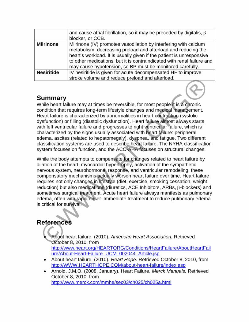
Milrinone Milrinone (IV) promotes vasodilation by interfering with calcium metabolism, decreasing preload and afterload and reducing the heart’s workload. It is usually given if the patient is unresponsive to other medications, but it is contraindicated with renal failure and may cause hypotension, so BP must be monitored carefully.
Nesiritide IV nesiritide is given for acute decompensated HF to improve stroke volume and reduce preload and afterload.
Summary While heart failure may at times be reversible, for most people it is a chronic condition that requires long-term lifestyle changes and medical management. Heart failure is characterized by abnormalities in heart contraction (systolic dysfunction) or filling (diastolic dysfunction). Heart failure almost always starts with left ventricular failure and progresses to right ventricular failure, which is characterized by the signs usually associated with heart failure: peripheral edema, ascites (related to hepatomegaly), dyspnea, and fatigue. Two different classification systems are used to describe heart failure. The NYHA classification system focuses on function, and the ACC-AHA focuses on structural changes.
While the body attempts to compensate for changes related to heart failure by dilation of the heart, myocardial hypertrophy, activation of the sympathetic nervous system, neurohormonal response, and ventricular remodeling, these compensatory mechanisms actually worsen heart failure over time. Heart failure requires not only changes in lifestyle (diet, exercise, smoking cessation, weight
reduction) but also medications (diuretics, ACE Inhibitors, ARBs, -blockers) and sometimes surgical treatment. Acute heart failure always manifests as pulmonary edema, often with rapid onset. Immediate treatment to reduce pulmonary edema is critical for survival.
References
• About heart failure. (2010). American Heart Association. Retrieved October 8, 2010, from http://www.heart.org/HEARTORG/Conditions/HeartFailure/AboutHeartFailure/About-Heart-Failure_UCM_002044_Article.jsp
• About heart failure. (2010). Heart Hope. Retrieved October 8, 2010, from http://WWW.HEARTHOPE.COM/about-heart-failure/index.asp
• Arnold, J.M.O. (2008, January). Heart Failure. Merck Manuals. Retrieved October 8, 2010, from http://www.merck.com/mmhe/sec03/ch025/ch025a.html
• Cohn, J.N. (2010). The changing face of heart failure. Medscape. Retrieved October 8, 2010, from http://cme.medscape.com/viewarticle/456645_3
• Heart disease: Heart valve disease. (2010). MedicineNet. Retrieved October 8, 2010, from http://www.medicinenet.com/heart_valve_disease/article.htm
• Heart failure. (2010). Heart Failure Online. Retrieved October 8, 2010, from http://www.heartfailure.org/eng_site/hf.asp
• Heart failure. (2010, January). National Heart Lung and Blood Institute. Retrieved October 10, 2010, from http://www.nhlbi.nih.gov/health/dci/Diseases/Hf/HF_WhatIs.html
• Heart failure. (2010, August). Texas Heart Institute. Retrieved October 9, 2010, from http://www.texasheart.org/HIC/Topics/Cond/CHF.cfm
• McPhee, S.J., & Papadakis, M.A. (2009). Current Medical Diagnosis and Treatment. San Francisco: McGraw Hill.
• Shier, D, Butler, J., & Lewis, R. Hole’s Human Anatomy & Physiology, 11 ed. New York: McGraw Hill.
• Smeltzer, S.C., Bare, B., Hinkle, J.L, & Cheever, K.H. (2009). Brunner & Suddarth’s Medical-Surgical Nursing 11th edition. Philadelphia: Lippincott, Williams, & Wilkins.
• What is heart failure? (2010). Cleveland Clinic. Retrieved October 10, 2010, from http://my.clevelandclinic.org/heart/disorders/heartfailure/hfwhatis.aspx
• Your guide to lowering your blood pressure with DASH. (2006, April). GPO: US Department of Health and Human Services. NHIBI. Retrieved October 8, 2010, from http://www.nhlbi.nih.gov/health/public/heart/hbp/dash/new_dash.pdf