heart and neck vessels - · pdf fileheart and neck vessels ... you learn the structure and...
TRANSCRIPT

Jarvis, Carolyn: PHYSICAL EXAMINATION AND HEALTH ASSESSMENT: Sixth Edition, Student Laboratory Manual. Copyright © 2012, 2008, 2004, 2000, 1996 by Saunders, an imprint of Elsevier Inc. All rights reserved. 163
C H A P T E R
19Heart and Neck Vessels
PURPOSE
This chapter helps you learn the structure and function of the heart, valves, and great vessels; understand the cardiac cycle; describe the heart sounds; understand the rationale and methods of examination of the heart; and accurately record the assessment. At the end of this chapter you should be able to perform a complete assessment of the heart and neck vessels.
READING ASSIGNMENT
Jarvis: Physical Examination and Health Assessment, 6th ed., Chapter 19, pp. 455–498.
__________________________________________________________________________________
MEDIA ASSIGNMENT
Jarvis: Physical Examination and Health Assessment DVD Series: Cardiovascular System: Heart and Neck Vessels.
Google the Internet with the term heart sounds. Listen to several websites for a good sampling of the most common heart sounds you will encounter.
__________________________________________________________________________________
GLOSSARY
Study the following terms after completing the reading assignment. You should be able to cover the defi ni-tion on the right and defi ne the term out loud.
Angina pectoris . . . . . . . . . . . . . acute chest pain that occurs when myocardial demand exceeds its oxygen supply
Aortic regurgitation . . . . . . . . . . (aortic insuffi ciency) incompetent aortic valve that allows backward fl ow of blood into left ventricle during diastole
Aortic stenosis . . . . . . . . . . . . . . calcifi cation of aortic valve cusps that restricts forward fl ow of blood during systole
Aortic valve . . . . . . . . . . . . . . . . . the left semilunar valve separating the left ventricle and the aorta
Apex of the heart . . . . . . . . . . . . tip of the heart pointing down toward the 5th left intercostal space

164 UNIT III Physical Examination
Jarvis, Carolyn: PHYSICAL EXAMINATION AND HEALTH ASSESSMENT: Sixth Edition, Student Laboratory Manual. Copyright © 2012, 2008, 2004, 2000, 1996 by Saunders, an imprint of Elsevier Inc. All rights reserved.
Apical impulse . . . . . . . . . . . . . . (point of maximal impulse, PMI) pulsation created as the left ventricle rotates against the chest wall during systole, normally at the 5th left inter-costal space in the midclavicular line
Base of the heart . . . . . . . . . . . . . broader area of heart’s outline located at the 3rd right and left intercostal space
Bell (of the stethoscope) . . . . . . cup-shaped endpiece used for soft, low-pitched heart sounds
Bradycardia . . . . . . . . . . . . . . . . . slow heart rate, <50 beats per minute in the adult
Clubbing . . . . . . . . . . . . . . . . . . . bulbous enlargement of distal phalanges of fi ngers and toes that occurs with chronic cyanotic heart and lung conditions
Coarctation of aorta . . . . . . . . . severe narrowing of the descending aorta, a congenital heart defect
Cor pulmonale . . . . . . . . . . . . . . right ventricular hypertrophy and heart failure due to pulmonary hypertension
Cyanosis . . . . . . . . . . . . . . . . . . . . dusky blue mottling of the skin and mucous membranes due to excessive amount of reduced hemoglobin in the blood
Diaphragm (of the stethoscope) . . . . . . . . . . . . . . . . fl at endpiece of the stethoscope used for hearing relatively high-pitched
heart sounds
Diastole . . . . . . . . . . . . . . . . . . . . the heart’s fi lling phase
Dyspnea . . . . . . . . . . . . . . . . . . . . diffi cult, labored breathing
Edema . . . . . . . . . . . . . . . . . . . . . . swelling of legs or dependent body part due to increased interstitial fl uid
Erb’s point . . . . . . . . . . . . . . . . . . traditional auscultatory area in the 3rd left intercostal space
First heart sound (S1) . . . . . . . . occurs with closure of the atrioventricular (AV) valves signaling the begin-ning of systole
Fourth heart sound (S4) . . . . . . (S4 gallop; atrial gallop) very soft, low-pitched ventricular fi lling sound that occurs in late diastole
Gallop rhythm . . . . . . . . . . . . . . the addition of a 3rd or a 4th heart sound makes the rhythm sound like the cadence of a galloping horse
Inching . . . . . . . . . . . . . . . . . . . . . technique of moving the stethoscope incrementally across the precordium through the auscultatory areas while listening to the heart sounds
LVH (left ventricular hypertrophy) . . . . . . . . . . . . . . . . increase in thickness of myocardial wall that occurs when the heart pumps
against chronic outfl ow obstruction (e.g., aortic stenosis)
MCL (midclavicular line) . . . . . imaginary vertical line bisecting the middle of the clavicle in each hemithorax
Mitral regurgitation . . . . . . . . . . (mitral insuffi ciency) incompetent mitral valve allows regurgitation of blood back into left atrium during systole
Mitral stenosis . . . . . . . . . . . . . . calcifi ed mitral valve impedes forward fl ow of blood into left ventricle during diastole
Mitral valve . . . . . . . . . . . . . . . . . left AV valve separating the left atria and ventricle
Palpitation . . . . . . . . . . . . . . . . . . uncomfortable awareness of rapid or irregular heart rate

CHAPTER 19 Heart and Neck Vessels 165
Jarvis, Carolyn: PHYSICAL EXAMINATION AND HEALTH ASSESSMENT: Sixth Edition, Student Laboratory Manual. Copyright © 2012, 2008, 2004, 2000, 1996 by Saunders, an imprint of Elsevier Inc. All rights reserved.
Paradoxical splitting . . . . . . . . . opposite of a normal split S2 so that the split is heard in expiration, and in inspiration the sounds fuse to one sound
Pericardial friction rub . . . . . . . high-pitched, scratchy extracardiac sound heard when the precordium is infl amed
Physiologic splitting . . . . . . . . . normal variation in S2 heard as two separate components during inspiration
Precordium . . . . . . . . . . . . . . . . . area of the chest wall overlying the heart and great vessels
Pulmonic regurgitation . . . . . . . (pulmonic insuffi ciency) backfl ow of blood through incompetent pul-monic valve into the right ventricle
Pulmonic stenosis . . . . . . . . . . . calcifi cation of pulmonic valve that restricts forward fl ow of blood during systole
Pulmonic valve . . . . . . . . . . . . . . right semilunar valve separating the right ventricle and pulmonary artery
Second heart sound (S2) . . . . . . occurs with closure of the semilunar valves, aortic and pulmonic, and signals the end of systole
Summation gallop . . . . . . . . . . . abnormal mid-diastolic heart sound heard when both the pathologic S3 and S4 are present
Syncope . . . . . . . . . . . . . . . . . . . . temporary loss of consciousness due to decreased cerebral blood fl ow (fainting), caused by ventricular asystole, pronounced bradycardia, or ven-tricular fi brillation
Systole . . . . . . . . . . . . . . . . . . . . . the heart’s pumping phase
Tachycardia . . . . . . . . . . . . . . . . . rapid heart rate, >100 beats per minute in the adult
Third heart sound (S3) . . . . . . . . soft, low-pitched ventricular fi lling sound that occurs in early diastole (S3 gallop) and may be an early sign of heart failure
Thrill . . . . . . . . . . . . . . . . . . . . . . palpable vibration on the chest wall accompanying severe heart murmur
Tricuspid valve . . . . . . . . . . . . . . right AV valve separating the right atria and ventricle
STUDY GUIDE
After completing the reading assignment and the audio-visual assignment, you should be able to answer the following questions in the spaces provided.
1. Defi ne the apical impulse and describe its normal location, size, and duration.

166 UNIT III Physical Examination
Jarvis, Carolyn: PHYSICAL EXAMINATION AND HEALTH ASSESSMENT: Sixth Edition, Student Laboratory Manual. Copyright © 2012, 2008, 2004, 2000, 1996 by Saunders, an imprint of Elsevier Inc. All rights reserved.
Which normal variations may affect the location of the apical impulse?
Which abnormal conditions may affect the location of the apical impulse?
2. Explain the mechanism producing normal fi rst and second heart sounds.
3. Describe the effect of respiration on the heart sounds.
4. Describe the characteristics of the fi rst heart sound and its intensity at the apex of the heart and at the base.
Which conditions increase the intensity of S1?
Which conditions decrease the intensity of S1?
5. Describe the characteristics of the second heart sound and its intensity at the apex of the heart and at the base.

CHAPTER 19 Heart and Neck Vessels 167
Jarvis, Carolyn: PHYSICAL EXAMINATION AND HEALTH ASSESSMENT: Sixth Edition, Student Laboratory Manual. Copyright © 2012, 2008, 2004, 2000, 1996 by Saunders, an imprint of Elsevier Inc. All rights reserved.
Which conditions increase the intensity of S2?
Which conditions decrease the intensity of S2?
6. Explain the physiologic mechanism for normal splitting of S2 in the pulmonic valve area.
7. Defi ne the third heart sound. When in the cardiac cycle does it occur? Describe its intensity, quality, location in which it is heard, and method of auscultation.
8. Differentiate a physiologic S3 from a pathologic S3.
9. Defi ne the fourth heart sound. When in the cardiac cycle does it occur? Describe its intensity, quality, location in which it is heard, and method of auscultation.
10. Explain the position of the valves during each phase of the cardiac cycle.
168 UNIT III Physical Examination
Jarvis, Carolyn: PHYSICAL EXAMINATION AND HEALTH ASSESSMENT: Sixth Edition, Student Laboratory Manual. Copyright © 2012, 2008, 2004, 2000, 1996 by Saunders, an imprint of Elsevier Inc. All rights reserved.
11. Defi ne venous pressure and jugular venous pulse.
12. Differentiate between the carotid artery pulsation and the jugular vein pulsation.
13. List the areas of questioning to address during the health history of the cardiovascular system.
14. Defi ne bruit, and discuss what it indicates.
15. Defi ne heave or lift, and discuss what it indicates.
16. State 4 guidelines to distinguish S1 from S2.
1. _______________________________________________________________________________
2. _______________________________________________________________________________
3. _______________________________________________________________________________
4. _______________________________________________________________________________
17. Defi ne pulse defi cit, and discuss what it indicates.

CHAPTER 19 Heart and Neck Vessels 169
Jarvis, Carolyn: PHYSICAL EXAMINATION AND HEALTH ASSESSMENT: Sixth Edition, Student Laboratory Manual. Copyright © 2012, 2008, 2004, 2000, 1996 by Saunders, an imprint of Elsevier Inc. All rights reserved.
18. Defi ne preload and afterload.
19. List the characteristics to explore when you hear a murmur, including the grading scale of murmurs.
20. Discuss the characteristics of an innocent or functional murmur.
Fill in the labels indicated on the following illustrations.

170 UNIT III Physical Examination
Jarvis, Carolyn: PHYSICAL EXAMINATION AND HEALTH ASSESSMENT: Sixth Edition, Student Laboratory Manual. Copyright © 2012, 2008, 2004, 2000, 1996 by Saunders, an imprint of Elsevier Inc. All rights reserved.

CHAPTER 19 Heart and Neck Vessels 171
Jarvis, Carolyn: PHYSICAL EXAMINATION AND HEALTH ASSESSMENT: Sixth Edition, Student Laboratory Manual. Copyright © 2012, 2008, 2004, 2000, 1996 by Saunders, an imprint of Elsevier Inc. All rights reserved.
REVIEW QUESTIONS
This test is for you to check your own mastery of the content. Answers are provided in Appendix A.
1. The precordium is:
a. a synonym for the mediastinum.b. the area on the chest where the apical
impulse is felt.c. the area on the anterior chest overlying
the heart and great vessels.d. a synonym for the area where the superior
and inferior venae cavae return unoxygenated venous blood to the right side of the heart.
2. Select the best description of the tricuspid valve.
a. left semilunar valveb. right atrioventricular valvec. left atrioventricular valved. right semilunar valve
3. The function of the pulmonic valve is to:
a. divide the left atrium and left ventricle.b. guard the opening between the right
atrium and right ventricle.c. protect the orifi ce between the right
ventricle and the pulmonary artery.d. guard the entrance to the aorta from the
left ventricle.
4. Atrial systole occurs:
a. during ventricular systole.b. during ventricular diastole.c. concurrently with ventricular systole.d. independently of ventricular function.
5. The second heart sound is the result of:
a. opening of the mitral and tricuspid valves.b. closing of the mitral and tricuspid valves.c. opening of the aortic and pulmonic valves.d. closing of the aortic and pulmonic valves.
6. The examiner has estimated the jugular venous pressure. Identify the fi nding that is abnormal.
a. patient elevated to 30 degrees, internal jugular vein pulsation at 1 cm above sternal angle
b. patient elevated to 30 degrees, internal jugular vein pulsation at 2 cm above sternal angle
c. patient elevated to 40 degrees, internal jugular vein pulsation at 1 cm above sternal angle
d. patient elevated to 45 degrees, internal jugular vein pulsation at 4 cm above sternal angle
7. The examiner is palpating the apical impulse. The normal size of this impulse:
a. is less than 1 cm.b. is about 2 cm.c. is 3 cm.d. varies depending on the size of the person.
8. The examiner wishes to listen in the pulmonic valve area. To do this, the stethoscope would be placed at the:
a. second right interspace.b. second left interspace.c. left lower sternal border.d. fi fth interspace, left midclavicular line.
9. Select the statement that best differentiates a split S2 from S3.
a. S3 is lower pitched and is heard at the apex.
b. S2 is heard at the left lower sternal border.c. The timing of S2 varies with respirations.d. S3 is heard at the base; timing varies with
respirations.
172 UNIT III Physical Examination
Jarvis, Carolyn: PHYSICAL EXAMINATION AND HEALTH ASSESSMENT: Sixth Edition, Student Laboratory Manual. Copyright © 2012, 2008, 2004, 2000, 1996 by Saunders, an imprint of Elsevier Inc. All rights reserved.
10. The examiner wishes to listen for a pericardial friction rub. Select the best method of listening.
a. with the diaphragm, patient sitting up and leaning forward, breath held in expiration
b. using the bell with the patient leaning forward
c. at the base during normal respirationd. with the diaphragm, patient turned to the
left side
11. When auscultating the heart, your fi rst step is to:
a. identify S1 and S2.b. listen for S3 and S4.c. listen for murmurs.d. identify all four sounds on the fi rst round.
12. You will hear a split S2 most clearly in what area?
a. apicalb. pulmonicc. tricuspidd. aortic
13. The stethoscope bell should be pressed lightly against the skin so that:
a. chest hair doesn’t simulate crackles.b. high-pitched sounds can be heard better.c. it does not act as a diaphragm.d. it does not interfere with amplifi cation of
heart sounds.
14. A murmur heard after S1 and before S2 is classifi ed as:
a. diastolic (possibly benign).b. diastolic (always pathologic).c. systolic (possibly benign).d. systolic (always pathologic).
15. When assessing the carotid artery, the examiner should palpate:
a. bilaterally at the same time, while standing behind the patient.
b. medial to the sternomastoid muscle, one side at a time.
c. for a bruit while asking the patient to hold his or her breath briefl y.
d. for unilateral distention while turning the patient’s head to one side.
16. Fill in the following blanks:
S1 is best heard at the _______ of the heart, whereas S2 is loudest at the ______ of the heart. S1 coincides with the pulse in the _________ and coincides with the ______ wave if the patient is on an ECG monitor.
Match column A to column B
Column A
17. _____ tough, fi brous, double-walled sac that surrounds and protects the heart
18. _____ thin layer of endothelial tissue that lines the inner surface of the heart chambers and valves
19. _____ reservoir for holding blood
20. _____ ensures smooth, friction-free movement of the heart muscle
21. _____ muscular pumping chamber
22. _____ muscular wall of the heart
Column B
a. pericardial fl uid
b. ventricle
c. endocardium
d. myocardium
e. pericardium
f. atrium

CHAPTER 19 Heart and Neck Vessels 173
Jarvis, Carolyn: PHYSICAL EXAMINATION AND HEALTH ASSESSMENT: Sixth Edition, Student Laboratory Manual. Copyright © 2012, 2008, 2004, 2000, 1996 by Saunders, an imprint of Elsevier Inc. All rights reserved.
23. Briefl y relate the route of a blood cell from the liver to tissue in the body.
24. List the major risk factors for heart disease and stroke identifi ed in the text.
SKILLS LABORATORY/CLINICAL SETTING
You are now ready for the clinical component of the cardiovascular system. The purpose of the clinical component is to practice the regional examination on a peer in the skills laboratory or a patient in the clinical setting and to achieve the following.
Clinical Objectives
1. Demonstrate knowledge of the symptoms related to the cardiovascular system by obtaining a regional health history from a peer or patient.
2. Correctly locate anatomic landmarks on the chest wall of a peer.
3. Using a grease pencil and with peer’s permission, outline borders of the heart and label auscultatory areas on a peer’s chest wall.
4. Demonstrate correct technique for inspection and palpation of the neck vessels.
5. Demonstrate correct techniques for inspection, palpation, and auscultation of the precordium.
6. Record the history and physical examination fi ndings accurately, reach an assessment of the health state, and develop a plan of care.
Instructions
Gather your equipment. Wash your hands. Clean the stethoscope endpiece with an alcohol wipe. Practice the steps of the examination of the cardiovascular system on a peer or on a patient in the clinical area. Record your fi ndings using the regional write-up sheet that follows. The front of the page is intended as a worksheet; the back of the page is intended for your narrative recording using the SOAP format.

174 UNIT III Physical Examination
Jarvis, Carolyn: PHYSICAL EXAMINATION AND HEALTH ASSESSMENT: Sixth Edition, Student Laboratory Manual. Copyright © 2012, 2008, 2004, 2000, 1996 by Saunders, an imprint of Elsevier Inc. All rights reserved.
NOTES

CHAPTER 19 Heart and Neck Vessels 175
Jarvis, Carolyn: PHYSICAL EXAMINATION AND HEALTH ASSESSMENT: Sixth Edition, Student Laboratory Manual. Copyright © 2012, 2008, 2004, 2000, 1996 by Saunders, an imprint of Elsevier Inc. All rights reserved.
REGIONAL WRITE-UP—CARDIOVASCULAR SYSTEM
Date _______________________
Examiner ___________________
Patient _________________________________________________ Age _________ Gender _________
Reason for visit ________________________________________________________________________
I. Health History No Yes, explain
1. Any chest pain or tightness? _____________________________________________2. Any shortness of breath? _____________________________________________3. Use more than one pillow to sleep? _____________________________________________4. Do you have a cough? _____________________________________________5. Do you seem to tire easily? _____________________________________________6. Facial skin ever turn blue or ashen? _____________________________________________7. Any swelling of feet or legs? _____________________________________________8. Awaken at night to urinate? _____________________________________________9. Any past history of heart disease? _____________________________________________
10. Any family history of heart disease? _____________________________________________ _____________________________________________11. Assess cardiac risk factors: _____________________________________________
II. Physical ExaminationA. Carotid arteries Inspect and palpate Grade R ________ L ________ (0 = absent, 1+ weak, 2+ normal, 3+ increased, 4+ bounding)B. Jugular venous system External jugular veins (circle one): �
collapsed supine meniscus visible at ____________________ bed elevated
Internal jugular venous pulsations (circle one):
�
not visible
visible at ____________________________ bed elevated
C. Precordium Inspect and palpate
1. Skin color and condition ________________________________________________________2. Chest wall pulsations ___________________________________________________________3. Heave or lift ___________________________________________________________________4. Apical impulse in the __________ at _______________________________________________
Size ________________ Amplitude ________________________________________________D. Auscultation
1. Identify anatomic areas where you will listen.2. Rate and rhythm _______________________________________________________________3. Identify S1 and S2 in diagram at right and note any variation. Fill in any murmur below: S1 S2 S1 S2
S1 _________________________________
S2 _________________________________
176 UNIT III Physical Examination
Jarvis, Carolyn: PHYSICAL EXAMINATION AND HEALTH ASSESSMENT: Sixth Edition, Student Laboratory Manual. Copyright © 2012, 2008, 2004, 2000, 1996 by Saunders, an imprint of Elsevier Inc. All rights reserved.
4. Listen in systole and diastole: Extra heart sounds ___________________________________________________________ Systolic murmur _____________________________________________________________ Diastolic murmur ____________________________________________________________
REGIONAL WRITE-UP—CARDIOVASCULAR SYSTEM
Summarize your fi ndings using the SOAP format.
Subjective (Reason for seeking care, health history)
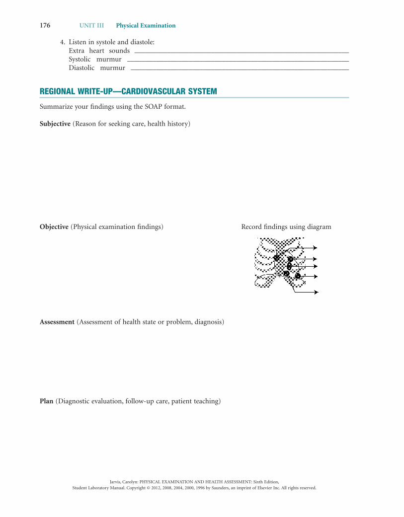
Objective (Physical examination fi ndings) Record fi ndings using diagram
Assessment (Assessment of health state or problem, diagnosis)
Plan (Diagnostic evaluation, follow-up care, patient teaching)