hearing sound how the ears work how the cochlea works auditory pathway hearing loss
TRANSCRIPT

HEARING• Sound• How the Ears Work• How the Cochlea Works• Auditory Pathway• Hearing Loss

Sound
• pressure variations (usually in air)• intensity: amplitude of sound waves

Sound Intensity
measured in deciBels (dB), sound pressure level compared to threshold
with every 6 dB increase, SPL doubles affects perceived loudness

Sound Frequency
• Number of vibrations per second • Measured in Hertz (Hz)• Affects perceived pitch

Sound Complexity
• Affects timbre (perceived sound quality)• Number of harmonics– multiples of fundamental frequency
• Attack• Decay

The Missing Fundamental
• If you play the harmonics of a fundamental without the fundamental itself, people will perceive the missing fundamental
• Example: play 400, 600, 800 Hz tones; a fundamental of 200 Hz will also be heard

How the Ear Works
• Outer Ear• Middle Ear• Inner Ear

Outer Ear
• Pinna - outer flap; helps slightly in amplifying and locating sounds
• External auditory canal - tube leading from pinna to eardrum; protects eardrum, resonates to frequencies around 3000 Hz

Outer Ear
• Tympanic membrane (eardrum) vibrates with sound waves
• Transmits sound energy to middle ear

Middle Ear
• Tympanic membrane moves ossicles– malleus (hammer)– incus (anvil)– stapes (stirrup)

Middle Ear
• Stapes pushes on oval window• Eustachian tube equalizes air pressure

Impedance Mismatch
• Sound intensity decreases when the sound travels from one medium to another (e.g., air to water)
• The cochlea is filled with fluid, so the sound intensity is decreased
• The middle ear compensates for this by amplifying the sound

Middle Ear Amplification
• Large surface area (tympanic membrane) to small surface area (oval window)
• Ossicles work as a lever system

Cochlea
• Part of inner ear, which also includes semicircular canals for vestibular sense
• Transduces sound energy to neural responses

Parts of the Cochlea
• Organ of Corti sits on basilar membrane, contains hair cells which are stimulated by sound
• Tectorial membrane is attached diagonally over Organ of Corti

The Traveling Wave
• The basilar membrane is narrow and stiff at the middle ear end, but wide and loose at the far end
• This causes it to vibrate most at one location, depending on frequency
• Higher frequencies cause vibration on middle ear end; lower frequencies cause vibration on far end

Hair Cells
• The traveling wave creates a shearing action of the tectorial membrane over the basilar membrane
• Stereocilia on top of hair cells are bent• Hair cells depolarize when stereocilia are bent
toward outer part of cochlea; hyperpolarize when bent inward

Inner Hair Cells• about 3,500• not attached to tectorial membrane• afferent fibers

Outer Hair Cells• about 12,000• attached to tectorial membrane• afferent and efferent fibers

Efferent Fibers• These fibers carry messages from the brain
which modulate activity in the cochlea• This may help code frequency more precisely• The efferent fibers explain cochlear
microphonics and otoacoustic emissions

Auditory Pathway
• Graded potentials in hair cells cause action potentials in bipolar cells
• Axons of bipolar cells (auditory nerve) carry information into the brain

Auditory Pathway
• Auditory nerve connects with cells in the cochlear nucleus in the hindbrain– lateral inhibition– frequency coding
• Superior olivary nucleus (brain stem)– some fibers cross; binaural information– sound localization

Auditory Pathway
• Inferior colliculus (brain stem)– tonotopic organization– regulate auditory reflexes (eye blink, middle
ear muscle tightening)
• Some fibers connect to superior colliculus

Auditory Pathway
• Medial geniculate nucleus of thalamus– complex feature analysis
• Primary Auditory Receiving Area– temporal lobe

Auditory Cortex Neurons
• simple tone cells• complex tone cells (clicks, species-specific
calls)• cells that respond to changing frequencies• cells that respond to binaural differences
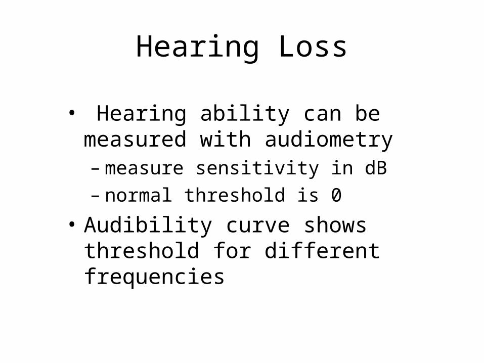
Hearing Loss
• Hearing ability can be measured with audiometry– measure sensitivity in dB– normal threshold is 0
• Audibility curve shows threshold for different frequencies

Hearing Loss
• presbycusis - loss of hearing with aging– high frequencies affected most– long term exposure to noise

Hearing Loss
• conduction deafness - problem with conduting sound to the cochlea
• nerve deafness - damage to cochlea or auditory nerve

Hearing Loss
• tinnitus - ringing in ears– auditory nerve damage– aspirin overdose– masking may help