healthcare infection surveillance of western australia/media/files/corporate...5. surveillance...
TRANSCRIPT
Healthcare Infection Surveillance of Western Australia (HISWA)
health.wa.gov.au
Surveillance ManualVersion 7, January 2020

This publication has been produced by the Healthcare Associated Infection Unit, Communicable Disease Control Directorate. © Department of Health, Western Australia January 2020
Contributors / Editors
McCann R, Stirling M, Parker C.
Contact Details
Healthcare Associated Infection Unit Communicable Disease Control Directorate Department of Health Western Australia
PO Box 8172 Perth Business Centre Western Australia 6849
Telephone: 08 9222 2131 Email: [email protected] Web: ww2.health.wa.gov.au/Health-for/Health-professionals/Infectious-diseases
1
Contents
Foreword 2
Module 1 – Introduction to surveillance of healthcare-associated infections 4
Module 2 – Surgical site infection surveillance 13
Module 3 – Methicillin-resistant Staphylococcus aureus (MRSA) healthcare associated infection 30
Module 4 – Clostridioides difficile infection 43
Module 5 – Vancomycin-resistant enterococci (VRE) sterile site infection 52
Module 6 – Staphylococcus aureus bloodstream infection 62
Module 7 – Central line-associated bloodstream infection 74
Module 8 – Haemodialysis access-associated bloodstream infection 89
Module 9 – Occupational exposure 99
Module 10 – Bed-day and separation data 105

2
ForewordHealthcare-associated infections (HAIs) are one of the most common causes of unintended harm suffered by health consumers. These infections cause the patient unnecessary pain and suffering and utilise significant human and financial resources within healthcare systems. It is increasingly recognised that HAIs are preventable adverse events rather than an inevitable complication of medical care. Establishment of baseline HAI rates and ensuring ongoing monitoring is essential to measure the effectiveness of infection prevention strategies that are implemented to reduce the occurrence of HAIs.
Both private and public healthcare facilities (HCFs) in Western Australia (WA) voluntarily commenced contributing data to the Healthcare Infection Surveillance WA (HISWA) program in 2005. The introduction of mandatory indicators for all public HCFs and private HCFs contracted to provide care for public patients commenced in 2007. Private HCFs continue to contribute data to HISWA voluntarily. The indicators collected for HISWA are described in Table 1.
The goals of the HISWA program are to ensure:
* all WA hospitals utilise standardised definitions and methodology
* ensure the validity of data through formal and informal validation exercises
* trends are identified and clinicians engaged to review clinical care to minimise infection risks and thus reduce the incidence of HAIs activities are aligned, where possible, with Australian and international surveillance programs to allow for relevant external benchmarking
* support is provided to surveillance personnel contributing data to HISWA.
HISWA data is analysed by staff at the Healthcare Associated Infection Unit (HAIU). Aggregated data and detailed hospital-specific reports are produced and distributed. All contributors are encouraged to internally review their own data to identify issues and trends in a timely manner. This surveillance manual contains the technical information to allow standardised definitions and methodology to be utilised by surveillance personnel reporting data to HISWA. If any hospital requires assistance with their surveillance program, the HAIU team are available to provide support.

3
Table 1: HISWA indicators
HISWA Indicators Data Collection Commenced
Requirements for Data
Submission
Status (Mandatory
Status Assigned)
Comments Any private hospital can
voluntarily submit data to HISWA where the indicator is
relevant to their facility
Healthcare-associated infections due to methicillin-resistant Staphylococcus aureus (MRSA)
July 2005
All data are required to be submitted within 30 days from the end of the reporting month.
SSI following hip and knee arthroplasty is subject to a 90 day surveillance period.
All data should be subject to internal validation processes prior to submission.
Mandatory (October 2007)
Mandatory for all public metropolitan, regional resource centres and integrated district hospitals.
Surgical site infection following hip and knee arthroplasty
July 2005 Mandatory (October 2007)
Mandatory for all public metropolitan, regional resource centres and integrated district hospitals where arthroplasty procedures are performed.
Healthcare-associated bloodstream infection due to Staphylococcus aureus (methicillin-sensitive and methicillin-resistant)
October 2007 Mandatory (October 2007)
Mandatory for all public metropolitan, regional resource centres and integrated district hospitals.
Hospital-identified Clostridioides difficile infection
January 2010 Mandatory (January 2010)
Mandatory for all public metropolitan, regional resource centres and integrated district hospitals.
Central line-associated bloodstream infections in adult intensive care units
July 2005 Mandatory (October 2009)
Mandatory for all public hospitals with adult intensive care units.
Haemodialysis access-associated bloodstream infection.
July 2005 Mandatory (July 2009)
Mandatory for all public metropolitan, regional resource centres and integrated district hospitals where haemodialysis is performed.
Healthcare worker occupational exposure to blood/body fluids
January 2008 Mandatory (January 2008)
Mandatory for all public metropolitan, regional resource centres and integrated district hospitals.
HISWA non-mandatory HAI indicators
Central line-associated bloodstream infections in haematology and oncology.
July 2005 Voluntary participation
Any private or public HCF where the indicator is relevant to the provision of care.
Surgical site infection following caesarean section
April 2011 Voluntary participation
Any private or public HCF performing these procedures
4
Module 1
Introduction to surveillance of healthcare-associated infections
5
Contents
Introduction 6
1. Surveillance overview 6
2. Rationale for surveillance 7
3. Types of HAI surveillance 7
3.1 Outcome surveillance 7
3.2 Process surveillance 7
4. Selection of surveillance indicators 7
5. Surveillance methodology 8
5.1 Active, prospective case-finding 8
5.2 Patient-based surveillance 8
5.3 Definitions 9
6. Data validation 9
7. Data entry to HISWA 10
8. Data analysis 10
8.1 Calculation of rates 10
8.2 The p-value 10
8.3 Confidence intervals 11
8.4 Risk stratification 11
8.5 Benchmarking 11
9. Interpretation of reports 11
9.1 WA aggregate rate 11
9.2 Cumulative WA aggregate rate 11
9.3 Cumulative hospital infections and rate 11
9.4 Rate previous two quarters 12
9.5 Trend 12
9.6 Comparator rate 12
9.7 Infection rates from small hospitals 12
10. Reporting and feedback 12
References 12
6
Introduction
1. Surveillance overview
Surveillance is the systematic collection, management, analysis, interpretation and reporting of data for use in the planning, implementation and evaluation of the provision of healthcare.1 The purpose of undertaking healthcare-associated infection (HAI) surveillance is to monitor and support improvement in the quality and safety of patient care within a healthcare facility (HCF).1
Data should not be collected just for the purpose of collecting data – the data need to be used to support the implementation of strategies that will reduce the risk of patients acquiring HAIs. Effective surveillance systems are the drivers for change and make it possible to evaluate the effectiveness of interventions. An effective surveillance system is one that provides timely feedback to HCF clinicians and managers to enable change to happen.2
Surveillance complements other prevention strategies including clinical interventions to improve the quality of care, adoption of evidence-informed practice and outbreak identification and management.
The National Safety and Quality Health Service (NSQHS) Standards requires HCFs to perform HAI surveillance in order to gather data on the incidence and prevalence of infection within their organisation.3 A robust surveillance strategy that collects data on HAIs relevant to the size and scope of the HCF, that monitors the surveillance data to guide risk reduction strategies and reports on the surveillance data to the key stakeholders, the governing bodies and the consumers, is required in order to comply with the NSQHS Standards.3
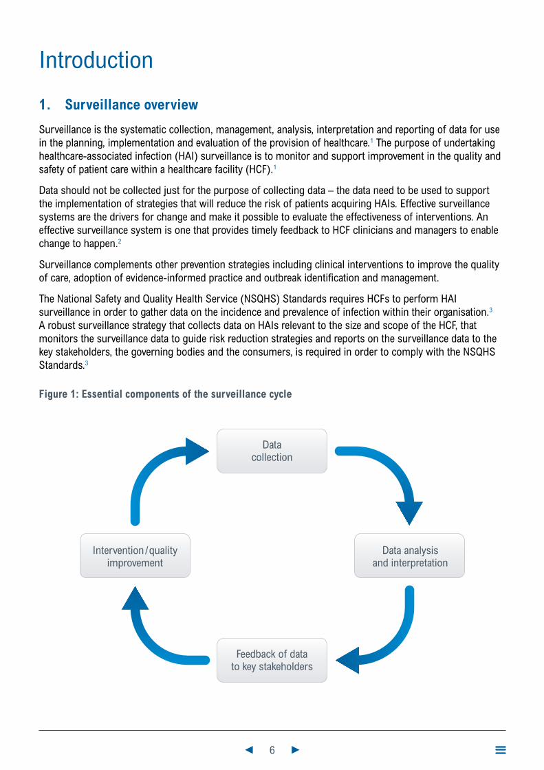
Figure 1: Essential components of the surveillance cycle
Datacollection
Feedback of datato key stakeholders
Intervention/qualityimprovement
Data analysisand interpretation

7
2. Rationale for surveillance
Surveillance of HAIs provides objective data on which to base decisions. Surveillance data enables us to determine whether a problem exists, identifies the size of the problem, and allows observation of trends over time. A sound surveillance system should:
* determine baseline HAI rates
* detect changes in rates or distribution of HAI
* facilitate investigation of significant increases in HAI rates
* determine the effectiveness of infection prevention measures
* monitor compliance with established infection prevention practices
* evaluate interventions and change in practice
* identify areas where research would be beneficial.1
3. Types of HAI surveillance
As it is not practical to conduct facility-wide surveillance for all HAI events, surveillance is often targeted, with a focus on specific sites of infection, specific populations, specific organisms, or specific locations within the HCF.1 There are two main methods of surveillance – outcome and process.1
3.1 Outcome surveillance
Outcome surveillance involves measuring adverse healthcare events, a proportion of which are preventable.1 Data may be expressed as:
* rates: time-series of HAI counts or proportions.
* point prevalence: the proportion of patients with HAIs at the time of the prevalence survey.
* incidence over time: the number of patients who develop a new HAI.
Examples of outcome surveillance include capturing the incidence of methicillin-resistant Staphylococcus aureus (MRSA) bacteraemia and surgical site infections (SSIs).1
3.2 Process surveillance
Process surveillance involves auditing actual practice against evidence-informed infection prevention strategies that are linked to improved outcomes.1 This methodology is useful because data can be captured quickly, and can capture instances of inappropriate care that did not actually result in patient harm.1 Improved processes should result in lower infection rates.
Examples of process surveillance include auditing compliance of antibiotic surgical prophylaxis or bundles of care for insertion of central lines and hand hygiene compliance.1
4. Selection of surveillance indicators
Infection prevention and control teams need to identify surveillance activities that will meet their facility’s priorities and objectives. The traditional hospital-wide surveillance, where data were collected on every infection identified, has been largely replaced by targeted surveillance that focuses on specific HAIs, organisms, medical devices or high-risk populations.

8
Jurisdictional surveillance allows aggregation of data from many HCFs, leading to a larger dataset with increased statistical value. Statewide trends can be identified to inform priorities for statewide infection prevention policies. Indicators selected for jurisdictional HAI surveillance are generally:
* procedures that are high volume or high risk for infection and are associated with high morbidity and mortality e.g. hip and knee arthroplasty
* medical device use in high-risk groups e.g. central venous catheters used in intensive care unit (ICU) patients
* significant organisms associated with antibiotic resistance and high morbidity and mortality.
5. Surveillance methodology
The value of surveillance is enhanced by providing high quality comparative data. For participating hospitals to make a valid comparison of their infection rates, the methodology used must be similar. HISWA aims for high sensitivity and specificity of reported HAIs. Sensitivity is based on false-negative HAIs i.e. true HAIs that are not reported and specificity is based on false-positive HAIs i.e. reported infections that do not meet the HAI surveillance definitions.
Processes are required to ensure that surveillance personnel automatically receive copies of all microbiology reports, in real-time, for patients presenting to their facility, including outpatient and emergency presentations. HISWA requires surveillance personnel to implement active, prospective, patient-based surveillance.4
The use of the flow charts provided in each indicator chapter is recommended to assist with each case review.
5.1 Active, prospective case-finding
* Active case-finding processes are required to identify patients who develop HAIs from the time of their admission until discharge, and on readmission with infection.
* All microbiological results relevant to a surveillance indicator should be investigated and interpreted in conjunction with information from clinical sources.
* Each case-finding method has some merit and limitations, therefore, in addition to the review of all relevant laboratory reports, a combination of case-finding methods that can be applied to eligible patients should be applied that include:
• total chart review for clinical data i.e. medical records, wound management plan, temperature chart, diagnostic and imaging reports e.g. x-ray, bone scan, ultrasound, biopsy and medication chart (antibiotics)
• liaison with clinical staff and regular ward rounds
• use of patient management systems for admission histories
• formal notification from clinical staff e.g. infection notification forms
• administration and coding reports e.g. ICD-10-AM
• pharmacy dispensing reports
• medical referrals e.g. for microbiologist or infectious disease physician
• the use of infection control management software where available.4
5.2 Patient-based surveillance
* Patient-based surveillance requires identification of all eligible patients for inclusion in the surveillance indicator. For example, in a reporting period:
• all patients undergoing a specific surgery must be counted for SSIs

9
• all patients that have had a central line in situ in ICU must be counted for ICU central line-associated bloodstream infection (CLABSI) surveillance.
* Surveillance personnel are required to determine the optimal method for obtaining denominator data for each surveillance indicator at their HCF. This may include the utilisation of:
• theatre management systems/theatre booking slips/coding reports
• medical records systems/business administration systems
• ward staff on wards relevant to the surveillance indicator
• the use of infection control management software where available.
5.3 Definitions
Standardised surveillance definitions are essential for successful data collection and analysis. The definitions developed by the National Healthcare Surveillance Network (NHSN) within the Centers for Disease Control and Prevention (CDC) in the United States of America are the most comprehensive and widely used definitions for HAI surveillance.4 Adoption of these definitions allows for benchmarking opportunities with large international datasets. Data collection for many of the HISWA indicators is based on the NHSN definitions in addition to those developed for the Australian Council for Healthcare Standards (ACHS).2
To improve the inter-rater reliability of HAI classification, contributors should ensure:
* surveillance personnel are trained in the use of surveillance definitions
* surveillance personnel consistently apply methodology for data collection and application of definitions
* infections are classified strictly according to the definition and only include HAI that fulfil the criteria in the definition
* liaison occurs with appropriate medical/surgical teams to assist in determining the source of the infection
* investigation of the patient’s hospitalisation history to identify the attributable HCF
* any queries or ambiguities in relation to the application of the surveillance definitions are referred to the Healthcare Associated infection Unit (HAIU).
6. Data validation
All HISWA contributors need to have internal validation processes in place to confirm the data they are submitting is reliable and valid. Surveillance personnel should ensure:
* prior to submission of data, that the clinical, laboratory and other diagnostic information collected meets the criteria in the definition and communication has occurred with relevant stakeholders e.g. review of all SSIs with a designated member of the surgical team
* they generate appropriate facility-specific reports to enable cross-checking of cases admitted for procedures and with infections e.g. ICD-10-AM reports
* they use HISWA hospital level raw data report i.e. data entered in the HISWA database, to cross-check with internal records prior to submission.
* they use consolidated laboratory reports and cross-check to ensure all relevant cases have been investigated.
* administrators providing bed-day data are informed of the data requirements outlined in Module 108.
* denominator data received from administrators and other external departments is cross-checked with data from previous months to identify potential outliers.
10
7. Data entry to HISWA
Prior to utilising the HISWA database, contributors should familiarise themselves with the HISWA information for new contributors. A username and password is assigned to each hospital to allow login to the database. Education and guidance on performing HISWA surveillance for new contributors can be arranged with members of the HAIU team by emailing [email protected]
All contributors have access to the HISWA Database Manual to assist with the technical details of data entry to the HISWA database. This can be accessed from the menu page of the database following login. All contributors need to ensure they:
* enter data accurately into the HISWA database
* save each record after data entry
* use the Raw Data Report in the Reports module to check both numerator and denominator data prior to finalising data
* have entered the HAIs in the appropriate modules when they meet the definition for multiple indicators e.g. a methicillin-resistant Staphylococcus aureus bloodstream infection (MRSA BSI) needs to be entered in the Specific Organism module and the Specific Organism Bloodstream module
* use the Finalisation Page as a means of checking data and advising HAIU that data submission is complete
* finalise data monthly for the previous month e.g. April data must be finalised by the last day of May.
8. Data analysis
Data analysis is an essential component of the surveillance cycle so that HAIs can be described and communicated in a meaningful way.
8.1 Calculation of rates
A rate indicates a relationship between two measurements with different units of measure and is used in HAI surveillance to describe HAIs in patient populations of different sizes and in different time periods.
A rate has three components:
* numerator: the number of infections
* denominator: the number of patients at risk
* constant: a multiple of 10 that results in a number greater than zero.
Mathematically, the rate is calculated as the numerator ÷ denominator x constant. Rates are generally expressed according to the denominator and the constant used e.g. per 100 surgical procedures or per 1,000 central line days.
8.2 The p-value
The p-value determines the probability that the difference between two rates has arisen by chance. If the probability is low (<0.05 or 5%) then the difference in rates is considered to be unlikely due to chance alone and therefore represents a significant difference.

11
8.3 Confidence intervals
HISWA rates are calculated with 95 per cent confidence intervals (CI) which provides an indication of the true infection rate. The CI displays the lowest and highest values that the true infection rate is likely to fall between 95 per cent of the time. As a general rule, a larger sample size results in a narrower CI and thus gives a better indication of the true rate.
8.4 Risk stratification
Risk stratification categorises patients at risk of infection into homogenous groups so that comparisons of infection rates can be made between groups with similar risk factors.2
Examples of risk stratification used by HISWA include:
* a risk index score for surgical patients based on their estimated risk of infection relative to other patients undergoing the same surgery
* categorisation of MRSA infections according to ICU and non-ICU settings
* categorisation of haemodialysis access device associated infections according to the type of access device
* categorisation of surgical procedures by elective or emergency status.
8.5 Benchmarking
Benchmarking involves comparing an infection rate with another point of reference which gives an indication of performance. Benchmarking should only be used as a guide and interpreted with caution due to potential variability in case mix, size of population and surveillance practice.
9. Interpretation of reports
The following information further assists with the interpretation of specific HISWA reports produced by the HAIU e.g. the hospital quarter report.
9.1 WA aggregate rate
This is an infection rate calculated from combined data submitted to HISWA from all contributing hospitals in WA for a specified period. It provides a useful benchmark with which individual hospitals can compare their infection rate for the same period.
9.2 Cumulative WA aggregate rate
The cumulative aggregate rate is the overall rate for WA since data collection commenced for that indicator. The cumulative aggregate rate is the total number of infections divided by the total relevant denominator for WA since reporting commenced.
9.3 Cumulative hospital infections and rate
The cumulative number of infections for a hospital is the total number of infections that have been reported for an indicator since their inclusion in the HISWA program. The cumulative hospital rate is the total number of infections divided by the total relevant denominator since reporting commenced.

12
9.4 Rate previous two quarters
This measure provides an internal benchmark to determine short term trends in the infection rate over time. It is the number of infections over the previous two quarters divided by the relevant denominator over the previous two quarters.
9.5 Trend
Trend is a term used to describe the general movement in rates over time. HISWA reports describe trends in terms of quarterly rates.
* rate this quarter greater than the previous quarter and indicated by
* rate this quarter less than the previous quarter and indicated by
* rate this quarter equal to the previous quarter and indicated by
9.6 Comparator rate
Where possible, a comparator rate from another Australian state or overseas country will be used for external benchmarking. Comparators are selected based on the use of the same definitions and methodology to HISWA and the sample size is sufficiently large to calculate a valid infection rate.
9.7 Infection rates from small hospitals
High infection rates and wide confidence intervals may be reported when there are small denominator numbers reported from small hospitals. This also means that a small increase in the number of infections can result in a large increase in the infection rate. Therefore rates should always be interpreted carefully and in conjunction with other information.
10. Reporting and feedback
Feedback of analysed data in a timely manner to key stakeholders is an important requirement of surveillance programs to drive change and improve outcomes and has been demonstrated to be effective in reducing infections when provided to clinicians.2
Surveillance results need to be communicated to appropriate committees and to the executive management who are accountable for patient safety and quality and have the ability to make changes within the facility.
References
1. National Health and Medical Research Council and Australian Commission on Safety and Quality in Healthcare. Australian Guidelines for the Prevention and Control of Infection in Healthcare. Canberra, ACT: Commonwealth of Australia; 2019.
2. The Australian Council on Healthcare Standards. Infection Control Version 5.1: Clinical Indicator User Manual. ACHS Clinical Indicator Program [Internet]. 2018. Available from: https://www.achs.org.au/media/154670/infection_control_v5.1.pdf.
3. Australian Commission on Safety and Quality in Health Care. National Safety and Quality Health Service Standards Second Edition. 2017.
4. National Healthcare Safety Network (NHSN). Patient safety component manual. 2020.
13
Module 2
Surgical site infection surveillance
14
Contents
Introduction 15
1. Methodology 15
1.1 Denominator data 15
1.2 Numerator data 15
1.3 Classification of SSI 15
2. Definitions 16
2.1 HISWA operative procedures 16
2.2 Primary and non-primary closure 16
2.3 Emergency and elective procedures 16
2.4 Types of SSI 16
2.5 Criteria for SSI 18
2.6 Specimen classification 20
2.7 Point of detection of SSI 20
2.8 Surveillance period 20
2.9 SSI risk index score 21
3. HISWA dataset 22
3.1 Numerator data fields 22
3.2 Numerator reporting instructions 23
3.3 Numerator reporting instructions for specific post-operative infection scenarios 23
3.4 Denominator data fields 23
3.5 Denominator reporting instructions 24
4. Specific information for HISWA operative procedures 24
4.1 Procedure inclusions and exclusions 24
5. Calculation of SSI rate 24
Appendix 1: HISWA operative procedures and ICD-10-AM codes 26
Appendix 2: Risk index score calculation for SSI 27
References 29

15
IntroductionA surgical site infection (SSI) is an infection that develops as a result of an operative procedure. They are associated with increased morbidity and mortality, prolonged hospital stay and increased healthcare costs.1,2 Surveillance of SSIs, coupled with prompt feedback of data to surgeons and key stakeholders, has been shown to be an important strategy to reduce SSIs.1,2
The World Health Organization Global Guidelines for the Prevention of Surgical Site Infection 2016 3 highlights the importance of a sound SSI surveillance system and along with the recently published CDC Guideline for the Prevention of Surgical Site Infection 2018 4 provide extensive evidence-informed resources for the prevention of SSIs.
The HISWA SSI surveillance module is based on the National Healthcare Safety Network (NHSN) Patient Safety Component Manual, Centres for Disease Control and Prevention (CDC) in the United States of America.5
1. Methodology
For participating hospitals to make a valid comparison of their SSI rates the methodology must be similar and infection definitions consistently applied. The preferred HISWA methodology is active, prospective, patient-based surveillance and this needs to be performed by trained infection prevention and control personnel. Refer to Module 1 for an introduction to surveillance of healthcare-associated infections (HAIs).
1.1 Denominator data
Patient-based surveillance requires identification of all eligible patients undergoing the selected operative procedure.1 (see Table 4: Procedure inclusions and exclusions) Eligible patients can be determined in liaison with operating theatre management systems/theatre bookings/theatre coding/medical record systems and notifications from theatre staff.
1.2 Numerator data
Patient-based surveillance requires monitoring of all patients undergoing a HISWA procedure for identification of an SSI within the designated surveillance period for that specific procedure i.e. either 30 or 90-day surveillance. (see Table 4: Procedure inclusions and exclusions) Active, prospective case-finding is required to monitor SSIs from the time of the surgical procedure and during the post-operative stay until discharge.2
Processes are required to detect patients who are readmitted to a hospital for treatment of SSIs.
1.3 Classification of SSI
To improve the classification inter-rater reliability, HISWA contributors should:
* classify SSI strictly according to the definitions
* liaise with the surgical team, other contributors and the Healthcare Associated Infection Unit (HAIU) via [email protected] for difficult classifications.

16
2. Definitions
2.1 HISWA operative procedures
A HISWA operative procedure is a procedure that is included in Appendix 1: HISWA operative procedures and ICD-10-AM codes and takes place during an operation where at least one incision, including laparoscopic approach, is made through the skin or mucous membranes, or re-operation via an incision that was left open during a prior procedure and takes place in an operating room including a caesarean section room, interventional radiology room, and cardiac catheterisation lab.1,5
Both types of incisional closure methods are included in HISWA operative procedures.
Both emergency and elective operative procedures are to be included for each procedure listed.
2.2 Primary and non-primary closure
Primary closure: is the closure of the skin level during the original surgery, regardless of the presence of wires, wicks, drains, or other devices or objects extruding through the incision. This category includes surgeries where any portion of the incision is closed at skin level by any means. For procedures which have multiple incisions or laparoscopic trocar sites, the procedure is classed as primary closed if any of the incisions are primarily closed.1
Non-primary closure: is the closure that leaves the skin level completely open following the surgery. The deep tissue layers may be closed by some means or the deep and superficial layers may both be left completely open. Wounds with non-primary closure may be described as “packed”, covered with plastic, or have “wound vacs” or other devices.1
2.3 Emergency and elective procedures
Elective: a planned procedure at a time to suit the patient and surgical team.
Emergency: a non-elective, unscheduled operative procedure that does not allow for the standard preoperative preparation normally done for a scheduled operation e.g. stabilisation of vital signs, pre-operative showering, adequate antiseptic skin preparation.
Emergency caesarean section: an unplanned procedure for reasons determined as compromising the mother or foetus requiring earlier than planned delivery.6
2.4 Types of SSI
An SSI can be classified as a superficial incisional, deep incisional or an organ/space infection, see Figure 1.1 HISWA data combines deep incisional and organ/space infections to allow for more meaningful statistical analysis and align with published reports from other jurisdictions.

17
Figure 1: Schematic of SSI anatomy and classification1
Skin
SubcutaneousTissue
Deep Soft Tissue(fascia and muscle)
Organ/Space Organ/SpaceSSI
SuperficialIncisionalSSI
DeepIncisionalSSI
2.4.1 Superficial SSI
A superficial incisional SSI involves only the skin and subcutaneous tissue of the incision and the date the SSI is identified occurs within 30 days of the operative procedure.1
2.4.2 Deep SSI
A deep incisional SSI involves deep soft tissues e.g. fascia and muscle layers and the date the SSI is identified occurs within 30 or 90 days of the operative procedure depending on operation type.1
2.4.3 Organ/space SSI
An organ/space SSI involves any part of the body deeper than the fascia or muscle layers that are opened or manipulated during the operative procedure and the date the SSI is identified occurs within 30 or 90 days of the operative procedure depending on operation type.1
Specific criteria must be met to be classified as an organ/space SSI event. The full listing of site-specific organ/space SSI and criteria are outlined in the CDC/NHSN Patient Safety Component Manual – Chapter 17 Surveillance Definitions for Specific Types of Infections 1
The CDC updates this document on an annual basis, therefore the reproduction of the contained text is not advised and HISWA contributors should source this document when required to apply the organ/site specific criteria.

18
2.5 Criteria for SSI
The criteria for each type of SSI is defined in Table 1: Criteria for superficial, deep and organ/space SSI.
Note: In Table 1 the term surgeon or attending physician* includes surgeon(s), infectious diseases or emergency physician, another physician involved in the case or physician’s designee e.g. nurse practitioner or physician’s assistant. The prescription of antimicrobials alone is not sufficient evidence of a diagnosis of SSI. These cases need to be carefully evaluated by the surveillance personnel to ensure the definition of an SSI has been met. If the reason for treatment has not been documented the case requires discussion with the surgeon or attending physician.
Table 1: Criteria for superficial, deep and organ/space SSI
To classify as a superficial incisional SSI the following criteria must be met:
Infection occurs within 30 days of the operative procedure (day one = day of procedure) and involves only skin or subcutaneous tissue of the incision and the patient has at least one of the following:
a. Purulent discharge from the superficial incision.
b. Organisms isolated from an aseptically obtained specimen from the superficial incision or subcutaneous tissue by culture or non-culture based microbiological testing method which is performed for purposes of clinical diagnosis or treatment.
Note: a negative culture result from a specimen does not meet this criterion
c. A superficial incision that is deliberately opened by a surgeon or attending physician and culture or non-culture based testing of the superficial incision or subcutaneous tissue is not performed and the patient has at least one of the following signs or symptoms: pain or tenderness; localised swelling; erythema or heat.
d. Diagnosis of superficial incisional SSI by the surgeon or attending physician.
Comments
Do not report the following as an SSI:
* a stitch abscess alone (minimal inflammation and discharge confined to the points of suture penetration)
* a localised stab wound e.g. drain incision site or pin site infection.
* diagnosis or treatment of cellulitis(redness, warmth, swelling) by itself, does not meet criterion d) for a superficial SSI. Conversely, an incision that is draining or that has organisms identified by culture or non-culture based testing is not considered a cellulitis.
* superficial incisions that are shown to be colonised with microorganisms by the collection of a wound swab and that are without clinical signs of infection.
* Note: a laparoscopic trocar site is considered a surgical site incision
Classify SSIs that involve both superficial and deep incisional sites as deep incisional.

19
To classify as deep incisional SSI the following criteria must be met:
Infection occurs within 30 or 90 days (depending on procedure type) of the operative procedure (day one = day of procedure) and involves deep soft tissues (fascial and muscle layers) and the patient has at least one of the following:
a. Purulent drainage from the deep incision.
b. A deep incision that spontaneously dehisces or is deliberately opened by a surgeon or attending physician and an organism(s) is identified from the deep soft tissues by culture or non-culture based microbiological testing method or a specimen is not obtained and the patient has at least one of the following signs or symptoms: fever (>38°C), localised pain or tenderness.
Note: A negative culture result from a specimen does not meet this criterion.
c. An abscess or other evidence of infection involving the deep incision that is detected on direct or histopathologic examination or imaging test.
Comments
Classify SSIs that involve both deep incisional and organ/space as organ/space SSIs
To classify as organ/space SSI the following criteria must be met:
Infection occurs within 30 or 90 days (depending on procedure type) of the operative procedure (day one = day of procedure) and involves any part of the body, deeper than the fascial/muscle layers that are opened or manipulated during the operative procedure and the patient has at least one of the following:
a. Purulent drainage from a drain that is placed into the organ/space e.g. closed suction.
b. Organisms identified from fluid or tissue in the organ/space by culture or non-culture microbiological testing method, performed for the purpose of clinical diagnosis or treatment.
c. An abscess or other evidence of infection involving the organ/space that is detected on direct or histopathologic examination or imaging test and meets at least one criterion for a specific organ/space infection site.
Comments
As an organ/space SSI involves any part of the body, excluding the skin incision, fascia, or muscle layers, that is opened or manipulated during the procedure, the criterion for infection at these body sites must be met in addition to the organ/space SSI criteria. Examples of specific organ/space infection sites are endometritis following a caesarean section or osteomyelitis following an arthroplasty procedure. Refer to CDC/NHSN Specific Classification of an Organ/Space SSI for infection criterion for body sites relevant to HISWA operative procedures.

20
2.6 Specimen classification
Classification of a specimen as either sterile or non-sterile assists in interpreting the clinical significance and determining if the criteria for classification as an SSI are met.
2.6.1 Sterile specimen
Sterile specimens are wound aspirates and tissue biopsies that are aseptically obtained i.e. obtained in a manner to prevent the introduction of organisms from surrounding tissues into the specimen being collected e.g. specimens collected intra-operatively. Sterile specimens are unlikely to be contaminated with skin micro-organisms and therefore positive results are significant evidence of infection.
2.6.2 Non-sterile specimen
Non-sterile specimens can be potentially contaminated and therefore positive results require a clinical assessment to determine if an infection is actually present and the organisms isolated are not representing skin flora or contamination during collection e.g. swabs of the incision, dehisced and debrided tissue.
2.7 Point of detection of SSI
Infections may be detected at three possible points and are reported accordingly.
2.7.1 Detected during initial admission
The SSI is detected during the initial hospitalisation following the procedure and prior to discharge of the patient from the hospital or Hospital-in-the-Home (HITH).
2.7.2 Detected on readmission
The SSI is detected on readmission to a hospital or to a HITH service for treatment of the SSI e.g. intravenous antimicrobial therapy, surgical washout, removal of a prosthesis and includes readmission to another hospital.
2.7.3 Detected and treated as an outpatient and other post-discharge surveillance
The SSI is detected and treated as an outpatient, and the patient is not admitted to a hospital or a HITH service for treatment of the SSI. This information may be identified by active post-discharge surveillance (PDS) or by notification from outpatient departments (clinic, emergency department) or general practitioners. Due to the lack of uniformity for PDS between healthcare facilities (HCFs), this data should be recorded by the facility and reported to HISWA but is not included in calculation of HISWA SSI rates used for benchmarking purposes.2 Note: If the SSI is detected by PDS and the patient is admitted to a HITH service then this shall be included as detected on readmission
2.8 Surveillance period
All eligible patients under surveillance for SSI must be followed up during the initial admission period until discharge and monitored for readmission. To detect an SSI, HISWA procedures are to be monitored for the following periods:
* caesarean section: follow-up period post-procedure is 30 days
* hip and knee arthroplasty: follow-up period post-procedure is 90 days.
21
2.9 SSI risk index score
The risk index score is a method of stratification of risk for infection associated with surgery. The higher the patient’s risk index score, the higher the risk the patient has of developing an SSI. Risk-adjusted rates allow statistical adjustment for differences across participating hospitals.
Risk index factors and scores are described in detail in Appendix 2: Risk index score calculation for SSI.
2.9.1 Calculation of risk index score
The risk index score consists of three risk factors that are host and procedure-related. These are the American Society of Anaesthesiology (ASA) classification, the duration of surgery and the surgical wound classification.
A score is assigned for each risk factor and the total score is calculated by adding the three scores together i.e. ASA + duration of surgery + surgical wound classification.
If an operative procedure is performed through the same incision within 24 hours e.g. for complications, the procedure duration times are combined and the higher surgical wound class and ASA score is reported, if they have changed.
2.9.2 Reporting of risk index
Hospitals performing more than 100 of each procedure type per year are required to calculate the risk index score for all eligible patients.
Hospitals performing less than 100 of each procedure type per year are not required to calculate the risk index score, however, risk index classification is encouraged to allow for more meaningful data analysis. Under the risk index exemption, eligible patients are classified as ‘All’. Do not submit a mixture of risk index and ‘All’ data.

22
3. HISWA dataset
3.1 Numerator data fields
Data described in Table 2 is required to be entered in the HISWA database.
Table 2: SSI numerator data fields and descriptors for HISWA database
Data field Descriptor
Patient ID Unique patient identifier* public hospital: medical record number* private hospital: patient initials or medical record number
Date of birth Patient date of birth
Procedure Select correct operative procedure from drop down list e.g. primary hip arthroplasty, revision knee arthroplasty, emergency caesarean section
Date of procedure
Date the operative procedure was performed
Date infection identified
Date the SSI infection criterion were met
Risk index score
Patient risk index classified as 0, 1, 2, 3, N/A (not available)For hospitals not assigning a risk index score use ‘All’
Point of detection
Detected during initial admissionDetected on readmissionDetected and treated as an outpatient or other post discharge surveillance
Infection classification
Superficial SSI
Deep SSI or organ/space infection 2.4.1 Superficial SSI
Specimen Sterile specimenNon-sterile specimenNot obtained
Organism 1 The 1st pathogenic organism isolated from a specimen
Organism 2 The 2nd pathogenic organism isolated from a specimen
Organism 3 The 3rd pathogenic organism isolated from a specimen
23
3.2 Numerator reporting instructions
If a patient has several procedures performed on different dates e.g. primary followed by revision, attribute the SSI to the procedure performed closest to the date of infection onset, unless there is evidence that the infection was associated with a different operation.
If during the post-operative period the surgical site has an invasive manipulation for diagnostic or therapeutic purposes e.g. needle aspiration and following this manipulation an SSI develops, this infection is not attributed to the operation. This does not apply to closed manipulation e.g. closed reduction of a dislocated hip or wound packing.
If the SSI is detected at a HCF that did not perform the initial procedure, contributors must inform the Healthcare Associated Infection Unit (HAIU) by emailing [email protected] The SSI will be assigned to the HCF where the initial procedure was performed.
* SSI detected at another HCF following transfer during the primary hospitalisation period are to be reported as detected on ‘initial admission’ for the HCF that performed the procedure
* SSI detected on readmission to another HCF are to be reported as detected on ‘readmission’ for the HCF that performed the procedure.
3.3 Numerator reporting instructions for specific post-operative infection scenarios
A SSI that meets the definitions should be reported without regard to post-operative accidents, falls inappropriate showering or bathing practices, or other occurrences that may or may not be attributable to patients’ intentional or unintentional postoperative actions.
A SSI should be reported regardless of the presence of certain skin infections e.g. dermatitis, blister, impetigo that occur near an incision.
A SSI should be reported regardless of the possible occurrence of a ‘seeding’ event from an unrelated procedure e.g. dental work.
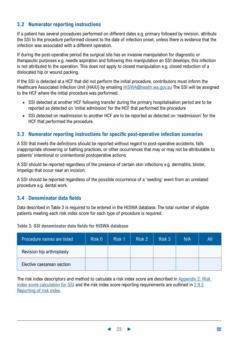
3.4 Denominator data fields
Data described in Table 3 is required to be entered in the HISWA database. The total number of eligible patients meeting each risk index score for each type of procedure is required.
Table 3: SSI denominator data fields for HISWA database
Procedure names are listed Risk 0 Risk 1 Risk 2 Risk 3 N/A All
Revision hip arthroplasty
Elective caesarean section
The risk index descriptors and method to calculate a risk index score are described in Appendix 2: Risk index score calculation for SSI and the risk index score reporting requirements are outlined in 2.9.2 Reporting of risk index.
24
3.5 Denominator reporting instructions
If a patient returns to the operating room within 24 hours of the original procedure for another procedure through the same incision, only one procedure is counted in the denominator. Combine the duration cut point for both procedures and use the wound classification that reflects the highest degree of contamination.
Bilateral procedures performed during the same episode of care in the operating room, are counted as two separate procedures.
If a patient dies in the operating room, do not count in the denominator.
4. Specific information for HISWA operative procedures
4.1 Procedure inclusions and exclusions
Refer to Table 4 for the numerator and denominator inclusions and exclusions for hip and knee arthroplasty and caesarean section procedures. Refer to Appendix 1: HISWA operative procedures and ICD-10-AM codes for specific procedure codes.
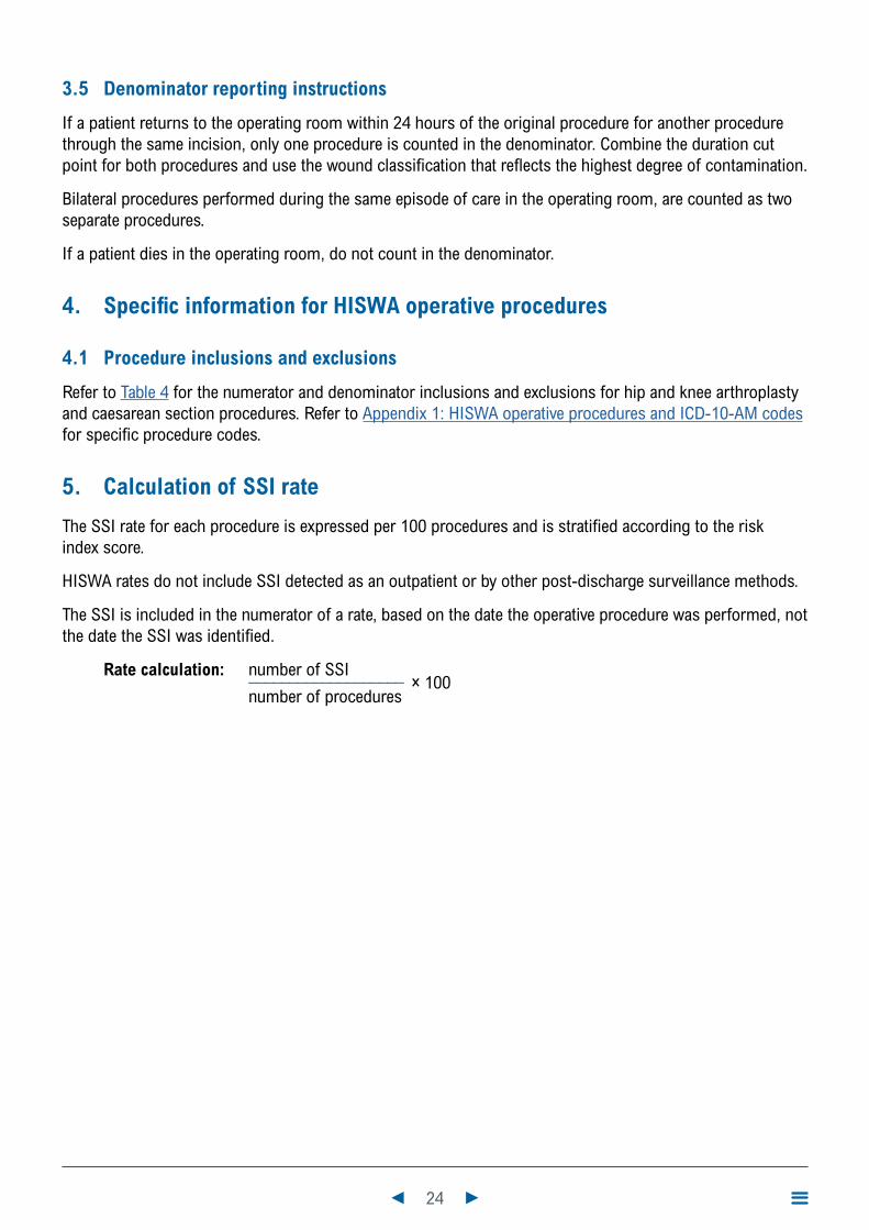
5. Calculation of SSI rate
The SSI rate for each procedure is expressed per 100 procedures and is stratified according to the risk index score.
HISWA rates do not include SSI detected as an outpatient or by other post-discharge surveillance methods.
The SSI is included in the numerator of a rate, based on the date the operative procedure was performed, not the date the SSI was identified.
Rate calculation: number of SSI × 100
___________________ number of procedures

25
Table 4: Procedure inclusions and exclusions
Procedure Include Exclude
Numerator
Hip and knee arthroplasty
* Superficial SSI detected up to 30 days after the procedure
* Deep or organ/space SSI detected within 90 days of the procedure
* SSI detected following a revision for infective reasons where SSI definitions are met again i.e. new infective episode with the same or different infecting organism
* Primary and non-primary closures
* Superficial SSI detected more than 30 days after procedure
* Deep or organ/space SSI detected more than 90 days after the procedure
Caesarean Section
* Superficial and deep or organ/space SSI detected up to 30 days after the procedure following both elective and emergency procedures
* Superficial and deep or organ/ space SSI detected more than 30 days after the procedure
Note: organ/space infections must meet the CDC/NHSN specific criteria
Denominator
Hip and knee arthroplasty
* All total, partial, primary and revision procedures as listed in Appendix 1: HISWA operative procedures and ICD-10-AM codes
* Both elective and emergency procedures are included
* Revision procedures for both mechanical and infective reasons
* Bilateral hip or knee procedures performed during the same trip to the operating room and counted as two separate procedures
* Primary and non-primary closures
* Procedures not listed in Appendix 1: HISWA operative procedures and ICD-10-AM codes e.g. hip-resurfacing, hemiarthroplasty of fractured neck of femur
Caesarean Section
* Classical and lower uterine segment caesarean section (LUSCS)
* Both emergency and elective procedures
* Procedures not listed in Appendix 1: HISWA operative procedures and ICD-10-AM codes
26
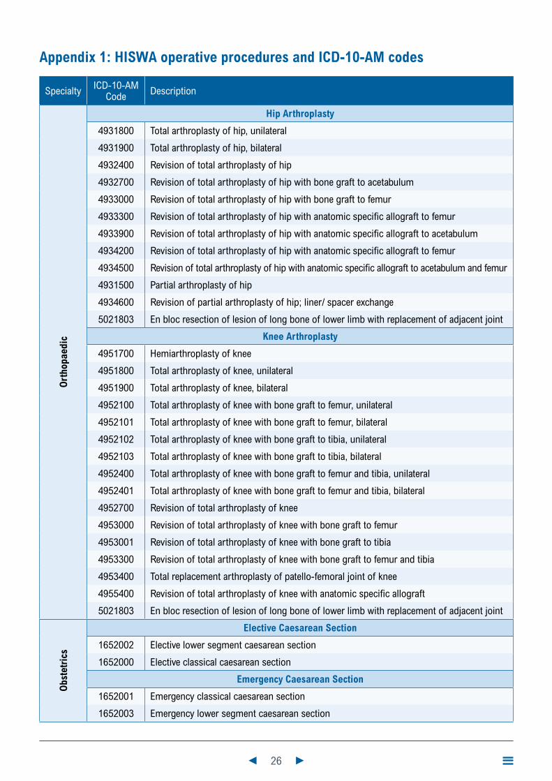
Appendix 1: HISWA operative procedures and ICD-10-AM codes
Specialty ICD-10-AM Code Description
Ort
hopa
edic
Hip Arthroplasty
4931800 Total arthroplasty of hip, unilateral
4931900 Total arthroplasty of hip, bilateral
4932400 Revision of total arthroplasty of hip
4932700 Revision of total arthroplasty of hip with bone graft to acetabulum
4933000 Revision of total arthroplasty of hip with bone graft to femur
4933300 Revision of total arthroplasty of hip with anatomic specific allograft to femur
4933900 Revision of total arthroplasty of hip with anatomic specific allograft to acetabulum
4934200 Revision of total arthroplasty of hip with anatomic specific allograft to femur
4934500 Revision of total arthroplasty of hip with anatomic specific allograft to acetabulum and femur
4931500 Partial arthroplasty of hip
4934600 Revision of partial arthroplasty of hip; liner/ spacer exchange
5021803 En bloc resection of lesion of long bone of lower limb with replacement of adjacent joint
Knee Arthroplasty
4951700 Hemiarthroplasty of knee
4951800 Total arthroplasty of knee, unilateral
4951900 Total arthroplasty of knee, bilateral
4952100 Total arthroplasty of knee with bone graft to femur, unilateral
4952101 Total arthroplasty of knee with bone graft to femur, bilateral
4952102 Total arthroplasty of knee with bone graft to tibia, unilateral
4952103 Total arthroplasty of knee with bone graft to tibia, bilateral
4952400 Total arthroplasty of knee with bone graft to femur and tibia, unilateral
4952401 Total arthroplasty of knee with bone graft to femur and tibia, bilateral
4952700 Revision of total arthroplasty of knee
4953000 Revision of total arthroplasty of knee with bone graft to femur
4953001 Revision of total arthroplasty of knee with bone graft to tibia
4953300 Revision of total arthroplasty of knee with bone graft to femur and tibia
4953400 Total replacement arthroplasty of patello-femoral joint of knee
4955400 Revision of total arthroplasty of knee with anatomic specific allograft
5021803 En bloc resection of lesion of long bone of lower limb with replacement of adjacent joint
Obs
tetr
ics
Elective Caesarean Section
1652002 Elective lower segment caesarean section
1652000 Elective classical caesarean section
Emergency Caesarean Section
1652001 Emergency classical caesarean section
1652003 Emergency lower segment caesarean section

27
Exclusions:
4752200 Hemiarthroplasty of femur- Austin Moore arthroplasty
9060700 Resurfacing of hip, unilateral,
9060701 Resurfacing of hip, bilateral
5021503 En bloc resection of lesion of soft tissue affecting the long bones of lower limb, with intercalary reconstruction using prosthesis
5021803 En bloc resection of lesion of long bone of lower limb with replacement of adjacent joint
Appendix 2: Risk index score calculation for SSI
1. ASA classification
The American Society of Anaesthesiology (ASA) classification system is a numerical quantification of disease severity in patients undergoing general anaesthesia. Studies have demonstrated that ASA class is a useful indicator of host susceptibility to infection for epidemiological purposes. A score of 0 can be entered when the ASA score cannot be established. Patients with an ASA score of 6 (organ retrieval in brain dead patients) are excluded.
ASA Class Description Risk Index Score
1 A normal healthy patient 0
2 A patient with mild systemic disease 0
3 A patient with severe systemic disease 1
4A patient with incapacitating systemic disease that is a constant threat to life
1
5A moribund patient who is not expected to survive for 24 hours with or without the operation
1
2. Duration of the operative procedure
The interval in hours and minutes between the time of skin incision and surgery finish time i.e. the time when all instrument and sponge counts are completed and verified as correct, all postoperative radiological studies in the OR are completed, all dressings and drains are secured, and the surgeons have completed all procedure-related activities on the patient. Duration cut points approximate the 75th percentile of the duration of surgery. Australian data (VICNISS)7 is used to determine the cut points. If a procedure is longer than the reported duration cut point then 1 risk point is scored.
Surgery duration cut point
Procedure Duration Cut Point
Hip arthroplasty 120 minutes
Knee arthroplasty 103 minutes
Caesarean section 48 minutes
28
3. Wound classification
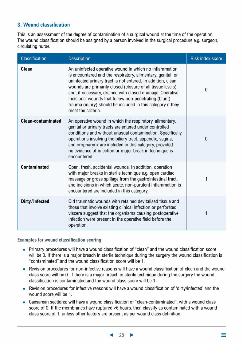
This is an assessment of the degree of contamination of a surgical wound at the time of the operation. The wound classification should be assigned by a person involved in the surgical procedure e.g. surgeon, circulating nurse.
Classification Description Risk index score
Clean An uninfected operative wound in which no inflammation is encountered and the respiratory, alimentary, genital, or uninfected urinary tract is not entered. In addition, clean wounds are primarily closed (closure of all tissue levels) and, if necessary, drained with closed drainage. Operative incisional wounds that follow non-penetrating (blunt) trauma (injury) should be included in this category if they meet the criteria.
0
Clean-contaminated An operative wound in which the respiratory, alimentary, genital or urinary tracts are entered under controlled conditions and without unusual contamination. Specifically, operations involving the biliary tract, appendix, vagina, and oropharynx are included in this category, provided no evidence of infection or major break in technique is encountered.
0
Contaminated Open, fresh, accidental wounds. In addition, operation with major breaks in sterile technique e.g. open cardiac massage or gross spillage from the gastrointestinal tract, and incisions in which acute, non-purulent inflammation is encountered are included in this category.
1
Dirty/ infected Old traumatic wounds with retained devitalised tissue and those that involve existing clinical infection or perforated viscera suggest that the organisms causing postoperative infection were present in the operative field before the operation.
1
Examples for wound classification scoring
* Primary procedures will have a wound classification of “clean” and the wound classification score will be 0. If there is a major breach in sterile technique during the surgery the wound classification is “contaminated” and the wound classification score will be 1.
* Revision procedures for non-infective reasons will have a wound classification of clean and the wound class score will be 0. If there is a major breach in sterile technique during the surgery the wound classification is contaminated and the wound class score will be 1.
* Revision procedures for infective reasons will have a wound classification of ‘dirty/infected’ and the wound score will be 1.
* Caesarean sections: will have a wound classification of “clean-contaminated”, with a wound class score of 0. If the membranes have ruptured >6 hours, then classify as contaminated with a wound class score of 1, unless other factors are present as per wound class definition.
29
References
1. The National Healthcare Safety Network (NHSN). CDC/NHSN surveillance definitions for specific types of infections. National Healthcare Safety Network (NHSN) Patient Safety Component Manual: Centers for Disease Control and Prevention; 2020.
2. Australian Commission on Safety and Quality in Health Care. Approaches to surgical site infection surveillance: For acute care settings in Australia. Sydney; 2017.
3. World Health Organization (WHO). Global guidelines for the prevention of surgical site infection, second edition. Geneva: WHO Press; 2018.
4. Berríos-Torres SI, Umscheid CA, Bratzler DW, et al. Centers for Disease Control and Prevention guideline for the prevention of surgical site infection, 2017. JAMA Surgery. 2017;152(8):784-91.
5. National Healthcare Safety Network (NHSN). Patient safety component manual. 2020.
6. The Royal Australian and New Zealand College of Obstetricians and Gynaecologists. Categorisation of urgency for caesarean section. 2019.
7. VICNISS. Surgical Site Infection(SSI): Protocol: Victoria Health; 2019 [Available from: https://www.vicniss.org.au/media/1507/ssi-protocol.pdf

30
Module 3
Methicillin-resistant Staphylococcus aureus (MRSA) healthcare associated infection
31
Contents
Introduction 32
1. Methodology 32
2. Definitions 32
2.1 MRSA infection 32
2.2 Criteria for MRSA healthcare-associated infection 33
2.3 Neutropenia 33
2.4 New MRSA HAI 33
2.5 Community-associated MRSA infection 33
2.6 Maternally-acquired MRSA infection 33
2.7 Colonisation 34
2.8 Specimen types 34
2.9 Specimen site of infection 34
2.10 Place of acquisition 35
2.11 Previous colonisation status 36
3. HISWA Dataset 36
3.1 Numerator data fields 36
3.2 Denominator data fields 36
4. Calculation of MRSA HAI rate 38
4.1 Inpatient MRSA HAI rate 38
4.2 Total MRSA HAI rate 38
Appendix 1: Methodology to identify MRSA HAI 39
Appendix 2: Clarification of MRSA-specific antibiotic therapy 40
Appendix 3: MRSA SSI 41
References 42
32
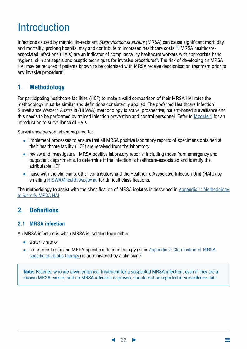
IntroductionInfections caused by methicillin-resistant Staphylococcus aureus (MRSA) can cause significant morbidity and mortality, prolong hospital stay and contribute to increased healthcare costs1,2. MRSA healthcare-associated infections (HAIs) are an indicator of compliance, by healthcare workers with appropriate hand hygiene, skin antisepsis and aseptic techniques for invasive procedures3. The risk of developing an MRSA HAI may be reduced if patients known to be colonised with MRSA receive decolonisation treatment prior to any invasive procedure4.
1. Methodology
For participating healthcare facilities (HCF) to make a valid comparison of their MRSA HAI rates the methodology must be similar and definitions consistently applied. The preferred Healthcare Infection Surveillance Western Australia (HISWA) methodology is active, prospective, patient-based surveillance and this needs to be performed by trained infection prevention and control personnel. Refer to Module 1 for an introduction to surveillance of HAIs.
Surveillance personnel are required to:
* implement processes to ensure that all MRSA positive laboratory reports of specimens obtained at their healthcare facility (HCF) are received from the laboratory
* review and investigate all MRSA positive laboratory reports, including those from emergency and outpatient departments, to determine if the infection is healthcare-associated and identify the attributable HCF
* liaise with the clinicians, other contributors and the Healthcare Associated Infection Unit (HAIU) by emailing [email protected] for difficult classifications.
The methodology to assist with the classification of MRSA isolates is described in Appendix 1: Methodology to identify MRSA HAI.
2. Definitions
2.1 MRSA infection
An MRSA infection is when MRSA is isolated from either:
* a sterile site or
* a non-sterile site and MRSA-specific antibiotic therapy (refer Appendix 2: Clarification of MRSA-specific antibiotic therapy) is administered by a clinician.2
Note: Patients, who are given empirical treatment for a suspected MRSA infection, even if they are a known MRSA carrier, and no MRSA infection is proven, should not be reported in surveillance data.

33
2.2 Criteria for MRSA healthcare-associated infection
An MRSA infection is considered to be an HAI if either criterion A or B is met:
* Criterion A: an infection acquired more than 48 hours after hospital admission or less than 48 hours after discharge and the infection was not present or incubating on admission i.e. no signs or symptoms of the MRSA infection were evident.
* Criterion B: an infection acquired 48 hours or less after admission and at least one of the following criteria is met:
1. Is a complication of the presence of an indwelling medical device e.g. intravascular line, cerebrospinal fluid shunt, urinary catheter and no other focus of infection is identified.
2. The infection is related to the surgical site and occurs within 30 or 90 days of a surgical procedure depending on the procedure type (refer to Appendix 3: MRSA SSI).
3. An invasive instrumentation or incision related to the infection was performed within 48 hours. If longer than 48 hours, there must be compelling evidence that the MRSA infection was related to the procedure.
4. Is associated with neutropenia contributed to by cytotoxic therapy.
2.3 Neutropenia
Neutropenia is defined as at least two separate calendar days with values of absolute neutrophil count (ANC) <500cells/mm3 (<0.5 x 10⁹/L) on or within a seven-day time period which includes the date the positive blood specimen was collected (day one), the three calendar days before and the three calendar days after.
2.4 New MRSA HAI
Only the first new MRSA HAI event for a single admission period is reported. The intention of this definition is to exclude ongoing episodes of infection that have been previously reported. However, if the admission period is prolonged e.g. > one month, count additional MRSA HAIs if a new infective event occurs and it is unrelated to a previously reported MRSA HAI event.
If a patient develops a non-sterile site infection and a sterile site infection during the same admission, then the sterile site HAI takes precedence and the non-sterile site HAI is not reported. If the non-sterile infection occurred in a previous admission, then it remains reported for that period. If a bloodstream infection (BSI) and another sterile site HAI occur, report the BSI only.
Note: the exception for repeat reporting is that the definition of a BSI requires that an additional MRSA BSI is reported if it has been more than 14 days since a previous positive MRSA BSI.
2.5 Community-associated MRSA infection
These events are when the infection manifests within 48 hours of admission and do not meet criterion A or B for classification as an HAI.
2.6 Maternally-acquired MRSA infection
Infections that arise in neonates <48 hours after delivery are not considered HAI unless there is compelling evidence that the infection was related to an intervention during passage through the birth canal e.g. wound secondary to vacuum extraction.
34
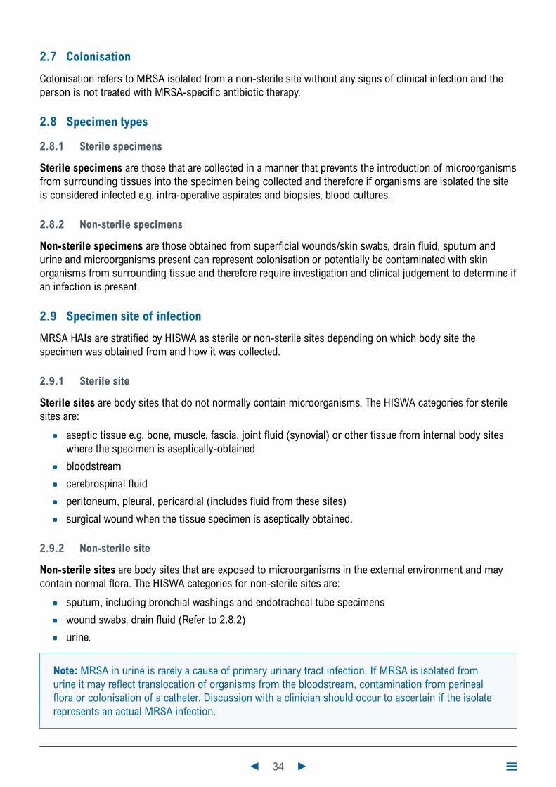
2.7 Colonisation
Colonisation refers to MRSA isolated from a non-sterile site without any signs of clinical infection and the person is not treated with MRSA-specific antibiotic therapy.
2.8 Specimen types
2.8.1 Sterile specimens
Sterile specimens are those that are collected in a manner that prevents the introduction of microorganisms from surrounding tissues into the specimen being collected and therefore if organisms are isolated the site is considered infected e.g. intra-operative aspirates and biopsies, blood cultures.
2.8.2 Non-sterile specimens
Non-sterile specimens are those obtained from superficial wounds/skin swabs, drain fluid, sputum and urine and microorganisms present can represent colonisation or potentially be contaminated with skin organisms from surrounding tissue and therefore require investigation and clinical judgement to determine if an infection is present.
2.9 Specimen site of infection
MRSA HAIs are stratified by HISWA as sterile or non-sterile sites depending on which body site the specimen was obtained from and how it was collected.
2.9.1 Sterile site
Sterile sites are body sites that do not normally contain microorganisms. The HISWA categories for sterile sites are:
* aseptic tissue e.g. bone, muscle, fascia, joint fluid (synovial) or other tissue from internal body sites where the specimen is aseptically-obtained
* bloodstream
* cerebrospinal fluid
* peritoneum, pleural, pericardial (includes fluid from these sites)
* surgical wound when the tissue specimen is aseptically obtained.
2.9.2 Non-sterile site
Non-sterile sites are body sites that are exposed to microorganisms in the external environment and may contain normal flora. The HISWA categories for non-sterile sites are:
* sputum, including bronchial washings and endotracheal tube specimens
* wound swabs, drain fluid (Refer to 2.8.2)
* urine.
Note: MRSA in urine is rarely a cause of primary urinary tract infection. If MRSA is isolated from urine it may reflect translocation of organisms from the bloodstream, contamination from perineal flora or colonisation of a catheter. Discussion with a clinician should occur to ascertain if the isolate represents an actual MRSA infection.

35
2.9.3 Wound specimens – non-sterile
Wound-surgical: MRSA HAIs related to surgery or invasive instrumentation and meets Criterion A or B (Refer to 2.2) and the specimen is obtained from a wound swab, drain site or another external surgical device e.g. external fixator, surgical wire. These should be entered as Specimen site: wound-surgical, Specimen: non-sterile.
Note: this includes infections related to surgery that don’t meet the criteria for an SSI but are HAIs e.g. an inpatient develops superficial MRSA infection of surgical incision >30 days post-procedure i.e. not an SSI by definition, but it is an HAI.
Wound-all other: MRSA HAIs in all other wound types including:
* all non-surgical wounds or skin and soft tissue infections e.g. decubitus ulcers
* device exit site infections e.g. intravenous cannulae, peritoneal dialysis catheter, suprapubic catheter
* infected burns and includes infections post-surgical debridement
* infections of the mucous membranes e.g. conjunctivitis, high vaginal swab
* infections of breast tissue due to mastitis i.e. MRSA isolated in breast milk.
2.10 Place of Acquisition
MRSA HAIs are categorised according to where the infection was likely acquired i.e. inpatient or non-inpatient healthcare setting. It does not relate to where the patient was physically located when the infection was identified e.g. outpatient department. For non-inpatient settings, the MRSA HAIs are associated with healthcare received as an outpatient, and meet Criterion B.
2.10.1 ICU or non-ICU (inpatient)
* MRSA HAI acquired as inpatients are stratified as intensive care unit (ICU) or non-ICU (all other wards/units outside of the ICU)
* ICU MRSA HAIs are those detected more than 48 hours after ICU admission or within 48 hours of discharge from ICU
* non-ICU MRSA HAI are associated with healthcare during a multi-day admission to non-ICU wards or Hospital-in-the-Home (HITH) or within 48 hours of discharge
* inpatient MRSA HAI also includes infections that meet Criterion B and are associated with a multi-day admission but are detected post-discharge e.g. a surgical patient develops an SSI caused by MRSA detected on readmission.
2.10.2 Non-inpatient higher-risk units – renal, haematology, oncology
* MRSA HAIs, in patients receiving care under these speciality units and who are not under the care of HITH, are acquired at home or following admission for day care at hospital outpatient settings or attendance at outpatient clinics e.g. haemodialysis, chemotherapy day-wards, day surgery.
2.10.3 Non-inpatient – other units
* MRSA HAIs acquired following admissions for day care in hospital outpatient settings or attendance at outpatient clinics who are not under the care of HITH or the higher-risk units e.g. an MRSA SSI in a general surgery patient following day surgery or an MRSA HAI following a facet joint injection at an outpatient clinic.
36
2.10.4 MRSA infections identified following care at another healthcare facility
* If an MRSA HAI is identified and is a result of care at another HCF or develops within 48 hrs of a transfer, contact the HAIU so that the infection can be attributed to the correct HCF.
2.11 Previous colonisation status
* Patients colonised with MRSA are at an increased risk of developing an MRSA HAI. The risk may be reduced if these patients receive decolonisation or suppression treatment.
* Report ‘Yes’ to previously colonised: if the patient has been previously identified to have colonisation or infection with any strain of MRSA prior to the HAI occurring.
* Report ‘No’ or ‘Unknown’ if it is the first time the patient has been identified with MRSA or their previous MRSA status is unknown.
3. HISWA dataset
3.1 Numerator data fields
Data described in Table 1 are required to be entered in the HISWA database.
3.1.1 Inclusions
* all strains of MRSA causing HAIs
* patients previously colonised with MRSA who develop a new MRSA HAI.
3.1.2 Exclusions
* community- associated MRSA infections
* maternally-acquired MRSA infections
* patients who are colonised only.
3.2 Denominator data fields
The denominator that is utilised is bed-days. Both multi-day and same-day bed-days are collected to allow for different rate calculations.
3.2.1 Inclusions
HISWA bed-day data for MRSA HAI includes:
* inpatient admissions to rehabilitation and aged care areas in an acute HCF.
* HITH bed-days
* same-day admissions e.g. haemodialysis units, day-surgery, procedure units.
3.2.2 Exclusions
HISWA bed-day data for MRSA HAI excludes:
* psychiatric wards/units
* unqualified newborns i.e. newborn who is nine days of age or less and does not require admission to a neonatal ICU and whose mother is the admitted patient
37
* boarders i.e. a person who is receiving food and/or accommodation but not medical care including newborns ≥10 days of age
* residential Aged Care Reporting Establishments co-located with public hospitals within the Western Australian Country Health Services (WACHS).
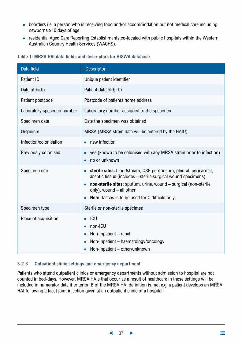
Table 1: MRSA HAI data fields and descriptors for HISWA database
Data field Descriptor
Patient ID Unique patient identifier
Date of birth Patient date of birth
Patient postcode Postcode of patients home address
Laboratory specimen number Laboratory number assigned to the specimen
Specimen date Date the specimen was obtained
Organism MRSA (MRSA strain data will be entered by the HAIU)
Infection/colonisation * new infection
Previously colonised * yes (known to be colonised with any MRSA strain prior to infection)
* no or unknown
Specimen site * sterile sites: bloodstream, CSF, peritoneum, pleural, pericardial, aseptic tissue (includes – sterile surgical wound specimens)
* non-sterile sites: sputum, urine, wound – surgical (non-sterile only), wound – all other
* Note: faeces is to be used for C.difficile only.
Specimen type Sterile or non-sterile specimen
Place of acquisition * ICU
* non-ICU
* Non-inpatient – renal
* Non-inpatient – haematology/oncology
* Non-inpatient – other/unknown
3.2.3 Outpatient clinic settings and emergency department
Patients who attend outpatient clinics or emergency departments without admission to hospital are not counted in bed-days. However, MRSA HAIs that occur as a result of healthcare in these settings will be included in numerator data if criterion B of the MRSA HAI definition is met e.g. a patient develops an MRSA HAI following a facet joint injection given at an outpatient clinic of a hospital.

38
4. Calculation of MRSA HAI rate
4.1 Inpatient MRSA HAI rate
The inpatient MRSA HAI rate is expressed as a rate per 10,000 multi-day bed-days.
Rate calculation: Number of inpatient MRSA HAI x 10,000
__________________________ Number of multi-day bed days
4.2 Total MRSA HAI rate
This rate reflects the total number (inpatient and non-inpatient) of MRSA HAIs
Rate calculation: Total number of MRSA HAI x 10,000
____________________________________ Number of multi-day and same-day bed days
39
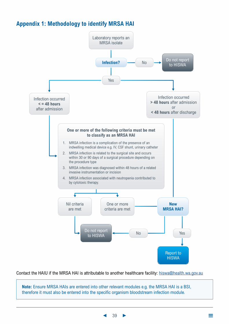
Appendix 1: Methodology to identify MRSA HAI
Laboratory reports anMRSA isolate
One or more of the following criteria must be metto classify as an MRSA HAI
1. MRSA infection is a complication of the presence of an indwelling medical device e.g. IV, CSF shunt, urinary catheter
2. MRSA infection is related to the surgical site and occurs within 30 or 90 days of a surgical procedure depending on the procedure type
3. MRSA infection was diagnosed within 48 hours of a related invasive instrumentation or incision
4. MRSA infection associated with neutropenia contributed to by cytotoxic therapy.
Infection?Do not report
to HISWA
Infection occurred< = 48 hours
after admission
Infection occurred> 48 hours after admission
or< 48 hours after discharge
No
Yes
NewMRSA HAI?
Do not reportto HISWA
Nil criteriaare met
One or morecriteria are met
YesNo
Report toHISWA
Contact the HAIU if the MRSA HAI is attributable to another healthcare facility: [email protected]
Note: Ensure MRSA HAIs are entered into other relevant modules e.g. the MRSA HAI is a BSI, therefore it must also be entered into the specific organism bloodstream infection module.
40
Appendix 2: Clarification of MRSA-specific antibiotic therapy
MRSA-specific antibiotic therapy is the use of antimicrobials that are clinically effective in the treatment of MRSA infections. MRSA antibiotic sensitivities may vary between strains and must always be checked from the laboratory report.
* MRSA-specific antibiotic therapy
• vancomycin
• teicoplanin
• linezolid
• quinupristin-dalfopristin (Synercid®)
• daptomycin
• ceftaroline
* Possible MRSA-specific antibiotic therapy – depending on sensitivity results
• fusidic acid
• rifampicin
• clindamycin
• co-trimoxazole
• quinolones (ciprofloxacin, moxifloxacin)
• doxycycline
• chloramphenicol ointment
* Antibiotics that are not MRSA-specific antibiotic therapy
All strains of MRSA are resistant to these groups of antibiotics and they are not suitable for treating MRSA infections. They include:
• all penicillin-based antibiotics e.g. benzylpenicillin, flucloxacillin, amoxycillin, Timentin®, Augmentin®
• all cephalosporins (except ceftaroline) e.g. cephalothin, cephalexin, cefotaxime, ceftazadine, ceftriaxone, cephazolin, cefepime, cefaclor
• carbapenems e.g. imipenem, meropenem
• others e.g. metronidazole, aztreonam.
* Antibiotics that are reported as sensitive on laboratory testing, but are not likely to be clinically effective against MRSA infection. They include:
• gentamicin, tobramycin, amikacin – as single therapy
• erythromycin, roxithromycin, clarithromycin and azithromycin.

41
Appendix 3: MRSA SSI
An MRSA infection is considered an HAI related to the surgical site when criteria for classification as a SSI are met (Refer to SSI module).
* SSIs are followed for the following periods where day 1 = the date of the procedure:
* 30 day period for all superficial SSI and 30 or 90 day period for deep and organ/space infections depending on the procedure. Common procedures are listed in Table 2.
Table 2: Procedures and surveillance periods for deep or organ/space SSI
30-day Surveillance
Abdominal aortic aneurysm repair Laminectomy
Limb amputation Liver transplant
Appendix surgery Neck surgery
Shunt for dialysis Kidney surgery
Bile duct, liver or pancreatic surgery Ovarian surgery
Carotid endarterectomy Prostate surgery
Gallbladder surgery Rectal surgery
Colon surgery Small bowel surgery
Caesarean section Spleen surgery
Caesarean section with tubal ligation Thoracic surgery
Gastric surgery Thyroid and/or parathyroid surgery
Heart Transplant Vaginal hysterectomy
Abdominal hysterectomy Exploratory laparotomy
Kidney transplant Other surgery not listed
90-day Surveillance
Breast surgery Cardiac surgery
Coronary artery bypass graft with both chest and donor site incisions
Coronary artery bypass graft with chest incision only
Craniotomy Spinal fusion
Open reduction of fracture Herniorrhaphy
Hip arthroplasty Pacemaker surgery
Knee arthroplasty Peripheral vascular bypass surgery
Refusion of spine Ventricular shunt

42
References
1. The National Healthcare Safety Network (NHSN). CDC/NHSN surveillance definitions for specific types of infections. National Healthcare Safety Network (NHSN) Patient Safety Component Manual: Centers for Disease Control and Prevention; 2020.
2. Christensen K, Coombs G, Ferguson J, Iredell J, Marshall C, Nimmo G, et al. Multiresistant organisms. 2008. In: Reducing harm to patient from health care associated infection: the role of surveillance [Internet]. Sydney: Australian Commission on Safety and Quality in Health Care.
3. The Australian Council on Healthcare Standards. Infection Control Version 5.1: Clinical Indicator User Manual. ACHS Clinical Indicator Program [Internet]. 2018. Available from: https://www.achs.org.au/media/154670/infection_control_v5.1.pdf
4. Institute for Healthcare Improvement. How-to Guide: Prevent Surgical Site Infections Cambridge, MA: www.ihi.org; 2012 [Available from: http://www.ihi.org/resources/Pages/Tools/HowtoGuidePreventSurgicalSiteInfection.aspx

43
Module 4
Clostridioides difficile infection
44
Contents
Introduction 45
1. Methodology 45
2. Definitions 46
2.1 Hospital-identified CDI 46
2.2 CDI case 46
2.3 Specimen descriptors 46
3. HISWA dataset 46
3.1 Numerator data fields 46
3.2 Denominator data fields 47
4. Calculation of hospital-identified CDI rate 47
5. Enhanced surveillance (optional) 48
5.1 Severe CDI case 48
5.2 Definitions of healthcare or community-associated CDI cases 48
5.3 Recurrent CDI cases 49
Appendix 1: Methodology for determining a HI-CDI case 50
References 51

45
IntroductionClostridioides difficile infection (CDI) (recently renamed from Clostridium difficile 1) is a common healthcare-associated infection (HAI) in the developed world and is strongly connected to the use of antibiotics.2,3 The severity of infection ranges from mild diarrhoea to pseudomembranous colitis, toxic megacolon and death4,5. Hyper virulent strains that are associated with epidemic spread and high rates of severe disease and death in other countries have now also been identified in Australia.3-5 Patients who acquire CDI whilst in a hospital may have their length of stay (LOS) increased by up to three times that of a patient without CDI.6
Identification of hospitalised patients with CDI using optimal surveillance systems and strict enforcement of infection prevention and control principles are the key to preventing transmission.4,5 CDI can be linked to prolonged or inappropriate use of antimicrobial therapies.7 Antimicrobial stewardship programs are an essential CDI prevention strategy to minimise the frequency and duration of antimicrobial use and to promote narrow-spectrum antibiotic prescribing.4,5
1. Methodology
Surveillance of hospital-identified CDI (HI-CDI) is the minimum requirement for both national5 and Healthcare Infection Surveillance Western Australia (HISWA) surveillance. Classification of HI-CDI cases is intended to be derived from laboratory reports and does not require case review by surveillance personnel. HISWA definitions are those endorsed by the Australian Commission on Safety and Quality in Health Care (ACSQHC)8. Refer to Module 1 for an introduction to HAI surveillance. The methodology to assist with the classification of HI-CDI is described in Appendix 1: Methodology for determining a HI-CDI case.
Surveillance personnel are required to:
* Implement processes to ensure they receive all laboratory reports that detect C.difficile from specimens of stools that take the shape of a container, obtained at their HCF, including from the emergency department and all other outpatient settings.
* Apply the definition of a HI-CDI case consistently.
* Additional surveillance of severe CDI, and healthcare or community-associated CDI cases, is recommended, however, it is optional for HISWA hospitals (Refer to section 5).
Note: HISWA HI-CDI data is not designed as a hospital performance indicator. The rate reported, measures the burden of CDI in the patient population, and includes both community acquired and healthcare acquired infections. The HISWA HI-CDI rate must not be used as a measure of performance or comparison between hospitals.

46
2. Definitions
2.1 Hospital-identified CDI
A HI-CDI is a case identified in a patient attending any area of a hospital i.e. admitted patients, emergency department, outpatient clinic, day surgery.
A HI-CDI case reflects the burden of CDI on a hospital and describes healthcare-associated infections, community-associated infections, as well as CDI of indeterminate or unknown origin (Refer to 5.2 Definitions of healthcare or community-associated CDI cases).
2.2 CDI case
A CDI case is defined as a case of diarrhoea i.e. an unformed stool that takes the shape of the container, in a person greater than two years of age at the date of specimen collection that meets the following criteria:
* the stool sample yields a positive result in a laboratory assay for C.difficile toxin A and/or B
or
* a toxin-producing C.difficile organism is detected in the stool sample by culture or other means.
and excludes
* cases where a known previous positive test has been obtained within the last eight weeks i.e. only include cases once in an eight week period.
* patients less than two years old at the date of specimen collection.
Note: An additional positive test obtained from a specimen collected from the same patient more than 8 weeks since the last positive test is regarded as a new case.
2.3 Specimen descriptors
Faecal samples that take the shape of the container are described in laboratory reports as semi-formed, watery, loose, liquid or fluid.
3. HISWA dataset
3.1 Numerator data fields
The numerator data fields for HI-CDI cases required to be entered into the HISWA database are described in Table 1.
3.1.1 Inclusions
* all events that meet the HI-CDI case definition.
3.1.2 Exclusions
* formed stools i.e. do not take the shape of the container, even if toxin positive
* recurrent cases in an eight week period
* patients less than two years old at the date of specimen collection.

47
Table 1: CDI numerator data fields and descriptors for HISWA database
Data field Descriptor
Patient ID Unique patient identifier
Date of birth Patient date of birth
Patient postcode Postcode of patient’s home address
Lab specimen number Laboratory number assigned to the specimen
Specimen date Date the specimen was obtained
Organism Clostridioides difficile
Infection/colonisation For every case enter: new infection
Previously colonised For every case enter: no / unknown
Specimen site For every case enter: Faeces
Specimen For every case enter: Non-sterile
Place of acquisition For every case enter: Hospital-identified CDI
3.2 Denominator data fields
The denominator that is utilised is bed-days and includes both multi-day and same-day bed-days.
3.2.1 Inclusions
HISWA bed-day data for HI-CDI includes:
* All inpatient wards or units within the HCF including psychiatric, rehabilitation and aged care. Do not include residential aged care facilities co-located on same site e.g. as per some Western Australian Country Health Service (WACHS) sites.
* HITH admissions
* Same-day admission wards or units e.g. haemodialysis units, day of surgery or procedure units.
3.2.2 Exclusions
* Boarders
* Patients less than two years of age
* Emergency and outpatient clinic attendance data is not included in bed-day counts provided to HISWA.
4. Calculation of hospital-identified CDI rate
The HI-CDI rate reflects the burden of CDI presenting to a HCF. CDI rates will be calculated and reported to HISWA using bed-days and expressed per 10,000 bed-days. Bed-days include both multi-day and same-day bed-days.
Rate calculation: Total number of HI-CDI cases x 10,000
__________________________________ Total number of bed-days at the hospital

48
5. Enhanced surveillance (optional)
National surveillance does not require classification of healthcare and community-associated CDI cases or severe and non-severe CDI. Surveillance of these classifications is optional, however, it is recommended for HISWA hospitals.
Enhanced surveillance requires an individual case review in addition to the routine review of laboratory reports required for HI-CDI surveillance.
HCFs may be requested to undertake enhanced surveillance for target periods and also if the rate of HI-CDI is high or increasing significantly at their facility.
HISWA definitions for enhanced surveillance align with recommended international definitions and are described in sections 5.1 Severe CDI case and 5.2 Definitions of healthcare or community-associated CDI cases.
5.1 Severe CDI case
* A severe CDI case is defined as a CDI case that meets any of the following criteria within 30 days of symptom onset:
• history of admission to an intensive care unit (ICU) for treatment of complication from CDI e.g. vasopressor therapy for shock
• history of surgery for treatment of toxic megacolon, perforation or refractory colitis
• death caused by CDI within 30 days of symptom onset8.
* Clinical criteria that have been associated with severe CDI include8:
• age >60 years of age
• temperature >38.3°C
• serum albumin <25g/L
• peripheral white blood cell count >15,000cells/microL
• deteriorating renal function
• elevated serum lactate
• endoscopic evidence of pseudomembranous colitis or treatment in the ICU
• subtotal colectomy procedure or diagnosis of toxic megacolon.
5.1.1 Calculation of incidence of severe CDI
For HCFs monitoring severe disease, this should be expressed as the proportion of total HI-CDI cases in the reporting period that were severe. The raw number, as well as the proportion, should be reported to aid interpretation. The proportion should be calculated for the reporting period as follows8:
Proportion calculation: Total number of patients with severe HI-CDI ____________________________________ Total hospital-identified CDI cases
5.2 Definitions of healthcare or community-associated CDI cases
Each CDI case is classified according to the place of probable exposure described below8 and in Figure 1: Timeline for healthcare or community-associated CDI definitions7.

49
Healthcare-associated CDI are classified as HCF onset or community-onset.
* HCF onset: when symptom onset or date and time of stool specimen collection is greater than 48 hrs after admission to an HCF.
* Community onset: when symptom onset was in the community or within 48 hrs of admission to an HCF, and symptom onset was less than four weeks after the last discharge from an HCF.
Cases can be further classified as:
* Community-associated CDI cases
• symptom onset or date and time of stool specimen was in the community or within 48 hrs of admission to an HCF provided the symptom onset was more than 12 weeks after the last discharge from an HCF
• record if the CDI case was admitted to an HCF from a residential care facility.
* Indeterminate onset
• criteria for community or healthcare-associated are not met e.g. CDI case with symptom onset in the community between four and 12 weeks of the last discharge from an HCF.
* Unknown
• exposure setting cannot be determined because of a lack of data to classify.
Figure 1: Timeline for healthcare or community-associated CDI definitions8
Admission Discharge
4 weeks
HCA CommunityOnset IndeterminateHCA HCF
Onset(*) CA-CDAD
48 h 8 weeksSymptom onset
Rates for healthcare-associated CDI cases are expressed per 10,000 bed-days (excluding same-day bed-days).
Rates for community-associated CDI cases are expressed per population rates.
Note: Healthcare-associated community onset cases should be:
* Attributed to the reporting period during which the case was discharged from the HCF before CDI symptom onset e.g. if the case was discharged on the 28 May and readmitted with CDI on 5 June, the case should be assigned to May.
* Attributed to the HCF from which the case was discharged, providing they were an inpatient at the HCF from more than 48 hours.
5.3 Recurrent CDI cases
A recurrent CDI case is an episode that occurs within eight weeks or less after the onset of a previous CDI episode, provided that CDI symptoms from the earlier episode have resolved with or without therapy. These cases are not included in the HI-CDI case definition and calculation, and monitoring is optional.

50
Appendix 1: Methodology for determining a HI-CDI case
Positive C. difficile toxin text ordetection of toxin-producingC. difficile in diarrhoeal stool.
Was the patient attending anyarea of an acute care facilitye.g. inpatient, emergency oroutpatient department, whenthe specimen was obtained?
Is the patient overtwo years of age?
Do not reportto HISWA
No
Yes
Report toHISWA
Yes
Yes
Yes
Is the specimen date at leasteight weeks after any previousHI-CDI case for this patient?
No
No
No
Contact the HAIU if the MRSA HAI is attributable to another healthcare facility: [email protected]

51
References
1. Lawson PA, Citron DM, Tyrrell KL, Finegold SM. Reclassification of Clostridium difficile as Clostridioides difficile (Hall and O’Toole 1935) Prévot 1938. Anaerobe. 2016;40:95-9.
2. Stuart RL, Marshall C, Harrington G, Sasko L, McLaws M-L, Ferguson J. ASI/ACIPC position statement- Infection control for patients with Clostridium difficile infection in healthcare facilities. Infection, Disease and Health. 2019;24(1):32-43.
3. Alfayyadh M, Collins DA, Tempone S, McCann R, Armstrong PK, Riley TV, et al. Recurrence of Clostridium difficile infection in the Western Australian population. Epidemiology and Infection. 2019;147.
4. Stuart RL, Marshall C. Clostridium difficile infection: a new threat on our doorstep. Medical Journal of Australia. 2011;194(7):331-33.
5. Cheng AC, Ferguson JK, Richards MJ, Robson JM, Gilbert GL, McGregor A, et al. Australasian Society for Infectious Diseases guidelines for the diagnosis and treatment of Clostridium difficile infection. Medical Journal of Australia. 2011;194(7):353-8.
6. Australian Commission on Safety and Quality in Health Care. Clostridium difficile infection 2017 data snapshot. Sydney: ACSQHC; 2019.
7. Australian Commission on Safety and Quality in Health Care. Clostridium difficile infection. Monitoring the national burden of Clostridium difficile. Sydney: ACSQHC; 2018.
8. Australian Commission on Safety and Quality in Health Care. Implementation guide for surveillance of Clostridium difficile infection. Sydney: ACSQHC; 2013.
52
Module 5
Vancomycin-resistant enterococci (VRE) sterile site infection
53
Contents
Introduction 54
1. Methodology 54
2. Definitions 54
2.1 VRE sterile site infection 54
2.2 VRE sterile site HAI 55
2.3 Inpatient or non-inpatient HAI 55
2.4 VRE sterile site community-associated infection 56
2.5 New VRE sterile site infection 56
2.6 Colonisation 56
2.7 Place of acquisition 56
2.8 Attributable HCF 56
2.9 Previous colonisation status 56
3. HISWA dataset 57
3.1 Numerator data fields 57
3.2 Denominator data fields 57
Appendix 1: VRE methodology flowchart 59
Appendix 2: VRE sterile site infection related to an SSI 60
References 61

54
Introduction Enterococci are opportunistic pathogens which can cause infections in patients who are vulnerable due to surgery, invasive devices, immunosuppression or extreme older age. Vancomycin is commonly used to treat infections caused by enterococci, but vancomycin-resistant enterococci (VRE) have become a major problem for healthcare in Australia.1
Bloodstream and other sterile site infections caused by VRE have been associated with significant morbidity and mortality for critically ill or immunocompromised patients. Surveillance of sterile site infections allows evaluation of strategies to reduce the spread of VRE and colonisation of patients receiving care in higher-risk units.
1. Methodology
HISWA surveillance is of both community-associated infection (CAI) and healthcare-associated infection (HAI) VRE sterile site infections. Surveillance personnel need to review all VRE positive laboratory reports, including those from emergency and outpatient departments.
Report new VRE infections from sterile sites (Refer to 2.1) that are community-associated or healthcare-associated infections (HAIs or CAIs).
If the VRE sterile site infection is a healthcare-associated infection (HAI), identify the attributable healthcare facility (HCF).
Currently, VRE sterile site infections are unable to be reported via the HISWA website.
A VRE surveillance form is available from the Healthcare Associated infection Unit (HAIU) website under HISWA tools and resources.When a new sterile site infection is identified, complete the form and send via email to [email protected]
2. Definitions
2.1 VRE sterile site infection
A VRE sterile site infection is when VRE is isolated from a specimen obtained aseptically from a sterile site. Sterile sites are body sites that do not normally contain microorganisms. Non-sterile sites are body sites that are exposed to microorganisms in the external environment and may also be colonised with normal flora.
The HISWA categories for sterile site specimen sites are:
* Blood
* Peritoneal: fluid and tissue from peritoneal space/peritoneum (includes abdominal fluid and ascites)
* Bone and joint: bone biopsy, bone marrow and synovial fluid and fluid aspirated or cultured from any specific joint including knee, ankle, elbow, hip, wrist synovial fluid
* Other Internal sites: specimens from body sites that are normally sterile where a specimen has been obtained surgically or by aspirate e.g. deep soft tissue (muscle and fascia), pleura, liver, pancreas, kidney, spleen, vascular tissue, heart, brain, lymph node, ovary.
Do not report VRE isolated from a specimen obtained from a non-sterile site e.g. wound, urine, and sputum.
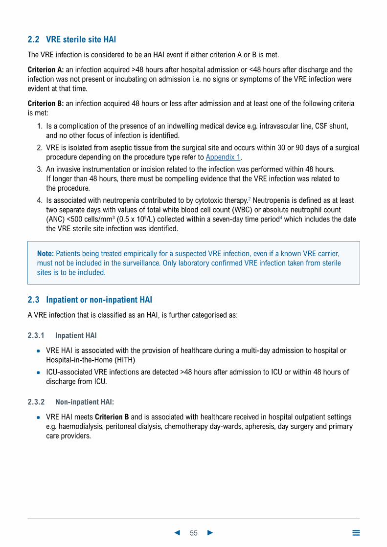
55
2.2 VRE sterile site HAI
The VRE infection is considered to be an HAI event if either criterion A or B is met.
Criterion A: an infection acquired >48 hours after hospital admission or <48 hours after discharge and the infection was not present or incubating on admission i.e. no signs or symptoms of the VRE infection were evident at that time.
Criterion B: an infection acquired 48 hours or less after admission and at least one of the following criteria is met:
1. Is a complication of the presence of an indwelling medical device e.g. intravascular line, CSF shunt, and no other focus of infection is identified.
2. VRE is isolated from aseptic tissue from the surgical site and occurs within 30 or 90 days of a surgical procedure depending on the procedure type refer to Appendix 1.
3. An invasive instrumentation or incision related to the infection was performed within 48 hours. If longer than 48 hours, there must be compelling evidence that the VRE infection was related to the procedure.
4. Is associated with neutropenia contributed to by cytotoxic therapy.2 Neutropenia is defined as at least two separate days with values of total white blood cell count (WBC) or absolute neutrophil count (ANC) <500 cells/mm3 (0.5 x 10⁹/L) collected within a seven-day time period4 which includes the date the VRE sterile site infection was identified.
Note: Patients being treated empirically for a suspected VRE infection, even if a known VRE carrier, must not be included in the surveillance. Only laboratory confirmed VRE infection taken from sterile sites is to be included.
2.3 Inpatient or non-inpatient HAI
A VRE infection that is classified as an HAI, is further categorised as:
2.3.1 Inpatient HAI
* VRE HAI is associated with the provision of healthcare during a multi-day admission to hospital or Hospital-in-the-Home (HITH)
* ICU-associated VRE infections are detected >48 hours after admission to ICU or within 48 hours of discharge from ICU.
2.3.2 Non-inpatient HAI:
* VRE HAI meets Criterion B and is associated with healthcare received in hospital outpatient settings e.g. haemodialysis, peritoneal dialysis, chemotherapy day-wards, apheresis, day surgery and primary care providers.
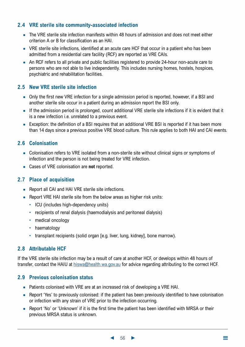
56
2.4 VRE sterile site community-associated infection
* The VRE sterile site infection manifests within 48 hours of admission and does not meet either criterion A or B for classification as an HAI.
* VRE sterile site infections, identified at an acute care HCF that occur in a patient who has been admitted from a residential care facility (RCF) are reported as VRE CAIs.
* An RCF refers to all private and public facilities registered to provide 24-hour non-acute care to persons who are not able to live independently. This includes nursing homes, hostels, hospices, psychiatric and rehabilitation facilities.
2.5 New VRE sterile site infection
* Only the first new VRE infection for a single admission period is reported, however, if a BSI and another sterile site occur in a patient during an admission report the BSI only.
* If the admission period is prolonged, count additional VRE sterile site infections if it is evident that it is a new infection i.e. unrelated to a previous event.
* Exception: the definition of a BSI requires that an additional VRE BSI is reported if it has been more than 14 days since a previous positive VRE blood culture. This rule applies to both HAI and CAI events.
2.6 Colonisation
* Colonisation refers to VRE isolated from a non-sterile site without clinical signs or symptoms of infection and the person is not being treated for VRE infection.
* Cases of VRE colonisation are not reported.
2.7 Place of acquisition
* Report all CAI and HAI VRE sterile site infections.
* Report VRE HAI sterile site from the below areas as higher risk units:
• ICU (includes high-dependency units)
• recipients of renal dialysis (haemodialysis and peritoneal dialysis)
• medical oncology
• haematology
• transplant recipients (solid organ [e.g. liver, lung, kidney], bone marrow).
2.8 Attributable HCF
If the VRE sterile site infection may be a result of care at another HCF, or develops within 48 hours of transfer, contact the HAIU at [email protected] for advice regarding attributing to the correct HCF.
2.9 Previous colonisation status
* Patients colonised with VRE are at an increased risk of developing a VRE HAI.
* Report ‘Yes’ to previously colonised: if the patient has been previously identified to have colonisation or infection with any strain of VRE prior to the infection occurring.
* Report ‘No’ or ‘Unknown’ if it is the first time the patient has been identified with MRSA or their previous MRSA status is unknown.

57
3. HISWA dataset
3.1 Numerator data fields
Table 1 VRE sterile site infection data fields and descriptors describe the numerator data fields for VRE sterile site infection required to be entered into the surveillance form provided for HISWA contributors.
3.1.1 Inclusions
* Patients previously colonised with VRE who develop a VRE sterile site infection.
* Both CAI and HAI VRE sterile site infections.
3.1.2 Exclusions
* VRE infections from non-sterile sites e.g wound, urine, sputum.
* Patients who are colonised with VRE and no VRE specific antimicrobial therapy is prescribed.
3.2 Denominator data fields
* Multi-day and same-day bed-day denominator data to calculate infection rates.

58
Table 1: VRE sterile site infection data fields and descriptors
Data field Descriptor
Case details
Reporting hospital Name of hospital or private dialysis unit – dropdown list
UMRN Unique patient identifier
First name
Surname
Date of birth ddmmyyyy
Admission diagnosis
Speciality
Date of admissionDate of dischargeDate of death
ddmmyyyy
Patient presented from HomeRehabilitationResidential Care Facility (RCF)PrisonPsychiatric hospitalTransfer from hospital within WATransfer from hospital outside WATransfer from hospital outside Australia
Has patient been hospitalised outside WA in the last 12 months?
Yes, No
If Yes, specify where:
If Yes, was a VRE screen performed on admission? Yes, No
Did the patient have a prior micro-alert F? Private hospitals can check via [email protected]
Did the patient have a prior micro-alert V? Private hospitals can check via [email protected]
Specimen Details
Date of specimen collection ddmmyyyy
Laboratory service provider Name of laboratory reporting the VRE infection
Laboratory specimen number Laboratory number assigned to the specimen
Organism Enterococcus faeciumEnterococcus faecalis
Van type Van A, Van B
Specimen type e.g. blood culture, tissue sample, bone chip
Reason for collection Screening – policy requirementScreening – VRE contactClinically indicated
Sterile site classification BloodPeritonealBone and jointPleural fluidOther internal sites
Surveillance classification HAI – inpatientHAI – non-inpatientCAI
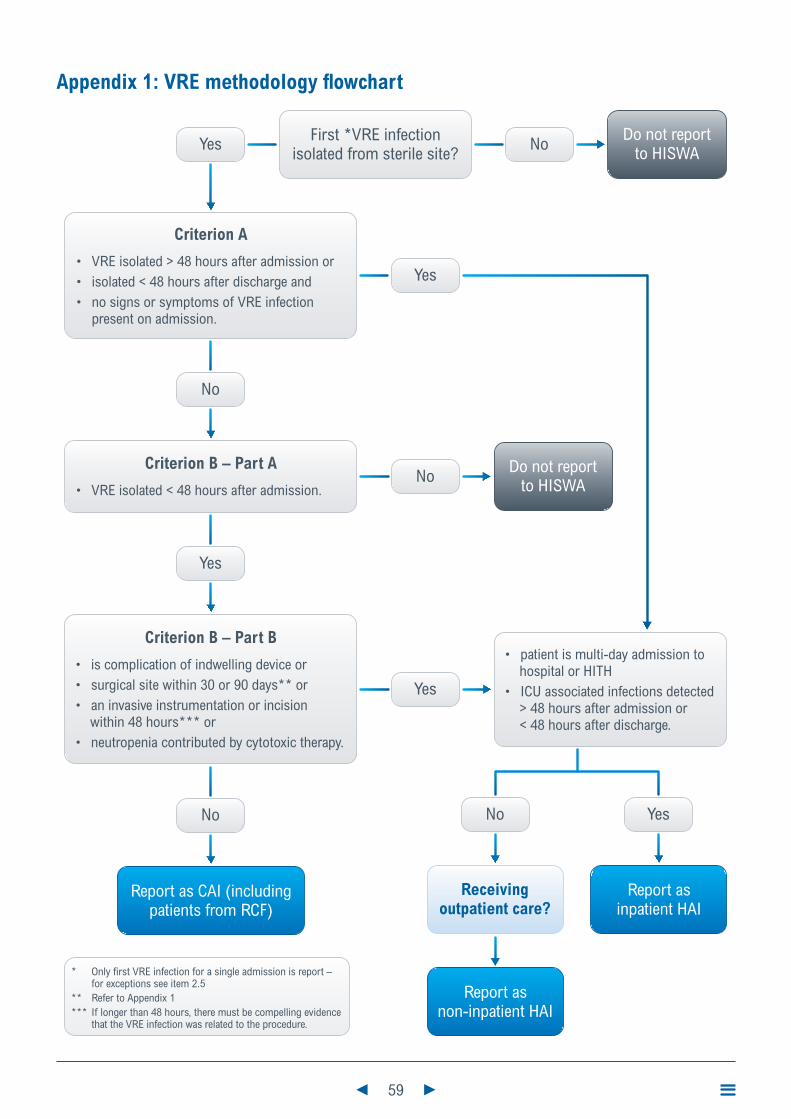
59
Appendix 1: VRE methodology flowchart
First *VRE infectionisolated from sterile site?
Do not reportto HISWANoYes
Yes
Receivingoutpatient care?
YesNo
Report as CAI (includingpatients from RCF)
Criterion A
• VRE isolated > 48 hours after admission or• isolated < 48 hours after discharge and• no signs or symptoms of VRE infection
present on admission.
Criterion B – Part B
• is complication of indwelling device or• surgical site within 30 or 90 days** or• an invasive instrumentation or incision
within 48 hours*** or• neutropenia contributed by cytotoxic therapy.
• patient is multi-day admission to hospital or HITH
• ICU associated infections detected > 48 hours after admission or < 48 hours after discharge.
Criterion B – Part A
• VRE isolated < 48 hours after admission.
No
No
Yes
Yes
No Do not reportto HISWA
Report asnon-inpatient HAI
Report asinpatient HAI
* Only first VRE infection for a single admission is report – for exceptions see item 2.5
** Refer to Appendix 1*** If longer than 48 hours, there must be compelling evidence
that the VRE infection was related to the procedure.

60
Appendix 2: VRE sterile site infection related to an SSI
VRE isolated from aseptic tissue obtained from a surgical site is considered a sterile site infection when criteria for classification as a SSI are met (Refer to SSI module).
SSIs are followed for the following periods where day one = the date of the procedure:
* 30 day period for superficial SSI for all procedures and 30 or 90 day period for deep and organ/space infections depending on the procedure.
Surveillance period for deep or organ/space SSI following surgical procedures
30-day Surveillance
Abdominal aortic aneurysm repair Laminectomy
Limb amputation Liver transplant
Appendix surgery Neck surgery
Shunt for dialysis Kidney surgery
Bile duct, liver or pancreatic surgery Ovarian surgery
Carotid endarterectomy Prostate surgery
Gallbladder surgery Rectal surgery
Colon surgery Small bowel surgery
Caesarean section Spleen surgery
Gastric surgery Thoracic surgery
Heart Transplant Thyroid and/or parathyroid surgery
Abdominal hysterectomy Vaginal hysterectomy
Kidney transplant Exploratory laparotomy
Other surgery not listed
90-day Surveillance
Breast surgery Cardiac surgery
Coronary artery bypass graft with both chest and donor site incisions
Coronary artery bypass graft with chest incision only
Craniotomy Spinal fusion
Open reduction of fracture Herniorrhaphy
Hip arthroplasty Pacemaker surgery
Knee arthroplasty Peripheral vascular bypass surgery
Refusion of spine Ventricular shunt
61
References
1. Australian Commission on Safety and Quality in Health Care (ACSQHC). AURA 2017: second Australian report on antimicrobial use and resistance in human health. Sydney; 2017.
2. Australian Infection Control Association (AICA). Multi-resistant organism (MRO) surveillance indicator definitions. 2004. In: Australian Council for Healthcare Standards, ACHS Infection Control Clinical Indicators Users Manual 2013 [Internet]. Available from: https://www.safetyandquality.gov.au/wp-content/uploads/2012/01/mrosurvlncdefine05.pdf
3. Minnesota Department of Health. Normally Sterile Sites: Invasive Bacterial Diseases 2017. Available from: http://www.health.state.mn.us/divs/idepc/dtopics/invbacterial/sterile.html
4. Centers for Disease Control and Prevention. CDC/NHSN surveillance definitions for specific types of infections. National Healthcare Safety Network (NHSN) Patient Safety Component Manual: NHSN; 2019.

62
Module 6
Staphylococcus aureus bloodstream infection

63
Contents
Introduction 64
1. Methodology 64
2. Definitions 64
2.1 Staphylococcus aureus bloodstream infection 64
2.2 Contaminants 65
2.3 Healthcare-associated SABSI (HA-SABSI) 65
2.3 Maternally-acquired SABSI 66
2.4 Focus of infection 66
2.5 Place of acquisition 67
2.6 Healthcare facility attribution 68
2.7 Classification of Staphylococcus aureus 68
3. HISWA dataset 69
3.1 Numerator data fields 69
3.2 Denominator data fields 69
4. Calculation of HA-SABSI rates 70
4.1 Calculation of total HA-SABSI 70
4.2 Calculation of inpatient-only HA-SABSI 70
Appendix 1: Methodology for classification of HA-SABSI 71
Appendix 2: HA-SABSI related to a SSI 72
References 73
64
Introduction Bloodstream infections (BSI) can cause significant illness, serious complications and high mortality, with more than half of BSIs associated with health-care.2,3 Staphylococcus aureus (S.aureus) is the most common cause of healthcare-associated BSI.3 The majority of healthcare-associated S.aureus BSIs (HA-SABSIs) are related to the presence of intravascular devices (IVDs) and these events are increasingly viewed as preventable adverse events.4,5 Quality improvement programs that have involved surveillance and implementation of policies to promote preventative strategies, have resulted in sustained reductions in HA-SABSI.2
1. Methodology
For participating hospitals to make a valid comparison of their HA-SABSI rates, the methodology must be similar and definitions consistently applied. Surveillance personnel are required to:
* Implement processes to ensure that all positive laboratory reports are received.
* Investigate all blood culture laboratory reports positive for both methicillin-sensitive and methicillin-resistant S.aureus, including those from emergency and outpatient departments, to determine if the S.aureus BSI (SABSI) is healthcare-associated and identify the attributable healthcare facility (HCF).
* Liaise with key stakeholders, clinical microbiologists/ infectious diseases physicians to assist with the classification of SABSI episodes.
* The methodology to assist with the classification of HA-SABSI is outlined in Appendix 1. Refer to Module 1 for an introduction to healthcare-associated infection (HAI) surveillance.
* HISWA HA-SABSI surveillance aligns with the Australian national definition developed by the Australian Commission on Safety and Quality in Health Care (ACSQHC).
Note: Surveillance personnel should take opportunities to promote best practice for blood culture collection to optimise BSI detection and classification as potential contaminants. Ideally blood culture specimens should be aseptically obtained from 2-4 blood draws from separate venepuncture sites, rather than through an intravascular device.1
Aseptic technique incorporating hand hygiene, the use of sterile gloves, and ensuring the skin or cannula hub and culture bottle tops are disinfected with an alcohol based disinfectant and allowed to dry prior to access is recommended.
2. Definitions
2.1 Staphylococcus aureus bloodstream infection
* A patient episode of SABSI is defined as a positive blood culture for S.aureus.6
* For surveillance purposes, only the first positive blood culture per patient within a 14 day period is counted. If the same patient has a further positive blood culture reported greater than 14 days after the last positive blood culture then an additional episode is counted (14-day rule).6
* The 14-day rule is to be applied to SABSI that occur in haemodialysis patients6 (not the 21 days specified for haemodialysis access-associated bloodstream infection surveillance).

65
2.2 Contaminants
* S. aureus is rarely a contaminant of blood cultures and therefore there are few false-positive isolates.6
* S. aureus positive blood culture will only be considered a contaminant, and not reported in the surveillance data if the clinical picture is unsupportive of infection and either a repeat blood culture is negative (within two days) and/or no antimicrobial treatment is given.
* It is recommended that attention is paid to aseptic technique, the volume of samples and that two or more samples of blood are collected (ideally from separate sites) on patients to reduce risk of contaminants and thus reduce false-positives.1
2.3 Healthcare-associated SABSI (HA-SABSI)
A patient episode of SABSI is considered to be healthcare-associated if either Criterion A or B are met:6
Criterion A: the patient’s first positive blood culture is collected more than 48 hours after hospital admission or less than 48 hours after discharge and a staphylococcal infection was not present or incubating on admission.
Note: incubating on admission means there were documented clinical signs or diagnostic evidence of staphylococcal infection on admission and provided there is no evidence of an association with a prior admission or medical procedure received in a HCF, then the episode was likely incubating on admission and is not counted as an HA-SABSI.
Criterion B: the patient’s first positive blood culture is collected less than or equal to 48 hours after admission and one or more of the following clinical criteria was met:
1. The SABSI is a complication of the presence of an indwelling medical device e.g. IVD, haemodialysis vascular access, cerebrospinal fluid shunt, feeding tube.
2. The SABSI is related to a surgical site infection (SSI) that occurs within 30 or 90 days of the procedure depending on the type of procedure. Refer to Appendix 2.
Note: include HA-SABSI related to SSI identified beyond the 30 or 90 day surveillance period, if first clinical signs of infection were identified within the surveillance period in a community or outpatient setting (e.g. patient was being treated for SSI by general practitioner etc.).
3. The SABSI is related to invasive instrumentation or an incision performed with 48 hours. If greater than 48 hours, there must be compelling evidence that the infection is related to the invasive procedure.
4. The SABSI is associated with neutropenia. Neutropenia is defined as at least two separate days with values of total white blood cell count (WBC) or absolute neutrophil count (ANC) <500 cells/mm3 (0.5 x 10⁹/L) collected within a seven-day time period which includes the date of the BSI (Day 1), the three calendar days before and the three calendar days after.7 For examples refer to Appendix 10.
If none of these criteria is met, then the episode of SABSI is considered to be community-associated.

66
2.3 Maternally-acquired SABSI
SABSI that arise in neonates less than 48 hours after delivery are not considered HAIs unless there is compelling evidence that it is related to a procedure or intervention during the birth.
2.4 Focus of infection
HA-SABSI are categorised according to the likely source of the infection. The following section can be used to clarify the application of Criterion B.
2.4.1 Intravascular device (IVD) related (clarifies Criterion B1)
* For central venous catheters, refer to central line-associated BSI (CLABSI) definitions (Module 7). For all other IVD, the IVD was present at some point within the 48 hours prior to the SABSI event, and there is no other identifiable focus of infection due to S. aureus at another body site.6 If the time period is greater than 48 hours there needs to be compelling evidence that the IVD is the cause of the infection e.g. pus at old IVD site.
* For haemodialysis patients- an HA-SABSI is haemodialysis access-associated if there is either clinical evidence of infection at the vascular access site or there is no other identifiable source of the SABSI.6
* An introducer used in intravascular procedures is considered an IVD, e.g. angiography, therefore, an HA-SABSI occurring within 48 hours of these procedures is IVD related unless there is an identifiable infection at another site related to the HA-SABSI.6
Note: if a patient is known or suspected to have accessed their own IVD and develops an HA-SABSI, and the infection meets the criteria, it is to be reported as an HA-SABSI.
2.4.2 Non intravascular device related (clarifies Criterion B1)
* The device was present at some point within the 48 hours prior to the SABSI event and there was clinical or microbiological evidence that the HA-SABSI was associated with the insertion site or an associated organ.6 Examples of non-IVDs include shunts, suprapubic catheters, chest tubes, urinary catheters, peritoneal dialysis catheters, gastrostomy/jejunostomy feeding tubes.6
2.4.3 Procedure-related (clarifies Criterion B2 and B3)
* A SABSI is related to an SSI that fulfils the surveillance criteria of an SSI (Refer Module 2) and occurs within 306 or 90 days of the procedure depending on the type of procedure (Refer to Appendix 2). Note the list of procedures are examples and does not include all surgical procedures that can be attributed to an HA-SABSI. The type of procedures in the 90-day list includes those where surgically implanted devices are permanently placed, such as joint prostheses, permanent pacemakers, breast implants, stents, grafts, surgical mesh, pins or wire.
* There is invasive instrumentation or incision performed within the previous 48 hours e.g. cardiac catheterisation, pacing wires (not implanted), endoscopic retrograde cholangiopancreatography (ERCP).6 If the time interval was longer, there must be compelling evidence that the HA-SABSI was related to the procedure.
* If there have been multiple incisions or instrumentation, then the HA-SABSI should be allocated to the most recent procedure.

67
2.4.4 Organ site focus
* There is clinical or bacteriological evidence that the HA-SABSI is a result of infection at a specific organ site e.g. skin and soft tissue, respiratory tract, urinary tract, gastrointestinal tract, and is not related to a procedure or an indwelling medical device.6
* To diagnose infection at a specific body site, refer to the CDC/NHSN Surveillance Definitions for Specific Types of Infection 7.
2.4.5 Neutropenia
The SABSI is associated with neutropenia contributed to by cytotoxic therapy. Neutropenia is defined as at least two separate days with values of total white blood cell count (WBC) or absolute neutrophil count (ANC) <500 cells/mm3 (0.5 x10⁹/L) collected within a seven-day time period which includes the date of the BSI (Day 1), the three calendar days before and the three calendar days after.7 (Refer to Appendix 4 in Module 7)
2.4.6 Unknown/disseminated focus
* The source of the HA-SABSI cannot be determined or there are multiple organ site foci of S. aureus infection i.e. disseminated infection.
2.5 Place of acquisition
* HA-SABSI are categorised according to healthcare settings where the infection was likely to have been acquired.
2.5.1 Inpatient
* An inpatient HA-SABSI event is associated with healthcare provided during a multi-day admission (overnight stay) to an HCF and meets either Criterion A or B of the HA-SABSI definition. These include Hospital-in-the-Home (HITH) patients.
* These events may occur during the multi-day admission or are detected on readmission following a multi-day admission e.g. HA-SABSI caused by an SSI detected on readmission.
2.5.2 Non-inpatient
* A non-inpatient HA-SABSI event is associated with healthcare received as an outpatient and meets Criterion B of the HA-SABSI definition.
* Non-inpatient HA-SABSI are related to the presence of indwelling medical devices, procedures, day surgery or treatments such as haemodialysis, apheresis, chemotherapy and IV therapy provided in an outpatient setting.
* Outpatient settings include day wards, day of surgery units, outpatient clinics, hospital home healthcare services (not HITH) or emergency departments.

68
2.6 Healthcare facility attribution
* If the HA-SABSI event develops 48 hours or less after transfer from one HCF to another, it is attributed to the transferring HCF.
* When a patient is transferred between HCFs with a peripheral IV line in situ and subsequently develops an HA-SABSI, it is attributed:
• to the transferring HCF if either the SABSI or an IV site infection occurs within 48 hours of the transfer unless there is other compelling evidence
• to the receiving hospital if the SABSI or an IV site infection occurs greater than 48 hours after the transfer unless there is other compelling evidence.
* An HA-SABSI associated with a central venous catheter or haemodialysis access device is attributed to the HCF or haemodialysis unit where the device was accessed prior to developing signs and symptoms of infection.
* If a surgical procedure or invasive instrumentation is the source of the HA-SABSI, it will be attributed to the hospital where the initial procedure was performed. If there have been recurrent procedures, the HA-SABSI will be attributed to the HCF where the most recent procedure occurred.
2.7 Classification of Staphylococcus aureus
* S.aureus infections are commonly treated with beta-lactam antibiotics that include penicillins, cephalosporins, carbapenems and monobactams.
* Beta-lactam resistance is due to the production of a beta-lactamase enzyme by some strains of S.aureus and is detected in the laboratory using methicillin or oxacillin.
* S.aureus isolates are classified according to methicillin sensitivity:
• methicillin-sensitive S.aureus (MSSA). S.aureus isolates that are sensitive to methicillin and therefore sensitive to flucloxacillin
- methicillin-sensitive = flucloxacillin sensitive
• methicillin-resistant S.aureus (MRSA). S.aureus isolates that are resistant to methicillin and therefore resistant to flucloxacillin
- methicillin-resistant = flucloxacillin resistant

69
3. HISWA dataset
3.1 Numerator data fields
The numerator data fields and information required to be entered into the HISWA database are described in Table 1.
Table 1: HA-SABSI numerator data fields and descriptors for HISWA database
Data field Descriptor
Patient ID Unique patient identifier
Date of birth Patient date of birth
Patient postcode Postcode of patients home address
Laboratory specimen number Laboratory number
Specimen date Date the specimen was obtained
Organism MSSA or MRSAMRSA and MSSA when both isolated from same pecimen
Acquisition InpatientNon-inpatient
Focus of infection IV line relatedNon-IV device relatedProcedure relatedNeutropeniaOther – Organ site focusUnknown/disseminated
Select the type of IVD or Non-IVD from the drop-down listIf IVD is a PIVC – enter time insitu in hours or unknownIf Procedure enter procedure name and date of the procedure
CIMS event raised YesNo
If Yes, enter SAC details and CIMS number or equivalent for private sector
3.2 Denominator data fields
The denominator that is utilised is bed-days. Both multi-day and same-day bed-days are collected to allow for different rate calculations.
3.2.1 Inclusions
HISWA bed-day data for HA-SABSI includes:
* All inpatients including those admitted to HITH, rehabilitation, aged care areas and psychiatric units /wards within an acute HCF.
* All same-day patients e.g. haemodialysis units, day surgery or procedure units.
* Psychiatric units/wards associated with acute psychiatric hospitals.
* Qualified newborns
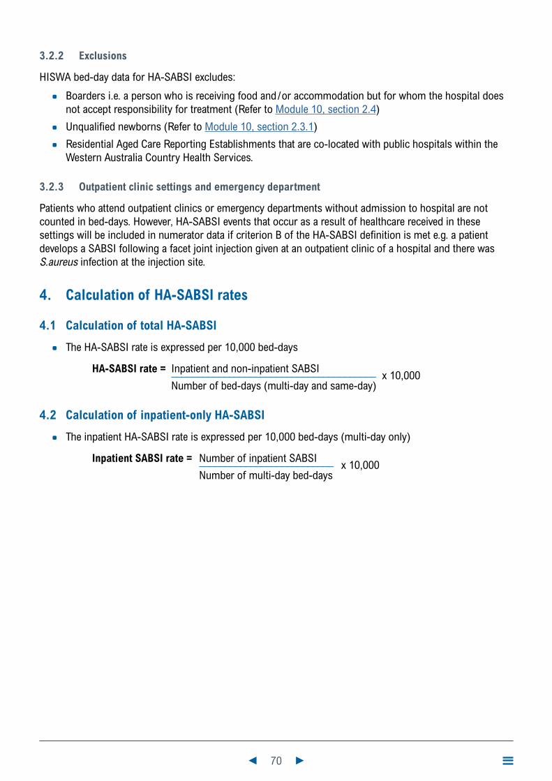
70
3.2.2 Exclusions
HISWA bed-day data for HA-SABSI excludes:
* Boarders i.e. a person who is receiving food and/or accommodation but for whom the hospital does not accept responsibility for treatment (Refer to Module 10, section 2.4)
* Unqualified newborns (Refer to Module 10, section 2.3.1)
* Residential Aged Care Reporting Establishments that are co-located with public hospitals within the Western Australia Country Health Services.
3.2.3 Outpatient clinic settings and emergency department
Patients who attend outpatient clinics or emergency departments without admission to hospital are not counted in bed-days. However, HA-SABSI events that occur as a result of healthcare received in these settings will be included in numerator data if criterion B of the HA-SABSI definition is met e.g. a patient develops a SABSI following a facet joint injection given at an outpatient clinic of a hospital and there was S.aureus infection at the injection site.
4. Calculation of HA-SABSI rates
4.1 Calculation of total HA-SABSI
* The HA-SABSI rate is expressed per 10,000 bed-days
HA-SABSI rate = Inpatient and non-inpatient SABSI x 10,000
_______________________________________ Number of bed-days (multi-day and same-day)
4.2 Calculation of inpatient-only HA-SABSI
* The inpatient HA-SABSI rate is expressed per 10,000 bed-days (multi-day only)
Inpatient SABSI rate = Number of inpatient SABSI x 10,000
__________________________ Number of multi-day bed-days

71
Appendix 1: Methodology for classification of HA-SABSI
Laboratory reports positiveStaphylococcus aureus blood culture
Is there >14 days since a previouspositive blood culture?
SABSI event identified > 48 hoursafter admission and not incubating
on admission, or < 48 hoursafter discharge
Do not reportto HISWANo
Yes
One or more of the following criteria must be met to be classified as a HA-SABSI:
1. SABSI is a complication of the presence of an indwelling medical device e.g. IV, CSF shunt, urinary catheter.
2. SABSI is related to the surgical site and occurs wihtin 30 or 90 days of a surgical procedure depending on the procedure type. (Refer to Appendix 7).
3. SABSI was diagnosed within 48 hours of a related invasive instrumentation or incision.
4. SABSI is associated with neutropenia contributed by cytotoxic therapy.
SABSI event identified < 48 hoursafter admission
Yes
Do not reportto HISWA
Yes to one ormore criteria
No toall criteria
Report toHISWA
Note: If the HA-SABSI is an MRSA HAI, please add to the Significant Organism Module. If relevant, also add to the Haemodialysis/CLABSI modules.
Contact the HAIU if the HA-SABSI is attributable to another healthcare facility: [email protected]

72
Appendix 2: HA-SABSI related to a SSI
An HA-SABSI is considered to be related to a surgical site when criteria for classification as an SSI are met (Refer to Module 2). SSIs are followed for the following periods where day one = the date of the procedure:
* 30 day period for superficial SSI for all procedures and 30 or 90 day period for deep and organ/space infections depending on the procedure. Common procedures are listed in Table 2.
Table 2: Surveillance period for deep or organ/space SSI following surgical procedures
30-day Surveillance
Abdominal aortic aneurysm repair Laminectomy
Limb amputation Liver transplant
Appendix surgery Neck surgery
Shunt for dialysis Kidney surgery
Bile duct, liver or pancreatic surgery Ovarian surgery
Carotid endarterectomy Prostate surgery
Gallbladder surgery Rectal surgery
Colon surgery Small bowel surgery
Caesarean section Spleen surgery
Gastric surgery Thoracic surgery
Heart Transplant Thyroid and/or parathyroid surgery
Abdominal hysterectomy Vaginal hysterectomy
Kidney transplant Exploratory laparotomy
Other surgery not listed
90-day Surveillance
Breast surgery Cardiac surgery
Coronary artery bypass graft with both chest and donor site incisions
Coronary artery bypass graft with chest incision only
Craniotomy Spinal fusion
Open reduction of fracture Herniorrhaphy
Hip arthroplasty Pacemaker surgery
Knee arthroplasty Peripheral vascular bypass surgery
Refusion of spine Ventricular shunt
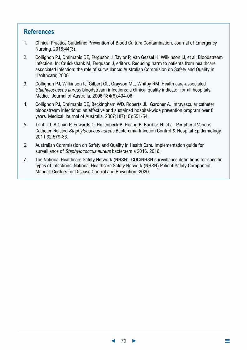
73
References
1. Clinical Practice Guideline: Prevention of Blood Culture Contamination. Journal of Emergency Nursing. 2018;44(3).
2. Collignon PJ, Dreimanis DE, Ferguson J, Taylor P, Van Gessel H, Wilkinson IJ, et al. Bloodstream infection. In: Cruickshank M, Ferguson J, editors. Reducing harm to patients from healthcare associated infection: the role of surveillance: Australian Commision on Safety and Quality in Healthcare; 2008.
3. Collignon PJ, Wilkinson IJ, Gilbert GL, Grayson ML, Whitby RM. Health care-associated Staphylococcus aureus bloodstream infections: a clinical quality indicator for all hospitals. Medical Journal of Australia. 2006;184(8):404-06.
4. Collignon PJ, Dreimanis DE, Beckingham WD, Roberts JL, Gardner A. Intravascular catheter bloodstream infections: an effective and sustained hospital-wide prevention program over 8 years. Medical Journal of Australia. 2007;187(10):551-54.
5. Trinh TT, A Chan P, Edwards O, Hollenbeck B, Huang B, Burdick N, et al. Peripheral Venous Catheter-Related Staphylococcus aureus Bacteremia Infection Control & Hospital Epidemiology. 2011;32:579-83.
6. Australian Commission on Safety and Quality in Health Care. Implementation guide for surveillance of Staphylococcus aureus bacteraemia 2016. 2016.
7. The National Healthcare Safety Network (NHSN). CDC/NHSN surveillance definitions for specific types of infections. National Healthcare Safety Network (NHSN) Patient Safety Component Manual: Centers for Disease Control and Prevention; 2020.

74
Module 7
Central line-associated bloodstream infection
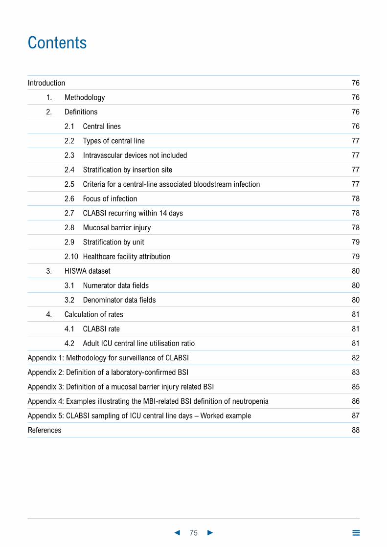
75
Contents
Introduction 76
1. Methodology 76
2. Definitions 76
2.1 Central lines 76
2.2 Types of central line 77
2.3 Intravascular devices not included 77
2.4 Stratification by insertion site 77
2.5 Criteria for a central-line associated bloodstream infection 77
2.6 Focus of infection 78
2.7 CLABSI recurring within 14 days 78
2.8 Mucosal barrier injury 78
2.9 Stratification by unit 79
2.10 Healthcare facility attribution 79
3. HISWA dataset 80
3.1 Numerator data fields 80
3.2 Denominator data fields 80
4. Calculation of rates 81
4.1 CLABSI rate 81
4.2 Adult ICU central line utilisation ratio 81
Appendix 1: Methodology for surveillance of CLABSI 82
Appendix 2: Definition of a laboratory-confirmed BSI 83
Appendix 3: Definition of a mucosal barrier injury related BSI 85
Appendix 4: Examples illustrating the MBI-related BSI definition of neutropenia 86
Appendix 5: CLABSI sampling of ICU central line days – Worked example 87
References 88

76
Introduction Central venous catheters (CVC), also referred to as central lines, serve a vital role in the management of critically ill patients, however, these predispose patients to preventable2 central line-associated bloodstream infections (CLABSI).3,4 CLABSI are serious infections that significantly increase morbidity, mortality and contribute to increased healthcare costs. CLABSI are viewed as preventable adverse events if evidence-based infection prevention practices are followed and integrated with monitoring and feedback of rates to key stakeholders.5 This approach should be taken by every healthcare facility (HCF) to achieve and maintain a zero CLABSI rate.4
1. Methodology
HISWA definitions are based on the CDC/NHSN CLABSI definitions6. For participating hospitals to make a valid comparison of their CLABSI rates the methodology must be similar and definitions consistently applied. Surveillance personnel are required to:
* Implement processes to ensure that all positive blood culture reports are received.
* Investigate all reported bloodstream infections (BSIs) to determine if definition criteria for a CLABSI are met and the attributable facility.
* Liaise with key stakeholders, clinical microbiologist / infectious diseases physicians to assist with the classification of CLABSI events.
* The methodology to assist with classification of CLABSI is described in Appendix 1. Refer to Module 1 for an introduction to surveillance of HAIs.
* HISWA CLABSI surveillance aligns with the Australian national definition developed by the Australian Commission on Safety and Quality in Health Care (ACSQHC).
Note: Surveillance personnel should take opportunities to promote best practice for blood culture collection to optimise BSI detection and classification as potential contaminants ie. blood specimens drawn for culture should be obtained from two to four blood draws from separate venepuncture sites, within a few hours of each other and not through an intravascular catheter.1
Aseptic technique incorporating the use of sterile gloves, and disinfecting culture bottle tops and the patient’s skin using an alcohol based disinfectant is recommended.
2. Definitions
2.1 Central lines
* A central line is defined as an intravascular catheter where the tip of the catheter terminates at or close to the heart or in one of the great vessels which is used for infusion, blood withdrawal or haemodynamic monitoring.6 The site of insertion or the type of catheter does not determine if a line qualifies as a central line for HISWA reporting purposes.
* The following are considered great vessels for CLABSI surveillance: aorta, pulmonary artery, superior/inferior vena cava, brachiocephalic veins, internal jugular veins, subclavian veins, external and common iliac veins, femoral veins and, in neonates, the umbilical artery/vein.6

77
2.2 Types of central line
The main types of central lines are:
* non-tunnelled CVCs: these are central lines placed in either the internal jugular or subclavian vein with the distal tip lying in the superior vena cava.
* tunnelled CVCs: the central line is tunnelled subcutaneously between the skin insertion site and the point where the catheter enters the blood vessel. Some have a cuff which sits in the subcutaneous tunnel and are referred to as cuffed catheters. These catheters are suitable for long term use.
* peripherally inserted central catheters (PICCs): these are central lines that are inserted percutaneously into peripheral veins e.g. basilic, brachial, cephalic. They are suitable for short, intermediate, and long term use.
* implanted ports: these central lines are surgically inserted, placed under the skin and accessed with specific port needles. They are for long term intermittent use.
An introducer is considered an intravascular catheter; however, if the location of its tip is in a great vessel, it may be considered a central line6. Central lines are sometimes described as permanent or temporary, however, HISWA do not stratify CLABSI by these terms.
2.3 Intravascular devices not included
The following are not considered central lines:
* pacemaker wires and other non-lumened devices inserted into central blood vessels or the heart, because fluids are not infused, pushed, nor withdrawn.6
* femoral arterial catheters, extracorporeal membrane oxygenation (ECMO), haemodialysis reliable outflow (HeRO) dialysis catheters and intra-aortic balloon pump (IABP) devices.6
2.4 Stratification by insertion site
Central lines are stratified by the insertion site for reporting and analysis.
* centrally-inserted (CI): the skin entry point is on the trunk of the patient
* peripherally-inserted (PI): the line is inserted through a limb vein e.g. PICC.
A higher risk of infection with CI lines is reported in some patient settings.3
2.5 Criteria for a central-line associated bloodstream infection
* First, the criteria for classification as a laboratory-confirmed BSI event must be met (Refer to Appendix 2.
* The date of the CLABSI event is the date the first positive blood culture was collected. For ‘same’ potential contaminants this is the date the first of two blood culture sets was collected.6
* A CLABSI is defined as a BSI, which is not related to an infection at another site, and on the date of the BSI event the central line had been in place for a period of >48 hours AND was in place on the date of the BSI event or within the previous 24 hours.7
* If a central line was in place for >48 hours and then removed, the CLABSI criteria must be fully met on the day the line was removed or within 24 hours of removal.7
* CLABSI may occur as inpatients or non-inpatients and both are included in surveillance. Non-inpatient CLABSI are present on admission or develop less than 48 hours after admission and are related to the receipt of health care.

78
2.6 Focus of infection
* The BSI definition requires that the organism cultured from the blood is not related to an infection at another site. A clinical assessment is required to determine if a focus of infection is present that is the likely cause of the BSI. This includes a review of medical records, laboratory, diagnostic and imaging reports. If an infection at another site is identified it must fulfil the infection criteria outlined in The National Healthcare Safety Network (NHSN) Surveillance Definitions for Specific Types of Infection6 found online at NHSN Patient Safety Component Manual.
* If a patient with both peripheral and central lines develops a BSI that can clearly be attributed to the peripheral line (e.g. pus at the peripheral line insertion site and the same pathogen from pus and BSI), it should not be reported as a CLABSI.
* Patients suspected or known to have accessed their own central lines that may have contributed to the CLABSI are not excluded from CLABSI surveillance. A facility must implement education and prevention efforts to protect the line.
2.7 CLABSI recurring within 14 days
* If the CLABSI criteria are met again within 14 days and the same organism(s) is identified, a clinical review should be undertaken to determine if the CLABSI is the same event or a new event. The clinical review should include consultation with a clinical microbiologist or infectious diseases physician and consider the following: completion of antimicrobial therapy, resolution of signs and symptoms with negative blood cultures, and central line change.
* If the original infection has resolved, and a new central line has been inserted and the CLABSI criteria are met again, a new CLABSI event should be reported.
* If the new CLABSI event occurs more than 14 days after the pervious event then it is always classified as a new event.
2.8 Mucosal barrier injury
* Oral and gastrointestinal mucosal barriers may break down as a result of chemotherapy and radiation treatment regimens. This mucosal barrier injury (MBI) can range from inflammation to ulceration and enables translocation of bacteria from the oral cavity or intestinal tract into the bloodstream and may cause a bloodstream infection.
* MBI-related BSI may occur in patients who are either:
* Severely neutropenic*, or
* A recipient of allogeneic haemopoietic stem cell transplant with either gastrointestinal graft versus host disease (GI GVHD) or diarrhoea.
* Refer to Appendix 2 for the definition of MBI-related BSI. A list of MBI organisms can be located in the NHSN Patient Safety Component Manual8.
* Neutropenia is defined as at least two separate days with values of total white blood cell count (WBC) or absolute neutrophil count (ANC) < 500 cells/mm3 (<0.5 × 10⁹/L) within a seven day time period which includes the date of the BSI (day one), the three calendar days before and the three calendar days after8. For examples refer to Appendix 3.
79
Note: In a neutropenic or allogeneic haemopoietic stem cell transplant patient with GI GVHD, who has a BSI caused by a MBI organism (with no other organism isolated, and the BSI is not related to infection at another site), the likely source of the BSI is MBI and not the central line.
Report MBI-related BSI to HISWA stratified by unit for monitoring, however, MBI-related BSI will not be included in CLABSI rate calculations.
2.9 Stratification by unit
HISWA CLABSI events are stratified according to specific higher-risk specialty units.
2.9.1 Adult haematology or oncology
Patients managed by these units often have central lines in situ following discharge from hospital. Therefore all CLABSI events that occur either during a hospital admission or as an outpatient are reported.
2.9.2 Adult ICU
* A CLABSI event that occurs more than 48 hours after admission to an adult ICU or within 48 hours of discharge from ICU are reported as ICU-associated.9
* High dependency unit, or step down unit patients should only be included if they are co-located within the ICU and managed by the same medical and nursing staff. They are to be included in the ICU surveillance and data.9
* When paediatric patients are admitted to an adult ICU on an ad-hoc basis, they should be included in the adult ICU surveillance.9
2.10 Healthcare facility attribution
* The CLABSI is attributable to the location where the patient was assigned on the date of the CLABSI event, unless the transfer rule (see below) is applicable.
* If all elements of a CLABSI are present within 48 hours of transfer from one location to another in the same facility or new facility, the CLABSI is attributed to the transferring location. This is called the transfer rule.9
* If a patient is transferred into a facility, with one central line in place, the date and time of the first access as an inpatient is considered when applying the transfer rule (not the date and time of transfer). “Access” is defined as line placement, infusion or withdrawal through the line.
* If a CLABSI develops in a non-inpatient setting, it will be attributed to the facility where the device was last accessed prior to the event.

80
3. HISWA dataset
3.1 Numerator data fields
The numerator data fields and information required to be entered into the HISWA database are described in Table 1.
3.1.1 Inclusions
* CLABSI occurring as inpatient and non-inpatients in ICU, Haematology and Oncology Units.
3.1.2 Exclusions
* MBI-related BSIs in patients who are neutropenic or a recipient of allogeneic haemopoietic stem cell transplant with either GI GVHD or diarrhoea. MBI-related BSIs are reported to HISWA but not included in CLABSI counts.
Table 1: CLABSI numerator data fields and descriptors for HISWA database
Data field Descriptor
Patient ID Unique patient identifier
Date of birth Patient date of birth
Lab specimen number Lab number assigned to the specimen
Specimen date Date the specimen was obtained
Type of central line The type of central line that was inserted in the patient:* centrally-inserted (CI) central line* peripherally-inserted (PI) central line
Place acquired Unit associated with the CLABSI andUnit associated with the MBI-related BSI:
* ICU* Haematology unit* Oncology unit * MBI – BSI ICU* MBI – BSI Haematology* MBI – BSI Oncology
Organism 1 The pathogenic organism isolated from a blood culture
Organism 2 The second pathogenic organism isolated from a blood culture
Organism 3 The third pathogenic organism isolated from a blood culture
3.2 Denominator data fields
The denominator that is utilised is central line-days and these are calculated either by tracking or tally methodologies.9
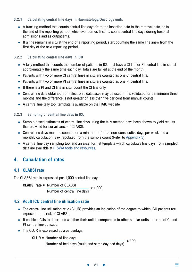
81
3.2.1 Calculating central line days in Haematology/Oncology units
* A tracking method that counts central line days from the insertion date to the removal date, or to the end of the reporting period, whichever comes first i.e. count central line days during hospital admissions and as outpatients.
* If a line remains in situ at the end of a reporting period, start counting the same line anew from the first day of the next reporting period.
3.2.2 Calculating central line days in ICU
* A tally method that counts the number of patients in ICU that have a CI line or PI central line in situ at approximately the same time each day. Totals are tallied at the end of the month.
* Patients with two or more CI central lines in situ are counted as one CI central line.
* Patients with two or more PI central lines in situ are counted as one PI central line.
* If there is a PI and CI line in situ, count the CI line only.
* Central line data obtained from electronic databases may be used if it is validated for a minimum three months and the difference is not greater of less than five per cent from manual counts.
* A central line tally tool template is available on the HAIU website.
3.2.3 Sampling of central line days in ICU
* Sample-based estimates of central line days using the tally method have been shown to yield results that are valid for surveillance of CLABSI.
* Central line days must be counted on a minimum of three non-consecutive days per week and a monthly calculation is extrapolated from the sample count (Refer to Appendix 5).
* A central line day sampling tool and an excel format template which calculates line days from sampled data are available at HISWA tools and resources.
4. Calculation of rates
4.1 CLABSI rate
The CLABSI rate is expressed per 1,000 central line days:
CLABSI rate = Number of CLABSI x 1,000
______________________ Number of central line days
4.2 Adult ICU central line utilisation ratio
* The central line utilisation ratio (CLUR) provides an indication of the degree to which ICU patients are exposed to the risk of CLABSI.
* It enables ICUs to determine whether their unit is comparable to other similar units in terms of CI and PI central line utilisation.
* The CLUR is expressed as a percentage:
CLUR = Number of line days x 100
___________________________________________ Number of bed days (multi and same day bed days)

82
Appendix 1: Methodology for surveillance of CLABSI
Laboratory reports positive blood culturein a patient with a central line in place
Is the definition of a MBI-related BSI met?Refer to Section 3.3 and Appendix 9
Is criterion 1 or 2 of the BSI definition met?Refer to Appendix B
Do not reportto HISWANo
No
Do not reportto HISWANo
No
Yes
Was the blood culture taken more than 14 days after a previous positive culture with the same organism?
Yes
Yes
Yes
The following criteria must be metto be classified as a CLABSI
1. The organism causing the BSI is not related to an infection at another site
2. On the date of the BSI event the central line had been in place for a period of >48 hours AND was in place on the date of the BSI event or within the previous 24 hours.
Review as perSection 3.2
Report MBI-relatedBSI to HISWA
Report toHISWA
Note: Ensure CLABSI are entered into other relevant modules e.g. If the BSI is a S.aureus
Contact the HAIU if the CLABSI is attributable to another healthcare facility: [email protected]

83
Appendix 2: Definition of a laboratory-confirmed BSI
A laboratory-confirmed BSI must meet either Criterion 1 or 2:
Criterion 1: recognised pathogen
* The patient has a recognised pathogen isolated from one or more positive blood cultures and is not related to an infection at another site.7,8
Comments for Criterion 1
* A recognised pathogen includes any organism that is not considered a potential contaminant.8
* Examples of recognised pathogens include: Staphylococcus aureus, Escherichia coli, Klebsiella spp, Pseudomonas aeruginosa, Proteus spp, Candida spp8 Streptococcus Spp (excluding viridans Streptococcus), Enterococcus Spp, Enterobacter Spp and Providencia Spp9.
* No signs or symptoms of infection are required to meet Criterion 1.8
Criterion 2: potential contaminant organisms
* The same (matching) potential contaminant organism is cultured from two or more blood cultures drawn on separate occasions within 24 hours.7,8 (Refer to Determining “same” potential contaminant organisms)
AND
* The patient has at least one of the following signs and symptoms: fever (>38°C); chills; or hypotension7,8 (within 24 hours of the date of the BSI event – see comments)
Comments for Criterion 2
* Organisms that can be considered as potential contaminants of blood cultures include those species that are part of the normal skin flora, such as diphtheroids [Corynebacterium spp.], Propionibacterium spp., coagulase-negative staphylococci [including S. epidermidis], viridans group streptococci, Aerococcus spp., Micrococcus spp. Potential contaminants may also include other bacteria that can be found transiently on the skin such as Bacillus [not B. anthracis] spp., Pseudomonas spp. [other than P.aeruginosa], Xanthomonas spp., Raltsonia spp.8
* CDC/NHSN uses the term: “common commensals” and the NHSN list of common commensals is to be used. This can be accessed in the NHSN Organisms List located in the NHSN Patient Safety Component Manual.8 Any organism that is considered a potential contaminant and is not on this list should be reviewed in liaison with a microbiologist/infectious diseases physician.
* An element refers to a specific component of infection and includes: positive blood culture(s); fever (>38°C), chills and hypotension.7,8 Criterion elements must occur within a timeframe that does not exceed a gap of 24 hours between any two elements e.g. positive blood cultures and fever.7,8 The same (matching) potential contaminant blood cultures represent a single element. The collection date of the first potential contaminant should be used to determine the date of the BSI event.7,8
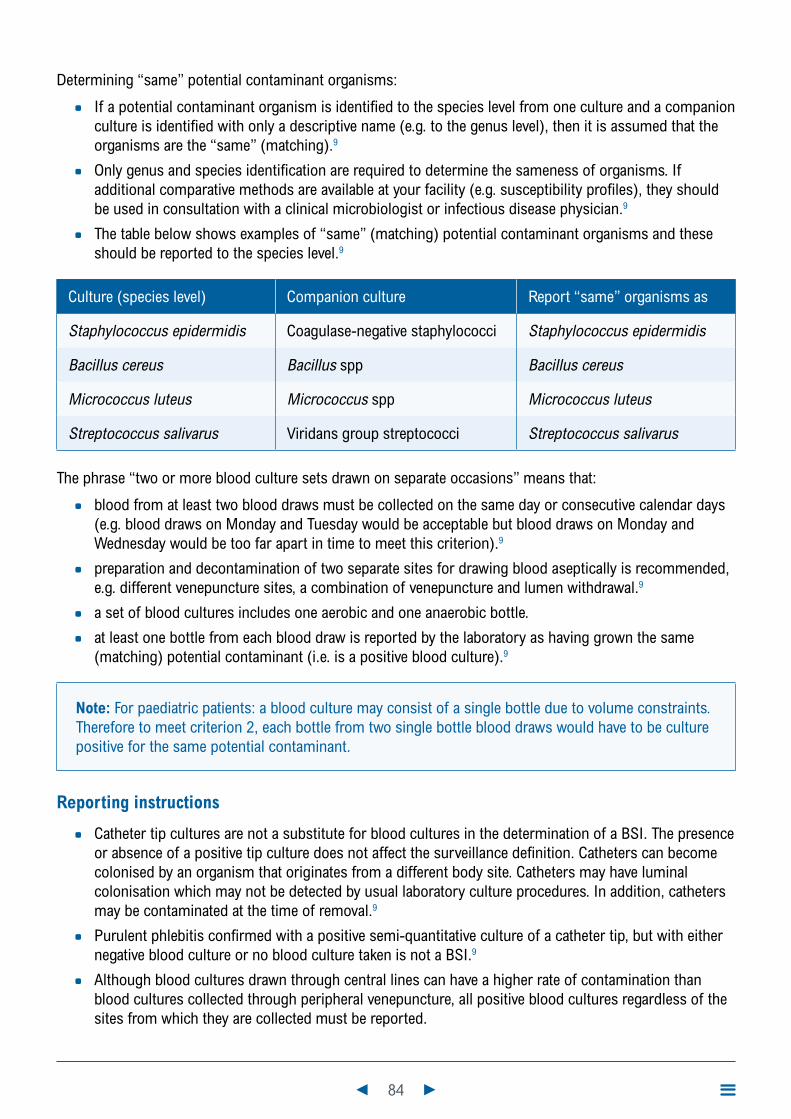
84
Determining “same” potential contaminant organisms:
* If a potential contaminant organism is identified to the species level from one culture and a companion culture is identified with only a descriptive name (e.g. to the genus level), then it is assumed that the organisms are the “same” (matching).9
* Only genus and species identification are required to determine the sameness of organisms. If additional comparative methods are available at your facility (e.g. susceptibility profiles), they should be used in consultation with a clinical microbiologist or infectious disease physician.9
* The table below shows examples of “same” (matching) potential contaminant organisms and these should be reported to the species level.9
Culture (species level) Companion culture Report “same” organisms as
Staphylococcus epidermidis Coagulase-negative staphylococci Staphylococcus epidermidis
Bacillus cereus Bacillus spp Bacillus cereus
Micrococcus luteus Micrococcus spp Micrococcus luteus
Streptococcus salivarus Viridans group streptococci Streptococcus salivarus
The phrase “two or more blood culture sets drawn on separate occasions” means that:
* blood from at least two blood draws must be collected on the same day or consecutive calendar days (e.g. blood draws on Monday and Tuesday would be acceptable but blood draws on Monday and Wednesday would be too far apart in time to meet this criterion).9
* preparation and decontamination of two separate sites for drawing blood aseptically is recommended, e.g. different venepuncture sites, a combination of venepuncture and lumen withdrawal.9
* a set of blood cultures includes one aerobic and one anaerobic bottle.
* at least one bottle from each blood draw is reported by the laboratory as having grown the same (matching) potential contaminant (i.e. is a positive blood culture).9
Note: For paediatric patients: a blood culture may consist of a single bottle due to volume constraints. Therefore to meet criterion 2, each bottle from two single bottle blood draws would have to be culture positive for the same potential contaminant.
Reporting instructions
* Catheter tip cultures are not a substitute for blood cultures in the determination of a BSI. The presence or absence of a positive tip culture does not affect the surveillance definition. Catheters can become colonised by an organism that originates from a different body site. Catheters may have luminal colonisation which may not be detected by usual laboratory culture procedures. In addition, catheters may be contaminated at the time of removal.9
* Purulent phlebitis confirmed with a positive semi-quantitative culture of a catheter tip, but with either negative blood culture or no blood culture taken is not a BSI.9
* Although blood cultures drawn through central lines can have a higher rate of contamination than blood cultures collected through peripheral venepuncture, all positive blood cultures regardless of the sites from which they are collected must be reported.

85
Appendix 3: Definition of a mucosal barrier injury related BSI
MBI-related BSI^
Criterion 1
Patient meets criterion 1 for laboratory-confirmed BSI,^ with at least one blood culture growing any of the following intestinal organisms with no other organisms isolated: Bacteriodes spp., Candida spp., Clostridium spp., Enterococcus spp., Fusobacterium spp., Peptostreptococcus spp., Prevotella spp., Veillonella spp., or Enterobacteriaceae. Refer to complete list of MBI organisms at:
http://www.cdc.gov/nhsn/PS-Analysis-resources/
and
patient meets at least one of the following:1. Is an allogeneic hematopoietic stem cell transplant recipient within the past
year with one of the following documented during same hospitalisation as positive blood culture:a. Grade III or IV gastrointestinal graft versus host disease (GI GVHD).b. ≥1 litre diarrhoea in a 24-hour period (or ≥20mL/kg in a 24-hour period
for patients <18 years or age) with onset on or within seven calendar days before the date the positive blood culture was collected.
2. Is neutropenic (see Definition Appendix 4).
MBI-related BSI^
Criterion 2
Patient meets criterion 2 for laboratory-confirmed BSI when the blood cultures are growing only viridans group streptococci with no other organisms isolated
and
patient meets as least one of the following:1. Is an allogeneic hematopoietic stem cell transplant recipient within the past
year with one of the following documented during same hospitalisation as positive blood culture:a. Grade III or IV gastrointestinal graft versus host disease (GI GVHD).b. ≥1 litre diarrhoea in a 24-hour period (or ≥20mL/kg in a 24-hour period
for patients <18 years or age) with onset on or within seven calendar days before the date the positive blood culture was collected.
2. Is neutropenic (see Definition Appendix 4).
Comments 1. MBI-related BSI Criterion 1 and 2 apply to patients of any age including those < one year of age
2. In MBI-related BSI Criterion 1 and 2, “No other organism isolated” means there is no isolation in a blood culture of another recognised pathogen (e.g., S. aureus) or two matching potential contaminants (e.g. coagulase negative staphylococci), other than listed in MBI-related BSI criterion 1 and 2 ,that would otherwise meet the CLABSI criteria. If this occurs, the infection should not be classified as MBI-related BSI.
3. Grade III/IV GI GVHD is defined as follows:• In adults: ≥1L diarrhoea/day or ileus with abdominal pain• In paediatric patients: ≥20ml/kg/day of diarrhoea.
Adapted from NHSN Bloodstream Infection Event (Central Line-Associated Bloodstream Infection and Non-Central Line-Bloodstream Infection) (2019)9
^ HISWA use the term “MBI-related BSI” instead of “MBI LCBI” used by CDC/NHSN
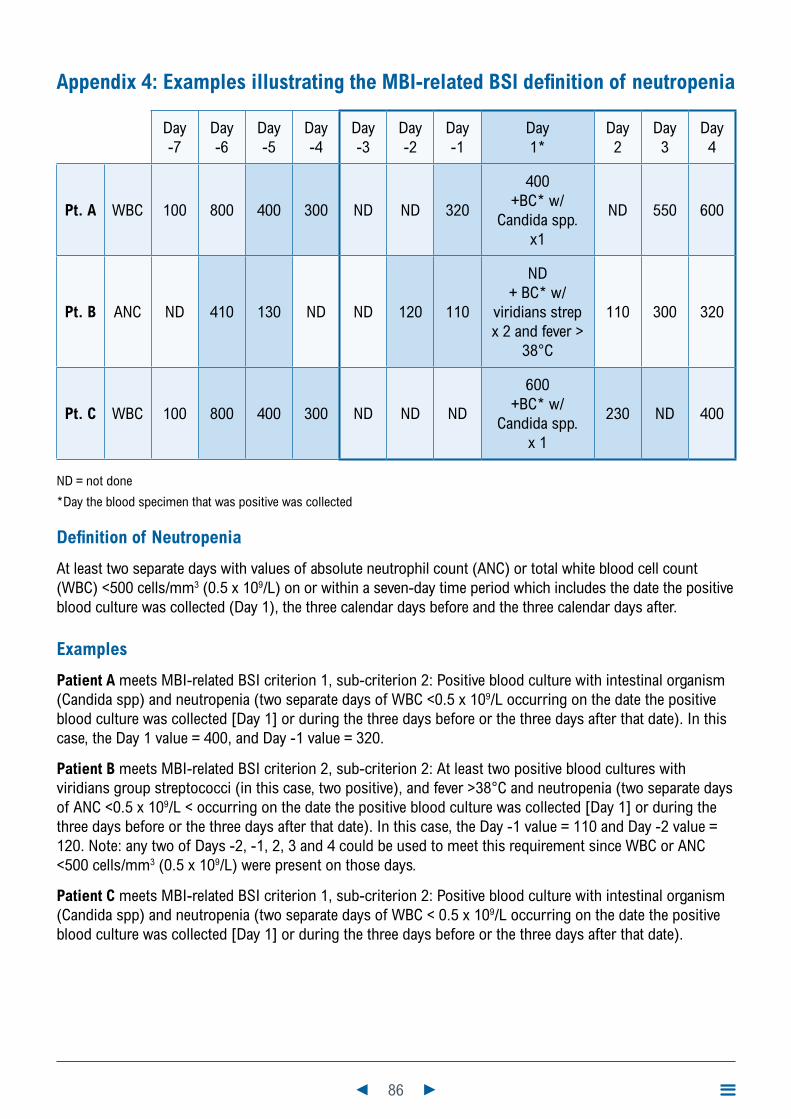
86
Appendix 4: Examples illustrating the MBI-related BSI definition of neutropenia
Day -7
Day -6
Day -5
Day -4
Day -3
Day -2
Day -1
Day 1*
Day 2
Day 3
Day 4
Pt. A WBC 100 800 400 300 ND ND 320
400 +BC* w/
Candida spp. x1
ND 550 600
Pt. B ANC ND 410 130 ND ND 120 110
ND + BC* w/
viridians strep x 2 and fever >
38°C
110 300 320
Pt. C WBC 100 800 400 300 ND ND ND
600 +BC* w/
Candida spp. x 1
230 ND 400
ND = not done
*Day the blood specimen that was positive was collected
Definition of Neutropenia
At least two separate days with values of absolute neutrophil count (ANC) or total white blood cell count (WBC) <500 cells/mm3 (0.5 x 10⁹/L) on or within a seven-day time period which includes the date the positive blood culture was collected (Day 1), the three calendar days before and the three calendar days after.
Examples
Patient A meets MBI-related BSI criterion 1, sub-criterion 2: Positive blood culture with intestinal organism (Candida spp) and neutropenia (two separate days of WBC <0.5 x 10⁹/L occurring on the date the positive blood culture was collected [Day 1] or during the three days before or the three days after that date). In this case, the Day 1 value = 400, and Day -1 value = 320.
Patient B meets MBI-related BSI criterion 2, sub-criterion 2: At least two positive blood cultures with viridians group streptococci (in this case, two positive), and fever >38°C and neutropenia (two separate days of ANC <0.5 x 10⁹/L < occurring on the date the positive blood culture was collected [Day 1] or during the three days before or the three days after that date). In this case, the Day -1 value = 110 and Day -2 value = 120. Note: any two of Days -2, -1, 2, 3 and 4 could be used to meet this requirement since WBC or ANC <500 cells/mm3 (0.5 x 10⁹/L) were present on those days.
Patient C meets MBI-related BSI criterion 1, sub-criterion 2: Positive blood culture with intestinal organism (Candida spp) and neutropenia (two separate days of WBC < 0.5 x 10⁹/L occurring on the date the positive blood culture was collected [Day 1] or during the three days before or the three days after that date).

87
Appendix 5: CLABSI sampling of ICU central line days – worked example
Central line sampling tool Instructions for line day data collection
* Patients with one or more central lines in situ on a day are counted only once as per these rules:
• if there are two or more CI central lines in situ count one CI central line
• if there are two or more PI central lines in situ count one PI central line
• if there is a PI and a CI central line in situ, count one CI central line only.
* Counts of central line days will cease on patient discharge from ICU even if the lines remain in situ.
* Count lines at approximately the same time each day.
* Counts of central lines can be performed daily or by sampling, preferably on three or more non-consecutive days per week.
Year: 2019 Month: August No. of patients with one or more
Day of month CI central lines PI central lines
1 20 523 22 545 15 4678 24 3910 25 31112 24 4131415 20 41617 22 41819 18 4202122 25 32324 22 32526 25 32728 22 329 24 33031 20 3Total line day counts 328 54
Calculations
Total number of central lines days (a) 328 54Number of days when counts performed (b) 15 15Average number of central lines per day (c) = a/b 21.9 3.6Number of days in the month (d) 31 31Total central line days for month (e) = c x d 678 112
88
References
1. Clinical Practice Guideline: Prevention of Blood Culture Contamination. Journal of Emergency Nursing. 2018;44(3).
2. Wise ME, Scott RD, Baggs JM, Edwards JR, Ellingson KD, Fridkin SK, et al. National Estimates of Central Line-Associated Bloodstream Infections in Critical Care Patients. Infection Control & Hospital Epidemiology. 2013;34(6):547-54.
3. Blot K, Bergs J, Vogelaers D, Blot S, Vandijck D. Prevention of Central Line-Associated Bloodstream Infections Through Quality Improvement Interventions: A Systematic Review and Meta-analysis. Clinical Infectious Diseases. 2014;59:96-105.
4. Marschall J, Mermel LA, Fakih M, Hadaway L, Kallen A, O’Grady NP, et al. Strategies to Prevent Central Line–Associated Bloodstream Infections in Acute Care Hospitals: 2014 Update. Infection Control & Hospital Epidemiology. 2014;35(7):753-71.
5. Umscheid C, Mitchell M, A Doshi J, Agarwal R, Williams K, J Brennan P. Estimating the Proportion of Healthcare-Associated Infections That Are Reasonably Preventable and the Related Mortality and Costs. Infection Control and Hospital Epidemiology. 2011;32(2):101-14.
6. The National Healthcare Safety Network (NHSN). CDC/NHSN surveillance definitions for specific types of infections. National Healthcare Safety Network (NHSN) Patient Safety Component Manual: Centers for Disease Control and Prevention; 2020.
7. Australian Commission on Safety and Quality in Health Care. National definition and calculation of central line associated blood stream infection 2019 [Available from: https://www.safetyandquality.gov.au/sites/default/files/2019-08/implementation-guide-for-surveillance-of-central-line-associated-blood-stream-infection-2019-final.pdf.
8. National Healthcare Safety Network (NHSN). Patient safety component manual. 2020.
9. Australian Commission on Safety and Quality in Health Care. Implementation guide: Surveillance of central line associated bloodstream infection. Commonwealth of Australia; 2019.
89
Module 8
Haemodialysis access-associated bloodstream infection

90
Contents
Introduction 91
1. Methodology 91
2. Definitions 91
2.1 Haemodialysis vascular access 91
2.2 Haemodialysis access site infection 91
2.3 Haemodialysis access-associated BSI 92
2.4 New access-associated BSI events 92
2.5 Attribution of BSI to an HD unit 92
2.6 Stratification by haemodialysis access type 92
3. HISWA dataset 93
3.1 Numerator data fields 93
3.2 Denominator data fields 93
4. Calculation of rates 94
Appendix 1: Methodology for determining HD-BSI 95
Appendix 2: Definition of a laboratory-confirmed BSI 96
Appendix 3: Haemodialysis collection tools 98
References 98

91
IntroductionHaemodialysis places patients at high risk for healthcare-associated infection due to the immunocompromised state intrinsic to end-stage renal disease2, the high prevalence of diabetes, and numerous human, environmental and procedural factors. The invasiveness of the haemodialysis procedure, which requires vascular access, is an established risk factor for bloodstream infection (BSI). Haemodialysis access-associated BSI is a serious complication that can result in significant morbidity and mortality.
1. Methodology
For haemodialysis (HD) units to make a valid comparison of access-associated BSI rates the methodology must be similar and definitions consistently applied. It is essential that communication occurs between hospital and satellite HD service providers to ensure that access-associated BSIs are identified and attributed to the correct unit.
Hospital surveillance personnel are required to:
* implement processes to ensure all positive blood culture reports from HD patients are received and investigated to determine if the BSI is access-associated and the attributable HD unit.
Satellite HD personnel are required to:
* contact the Healthcare Associated Infection Unit (HAIU) [email protected] if a patient is transferred to a hospital directly from dialysis for investigation of infection and report if blood cultures or access site specimens were obtained prior to transfer.
Methodology to assist with classification of HD access-associated BSI is described in Appendix 1. Refer to Module 1 for an introduction to healthcare-associated infection (HAI) surveillance.
Note: Surveillance personnel should take opportunities to promote best practice for blood culture collection to optimise BSI detection and classification as potential contaminants. Ideally blood culture specimens should be aseptically obtained from two to four blood draws from separate venepuncture sites, rather than through an intravascular device.1
Aseptic technique incorporating hand hygiene, the use of sterile gloves, and ensuring the skin or cannula hub and culture bottle tops are disinfected with an alcohol based disinfectant and allowed to dry prior to access is recommended.
2. Definitions
2.1 Haemodialysis vascular access
* Refers to any intravascular access utilised for the purpose of haemodialysis e.g. cuffed or non-cuffed central venous catheters, arterio-venous grafts or fistulae.
2.2 Haemodialysis access site infection
* An access site infection is defined as the presence of one or more of the following symptoms at the access site: purulent discharge, increased swelling or redness.4

92
2.3 Haemodialysis access-associated BSI
* Firstly, the criteria for classification as a BSI must be met (Refer to Appendix 2: Definition of a laboratory-confirmed BSI).
* An HD access-associated BSI is defined as a BSI in a patient where the source of the BSI is an access site infection or is unknown.4
• if an access site infection is present the BSI is classified as access-associated
• where there is no access site infection, active investigation must be taken to determine the presence or absence of a focus of infection at another site. This includes a review of the medical record, laboratory, diagnostic and imaging reports.
• If a focus of infection at another site other than the access device is considered the likely source of the BSI the infection must fulfil the infection criteria for that site outlined in the CDC NHSN Surveillance Definitions for Specific Types of Infection.5
Note: Haemodialyis patient’s often have chronic vascular wounds e.g. leg ulcers, which are colonised with micro-organisms and are not clinically infected. If the same organism is identified in a BSI, it is unlikely that the colonised wound is the source of the BSI.
Rather, it is probable that these organisms have been transmitted to the access site resulting in a BSI or access site infection. Therefore, if there are no other sources of infection, these cases are classified as a HD-BSI.
2.4 New access-associated BSI events
* There must be 21 days or more between positive blood cultures with the same organism for an HD access-associated BSI to be counted as a new event four i.e. BSIs that occur less than 21 days apart and with the same organism are considered ongoing infection and are not counted as a new event. The exception to this rule is when a Staphylococcus aureus BSI occurs in an HD patient and then a 14-day rule is applied between infection episodes. Refer to Module 6.
2.5 Attribution of BSI to an HD unit
* An access-associated BSI will be attributed to the HD unit where the access device was last accessed prior to developing signs and symptoms of the BSI unless there is compelling evidence to the contrary.
2.6 Stratification by haemodialysis access type
* Haemodialysis access types are stratified for reporting and analysis and are listed in order of increasing risk of infection:
• arteriovenous fistula (AVF) – the connection of an artery and a vein using the patient’s own blood vessels
• arteriovenous graft (AVG) – the connection of an artery and a vein using synthetic or native grafts (graft types are combined for reporting)
• cuffed catheters – permanent or semi-permanent, tunnelled central lines e.g. Hickman
• non-cuffed catheters – temporary, non-tunnelled central lines.4

93
3. HISWA dataset
3.1 Numerator data fields
The numerator data fields for HD access-associated BSI required to be entered into the HISWA database are described in Table 1.
Table 1: Haemodialysis access-associated BSI numerator data fields and descriptors for HISWA database
Data field Descriptor
Patient ID Unique patient identifier
Date of birth Patient date of birth
Laboratory specimen number Laboratory number assigned to the specimen
Specimen date Date the specimen was obtained
Type of access Type of access:
* AVF
* AVG (native and synthetic)
* Non-cuffed catheter
* Cuffed catheter
Organism 1 The pathogenic organism isolated from a blood culture
Organism 2 The 2nd pathogenic organism isolated from a blood culture
Organism 3 The 3rd pathogenic organism isolated from a blood culture
3.2 Denominator data fields
* The denominator used is the number of patient-months, stratified by the type of vascular access type.
* The data fields required to be entered into the HISWA database each month are described in Table 2.
Table 2: Haemodialysis access-associated BSI denominator data fields for HISWA database
Access type Number of patient-months
AVF
AVG (synthetic and native combined)
Cuffed catheter
Non-cuffed catheter

94
3.2.1 Denominator data collection
* The number of patients who received HD on the first two working days of each month, stratified by access type, are counted.
* Links to denominator data collection tools for satellite and in-centre dialysis units is available in Appendix 3. These links are also available on the HAIU website in HISWA tools and resources.
• Each HD patient is only counted once each month on the specified collection date.4
• If the patient has multiple vascular access types, count only the access type with the highest risk of infection,4 e.g. catheters have a higher risk than AVF or AVG. Refer to section 2.6. Stratification by haemodialysis access type.
• Non-cuffed catheters are not included in counts from Satellite HD units, as utilisation in this setting is rare.
3.2.2 Inclusions
The following patients are included in the surveillance:
* Chronic adult HD patients
* Patients receiving HD as “visitors” to another HD unit within Western Australia.
3.2.3 Exclusions
The following patients are excluded in the surveillance:
* Patients with acute renal failure requiring HD.
* HD patients who are short term visitors from outside WA, i.e. less than one week.
4. Calculation of rates
* The haemodialysis access-associated BSU rate is expressed per 100 patient-months, stratified by access type, and can be interpreted as the proportion of patients with each access type who develop a BSI each month.
BSI rate = Number of access-associated BSI x 100
____________________________ Number of patient-months

95
Appendix 1: Methodology for determining HD-BSI
Laboratory reports positive blood culturein a haemodialysis patient
Was the blood culture >21 days since a previous positive culture with the same organism
Is criterion 1 or 2 of the BSI definition met?Do not report
to HISWA
No
Yes
No
Yes
Yes
The following criteria must be met to classify the BSI as haemodialysis access-associated
1. Infection is present at the access site2. The source of the BSI is unknown.
Report toHISWA
No to both
Yes to either
Note: Ensure haemodialysis access-associated BSIs are entered into other relevant modules e.g. if the BSI is a MRSA, ensure it is entered into the specific organism module.
Contact the HAIU if the haemodialysis access-associated BSI is attributable to another healthcare facility: [email protected]

96
Appendix 2: Definition of a laboratory-confirmed BSI
A laboratory-confirmed BSI must meet either Criterion 1 or 2:
Criterion 1: recognised pathogen
* The patient has a recognised pathogen isolated from one or more positive blood cultures
Comments for Criterion 1
* a ‘recognised pathogen’ includes any organism that is not considered a potential contaminant.
* examples of recognised pathogens include: Staphylococcus aureus, Escherichia coli, Klebsiella pneumoniae, Pseudomonas aeruginosa, Proteus spp, Candida spp.
Criterion 2: potential contaminant organisms
* The same (matching) potential contaminant organism is cultured from two or more blood cultures drawn on separate occasions. (Refer to Appendix 12 “Interpreting ‘same” potential contaminants)
AND
* the patient has at least one of the following signs and symptoms: fever (>38°C); chills; or hypotension (within 24 hours of the date of the BSI event – see comments)
Note: Other specific signs and symptoms for patients aged one year or less are not listed as paediatric patients are not included in HISWA surveillance. Refer to CDC/NHSN module3
Comments for Criterion 2
* Organisms that can be considered as potential contaminants of blood cultures include those species that are part of the normal skin flora, such as diphtheroids [Corynebacterium spp.], Propionibacterium spp., coagulase-negative staphylococci [including S. epidermidis], viridans group streptococci, Aerococcus spp., Micrococcus spp. Potential contaminants may also include other bacteria that can be found transiently on the skin such as Bacillus [not B. anthracis] spp., Pseudomonas spp. [other than P.aeruginosa], Xanthomonas spp., Raltsonia spp.
* CDC/NHSN uses the term: “common commensals” and the NHSN list of common commensals is to be used. This can be accessed at: http://www.cdc.gov/nhsn/PS-Analysis-resources/. Any organism that is considered a potential contaminant and is not on this list should be reviewed in liaison with a microbiologist/infectious diseases physician.
* An element refers to a specific component of infection and includes: positive blood culture(s); fever (>38°C), chills and hypotension. Criterion elements must occur within a timeframe that does not exceed a gap of 24 hours between any two elements e.g. positive blood cultures and fever. The same (matching) potential contaminant blood cultures represent a single element. The collection date of the first potential contaminant should be used to determine the date of the BSI event.

97
Determining “same” potential contaminant organisms
* If potential contaminant organisms are identified to the species level from one culture and a companion culture is identified with only a descriptive name (e.g. to the genus level), then it is assumed that the organisms are the “same” (matching).
* Only genus and species identification are required to determine the sameness of organisms. If additional comparative methods are available at your facility (e.g. susceptibility profiles), they should be used in consultation with a clinical microbiologist or infectious disease physician.
* Table 3 below shows examples of “same” potential contaminant organisms and these should be reported to the species level.
Table 3
Culture (species level) Companion culture Report “same” organisms as
Staphylococcus epidermidis Coagulase-negative staphylococci Staphylococcus epidermidis
Bacillus cereus Bacillus spp Bacillus cereus
Micrococcus luteus Micrococcus spp Micrococcus luteus
Streptococcus salivarus Viridans group streptococci Streptococcus salivarus
* The phrase “two or more blood cultures drawn on separate occasions” means that:
• Blood from at least two blood draws must be collected on the same day or consecutive calendar days (e.g. blood draws on Monday and Tuesday would be acceptable but blood draws on Monday and Wednesday would be too far apart in time to meet this criterion).6
• Preparation and decontamination of two separate sites for drawing blood using an aseptic non-touch technique is recommended, e.g. different venepuncture sites, a combination of venepuncture and lumen withdrawal.
• At least one bottle from each blood draw is reported by the laboratory as having grown the same (matching) potential contaminant (i.e. is a positive blood culture).
Reporting instructions
* Catheter tip cultures are not a substitute for blood cultures in the determination of a BSI. The presence or absence of a positive tip culture does not affect the surveillance definition. Catheters can become colonised by an organism that originates from a different body site. Catheters may have luminal colonisation which may not be detected by usual laboratory culture procedures. In addition, catheters may be contaminated at the time of removal.
* Purulent phlebitis confirmed with a positive semi-quantitative culture of a catheter tip, but with either negative blood culture or no blood culture taken is not a BSI.
* Although blood cultures drawn through central lines can have a higher rate of contamination than blood cultures collected through peripheral venepuncture, all positive blood cultures regardless of the sites from which they are collected must be reported.

98
Appendix 3: Haemodialysis collection tools
HD-BSI Denominator Data Collection Tool for Satellite Dialysis Units
HD-BSI Denominator Data Collection Tool for Incentres
References
1. Clinical Practice Guideline: Prevention of Blood Culture Contamination. Journal of Emergency Nursing. 2018;44(3).
2. Jardine M, Commons RJ, De Zoysa JR, Wong MG, Gilroy N, Green J, et al. Kidney Health Australia- Caring for Australasians with renal impairment guideline recommendations for infection control for haemodialysis units. Nephrology. 2019;24:951-7.
3. Australian Commission on Safety and Quality in Health Care. Implementation guide for surveillance of Staphylococcus aureus bacteraemia 2016. 2016.
4. (CDC) CfDCaP. National Healthcare Safety Network (NHSN) Dialysis Event Surveillance Protocol. 2018.
5. The National Healthcare Safety Network (NHSN). CDC/NHSN surveillance definitions for specific types of infections. National Healthcare Safety Network (NHSN) Patient Safety Component Manual: Centers for Disease Control and Prevention; 2020.
6. Australian Commission on Safety and Quality in Health Care. Implementation guide: Surveillance of central line associated bloodstream infection. Commonwealth of Australia; 2019.
99
Module 9
Occupational exposure

100
Contents
Introduction 101
1. Methodology 101
2. Definitions 101
2.1 Occupational exposure 101
2.2 Parenteral exposure 101
2.3 Non-parenteral exposure 101
2.4 Blood and body fluids 102
2.5 Classification of healthcare workers 102
3. HISWA dataset 103
3.1 Numerator data fields 103
3.2 Denominator data fields 103
4. Calculation of rates 104
References 104
101
IntroductionOccupational exposure occurs when a healthcare worker (HCW) is put at risk of acquiring a blood-borne viral (BBV) disease, such as hepatitis B (HBV), hepatitis C (HCV) or human immunodeficiency virus (HIV), through exposure to an infected patient’s blood or body fluids.1-3 Occupational exposures are increasingly regarded as preventable. In addition to education and adherence to standard precautions, the use of safety-engineered medical devices (SEMDs) is an effective measure in eliminating the risk of some exposures.3
1. Methodology
* All HCFs should have incident monitoring systems in place for the reporting and management of occupational exposures.
* All occupational exposures to BBVs where a risk assessment has been performed and follow-up is deemed necessary are to be reported to Healthcare Infection Surveillance Western Australia (HISWA).
* The minimal data on each occupational exposure is reported to HISWA. Hospitals should collect additional information to ensure a risk management approach is undertaken to prevent occupational exposures.
2. Definitions
2.1 Occupational exposure
* An occupational exposure is an incident that occurs during the course of a person’s paid or unpaid work where there is a risk of acquiring a BBV following exposure, typically via broken skin, eyes, mucous membranes or parenteral contact, to another person’s blood, tissue, or body fluids that are potentially infected with a BBV.4
* Contact between blood or body fluids and intact skin is not considered occupational exposure.
2.2 Parenteral exposure
* Parenteral (or percutaneous) exposures include:
• any incident where there is a penetration of the skin or mucous membranes with a sharp object including but not limited to needles, scalpels, broken glass, broken capillary tubes, surgical instruments, wires, spicules of bone and teeth, that may be contaminated with blood, tissue or other potentially infectious body fluids.1
• penetration of skin through a dirty/contaminated glove with a clean sharp object.
• human bites if the HCW skin is broken.
2.3 Non-parenteral exposure
* Non-parenteral (or non-percutaneous) exposures include:
• any incident where a HCW’s mucous membranes e.g. eyes, nose, mouth, or where non-intact skin e.g. skin abrasions, open wounds or skin that is damaged with dermatitis, is exposed to blood, tissue or other potentially infectious body fluids.4

102
2.4 Blood and body fluids
* The following body fluids are considered a potential risk for BBV transmission:
• blood, serum, plasma and all tissue or body fluids visibly contaminated with blood
• pleural, amniotic, pericardial, peritoneal, synovial and cerebrospinal fluids, uterine/vaginal secretions or semen
• laboratory specimens containing concentrated BBV.5,6
* Faeces, nasal secretions, saliva, sputum, sweat, tears, urine, and vomitus carry a minimal risk of BBV infection unless they are visibly contaminated with blood or where there is no obvious blood but there is potential for blood contamination.6,7
2.5 Classification of healthcare workers
All HCWs, students, contractors and volunteers are included in the surveillance and classified according to Table 1.
Table 1: Classification of HCW occupations and descriptors
HISWA Classification Descriptor
Doctor (include student) All medical officers, specialist clinicians, dentists, visiting and student doctors.
Nurse (include student) All nurses – registered, enrolled; student; midwife; nursing assistant, dental nurses.
Allied Health (include student)
Clinical healthcare professionals distinct from medicine, dentistry and nursing e.g. social work, dietetics, podiatry, pharmacy, audiology occupational therapy, physiotherapy, radiography, psychology, speech pathology and prosthetics and student allied health.
Patient Support Services Other HCWs providing services that support clinical patient care e.g. patient care assistants, ward orderlies, phlebotomists*, all technicians (laboratory, theatre, respiratory, orthopaedic, pathology and anaesthetic) and CSSD/TSSU staff.
* phlebotomists employed by contracted services i.e. Pathwest should not be included as their occupational exposures should be recorded by their employer.
Environmental Services HCWs mainly involved in maintaining equipment and the environment e.g. housekeeping, catering, cleaning, laundry workers, waste management, plumbers, engineers, carpenters, maintenance, visiting contractors.
Other Other employees/workers who do not fit into the above classifications e.g. administrative, clerical, information technology, chaplains, volunteers, transport, security.

103
3. HISWA dataset
3.1 Numerator data fields
The numerator data fields required to be entered into the HISWA database are described in Table 2.
Table 2: Occupational exposure data fields and descriptors for HISWA database
Data field Descriptor
Identifier HCW identifier – initials or DOB
Exposure date The date of the occupational exposure incident
Occupation The classification of the HCW reporting the occupational exposure as per Table 1
Type of exposure * parenteral
* non-parenteral
3.1.2 Inclusions
* Report all occupational exposures where a risk assessment has been performed and follow-up is required.
* Report occupational exposures from staff working in all inpatient and outpatient departments of a hospital, including:
• emergency and outpatient departments, day wards and units e.g. dialysis
• psychiatric hospitals, and psychiatric units within hospitals
• HITH
• rehabilitation wards within hospitals.
3.1.3 Exclusions
* Do not report exposure to faeces, nasal secretions, saliva, sputum, sweat, tears, urine and vomitus to non-intact skin or mucous membranes unless visibly contaminated with blood or there is the potential for blood contamination.
* Do not report occupational exposures that are not officially reported and documented e.g. anecdotal reports.
* Do not report occupational exposures from visitors who are not employees or contractors e.g. patient visitors.
3.2 Denominator data fields
The denominator used is the total number of bed-days for the HCF (multi-day and same-day bed-days). Emergency department and outpatient clinic presentations are not included in bed-day data.

104
4. Calculation of rates
The occupational exposure rate is expressed per 10,000 bed-days.
Occupational exposure rate = Number of exposure x 10,000
______________________________________ Total number of multi and same-day bed-days
References
1. Australian Council on Healthcare Standards. Infection Control: Clinical Indicator User Manual Version 5. Ultimo, NSW: ACHS Performance an Outcomes Service; 2018.
2. Jagger J, Perry J. Avoiding blood and body fluid exposures. Nursing. 2002;32(8):68.
3. Jagger J. Caring for Healthcare Workers A Global Perspective. Infection Control & Hospital Epidemiology. 2015;28(1):1-4.
4. King S, Murphy C. Health-care worker bloodborne virus exposure. In: Cruickshank M, Ferguson J, editors. Reducing harm to patients from health care associated infection: the role of surveillance. Sydney, NSW: ACSQHC; 2008.
5. Centers for Disease Control and Prevention. Healthcare personnel safety component protocol: Healthcare personnel exposure module. 2012. In: The National Healthcare Safety Network (NHSN) Manual [Internet]. Atlanta, GA. Available from: https://www.cdc.gov/nhsn/pdfs/hps-manual/hps_manual-exp-plus-flu-portfolio.pdf.
6. Kuhar DT, Henderson DK, Struble KA, Heneine W, Thomas V, Cheever LW, et al. Updated US Public Health Service Guidelines for the Management of Occupational Exposures to Human Immunodeficiency Virus and Recommendations for Postexposure Prophylaxis. Infection Control & Hospital Epidemiology. 2015;34(9):875-92.
7. Pintilie H, Brook G. Commentary: A review of risk of hepatitis B and C transmission through biting or spitting. Journal of Viral Hepatitis. 2018;25(8).
105
Module 10
Bed-day and separation data
106
Contents
Introduction 107
1. Roles and responsibilities 107
2. Definitions 107
2.1 Bed-days 107
2.2 Separations 107
2.3 Newborns 108
2.4 Boarders 108
2.5 Contracted services 108
3. HISWA data fields 109
4. HISWA data field definitions 110
5. National surveillance data 112
5.1 Variations between HISWA and national data 112
References 112
107
IntroductionAll healthcare facilities (HCFs) and haemodialysis units (both privately operated and public units) are required to submit data for a suite of mandatory surveillance indicators to Healthcare Infection Surveillance Western Australia (HISWA).1 Collection of accurate bed-day and separation data is essential to calculate infection rates for these indicators.
1. Roles and responsibilities
* Administrators responsible for the management of patient information data are required to provide monthly bed-day and separation data to surveillance personnel as outlined in this module.
* Surveillance personnel are required to check bed-day and separation data and submit to HISWA within 30 days from the end of the reporting month.
* Surveillance personnel are to regularly liaise with administrators providing the data to ensure HISWA requirements are being met.
* The mandatory indicators and reporting requirements are outlined in MP-0108/19 Healthcare Associated Infection Surveillance in Western Australia Policy.2
2. Definitions
2.1 Bed-days
* Bed-days are defined and calculated as multi-day and same-day. Bed-days are calculated by subtracting admission date from separation date and are attributable to the month the patient was separated.3
* Hospital-in-the-Home (HITH) patients are considered admitted patients to a virtual non-ICU ward and are included in all bed-day data.
2.1.1 Multi-day bed-days
A count of beds that are occupied by overnight patients admitted to the hospital for a minimum of one night.3
2.1.2 Same-day bed-days
A count of beds/chairs that are occupied by patients that are admitted as same-day patients i.e. the patient is admitted to and separated from the HCF on the same date.3
2.2 Separations
* Separations are defined as formal and statistical.3
* Separations submitted to HISWA include both formal and statistical separations.
* HITH patients are included in all separation data.
2.2.1 Formal separations
This is the administrative process by which an HCF records the cessation of inpatient treatment and/or care and/or accommodation of a patient.3
108
2.2.2 Statistical separations
This is the administrative process by which an HCF records the cessation of an episode of care for a patient within the one hospital stay i.e. there is a change of care type category (not a change of ward, treatment or client status).3
2.3 Newborns
A newborn is a child who is aged nine days or less.3
2.3.1 Unqualified newborns
A newborn who meets at least one of the following criteria:
* is a single live birth or the first live-born infant in a multiple birth, whose mother is currently an admitted patient
* is not admitted to an intensive care facility in a hospital for the provision of special care.3
If the unqualified newborn remains in hospital after day nine, then the newborn becomes a Boarder.3
2.3.2 Qualified newborns
A newborn who meets at least one of the following criteria:
* is the second or subsequent live born infant of a multiple birth whose mother is an admitted patient
* is admitted to a level two neonatal intensive care unit (NICU) for the provision of special care
* remains in a hospital without the mother
* is admitted to the hospital without the mother.3
2.4 Boarders
A boarder is a person who is receiving food and/or accommodation but for whom the hospital does not accept responsibility for treatment and/or care including:
* family members of an admitted child who are provided with accommodation
* healthy newborns more than nine days of age who do not require acute care and belong to a mother who is admitted to the hospital or transferred to another hospital.3
2.5 Contracted services
An episode of care for an admitted patient who’s treatment and care is provided under an arrangement between a hospital that purchases the care (funding establishment) and a provider of the admitted service (contracted service provider).3
* For the purposes of HISWA surveillance, the denominator bed-day/separation data for an admitted episode of care should be counted for the contracted service provider and not the funding hospital.

109
Examples:
1. Hospitals (funding hospital) purchase care for public dialysis patients (contracted patients) at private dialysis units (contracted service provider). Admissions to the private dialysis units for treatment should not be counted in the bed-day /separation data of the funding hospitals.
2. Public hospitals (funding hospital) purchase care for public patients (contracted patients) in private hospitals (contracted service provider). Admissions to private hospitals contracted to provide care to public patients should be counted in the bed-day/separation data of the private hospital.
Note: contracted patients (activity funded by other service providers) will be counted in the denominator data of the contracted service provider only.
3. HISWA data fields
Data fields required to be entered into the HISWA database are outlined in Table 1 and HISWA data field definitions are described in Table 2 and Table 3.
Table 1: Monthly bed-day data required for HISWA
Month ICU (all ages)
Non-ICU (all ages)
Psychiatric (all ages)
Unqualified Newborns
Patients < 2 years of age*
Multi-day bed-days
Same-day bed-days
Multi-day separations (formal and statistical)
Same-day separations (formal and statistical)
* Patients < 2 years of age on the date of their admission

110
4. HISWA data field definitions
Table 2: HISWA bed-day data fields
Definitions ICU (all ages)
Non-ICU wards/units (all ages)
Psychiatric wards/units (all ages)
Unqualified Newborns
Patients < 2 years of age*
Multi-day bed-days
admitted patients with an overnight stay in ICU
admitted patients with overnight stay in non-ICU wards/units i.e. excluding ICU and psychiatric units
admitted patients with overnight stay in a psychiatric ward/unit
unqualified newborns where the mother is admitted with an overnight stay in ICU, non-ICU and psychiatric units
patients < 2 years of age admitted with an overnight stay in ICU, non-ICU or psychiatric units
Inclusions qualified newborns
HITH, qualified newborns
HITH, qualified newborns
HITH HITH, qualified newborns
Exclusions same-day admissions, unqualified newborns, universal exclusions**
same-day admissions, unqualified newborns, universal exclusions**
same-day admissions, unqualified newborns, universal exclusions**
same-day admissions qualified newborns, universal exclusions**
same-day admissions, unqualified newborns, universal exclusions**
Same-day bed-days
patients separated from the HCF directly from ICU on the same-day of admission. This does not include transfers from the ICU to wards
patients separated from non-ICU wards/units i.e. excluding ICU and psychiatric units, on the same day of admission
patients separated from psychiatric wards/units on the same day of admission
unqualified newborns where the mother is separated on the same-day of admission from ICU, non-ICI and psychiatric units
patients < 2 years of age separated from the ICU, non-ICU and psychiatric units on the same-day of admission
Inclusions qualified newborns
HITH, qualified newborns
HITH, qualified newborns
HITH HITH, qualified newborns
Exclusions multi-day admissions, unqualified newborns, universal exclusions**
multi-day admissions, unqualified newborns, universal exclusions**
multi-day admissions, unqualified newborns, universal exclusions**
multi-day admissions qualified newborns, universal exclusions**
multi-day admissions, unqualified newborns, universal exclusions**
* Patients < 2 years of age on the date of their admission
** Universal exclusions are exclusions from all HISWA data and include: boarders, contracted patients, Rehabilitation in the Home (RITH), organ procurement, small hospitals and residents of Residential Aged Care Reporting Establishments within WACHS.
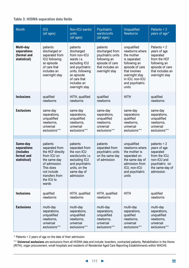
111
Table 3: HISWA separation data fields
Month ICU (all ages)
Non-ICU wards/units (all ages)
Psychiatric wards/units (all ages)
Unqualified Newborns
Patients < 2 years of age*
Multi-day separations (formal and statistical)
patients discharged or separated from ICU following an episode of care that includes an overnight stay
patients discharged from non-ICU wards i.e. excluding ICU and psychiatric units, following an episode of care that includes an overnight stay
patients discharged from psychiatric units following an episode of care that includes an overnight stay
unqualified newborns where the mother is separated following an episode of care that involves overnight stay in ICU, non-ICU and psychiatric units
Patients < 2 years of age separated from the HCF following an episode of care that includes an overnight stay
Inclusions qualified newborns
HITH, qualified newborns
qualified newborns
HITH qualified newborns
Exclusions same-day separations, unqualified newborns, universal exclusions**
same-day separations, unqualified newborns, universal exclusions**
same-day separations, unqualified newborns, universal exclusions**
same-day separations qualified newborns, universal exclusions**
same-day separations, unqualified newborns, universal exclusions**
Same-day separations (includes formal and statistical)
patients separated from the HCF directly from ICU on the same-day of admission. This does not include transfers from the ICU to wards
patients separated from the non-ICU wards/units i.e. excluding ICU and psychiatric units, on the same day of admission
patients separated from psychiatric units on the same day of admission
unqualified newborns where the mother is separated on the same day of admission from ICU, non-ICU and psychiatric units
patients < 2 years of age separated from the ICU, non-ICU and psychiatric on the same-day of admission
Inclusions qualified newborns
HITH, qualified newborns
HITH, qualified newborns
HITH qualified newborns
Exclusions multi-day separations, unqualified newborns, universal exclusions**
multi-day separations, unqualified newborns, universal exclusions**
multi-day separations, unqualified newborns, universal exclusions**
multi-day separations, qualified newborns, universal exclusions**
multi-day separations, unqualified newborns, universal exclusions**
* Patients < 2 years of age on the date of their admission
** Universal exclusions are exclusions from all HISWA data and include: boarders, contracted patients, Rehabilitation in the Home (RITH), organ procurement, small hospitals and residents of Residential Aged Care Reporting Establishments within WACHS.

112
5. National surveillance data
Patient-days are the standard denominator used for national reporting of HAI surveillance data. Patient-day denominator data for public HCFs in WA will be obtained from the state information management systems for national reporting as required.4
Patient-days are calculated by counting the total patient-days of those patients separated during the specified period, including those admitted before the specified period. Patient-days of those patients admitted during the specified period who did not separate until another reporting period are not counted until the period of separation.4
5.1 Variations between HISWA and national data
* HISWA uses bed-days to calculate rates. The yearly variance between calculations of patient-days and bed-days is reported to be less than one percent, however, the monthly variation can be quite significant for smaller hospitals.4
References
1. Government of Western Australia. HISWA Surveillance Program and reporting requirements. 2019.
2. Government of Western Australia. Healthcare Associated Infection Surveillance in Western Australia Policy 2019 [Available from: https://ww2.health.wa.gov.au/About-us/Policy-frameworks/Public-Health/Mandatory-requirements/Communicable-Disease-Control/Infection-Prevention-and-Control/Healthcare-Associated-Infection-Surveillance-in-Western-Australia-Policy
3. Western Australian Department of Health. Hospital Morbidity Data System Reference manual part A: Contacts, Hospital Responsibilities, Data element Definitions. 2017.
4. Australian Commission on Safety and Quality in Health Care (ACSQHC). Data Set Specification. Surveillance of healthcare associated infections: Staphylococcus aureus bacteraemia & Clostridium difficile infection. Version 4.0. 2012.


health.wa.gov.au
Produced by the Communicable Disease Control Directorate© Department of Health 2020
Copyright to this material is vested in the State of Western Australia unless otherwise indicated. Apart from any fair dealing for the purposes of private study, research, criticism or review, as permitted under the provisions of the Copyright Act 1968, no part may be reproduced or re-used for any purposes whatsoever without written permission of the State of Western Australia.
CD
C-0
1368
0 A
PR
’20
This document can be made available in alternative formats on request for a person with disability.