health effects support document for the cyanobacterial ... · health effects support document for...
TRANSCRIPT
Health Effects Support Document for the Cyanobacterial Toxin
Anatoxin-A
Office of Water Mail Code 4304T
EPA- 820R15104 June 2015
United States Environmental Protection Agency
Health Effects Support Document for the Cyanobacterial Toxin
Anatoxin-A
U.S. Environmental Protection Agency Office of Water (4304T)
Health and Ecological Criteria Division Washington, DC 20460
EPA Document Number: 820R15104 Date: June 15, 2015
FOREWORD
The Safe Drinking Water Act (SDWA), as amended in 1996, requires the Administrator of the U.S. Environmental Protection Agency (EPA) to establish a list of unregulated microbiological and chemical contaminants that are known or anticipated to occur in public water systems and that may need to be controlled with a national primary drinking water regulation. The SDWA also requires that the Agency make regulatory determinations on at least five contaminants on the list every five years. For each contaminant on the Contaminant Candidate List (CCL), the Agency will need to obtain sufficient data to conduct analyses on the extent of occurrence and the risk posed to populations via drinking water. Ultimately, this information will assist the Agency in determining the appropriate course of action (e.g., develop a regulation, develop guidance or make a decision not to regulate the contaminant in drinking water).
This document presents information, including occurrence, toxicology and epidemiology
data, for the cyanobacterial toxin anatoxin-a to be considered in the development of a Drinking Water Health Advisory (DWHA). DWHAs serve as the informal technical guidance for unregulated drinking water contaminants to assist federal, state and local officials, and managers of public or community water systems in protecting public health as needed. They are not to be construed as legally enforceable federal standards.
To develop the Health Effects Support Document (HESD) for anatoxin-a, a
comprehensive literature search was conducted from January 2013 to May 2014 using Toxicology Literature Online (TOXLINE), PubMed component, and Google Scholar to ensure the most recent published information on anatoxin-a was included in this document. The literature search included the following terms: anatoxin-a, human toxicity, animal toxicity, in vitro toxicity, in vivo toxicity, occurrence, environmental fate, mobility, and persistence. EPA assembled available information on occurrence; environmental fate; mechanisms of toxicity; acute, short term, subchronic and chronic toxicity and cancer in humans and animals; toxicokinetics; and exposure.
Additionally, EPA relied on information from the following risk assessments in the development of the anatoxin-a’s HESD:
• Health Canada (2012) Toxicity Profile for Cyanobacterial Toxins • Enzo Funari and Emanuela Testai (2008) Human Health Risk Assessment Related to
Cyanotoxins Exposure • Tai Nguyen Duy, Paul Lam, Glen Shaw and Des Connell (2000) Toxicology and Risk
Assessment of Freshwater Cyanobacterial (Blue-Green Algal) Toxins in Water Development of the HESD for anatoxin-a follows the general guidelines for risk assessment as set forth by the National Research Council (1983) and EPA’s (2014) Framework for Human Health Risk Assessment to Inform Decision Making. EPA guidelines that were used in the development of this assessment include the following:
• Guidelines for the Health Risk Assessment of Chemical Mixtures (U.S. EPA, 1986a) • Guidelines for Mutagenicity Risk Assessment (U.S. EPA, 1986b)
iii Health Effects Support Document for Anatoxin-a - June 2015
• Recommendations for and Documentation of Biological Values for Use in Risk Assessment (U.S. EPA, 1988)
• Guidelines for Developmental Toxicity Risk Assessment (U.S. EPA, 1991) • Interim Policy for Particle Size and Limit Concentration Issues in Inhalation Toxicity
Studies (U.S. EPA, 1994a) • Methods for Derivation of Inhalation Reference Concentrations and Application of
Inhalation Dosimetry (U.S. EPA, 1994b) • Use of the Benchmark Dose Approach in Health Risk Assessment (U.S. EPA, 1995) • Guidelines for Reproductive Toxicity Risk Assessment (U.S. EPA, 1996) • Guidelines for Neurotoxicity Risk Assessment (U.S. EPA, 1998) • Science Policy Council Handbook: Peer Review (2nd edition) (U.S. EPA, 2000a) • Supplemental Guidance for Conducting Health Risk Assessment of Chemical Mixtures
(U.S. EPA, 2000b) • A Review of the Reference Dose and Reference Concentration Processes (U.S. EPA,
2002) • Guidelines for Carcinogen Risk Assessment (U.S. EPA, 2005a) • Supplemental Guidance for Assessing Susceptibility from Early-Life Exposure to
Carcinogens (U.S. EPA, 2005b) • Science Policy Council Handbook: Peer Review (U.S. EPA, 2006a) • A Framework for Assessing Health Risks of Environmental Exposures to Children (U.S.
EPA, 2006b) • Exposure Factors Handbook 2011 Edition (U.S. EPA, 2011) • Benchmark Dose Technical Guidance Document (U.S. EPA, 2012) • Child-Specific Exposure Scenarios Examples (U.S. EPA, 2014a) • Framework for Human Health Risk Assessment to Inform Decision Making (U.S. EPA,
2014b)
iv Health Effects Support Document for Anatoxin-a - June 2015
AUTHORS, CONTRIBUTORS AND REVIEWERS
Authors Lesley V. D’Anglada, Dr.P.H. (Lead) Joyce M. Donohue, Ph.D. Jamie Strong, Ph.D. Office of Water, Office of Science and Technology Health and Ecological Criteria Division U.S. Environmental Protection Agency, Washington, DC Belinda Hawkins, Ph.D., DABT Office of Research and Development, National Center for Environmental Assessment U.S. Environmental Protection Agency, Cincinnati, OH The following contactor authors supported the development of this document: Anthony Q. Armstrong, M.S. Carol S. Wood, Ph.D., DABT Oak Ridge National Laboratory, Oak Ridge, TN The Oak Ridge National Laboratory is managed and operated by UT-Battelle, LLC. for the U.S. Department of Energy under Contract No. DE-AC05-00OR22725. The following contractor authors developed earlier unpublished drafts that contributed significantly to this document: Carrie Fleming, Ph.D. (former Oak Ridge Institute for Science and Education participant) Oak Ridge National Laboratory, Oak Ridge, TN Stephen Bosch, B.S. Marc Odin, M.S., DABT David Wohlers, Ph.D. SRC, Inc., Syracuse, NY Robyn Blain, Ph.D. Audrey Ichida, Ph.D. Kaedra Jones, MPH William Mendez, Ph.D. Pam Ross, MPH ICF International, Fairfax, VA
Reviewers Internal Peer Reviewers Neil Chernoff, Ph.D. Office of Research and Development, U.S. EPA
v Health Effects Support Document for Anatoxin-a - June 2015
Armah A. de la Cruz, Ph.D. Office of Research and Development, U.S. EPA Elizabeth Hilborn, DVM, MPH, DACVPM Office of Research and Development, U.S. EPA Heath Mash, Ph.D. Office of Research and Development, U.S. EPA Nicole Shao, M.S. Office of Research and Development, U.S. EPA Jody Shoemaker, Ph.D. Office of Research and Development, U.S. EPA External Peer Reviewers Lorraine Backer, Ph.D., MPH Centers for Disease Control and Prevention Wayne W. Carmichael, Ph.D. Wright State University Richard Charron, M.S. Water and Air Quality Bureau, Health Canada Michele Giddings, B.S. Water and Air Quality Bureau, Health Canada Ian Stewart, Ph.D. South Australian Government’s R&D Institute
(SARDI)
vi Health Effects Support Document for Anatoxin-a - June 2015
TABLE OF CONTENTS FOREWORD .................................................................................................................................. III
TABLE OF CONTENTS .............................................................................................................. VII
LIST OF TABLES .......................................................................................................................... IX
LIST OF FIGURES ........................................................................................................................ IX
ABBREVIATIONS AND ACRONYMS ........................................................................................ X
EXECUTIVE SUMMARY .............................................................................................................. 1
1.0 IDENTITY: CHEMICAL AND PHYSICAL PROPERTIES .............................................. 3
2.0 TOXIN SYNTHESIS AND ENVIRONMENTAL FATE ................................................... 6
2.1 Cyanotoxin Synthesis........................................................................................................ 6 2.2 Environmental Factors that Affect the Fate of Cyanotoxins ............................................ 6
2.2.1 Nutrients ........................................................................................................................ 6 2.2.2 Light Intensity ............................................................................................................... 7 2.2.3 Temperature .................................................................................................................. 7 2.2.4 Other Environmental Factors ........................................................................................ 8
2.3 Environmental Fate of Anatoxin-a.................................................................................. 10 2.3.1 Hydrolysis ................................................................................................................... 10 2.3.2 Photolysis .................................................................................................................... 10 2.3.3 Metabolism ................................................................................................................. 11 2.3.4 Transport ..................................................................................................................... 11
2.4 Summary ......................................................................................................................... 11
3.0 CYANOTOXIN OCCURRENCE AND EXPOSURE IN WATER .................................. 12
3.1 General Occurrence of Cyanobacteria in Water ............................................................. 12 3.2 Anatoxin-a Occurrence in Surface Water ....................................................................... 12 3.3 Anatoxin-a Occurrence in Drinking Water ..................................................................... 14
4.0 OCCURRENCE IN MEDIA OTHER THAN WATER ..................................................... 15
4.1 Occurrence in Soil and Edible Plants .............................................................................. 15 4.2 Occurrence in Fish and Shellfish .................................................................................... 15 4.3 Occurrence in Dietary Supplements ............................................................................... 16
5.0 TOXICOKINETICS ........................................................................................................... 17
5.1 Absorption....................................................................................................................... 17 5.2 Distribution ..................................................................................................................... 17 5.3 Metabolism ..................................................................................................................... 17 5.4 Excretion ......................................................................................................................... 17 5.5 Pharmacokinetic Considerations ..................................................................................... 17
6.0 HAZARD IDENTIFICATION ........................................................................................... 18
6.1 Case Reports and Epidemiology Studies ........................................................................ 18 6.2 Animal Studies ................................................................................................................ 18
6.2.1 Acute Toxicity ............................................................................................................ 18
vii Health Effects Support Document for Anatoxin-a - June 2015
6.2.1.1 Oral Exposure ..................................................................................................... 18 6.2.1.2 Other Exposure Routes ....................................................................................... 19
6.2.2 Short Term Studies ..................................................................................................... 20 6.2.3 Subchronic Studies...................................................................................................... 20
6.2.3.1 Oral Exposure ..................................................................................................... 20 6.2.3.2 Other Exposure Routes ....................................................................................... 21
6.2.4 Chronic Toxicity ......................................................................................................... 25 6.3 Carcinogenicity ............................................................................................................... 25 6.4 Other Key Data ............................................................................................................... 25
6.4.1 Mutagenicity and Genotoxicity................................................................................... 25 6.4.2 Immunotoxicity ........................................................................................................... 25
6.5 Physiological or Mechanistic Studies ............................................................................. 26 6.5.1 Noncancer Effects ....................................................................................................... 26 6.5.2 Cancer Effects ............................................................................................................. 26 6.5.3 Interactions with Other Chemicals .............................................................................. 27 6.5.4 Structure Activity Relationship................................................................................... 27
6.6 Hazard Characterization .................................................................................................. 28 6.6.1 Synthesis and Evaluation of Major Noncancer Effects .............................................. 28 6.6.2 Synthesis and Evaluation of Major Carcinogenic Effects .......................................... 29
6.6.2.1 Mode of Action and Implications in Cancer Assessment ................................... 29 6.6.2.2 Weight of Evidence Evaluation for Carcinogenicity .......................................... 29 6.6.2.3 Potentially Sensitive Populations ........................................................................ 29
7.0 DOSE-RESPONSE ASSESSMENT .................................................................................. 30
7.1 Dose-Response for Noncancer Effects ........................................................................... 30 7.1.1 RfD Determination...................................................................................................... 30 7.1.2 RfC Determination ...................................................................................................... 31
7.2 Dose-Response for Cancer Effects ................................................................................. 31
8.0 RESEARCH GAPS ............................................................................................................ 32
9.0 REFERENCES ................................................................................................................... 33
viii Health Effects Support Document for Anatoxin-a - June 2015
LIST OF TABLES Table 1-1. Chemical and Physical Properties of Anatoxin-a .......................................................... 5
LIST OF FIGURES Figure 1-1. Structures of Anatoxin-a and Homoanatoxin-a (Mann et al., 2011) ............................ 4 Figure 2-1. Environmental factors influencing cyanobacterial blooms ........................................ 10
ix Health Effects Support Document for Anatoxin-a - June 2015
ABBREVIATIONS AND ACRONYMS ADHD Attention deficit hyperactivity disorder AFA Aphanizomenon flos-aquae ALP alkaline phosphatase AMPHITOX Amphibian Embryo-Larval Toxicity Test AST Aspartate aminotransferase ATP Adenosine triphosphate BGAS Bluegreen algae supplements CAS Chemical Abstracts Service CCL Contaminant Candidate List CDC Centers for Disease Control CE CI Confidence Interval CNS Central Nervous System DNA Deoxyribonucleic Acid DWHA Drinking Water Health Advisories ED50 Median effective dose ELISA Enzyme-linked Immunosorbent assay EPA U.S. Environmental Protection Agency FEL Frank effect level g Gram GD Gestation day HA Health Advisory HAB Harmful algal bloom HESD Health Effects Support Document HPLC High Pressure Liquid Chromatography HPLC/FD High Pressure Liquid Chromatography Fluorescence Detection ILS Integrated Laboratory Systems i.p. Intraperitoneal kg Kilogram Kow Octanol:water partition coefficient Koc Organic carbon:water partition coefficient L Liter LC-MS/MS Liquid chromatography tandem mass spectrometry LD50 Lethal dose50 LOAEL Lowest-observed-adverse-effect level LPS Lipopolysaccharides µg Microgram µM Micromole mg Milligram mL Milliliter N Nitrogen N/A Not Applicable ng Nanogram NOAEL No-observed-adverse-effect level
x Health Effects Support Document for Anatoxin-a - June 2015
P Phosphorus PND Postnatal days RBC Red blood cell RfD Reference dose SDWA Safe Drinking Water Act TOXLINE Toxicology Literature Online USGS U.S. Geological Survey WHO World Health Organization WSDE Washington State Department of Ecology
xi Health Effects Support Document for Anatoxin-a - June 2015
EXECUTIVE SUMMARY
The U.S. Environmental Protection Agency (EPA) has prepared this Health Effects Support Document (HESD) for anatoxin-a to be considered in developing a Health Advisory (HA). The available data on toxicity are not adequate to derive a health-based value for anatoxin-a at the present time. EPA will reevaluate the ability to derive an HA for anatoxin-a as new information becomes available.
Anatoxin-a is produced by a variety of cyanobacteria species including: Chrysosporum
(Aphanizomenon) ovalisporum, Cuspidothrix, Cylindrospermopsis, Cylindrospermum, Dolichospermum, Microcystis, Oscillatoria, Planktothrix, Phormidium, Anabaena flos-aquae, A. lemmermannii Raphidiopsis mediterranea (strain of Cylindrospermopsis raciborskii), Tychonema and Woronichinia (Funari and Testai, 2008; Moustaka-Gouni et al., 2009). Anatoxin-a is weakly sorbed to sandy sediments and sorbs most strongly to clay-rich and organic-rich sediment (Klitzke et al., 2011). Anatoxin-a undergoes rapid photochemical degradation in sunlight, with higher pH favoring degradation reactions (Stevens and Krieger, 1991a). A half-life of 1 to 2 hours at pH ranges from 8 to 9 have been reported. In the absence of sunlight, half-lives of anatoxin-a can range from several days to several months (Stevens and Krieger, 1991a).
Anatoxin-a is highly soluble in water and has been found in surface waters around the world including the U.S. Limited information is available on anatoxin-a in finished drinking water, and reported concentrations are rare and vary widely depending on the water body sampled and the analytical method used.
Deaths in domestic animals, livestock and waterfowl that consumed water containing cyanotoxins including anatoxin-a from cyanobacteria blooms have been reported. The signs of toxicity were mostly neurologic, with deaths due to respiratory paralysis. Very limited information was available on anatoxin-a accumulation in plants and fish.
No quantitative data were located regarding the rate or extent of absorption, tissue distribution, metabolism or excretion of anatoxin-a in humans or animals. In oral toxicity studies, animals demonstrated acute clinical signs of neurotoxicity such as loss of coordination, muscular twitching and death from respiratory paralysis within several minutes of exposure (Stevens and Krieger, 1991a; Fitzgeorge et al., 1994). Based on these studies, anatoxin-a is rapidly absorbed from the gastrointestinal tract and distributed in the blood.
Literature on the toxicity from oral exposure to anatoxin-a is limited and the majority of studies are in vitro experimental studies on its mode of neurotoxic action. These studies have established that anatoxin-a binds to acetylcholine receptors and mimics the action of acetylcholine at neuromuscular nicotinic receptors which causes neurological effects (Wonnacott and Gallagher, 2006). With sufficient exposure, acetylcholine accumulation occurs at skeletal myoneural junctions, at cholinergic neuroeffector junctions (muscarinic effects) and in autonomic ganglia (nicotinic effects).
1 Health Effects Support Document for Anatoxin-a - June 2015
Information on the short-term oral toxicity of anatoxin-a is available from 5-day and 28-day systemic toxicity studies in mice, and a developmental toxicity study in mice (Fawell and James, 1994; Fawell et al., 1999). A NOAEL (No Observed Adverse Effect Level) of 0.1 mg/kg-day was derived from the 28-day study that tested groups of 10 mice per sex at dose levels of 0, 0.1, 0.5 and 2.5 mg/kg-day. The study demonstrated mortality at doses ≥0.5 mg/kg-day since one of 10 animals died in each of the two highest dose groups at days 10 and 14 of dosing, respectively. The authors could not identify the cause of death for the animals that died. Other effects reported in treated animals, such as minor changes in hematology and blood chemistry, were not considered toxicologically significant by the authors. Therefore, these findings are not considered sufficient to support derivation of a short-term oral reference dose (RfD) for anatoxin-a.
One seven-week drinking water study in rats provides information on the subchronic oral
toxicity of anatoxin-a (Astrachan and Archer, 1981; Astrachan et al., 1980). The authors identified a NOAEL of 0.05 mg/kg-day with a LOAEL (Lowest Observed Adverse Effect Level) of 0.5 mg/kg-day for increased white blood cell counts that persisted for 5 weeks. There were no effects on red cell counts. This study was limited because it only included two dose levels, evaluated only a few endpoints, provided limited quantitative data, and used partially purified extract. The toxicological significance of the increased white cell count is also unclear. Fawell et al. (1999) reported minor, statistically significant changes in red blood cell hemoglobin and mean cell hemoglobin concentrations at a LOAEL of 0.5 mg/kg-day, but considered them to lack toxicological significance.
Because neither the mortality in the Fawell et al. (1999) study nor the white blood cell
end point in Astrachan and Archer (1981) were replicated in other studies, the data do not support derivation of an RfD.
There are no data available to evaluate the carcinogenicity of anatoxin-a in humans.
Additionally, there is no dose-response or mode of action information available regarding the carcinogenicity of anatoxin-a from studies in animals. Thus, available data do not support assessment of the carcinogenic potential of anatoxin-a at this time.
2 Health Effects Support Document for Anatoxin-a - June 2015
1.0 IDENTITY: CHEMICAL AND PHYSICAL PROPERTIES Cyanobacteria, formerly known as blue-green algae (Cyanophyceae), are a group of bacteria containing chlorophyll-a that can carry out the light and dark phases of photosynthesis (Castenholz and Waterbury, 1989). In addition to chlorophyll-a, other pigments such as carotene, xanthophyll, blue c phycocyanin and red c phycoerythrin are also present in cyanobacteria (Duy et al., 2000). Most cyanobacteria are aerobic photoautotrophs, requiring only water, carbon dioxide, inorganic nutrients and light for survival, but others have heterotrophic properties and can survive long periods in complete darkness (Fay, 1965). Some species also are capable of nitrogen fixation (i.e., diazotrophy) (Duy et al., 2000) producing inorganic nitrogen compounds to synthesize nitrogen-containing biomolecules, such as nucleic acids and proteins. Cyanobacteria can form symbiotic associations with animals and plants, such as fungi, bryophytes, pteriodophytes, gymnosperms and angiosperms, supporting their growth and reproduction (Sarma, 2013; Hudnell, 2008; Hudnell, 2010; Rai, 1990). Cyanobacteria can be found in unicellular, colony and multicellular filamentous forms. The unicellular form occurs when the daughter cells separate after binary fission reproduction. These cells can aggregate into irregular colonies held together by a slimy matrix secreted during colony growth (WHO, 1999). The filamentous form occurs when repeated cell divisions happen in a single plane at right angles to the main axis (WHO, 1999). Reproduction is asexual.
Cyanobacteria are considered gram-negative, even though the peptidoglycan layer is thicker than most gram-negative bacteria. However, studies using electron microscopy show that cyanobacteria possess properties of both gram-negative and gram-positive bacteria (Stewart et al., 2006). Compared to heterotrophic bacteria, the cyanobacterial lipopolysaccharides (LPS) have little or no 2-keto-3-deoxy-D-manno-octonic acid, and they lack phosphate groups, glucosamine and L-glycero-D-mannoheptose. Cyanobacteria also have long-chain saturated and unsaturated fatty acids.
Under the optimal pH, nutrient availability, light and temperature conditions,
cyanobacteria can reproduce quickly forming a bloom. Studies of the impact of environmental factors on cyanotoxin production are ongoing, including such factors as nutrient (nitrogen, phosphorus and trace metals) concentrations, light, temperature, oxidative stressors and interactions with other biota (viruses, bacteria and animal grazers), as well as the combined effects of these factors (Paerl and Otten 2013a; 2013b). Fulvic and humic acids also have been reported to encourage cyanobacteria growth (Kosakowska et al., 2007).
Cyanobacteria can produce a wide range of bioactive compounds, some of which have
beneficial or therapeutic effects. These bioactive compounds have been used in pharmacology, as dietary supplements and as mood enhancers (Jensen et al., 2001). Other cyanobacteria can produce bioactive compounds that may be harmful, called cyanotoxins. The most commonly recognized bioactive compounds produced by cyanobacteria fall into four broad groupings: cyclic peptides, alkaloids, amino acids and LPS. Anatoxin-a is in the alkaloid group (WHO, 1999).
3 Health Effects Support Document for Anatoxin-a - June 2015
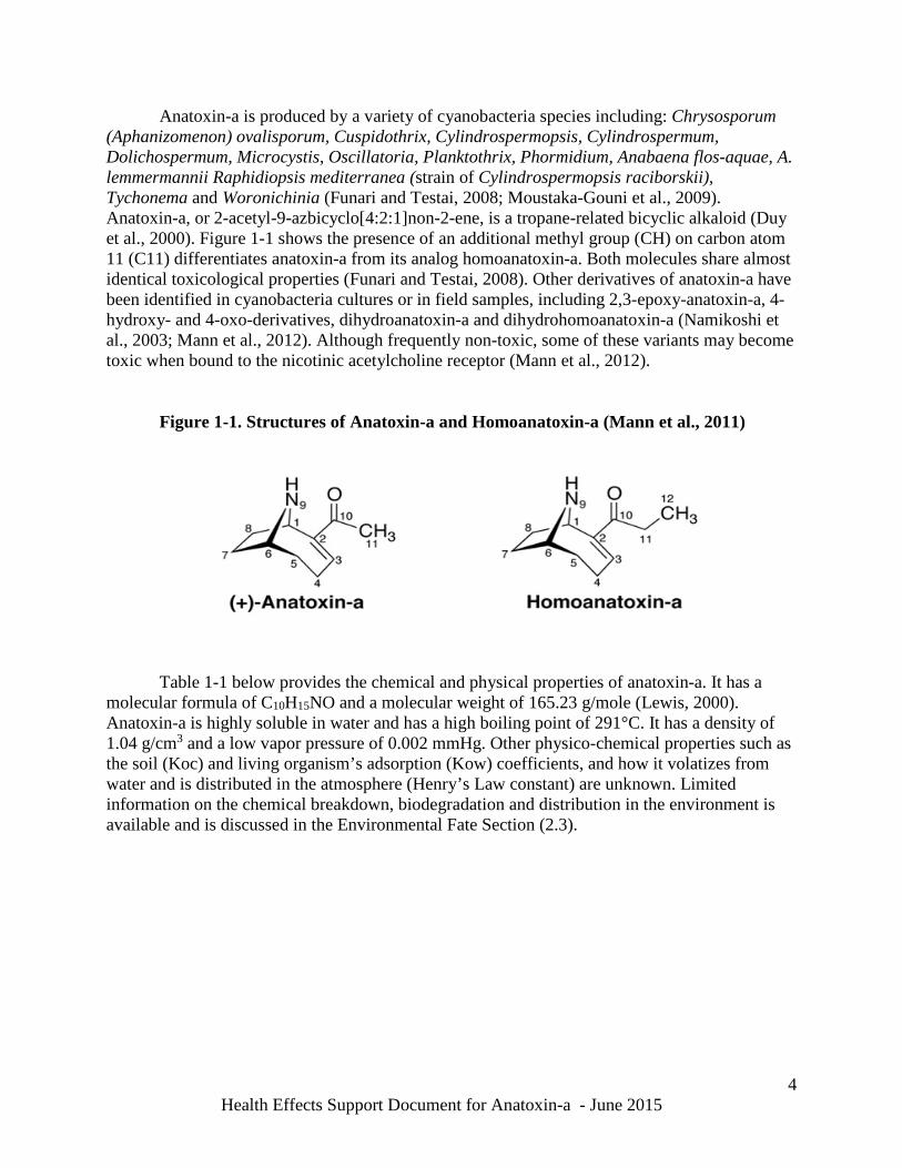
Anatoxin-a is produced by a variety of cyanobacteria species including: Chrysosporum (Aphanizomenon) ovalisporum, Cuspidothrix, Cylindrospermopsis, Cylindrospermum, Dolichospermum, Microcystis, Oscillatoria, Planktothrix, Phormidium, Anabaena flos-aquae, A. lemmermannii Raphidiopsis mediterranea (strain of Cylindrospermopsis raciborskii), Tychonema and Woronichinia (Funari and Testai, 2008; Moustaka-Gouni et al., 2009). Anatoxin-a, or 2-acetyl-9-azbicyclo[4:2:1]non-2-ene, is a tropane-related bicyclic alkaloid (Duy et al., 2000). Figure 1-1 shows the presence of an additional methyl group (CH) on carbon atom 11 (C11) differentiates anatoxin-a from its analog homoanatoxin-a. Both molecules share almost identical toxicological properties (Funari and Testai, 2008). Other derivatives of anatoxin-a have been identified in cyanobacteria cultures or in field samples, including 2,3-epoxy-anatoxin-a, 4-hydroxy- and 4-oxo-derivatives, dihydroanatoxin-a and dihydrohomoanatoxin-a (Namikoshi et al., 2003; Mann et al., 2012). Although frequently non-toxic, some of these variants may become toxic when bound to the nicotinic acetylcholine receptor (Mann et al., 2012).
Figure 1-1. Structures of Anatoxin-a and Homoanatoxin-a (Mann et al., 2011)
Table 1-1 below provides the chemical and physical properties of anatoxin-a. It has a molecular formula of C10H15NO and a molecular weight of 165.23 g/mole (Lewis, 2000). Anatoxin-a is highly soluble in water and has a high boiling point of 291°C. It has a density of 1.04 g/cm3 and a low vapor pressure of 0.002 mmHg. Other physico-chemical properties such as the soil (Koc) and living organism’s adsorption (Kow) coefficients, and how it volatizes from water and is distributed in the atmosphere (Henry’s Law constant) are unknown. Limited information on the chemical breakdown, biodegradation and distribution in the environment is available and is discussed in the Environmental Fate Section (2.3).
4 Health Effects Support Document for Anatoxin-a - June 2015
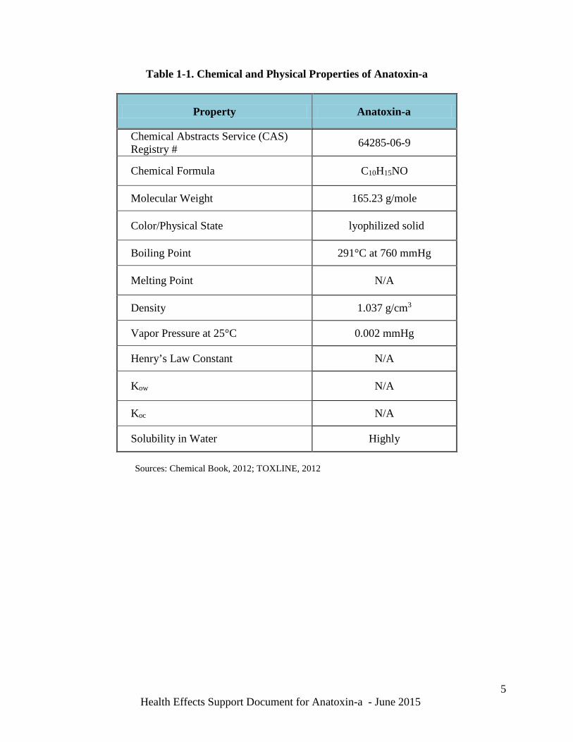
Table 1-1. Chemical and Physical Properties of Anatoxin-a
Property Anatoxin-a
Chemical Abstracts Service (CAS) Registry # 64285-06-9
Chemical Formula C10H15NO
Molecular Weight 165.23 g/mole
Color/Physical State lyophilized solid
Boiling Point 291°C at 760 mmHg
Melting Point N/A
Density 1.037 g/cm3
Vapor Pressure at 25°C 0.002 mmHg
Henry’s Law Constant N/A
Kow N/A
Koc N/A
Solubility in Water Highly
Sources: Chemical Book, 2012; TOXLINE, 2012
5 Health Effects Support Document for Anatoxin-a - June 2015
2.0 TOXIN SYNTHESIS AND ENVIRONMENTAL FATE 2.1 Cyanotoxin Synthesis
Toxin production varies between blooms and within an individual bloom over time (Duy
et al., 2000). Cyanotoxins can be produced by more than one species of cyanobacteria and some species may produce more than one toxin at a time, resulting in blooms with different cyanotoxins (Funari and Testai, 2008). The toxicity of a particular bloom is complex, determined by the mixture of species and the variation of strains with toxic and nontoxic genotypes involved (WHO, 1999). Generally, toxins in cyanobacteria are retained within the cell unless conditions favor cell wall lysis (ILS, 2000).
Mann et al. (2012) identified the ana genes as the gene cluster responsible for the biosynthesis of anatoxin-a and homoanatoxin-a. These analogs are formed by methylation (corresponding to the C12 methyl group) of an intermediate tethered to the polyketide synthase AnaG. However, the mechanism responsible for the difference in ratio of the concentration of anatoxin-a over homoanatoxin-a has not yet been described. 2.2 Environmental Factors that Affect the Fate of Cyanotoxins
Cyanotoxin concentrations depend on the dominance and diversity of strains within the bloom along with environmental and ecosystem influences on bloom dynamics as shown in Figure 2-1 below (Hitzfeld et al., 2000; WHO, 1999). Cyanotoxin production is strongly influenced by the environmental conditions that promote growth of particular cyanobacterial species and strains. Nutrient concentrations, light intensity, temperature, and other environmental factors affect growth and the population dynamics of cyanobacteria production, as described below. Although environmental conditions affect the formation of blooms, the number of cyanobacteria and the concentration of toxins produced are not always closely related.
2.2.1 Nutrients
Nutrient concentrations are key environmental drivers that influence the proportion of
cyanobacteria in the phytoplankton community, the cyanobacterial biovolume, toxin production, and the impact that cyanobacteria may have on ecosystem function and water quality. Cyanobacteria production and toxin concentrations are dependent on nutrient levels (Wang et al., 2002); however, different cyanobacteria species use organic and inorganic nutrient forms differently. Loading of nitrogen (N) and/or phosphorus (P) to water bodies from agricultural, industrial and urban sources influence the development of cyanobacterial blooms and may be related to cyanotoxin production (Paerl et al., 2011).
Smith (1983) first described a strong relationship between the relative amounts of N and
P in surface waters and cyanobacterial blooms. Smith proposed that cyanobacteria should be superior competitors under conditions of N-limitation because of their unique capacity for N-fixation. While the dominance of N-fixing cyanobacteria at low N:P ratios has been demonstrated in mesocosm- and ecosystem-scale experiments in prairie and boreal lakes (Schindler et al., 2008, and references therein), the hypothesis has been debated and challenged
6 Health Effects Support Document for Anatoxin-a - June 2015
for its inability to reliably predict cyanobacterial dominance (Downing et al., 2001). Eutrophic systems already subject to bloom events are prone to further expansion of these blooms due to additional N inputs, especially if these nutrients are available from internal sources. Recent surveys of cyanobacterial and algal productivity in response to nutrient pollution across geographically diverse eutrophic lakes, reservoirs, estuarine and coastal waters, and in different experimental enclosures of varying sizes demonstrate that greater stimulation is routinely observed in response to both N and P additions. Further, this evidence suggests that nutrient colimitation is widespread (Elser et al., 2007; Lewis et al., 2011; Paerl et al., 2011). These results strongly suggest that reductions in both N and P inputs are needed to stem eutrophication and cyanobacterial bloom expansion.
2.2.2 Light Intensity
Sunlight availability and turbidity have a strong influence on the cyanobacteria species
that predominate, as well as the depth at which they occur (Falconer et al., 2005; Carey et al., 2012). For example, Cylindrospermopsis forms dense layers of filaments at the lower bound of the euphotic zone in deeper rivers, lakes and reservoirs. The relationship of light intensity to toxin production in blooms is somewhat unclear and continues to be investigated (Duy et al., 2000). Some scientists have found evidence that toxin production increases with high light intensity (Watanabe and Oishi, 1985), while others have found little variation in toxicity at different levels of light intensity (Codd and Poon, 1988; Codd, 1995). Deep water mixing and low light also have been associated with an increase in dominance of C. raciborskii, a toxin-producing species (O’Brien et al., 2009).
Recently, Kosten et al. (2011) reported results from a survey of 143 lakes along a
latitudinal transect (between 5-55°S and 38-68°N) ranging from subarctic Europe to southern South America. They found that the percentage, or biovolume, of the total phytoplankton attributable to cyanobacteria was greater in lakes with high rates of light absorption. Kosten et al. (2011) could not establish cause and effect from these field data; however, other controlled experiments and field data support the importance of light availability on the competitive balance among a large group of shade-tolerant cyanobacteria species, mainly Oscillatoriales and other phytoplankton species (Smith, 1986; Scheffer et al., 1997). Results from Kosten et al. (2011) also suggest that higher temperatures can interact with nutrient loading and underwater light conditions to determine the proportion of cyanobacteria in the phytoplankton community in shallow lakes.
2.2.3 Temperature
The increasing body of laboratory and field data (Weyhenmeyer, 2001; Huisman et al.,
2005; Reynolds, 2006; De Senerpont Domis et al., 2007; Jeppesen et al., 2009; Wagner and Adrian, 2009; Kosten et al., 2011; Carey et al., 2012) suggest that an increase in temperature may influence cyanobacterial dominance in the phytoplankton community. Kosten et al. (2011) demonstrated that during the summer, the percentage of the total phytoplankton biovolume attributable to cyanobacteria increased steeply with temperature in shallow lakes sampled along a latitudinal transect ranging from subarctic Europe to southern South America.
7 Health Effects Support Document for Anatoxin-a - June 2015
The relationship between temperature and cyanobacterial dominance may be explained in part by the competitive advantage of cyanobacteria under higher temperatures. Warmer temperatures favor surface bloom-forming cyanobacteria genera because they are heat-adapted and their maximal growth rates occur at relatively high temperatures, often in excess of 25°C (Robarts and Zohary 1987; Reynolds, 2006). At these elevated temperatures, cyanobacteria routinely out-compete eukaryotic algae (Elliott, 2010; Paerl et al., 2011). Specifically, as the growth rates of the eukaryotic taxa decline in response to warming, cyanobacterial growth rates reach their optima.
Another possible factor favoring cyanobacteria with higher temperatures is based on a set of temperature-induced mechanisms that alter underwater light levels favorably for cyanobacteria (Kosten et al., 2011; Carey et al., 2012).
Indirectly, warming within the water column may increase nutrient concentrations by enhancing the rate of mineralization (Gudasz et al., 2010; Kosten et al., 2009, 2010) and by temperature or anoxia-mediated sediment phosphorus release (Jensen and Andersen, 1992; Søndergaard et al., 2003). Thus, temperature may increase cyanobacteria biomass indirectly through its effect on nutrient concentrations. Others have suggested that warmer conditions may raise total phytoplankton biomass through an alteration of top-down regulation by grazers (Jeppesen et al., 2009, 2010; Teixeira-de Mello et al., 2009).
Rising global temperatures and changing precipitation patterns both stimulate
cyanobacteria blooms. Warmer surface waters, especially in areas of reduced precipitation, are prone to intense vertical stratification. The degree of vertical stratification depends on the density difference between the warm surface layer and the underlying cold water. The density difference also is influenced by the relative amount of precipitation. As temperatures rise due to climate change, waters are expected to stratify earlier in the spring and the stratification will persist longer into the fall (Paerl and Otten, 2013b). The increase in water column stability associated with higher temperatures also may favor cyanobacteria (Wagner and Adrian, 2009; Carey et al., 2012).
2.2.4 Other Environmental Factors
Cyanobacterial blooms have been shown to intensify and persist at pH levels between six
and nine (WHO, 2003). When blooms are massive or persist for a prolonged period they can become harmful. Kosten et al. (2011) noted the impact of pH on cyanobacteria abundance in lakes along a latitudinal transect from Europe to southern South America. The percentage of cyanobacteria in the 143 shallow lakes sampled was well correlated with pH, with an increased proportion of cyanobacteria at higher pH.
Cyanobacteria have a competitive advantage over other phytoplankton species because
they are efficient users of carbon dioxide (Shapiro, 1984; Caraco and Miller, 1998). This characteristic is especially advantageous for cyanobacteria under conditions of higher pH when the concentration of carbon dioxide in the water column is diminished due to photosynthetic activity. Although this could explain the positive correlation observed between pH and the proportion of cyanobacteria, the high proportion of cyanobacteria at high pH could be the result of an indirect nutrient effect as described previously (see discussion in Temperature Section
8 Health Effects Support Document for Anatoxin-a - June 2015
2.2.3). As photosynthesis intensifies, pH increases due to carbon dioxide uptake from the water. Thus, higher water column pH may be correlated with a higher proportion of cyanobacteria because of higher photosynthetic rates, which can be linked with high nutrient concentrations (Duy et al., 2000) that stimulate phytoplankton growth and bloom formation.
Most phytoplankton-cyanobacteria blooms occur in late summer and early fall when
deeper lakes or reservoirs are vertically stratified and phytoplankton species may be stratified as well. Vertical phytoplankton biomass structure and cyanotoxin production can be influenced by seasonal changes as well as severe weather conditions (e.g., strong wind or rainfall), and also by runoff. At times, the hypolimnion (bottom layer of the water column) can have a higher phytoplankton-cyanobacteria biomass and display different population dynamics than the epilimnion (upper layer of the water column). Conversely, seasonal effects of increasing temperatures and changes in wind patterns may favorably influence the upper water column cyanobacterial community. This vertical variability is common and attributed to four causes, each of which may occur at different times, including: (a) sinking of dead/dying cells; (b) density stratification of the water column, especially nutrient concentrations and light, which affects all aspects of cyanobacteria growth; (c) increased nutrient supply from organic-rich bottom sediment (even when the water body is not density-stratified), encouraging cyanobacteria growth at or near the bottom sediment; and (d) species-specific factors (Drake et al., 2010). In addition, there are microbial interactions that may occur within blooms, such as competition and adaptation between toxic and nontoxic cyanobacterial strains, as well as impacts from viruses. Each of these factors can cause fluctuations in bloom development and composition. When the composition of the cyanobacteria bloom changes, so do the toxins present and their concentrations (Honjo et al., 2006; Paerl and Otten, 2013b). The concentration of cyanotoxins observed in a water body when a bloom collapses, such as from cell aging or algaecide treatment, depends on dilution of the toxin due to water column mixing, the degree of adsorption to sediment or particulates and the rate of toxin biodegradation (Funari and Testai, 2008).
In summary, there is a complex interplay of environmental factors that dictates the spatial
and temporal changes in the concentration of cyanobacteria cells and their toxins with respect to the dominant species as illustrated in Figure 2-1 (Paerl and Otten, 2013b). Factors such as the N:P ratio, organic matter availability, temperature, and light attenuation, as well as other physico-chemical processes, can play a role in determining harmful algal bloom (HAB) composition and toxin production (Paerl and Huisman, 2008; Paerl and Otten, 2013b). Dynamics of microflora competition as blooms develop and collapse can also impact cyanotoxin concentrations in surface waters. In addition, impacts of climate change, including potential warming of surface waters and changes in precipitation, could result in changes in ecosystem dynamics that lead to more frequent formation of cyanobacteria blooms and their associated toxins (Paerl and Huisman, 2008; Paerl et al., 2011; Paerl and Otten, 2013b).
9 Health Effects Support Document for Anatoxin-a - June 2015
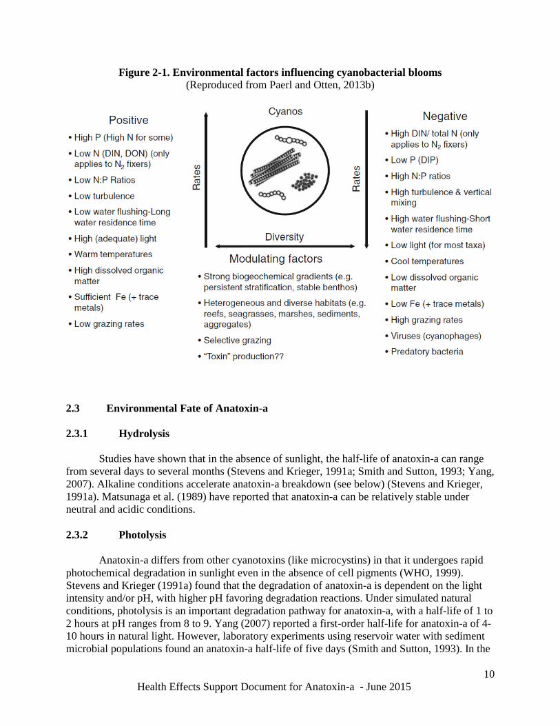
Figure 2-1. Environmental factors influencing cyanobacterial blooms (Reproduced from Paerl and Otten, 2013b)
2.3 Environmental Fate of Anatoxin-a 2.3.1 Hydrolysis
Studies have shown that in the absence of sunlight, the half-life of anatoxin-a can range from several days to several months (Stevens and Krieger, 1991a; Smith and Sutton, 1993; Yang, 2007). Alkaline conditions accelerate anatoxin-a breakdown (see below) (Stevens and Krieger, 1991a). Matsunaga et al. (1989) have reported that anatoxin-a can be relatively stable under neutral and acidic conditions.
2.3.2 Photolysis
Anatoxin-a differs from other cyanotoxins (like microcystins) in that it undergoes rapid photochemical degradation in sunlight even in the absence of cell pigments (WHO, 1999). Stevens and Krieger (1991a) found that the degradation of anatoxin-a is dependent on the light intensity and/or pH, with higher pH favoring degradation reactions. Under simulated natural conditions, photolysis is an important degradation pathway for anatoxin-a, with a half-life of 1 to 2 hours at pH ranges from 8 to 9. Yang (2007) reported a first-order half-life for anatoxin-a of 4-10 hours in natural light. However, laboratory experiments using reservoir water with sediment microbial populations found an anatoxin-a half-life of five days (Smith and Sutton, 1993). In the
10 Health Effects Support Document for Anatoxin-a - June 2015
same study, the authors found anatoxin-a for at least 21 days at pH 4, and detectable levels after 14 days at pH 8 and 10.
2.3.3 Metabolism Anatoxin-a can be readily degraded by bacteria associated with cyanobacterial filaments;
however, there is less information available for anatoxin-a than for other cyanotoxins (i.e. microcystins). 2.3.4 Transport
Anatoxin-a is weakly sorbed to sandy sediments. The strongest sorption is to clay-rich
and organic-rich sediment. Researchers found that sorption follows a non-linear Langmuir model, such that it is linear at lower concentrations with sorption decreasing at higher concentrations. Organic matter promotes sorption of the anatoxin-a molecule due to the availability of negatively charged sites (Klitzke et al., 2011).
2.4 Summary
Anatoxin-a is produced by a variety of cyanobacteria. Factors such as nutrient levels, pH, light intensity and temperature influence the growth of these cyanobacteria and could encourage toxin production. The half-life of anatoxin-a in the absence of sunlight ranges from several days to several months. However, in sunlight anatoxin-a undergoes rapid photochemical degradation even in the absence of cell pigments. Degradation is dependent on pH, with higher pH favoring more rapid degradation reactions. The half-life of anatoxin-a in sunlight is 1 to 2 hours at a pH of 8 to 9. Anatoxin-a can be degraded by bacteria associated with cyanobacterial filaments. It is weakly sorbed to sandy sediment, but has strong sorption to clay- and organic-rich sediment. Organic matter promotes sorption of the anatoxin-a molecule due to the availability of negatively charged sites.
11 Health Effects Support Document for Anatoxin-a - June 2015
3.0 CYANOTOXIN OCCURRENCE AND EXPOSURE IN WATER
The presence of detectable concentrations of cyanotoxins in the environment is closely associated with blooms of cyanobacteria. Cyanobacteria flourish in various natural environments including salty, brackish or fresh water, cold and hot springs, and in environments where no other microalgae can exist, including desert sand, volcanic ash and rocks (Jaag, 1945; Dor and Danin, 1996). Cyanobacteria also form symbiotic associations with aquatic animals and plants, and cyanotoxins are known to bioaccumulate in common aquatic vertebrates and invertebrates (Ettoumi et al. 2011).
Currently, there is no national database recording freshwater harmful algal bloom (HAB) events. Instead, state and local governments document HAB occurrences in various ways depending on the monitoring methods used and the availability of laboratories capable of conducting algal toxin analyses.
Human exposure to cyanotoxins, including anatoxin-a, may occur by direct ingestion of
toxin-contaminated water or food, and by inhalation and dermal contact during bathing, showering or during recreational activities in water bodies contaminated with the toxins. Anatoxin-a can be dissolved in drinking water either by the breakdown of a cyanobacterial bloom or by cell lysis. Exposure through drinking water can occur if there are toxins in the water source and the existing water treatment technologies were not designed for removal of cyanotoxins. Because children consume more water per unit body weight than do adults, children potentially may receive a higher dose than adults. Exposures are usually not chronic; however, they can be repeated in regions where cyanobacterial blooms are more extensive or persistent. As described above, anatoxin-a is not highly persistent; thus exposure to anatoxin-a from ambient surface waters is more likely to be acute or subacute. People, particularly children, recreating close to lakes and beach shores also can be at potential risk from exposure to nearshore blooms.
Livestock and pets potentially can be exposed to higher concentrations of cyanobacterial
toxins than humans because they are more likely to consume scum and mats while drinking cyanobacteria-contaminated water (Backer et al., 2013). Dogs are particularly at risk as they may lick cyanobacteria from their fur after swimming in a water body with an ongoing bloom.
3.1 General Occurrence of Cyanobacteria in Water Species of cyanobacteria are predominantly found in eutrophic (nutrient-rich) water
bodies in freshwater and marine environments (ILS, 2000), including salt marshes. Most marine cyanobacteria of known public health concern grow along the shore as benthic vegetation between the low- and high-tide water marks. The marine planktonic forms have a global distribution. They also can be found in hot springs (Castenholz, 1973; Mohamed, 2008), mountain streams (Kann, 1988), Arctic and Antarctic lakes (Skulberg, 1996) and in snow and ice (Laamanen, 1996). 3.2 Anatoxin-a Occurrence in Surface Water
12 Health Effects Support Document for Anatoxin-a - June 2015
Gas vacuoles of A. ovalisporum and C. raciborskii act to regulate the position of the cyanobacteria in the water column. These species of cyanobacteria do not form a floating scum, but concentrate (with densities up to 100,000 cells/mL) several meters below the surface. Because the cells remain suspended in the water column, potentially toxin-producing blooms of these cyanobacteria may not be readily observable.
Reported concentrations of anatoxin-a are limited and vary widely depending on the
water body sampled and the analytical method used. Anatoxin-a has been found in surface waters around the world including in the U.S. (Carmichael et al., 1975; Carmichael et al., 2001). Concentrations of anatoxin-a in surface freshwater cyanobacterial blooms or surface freshwater samples reported worldwide from 1985 to 1996 ranged from 0.4 to 4,400 µg/g dry-weight. Reported water-volume concentrations of extracellular and intracellular anatoxin-a ranged from 0.02 to 0.36 µg/L (WHO, 1999).
Monitoring and analysis of U.S. surface water described below has shown concentrations
of anatoxin-a ranging from below the detection limit (0.05 µg/L) to 1,929 µg/L. In 2006, the U.S. Geological Survey (USGS) conducted a targeted study of cyanotoxins
in Midwestern waters (Loftin et. al., 2008; Graham et al., 2010). Twenty-three samples were collected from lakes in the Midwest (MN, IA, MO, KS) over a 1-week period in August 2006 and were analyzed by liquid chromatography-tandem mass spectrometry (LC-MS/MS). Anatoxin-a was detected in about a third of the samples at concentrations of 0.05 to 10 µg/L.
Yang (2007) analyzed anatoxin-a using high performance liquid chromatography with
fluorescence detection (HPLC/FD) and reported periodic detections of anatoxin-a at concentrations above 0.1 μg/L in samples collected from western Lake Erie, along the southern shoreline of Lake Ontario and in Lake Champlain. Higher concentrations exceeding 1 μg/L of anatoxin-a were reported in samples collected from Onondaga Lake and Lake Agawam, smaller inland lakes in New York State (Yang, 2007).
Hedman et al. (2008) sampled surface waters in Wisconsin using LC-MS/MS and
detected anatoxin-a in 4 of 74 samples with concentrations ranging from 0.68 to 1,750 µg/L. Ohio EPA (2010) reported anatoxin-a concentrations ranging from below the detection
limit to 15 µg/L in Grand Lake St. Mary’s, Ohio using enzyme-linked immunosorbent assay (ELISA).
The Washington State Department of Ecology used LC-MS/MS to test for anatoxin-a and
other toxins in Washington’s lakes, ponds and streams from 2009 to 2011 (WSDE, 2012). In 2009, of the 32 lakes tested for anatoxin-a, 44% of lakes had detectable concentrations ranging from 0.05 to 144 µg/L. In 16% of the lakes tested, the concentration of anatoxin-a was above the recreational guidance level established by the state of 1 μg/L. In 2010, 41 lakes were tested for anatoxin-a; 24% of lakes had anatoxin-a concentrations ranging from 0.05 to 538 µg/L. In 12% of the lakes sampled, the concentration of anatoxin-a was above the recreational guidance level of 1 µg/L. In samples taken in 2011 from 46 lakes, anatoxin-a concentrations varied from 0.05 to
13 Health Effects Support Document for Anatoxin-a - June 2015
1,929 μg/L in 57% of the lakes tested. In 13% of the lakes sampled in 2011, levels were above the state recreational guideline of 1 μg/L.
Al-Sammak et al. (2014) detected anatoxin-a in samples collected from 12 reservoirs in
Nebraska between 2009 and 2010. Samples were analyzed using HPLC/FD for the preliminary analysis of all extracts, and LC-MS/MS was used for confirmation. Anatoxin-a was detected in 31 of the 67 samples at concentrations ranging from 0.05 μg/L (detection limit) to 35 μg/L. 3.3 Anatoxin-a Occurrence in Drinking Water
Data on the presence of cyanotoxins, including anatoxin-a, in drinking water and finished drinking water are scarce and generally not published. In drinking water, the occurrence of cyanotoxins depends on their level in the raw source water and the effectiveness of the drinking water treatment. Currently, there is no national regulatory program in place to monitor for the occurrence of cyanotoxins in drinking water in the U.S. In 2008, anatoxin-a was detected in three samples of finished water in Florida ranging from below the detection limit to 8.46 µg/L (Burns, 2008). Methods employed to characterized algal toxins included ELISA, protein phosphatase inhibition assay (PPIA), HPLC, and LC/MS/MS (no detection limits were reported).
3.4 Summary Anatoxin-a -producing cyanobacteria occur in freshwater systems around the world and
in the U.S. No national database recording freshwater anatoxin-a is available. The available data for the occurrence of anatoxin-a in surface waters and drinking water is by published literature and reports such as the USGS. A survey done by USGS in 2006 of 23 lakes in the Midwestern U.S., found that anatoxin-a was detected in about a third of the samples at concentrations from 0.05 to 10 µg/L. Data on the presence of anatoxin-a, in drinking water and finished drinking water are scarce and generally not published. In Washington State, samples taken in 2011 from 46 lakes had anatoxin-a concentrations from 0.05 to 1,929 μg/L. A survey conducted in 1999 in Florida, found that anatoxin-a occurred in three finished water samples ranging from below the detection limit to 8.46 µg/L
Exposure to anatoxin-a from contaminated drinking water sources may occur mostly via oral exposure (e.g. ingestion of contaminated drinking), dermal exposure (contact of exposed parts of the body with water containing toxins); and inhalation exposure. Exposure to anatoxin-a during recreational activities may be due through direct contact, inhalation and/or ingestion. Exposures are usually not chronic with the exception of regions with extensive and persistent cyanobacterial blooms. Since anatoxin-a is not highly persistent, exposure from ambient surface waters is more likely to be acute or subacute. Since children consume more water per unit body weight than do adults, children may potentially receive a higher dose. Pets, livestock and wildlife are also potentially exposed to cylindrospermopsin when consuming scum and mats, and drinking cyanobacteria-contaminated water.
14 Health Effects Support Document for Anatoxin-a - June 2015
4.0 OCCURRENCE IN MEDIA OTHER THAN WATER
4.1 Occurrence in Soil and Edible Plants
Cyanobacteria are highly adaptable and have been found to colonize infertile substrates, such as volcanic ash and desert sand (Jaag, 1945; Dor and Danin, 1996; Metcalf et al., 2012). They also have been found in soil, at the surface or several centimeters below the surface, where they play a functional role in nutrient cycling. Cyanobacteria are known to survive on rocks or tree trunks, and in snow and ice (Adhikary, 1996). They have been reported in deeper soil layers likely transported by percolating water or burrowing animals. Some freshwater species are halotolerant (salt tolerant) and have been found in saline environments such as salt works or salt marshes (WHO, 1999). Cyanobacterial cells can bioaccumulate in zooplankton (Watanabe et al., 1992). As a result of higher trophic level grazing, the damaged or residual cyanobacterial cells may settle out of the water column and accumulate in sediment where breakdown by sediment bacteria and protozoa can release their toxins (Watanabe et al., 1992).
Al-Sammak et al. (2014) detected anatoxin-a in aquatic plant samples collected from 12 reservoirs sampled in Nebraska from 2009 to 2010. Both bound and free anatoxin-a were measured in 18 of 48 plant samples analyzed. The bound anatoxin-a concentrations ranged from 1.47 to 8.01 µg/g. Concentrations of free anatoxin-a ranged from 0.26 to 0.61 µg/g. Plant detections generally co-occurred with detections in water and, in the water samples, bound anatoxin-a concentrations were generally higher than free concentrations. 4.2 Occurrence in Fish and Shellfish
Cyanotoxins can bioaccumulate in common aquatic vertebrates and invertebrates, including fish, snails (Carbis et al., 1997; Beattie et al., 1998; Berry et al., 2012) and mussels (Eriksson et al., 1989; Falconer and Yeung, 1992; Prepas et al., 1997; Watanabe et al., 1997; Funari and Testai, 2008). Bioconcentration in fish has been reported (Osswald et al., 2011) with bioconcentration factors ranging from 30 to 47 based on fresh weight. Human exposure to cyanotoxins may occur if fish are consumed from reservoirs with existing blooms of toxin-producing cyanobacteria (Magalhães et al., 2001).
The health risk from consumption depends on the bioaccumulation of toxins in edible
fish tissue. Because fish are generally more tolerant of cyanobacterial toxins than mammals, they tend to accumulate them over time (ILS, 2000). Very limited information was available regarding anatoxin-a accumulation in fish. Osswald et al. (2007) exposed juvenile common carp, Cyprinus carpio, to freeze-dried cells of Anabaena sp. at a cell density of 105 or 107 cells/mL for four days. Toxin content measured in extracts from whole fish was 0.005 and 0.073 µg/g fresh weight, respectively. In a study by Al-Sammak et al. (2014), anatoxin-a was not detected in any fish samples collected from 248 fish, including bottom feeding fish such as carp and catfish, from 12 Nebraska lakes.
15 Health Effects Support Document for Anatoxin-a - June 2015
4.3 Occurrence in Dietary Supplements
Extracts from Arthrospira (Spirulina spp.) and Aphanizomenon flos-aquae (AFA) have been used as dietary bluegreen algae supplements (BGAS) (Funari and Testai, 2008). These supplements are reported to have beneficial health effects including supporting weight loss, and increasing alertness, energy and mood elevation for people suffering from depression (Jensen et al., 2001). In children, they have been used as an alternative, natural therapy to treat attention deficit hyperactivity disorder (ADHD). Heussner et al. (2012) did not detect anatoxin-a in 18 commercially available BGAS analyzed for the presence of toxins. However, Rellán et al. (2009) reported that three of 39 samples (7.7%) of BGAS contained anatoxin-a at concentrations ranging from 2.50 to 33 µg/g. 4.4 Summary
Anatoxin-a could be detected in aquatic animals and edible plants. Very limited
information was available on anatoxin-a accumulation in fish. No cases of toxicity in humans following ingestion of fish or shellfish exposed to cyanotoxins have been documented.
Anatoxin-a have been found in algal supplements ranging from 2.5 to 33 µg/g. Exposure
for the general population is mostly through the ingestion of drinking water and incidental ingestion when recreating in a contaminated water source.
16 Health Effects Support Document for Anatoxin-a - June 2015
5.0 TOXICOKINETICS Data on the toxicokinetics of anatoxin-a are negligible. Absorption and distribution are demonstrated only by the rapid appearance of neurotoxicity and the systemic effects observed after exposures in repeat dose studies. 5.1 Absorption
No information regarding the absorption of anatoxin-a in humans or animals was identified. However, acute oral toxicity studies in animals demonstrate that it can be absorbed rapidly by the gastrointestinal tract. Symptoms of clinical neurotoxicity such as muscular twitching, loss of coordination and death from respiratory paralysis occur within minutes of exposures (Stevens and Krieger, 1991a; Fitzgeorge et al., 1994). 5.2 Distribution
The rapid appearance of symptoms following exposure is consistent with rapid uptake from the gastrointestinal tract and serum distribution to the liver, brain and central nervous system. In a study by Fitzgeorge et al. (1994), deaths in mice occurred by respiratory paralysis within 2 minutes of gavage administration of doses greater than 5 mg/kg. In bioassay studies, Stevens and Krieger (1991a) found that lethal doses (concentrations not reported) manifested the same signs of respiratory paralysis as control solutions of anatoxin-a, and that the breakdown products of anatoxin-a are less toxic than the parent compound. 5.3 Metabolism
No information on the metabolism of anatoxin-a was identified. 5.4 Excretion
No information regarding the excretion of anatoxin-a was identified. 5.5 Pharmacokinetic Considerations No data on half-life or other quantitative pharmacokinetic parameters for anatoxin-a were identified. The interactions with the nicotinergic acetylcholine receptor are known to be enanteromerically specific [(+) isomer only]. The (-) isomer also has toxic properties based on lethality studies. However, the (-) isomer lacks the direct neurotoxicity of the (+) isomer.
17 Health Effects Support Document for Anatoxin-a - June 2015
6.0 HAZARD IDENTIFICATION 6.1 Case Reports and Epidemiology Studies
Information on epidemiology studies or confirmed case reports of human poisoning from exposure to anatoxin-a are not available.
Non-lethal human poisonings, usually manifested as acute gastrointestinal disorders such
as nausea, vomiting and diarrhea, have been related to ingesting water with unspecified species of Microcystis and Anabaena (producers of anatoxin-a) as later detected in the victims’ feces (Schwimmer and Schwimmer, 1968). Allergic reactions (e.g., skin papulo-vesicular eruptions) have been related to swimming in water with a bloom of Anabaena (Schwimmer and Schwimmer, 1968). However, anatoxin-a detections were not reported.
Anatoxin-a has been associated with poisonings and deaths of livestock, dogs and ducks
after exposure to water contaminated with cyanotoxins (Carmichael and Gorham, 1978; Edwards et al., 1992; Gunn et al., 1992; Puschner et al., 2008; Stewart et al., 2008). Quantitative exposure data were not reported but clinical signs were mostly neurologic and deaths due to respiratory paralysis, characteristic adverse effects of anatoxin-a. In the U.S., 368 cases of cyanotoxin poisonings associated with dogs were identified in a review done by the Centers for Disease Control (CDC) from the 1920s to 2012 (Backer et al., 2013). A retrospective review of veterinary biopsy and necropsy case files between 1984 and 2012 found that of the 71 cases of dogs deaths, 45 (4%) were either suspected or confirmed cyanotoxins poisoning. Two dogs (3%) were confirmed with anatoxin-a poisoning. Both dogs died within 20 to 30 minutes of onset of illness after exposure to cyanobacteria in a backyard pond. Anatoxin-a was identified in the kidney by biochemical testing (Backer et al., 2013). 6.2 Animal Studies
6.2.1 Acute Toxicity 6.2.1.1 Oral Exposure
Stevens and Krieger (1991b) used a single dose gavage in adult male Swiss Webster ND-
4 mice to determine an LD50 of 16.2 mg/kg (Confidence Interval [CI] of 95%: 15.4-17.0) for synthetic (+)-anatoxin-a hydrochloride ( >98% pure commercial product), which is equivalent to 13.3 mg anatoxin-a/kg (95% CI: 12.8-14.1). When using a lysate solution of lyophilized A. flos-aquae (NRC-44-1) cells, an LD50 value of 6.7 mg/anatoxin-a kg (95% CI: 6.3-7.1) was determined. The LD50 values were determined using the method of moving averages for four doses with six animals per dose (Stevens and Krieger, 1991b). A single dose gavage study in newly weaned CBA/BalbC mice of unspecified sex determined an LD50 of >5 mg/kg for anatoxin-a; the study authors used a “suitably purified” but an unspecified form of commercial product (Fitzgeorge et al., 1994). Deaths due to neurotoxicity, expressed as muscular twitching, loss of coordination and death by respiratory paralysis, occurred within 2 minutes of administration (Fitzgeorge et al., 1994).
18 Health Effects Support Document for Anatoxin-a - June 2015
A 5-day gavage, range-finding study was conducted to determine the maximum tolerated
dose for use in a 28-day study (Section 6.2.2) (Fawell and James, 1994; Fawell et al., 1999). Doses of 1.5, 3, 7.5 or 15 mg/kg-day (equivalent to 1.2, 2.5, 6.2 or 12.3 mg anatoxin-a/kg-day) using aqueous (+)-anatoxin-a hydrochloride (commercial product, purity not reported) were administered to 2 male and 2 female Crl:CD-1(ICR)BR mice groups (no control group included). After 24 hours of administering the lower dose (1.2 mg/kg-day), the 6.2 and 12.3 mg/kg-day dosing started. After 5 days, the 2.5 mg/kg-day (intermediate level) dosing was administered. Evaluation of clinical signs, food consumption and body weight were done and the surviving animals were necropsied. During the first 4 days, all mice in the high-dose group died (within 5 minutes of dosing), and one female mouse from the 6.2 mg/kg-day group died. These deaths happened within 5 minutes of dosing. The male mice in the 6.2 mg/kg-day dose group were hyperactive following the third dose. The rest of the surviving animals in this group (6.2 mg/kg-day) did not express any abnormal clinical signs and no other signs of neurotoxicity were reported. The 6.2 mg/kg-day dose was identified as the Frank Effect Level (FEL) based on the death of one of the two female mice. The maximum tolerated dose was established as 3 mg/kg/day anatoxin-a hydrochloride (2.5 mg/kg/day anatoxin-a). 6.2.1.2 Other Exposure Routes
A single dose intraperitoneal (i.p.) study in mice identified an LD50 of 0.25 mg/kg (95%
CI: 0.24-0.28) for (+)-anatoxin-a hydrochloride (commercial product, >98% pure) equivalent to 0.21 mg anatoxin-a/kg (Stevens and Krieger, 1991b). In another i.p. study, Fitzgeorge et al., (1994) determined an LD50 of 0.375 mg/kg for commercial anatoxin-a (form and purity not reported).
Single i.p. injections of (+)-, racemic or (-)-anatoxin-a hydrochloride (all >95% pure)
were administered in male BalbC mice (Valentine et al., 1991). After observing for 30 minutes, LD50 values were determined as 386 μg/kg (95% CI: 365-408) for (+)-anatoxin-a hydrochloride equivalent to 0.32 mg anatoxin-a/kg and 913 μg/kg (95% CI: 846-985) for racemic anatoxin-a hydrochloride equivalent to 0.76 mg anatoxin-a/kg. According to the authors, this two-fold potency difference is consistent with mechanistic data indicating that (+)-anatoxin-a is the biologically active enantiomer.
An i.p. 2-day study in 18 female CD-1 mice was performed to determine a maximum
dose to evaluate neurodevelopmental toxicity (Section 6.2.3) (Rogers et al., 2005). Dosages of anatoxin-a (commercial product, >90% purity) in distilled water were 10, 100, 200, 250, 300 and 400 μg/kg (0.008, 0.08, 0.17, 0.21, 0.25 and 0.33 mg anatoxin-a/kg-day). Group sizes ranged from 1 in the 400 μg/kg dose group to 6 in the 100 μg/kg group (Personal communication). After 5 to 6 minutes of administering the higher dose, mice expressed decreased motor activity, altered gait, difficulty breathing and convulsions. Anatoxin-a was 100% lethal at the 400 μg/kg after 10 minutes. Mice receiving 100 or 200 µg/kg survived and received a second dose of racemic anatoxin-a the following day. All mice survived after the second dose. Clinical signs of toxicity after 10 minutes of administering the lower doses included decreased activity level, altered gait and breathing irregularities. At the lower doses, mice did not have convulsions and recovery was observed by 15 to 20 minutes after treatment (Rogers et al., 2005).
19 Health Effects Support Document for Anatoxin-a - June 2015
6.2.2 Short Term Studies
In the 28-day study, four groups of 10 male and 10 female mice were dosed by gavage once a day for 28 days with 0 (vehicle control), 0.12, 0.6 or 3 mg/kg-day (corresponding to 0.098, 0.49 and 2.46 mg anatoxin-a/kg-day) (Fawell and James, 1994; Fawell et al., 1999). Histological and blood analysis examinations were performed in the control and dose groups, and microscopic examinations were done to all tissues. During the study, three deaths were reported within 2.5 hours of dosing: one from each of the high dose groups (a male from the 0.49 mg anatoxin-a/kg-day group and a female from the 2.46 mg anatoxin-a/kg-day group), but no cause for these deaths was determined. The authors did not demonstrate any clear clinical signs of general toxicity, such as changes in body weight, altered food consumption or unusual necropsy findings. The third death was not related to treatment. The animal was sacrificed after showing signs of having been attacked by its cage mates.
The only adverse clinical signs observed among the survivors, although not considered
toxicologically significant, were a significant increase in mean cell hemoglobin concentration in males at >0.1 mg/kg-day and in females at >0.5 mg/kg-day, and an increase in serum sodium in females at >0.5 mg/kg-day. No significant changes were observed in serum levels for liver enzymes, albumin, BUN or sodium. The study authors determined a NOAEL (No Observed Adverse Effect Level) of 0.1 mg/kg-day (0.098 mg anatoxin-a hydrochloride/kg-day) based on the deaths in the higher dose groups (Fawell and James, 1994; Fawell et al., 1999).
6.2.3 Subchronic Studies 6.2.3.1 Oral Exposure Anatoxin-a extracted from the culture media of A. flos-aquae (NRC-44-1) cells and partially purified by high pressure liquid chromatography (HPLC) in a 30% perchloric acid/70% methanol solvent (purity not quantified) was administered in drinking water to groups of 20 female Sprague-Dawley rats (Astrachan and Archer, 1981; Astrachan et al., 1980). Doses of 0, 0.51, or 5.1 mg/kg were administered for 7 weeks with an estimated daily intake of anatoxin-a in the low dose group of 0.05 mg/kg-day and 0.5 mg/kg-day in the high dose group. Daily intake was estimated assuming that the test rats consumed 0.1 mL/g body weight per day (based on a preliminary water consumption study). The authors evaluated food consumption, body weight, red and total white blood cell counts, and serum enzyme activities throughout the study. At the end of the study, the authors evaluated hepatic mixed function oxidase activity (aldrin epoxidation in vitro), organ weights (liver, kidneys and spleen), and gross pathology and histology (liver, kidneys, spleen, adrenals, heart, lungs and brain). No clinical signs attributed to treatment were observed and a NOAEL of 0.5 mg/kg-day was identified by the authors. Graphic data were reported for the hematological effects and liver enzymes (Astrachan and Archer, 1981). There were no apparent differences in the red blood cell (RBC) counts (mm3x 10-6), alkaline phosphatase (ALP), and aspartate aminotransferase (AST). There was a dose- and duration-related increase in white cell counts (mm3 x 10-3). White cell counts for the low dose group reached normal levels by week 3, but not until week 7 for the high
20 Health Effects Support Document for Anatoxin-a - June 2015
dose group. Organ weights were similar and no gross or histological tissue abnormalities were observed. The graphic presentation of the hematology data does not support determination of statistical significance for the effects on the white cell counts. They remained about 30 to 50% higher (estimate from the figure in the report) than the controls over the first 5 weeks of the study. The high dose can be considered as a LOAEL (Lowest Observed Adverse Effect Level) for the white blood cell effects. At one week the elevation of the white cell count was approximately equivalent to that for the high dose, but at 3 weeks was comparable to controls. There are insufficient data from other studies to determine whether the white cell effects should be regarded as toxicologically adverse. 6.2.3.2 Other Exposure Routes Neurotoxicity
In a neurodevelopmental study, racemic (+/-)-anatoxin-a hydrochloride (commercial
product, ≥90% purity) was administered to groups of 8 to 11 time-pregnant CD-1 mice (Rogers et al., 2005). Doses of 0 (control), 125 or 200 μg/kg-day equivalent to 0, 0.09 or 0.15 mg anatoxin-a/kg-day on gestation days (GD) 8-12 or 13-17 were administered via i.p. injection in distilled water. After all mice gave birth, body weight and viability of the pups were determined on postnatal days (PND) 1 and 6. Immediately after treatment, toxicity in the pregnant mice was observed at 0.15 mg/kg-day expressed as decreased motor activity. PND evaluation did not find effects on pup viability (number of live pups) on PND 1 or 6 in mice treated on GD 8-12 or 13-17. No effects were observed on pup body weight on PND 1 or 6 in mice treated on GD 8-12 either. However, a statistically significant dose-related trend for reduced body weight was observed in pups treated on GD 13-17 on PND 1 (p<0.05) only. On PND 1, body weights in the pups exposed on GD 13-17 showed a trend (7.1 and 8.7% less than controls) at the two higher doses (0.09 and 0.15 mg/kg-day, respectively) but the authors’ reported differences from controls were not significant. The authors attributed the trend in reduced pup body weight to random variability in litter size (GD 13-17 controls were noticeably smaller than the treated groups; p=0.09). A difference in litter size would have an impact in both birth weight and growth on PND 1 and 6 since pups in smaller litters are larger at birth (McCarthy, 1967) and will grow more rapidly postnatally (Rogers et al., 2003). A NOAEL for the racemic mixture was identified as 0.09 mg/kg-day for the dams based on decreased post treatment motor activity and a LOAEL of 0.15 mg/kg-day (Rodgers et al., 2005).
Righting reflex, negative geotaxis and hanging grip time were evaluated only on PND 6,
12 and/or 20 in pups from dams exposed on GD 13-17 (Rogers et al., 2005). Righting reflex (measurement of the time a pup takes to turn from his back to an upright position) was tested on PND 6 and 12; negative geotaxis (time to rotate on an inclined screen facing downhill to facing up the incline) was tested on PND 6, 12 and 20; and hanging grip time (time when a pup let go after the pup grasped a bar with their front feet to hang) was tested on PND 12 and 20. The reason for testing only the pups exposed on the GD 13-17 was because this gestational interval follows the onset of neurogenesis in the mouse brain (Rice and Barone, 2000). The litters from the exposed dams were normalized to eight pups (including four male and four female pups) on PND 6, and on each test day a randomly selected male and female pup from each litter was evaluated.
21 Health Effects Support Document for Anatoxin-a - June 2015
Based on the results from the testing (righting reflex, negative geotaxis and hanging grip
time), postnatal neurotoxicity was not observed (Rogers et al., 2005). Results showed no statistically significant differences between exposed and control groups and no dose-related differences. However, a non-statistically significant (p<0.086) dose-related trend was observed for slower righting reflex in males in the righting reflex test on PND 6. A significant (p value not reported) sex-difference was observed in terms of a slower reflex in females than in males in all treatment groups on PND 6. No sex-difference or treatment differences in righting reflex were observed on PND 12.
Turning times did not decrease as expected from PND 6 to 20 in the negative geotaxis
test (Rogers et al., 2005). In addition, control and treated pups fell off the screen before turning. Data from those mice that stayed on the inclined screen showed no significant differences across treatment groups in both the number of fallen mice and the average turning times. Also, no treatment-related differences in hanging grip time on either test day were observed. In the hanging grip time test, the authors found that the hang time in females increased significantly from PND 12 to 20, but males did not show an expected increase in hanging grip time. The investigators indicated that random variability in the tested population may be the reason for the sex-difference (Rogers et al., 2005).
To evaluate the effect of prenatal exposure to anatoxin-a on the motor activity of adult
mice and their responses to nicotine challenge, mouse pups already exposed to 0, 0.09 or 0.15 mg anatoxin-a/kg-day on GD 8-12 or 13-17 in the Rogers et al. (2005) study were tested as adults by MacPhail et al. (2005). Motor activity was measured on approximately 8-month-old offspring during 30-minute sessions using a photocell device. Doses of 0, 0.1, 0.3, 1.0 or 3.0 mg/kg nicotine in saline were administered subcutaneously to groups of 12 male and 12 female mice approximately 5 minutes before testing motor activity. These mice were assigned to the nicotine dose groups regardless of the gestational period during which they received anatoxin-a. A dose-related decrease was observed in both horizontal and vertical activity. In both sexes, 0.65 mg/kg nicotine was identified as the effective dose in 50% (ED50) of the animals.
Adult offspring from mice exposed to the racemic anatoxin-a on GD 13-17 were given
nicotine at the ED50 or saline vehicle about 5 minutes before testing the motor activity (MacPhail et al., 2005). Both treatments (nicotine ED50 and saline vehicle) were separated by 1 week. The group sizes were composed of 10 mice per gender for each dose with the exception of the high-dose anatoxin-a female group, which consisted of 9 mice. Although no quantitative data were provided, graphical displays show no differences in horizontal or vertical motor activity between the anatoxin-a-exposed mice and the controls. No dose-response for either male or female mice to the nicotine challenge was observed (MacPhail et al., 2005).
The Irwin Screen was used to evaluate neurobehavioral effects of anatoxin-a in mice
(Fawell and James, 1994; Fawell et al., 1999). The Irwin Screen is a systematic observational procedure used to assess Central Nervous System (CNS) effects such as motor activity, sensory/motor reflex responses, coordination and behavioral changes. Intravenous injections of (+)-anatoxin-a hydrochloride (commercial product, purity not reported) were administered in doses of 10, 30 or 100 μg/kg (equivalent to 8, 25 or 82 μg anatoxin-a/kg) to 6 mice with a
22 Health Effects Support Document for Anatoxin-a - June 2015
positive control to evaluate cholinergic effects of 300 µg/kg of nicotine. After dosing, the mice were observed at 15 and 30 minutes, and 1, 2 and 4 hours (Fawell and James, 1994; Fawell et al., 1999). Within 1 minute of dosing, all the mice (6 in total) in the high dose group (82 μg anatoxin-a/kg) died exhibiting symptoms of cholinergic stimulation and CNS effects such as increased respiration, salivation, micturition (urination), hyperactivity and Straub tail. Two animals in the 25 μg anatoxin-a/kg dose group died and those that survived showed increased salivation, respiration and hyperactivity. No effects were observed in the low dose group (8 μg/kg anatoxin-a/kg).
A rota-rod test to evaluate sensorimotor coordination based on the ability of the animal to
remain on a rotating rod also was performed on CD-1 strain mice (Fawell and James, 1994; Fawell et al., 1999). Seven groups of male mice were dosed with 0, 30, 50 or 60 mg/kg anatoxin-a as the hydrochloride salt (equivalent to 25, 41 or 49 mg/kg anatoxin-a), and 300, 500 or 5,000 mg/kg of nicotine (standard) and with phosphate buffered saline (vehicle control). Fifteen minutes after dosing, mice were placed onto the rota-rod for a 3-minute period and the authors recorded the time taken to fall off. Within 1 minute of dosing, only the highest doses caused clinical signs of neurotoxicity and death. Death was observed in 3 of 3, 2 of 6 and 1 of 6 of the mice dosed with 49, 41 and 25 mg/kg anatoxin-a, respectively, and in 4 of 5 animals dosed with the highest nicotine dose (5,000 mg/kg). Prior to death, mice showed symptoms of CNS effects and cholinergic stimulation. No exposure-related effects were observed in the rest of the dosed animals with the exception of 2 of 6 animals dosed with 500 mg/kg of nicotine that showed elevated respiration rates for 1 minute (Fawell and James, 1994; Fawell et al., 1999).
The effects of anatoxin-a on operant performance was evaluated in adult male Long
Evans rats (MacPhail et al., 2007). Groups of 8 rats trained to respond under a multiple variable-ratio variable-interval schedule of food reinforcement were given (+)-anatoxin-a fumarate in subcutaneous injections at 0 (control), 0.05, 0.075, 0.1, 0.15 or 0.2 mg/kg doses equivalent to 0, 0.03, 0.045, 0.06, 0.09 and 0.12 mg anatoxin-a/kg, respectively) weekly for four weeks. Dose-related decreases in performance were observed at 0.06, 0.09 and 0.12 mg/kg doses. At the two highest doses, mild tremors were observed. Over subsequent weeks, diminished effects were observed indicative of tolerance development. In an operant conditioning procedure, those rats that were administered a single dose of 0.1 mg/kg (+)-anatoxin-a fumarate (0.06 mg anatoxin-a/kg) showed decreased locomotor activity and a partial nicotine-like discriminative stimulus effect in animals trained to discriminate nicotine from saline (MacPhail et al., 2007). Anatoxin-a also decreased the response and reinforcement rates in rats in multiple-schedule operant performance tests (Jarema and MacPhail, 2003). However, upon repeated administration, substantial tolerance was developed.
Reproductive/Developmental Toxicity
Fawell et al. (1999) reported the results of a developmental toxicity screening study of
anatoxin-a in timed-pregnant female CD-1 mice. Gavage dosing occurred with either vehicle or 2.5 mg/kg/day anatoxin-a as the hydrochloride salt on GD 6-15. Maternal body weights and clinical signs were recorded. On GD 18, the mice were sacrificed. Live and dead fetuses were counted, weighed, sexed and observed for external abnormalities. The raw data were not provided. The authors reported a lack of maternal toxicity. There was no effect of treatment on
23 Health Effects Support Document for Anatoxin-a - June 2015
mean fetal weight, sex ratio or post implantation losses. There were also no treatment-related major fetal abnormalities.
In a reproductive study, male mice were administered doses of 0 (control), 0.05, 0.1 and
0.15 mg/kg-day anatoxin-a for seven consecutive days (Yavasoglu et al., 2008). Commercially available (+/-)-anatoxin-a fumarate diluted in physiological saline (0.9%; controls) was administered by the i.p. route to 10 males in each treatment group. Although there were no significant changes in body weight gain or with absolute and relative testes weights, a statistically significant (p<0.01) reduction in absolute and relative weights of cauda epididymis was observed in the 0.1 and 0.15 mg/kg treatment groups. A statistically significant (p<0.01) dose-dependent reduction in sperm count in the cauda epididymis was observed in all treatment groups compared to control. Histopathological examination of the testes revealed dose-dependent degeneration in seminiferous tubules, sloughing of germ cells into tubular lumen, vacuolization in Sertoli cells, intercellular disassociation of spermatogenetic cell lines and loss of germ cells. Epithelial thickness of seminiferous tubules decreased significantly in all treatment groups in a dose-dependent manner. The LOAEL was identified as 50 µg/kg based on reduced sperm count in cauda epididymis (Yavasoglu et al., 2008).
In a developmental toxicity screening study, groups of 10 and 12 pregnant Crl:CD-
1(ICR) BR mice were administered by gavage doses 0 (vehicle control) or 3 mg/kg-day of aqueous (+)-anatoxin-a hydrochloride (commercial product, purity not reported) equivalent to 0 or 2.5 mg anatoxin-a/kg, respectively, on GD 6-15 (Fawell and James, 1994; Fawell et al., 1999). Until GD 18, clinical signs and body weights were recorded, and at that time maternal animals were sacrificed and necropsied to assess the numbers of implantations and live fetuses, post implantation loss and fetal body weight, sex ratio and external abnormalities. No treatment-related maternal effects or effects in the fetuses were observed. However, mean fetal weight in the treated group was marginally lower than in controls (data not reported). Based on the absence of adverse effects in dams and fetuses, the NOAEL for maternal and developmental toxicity was 2.5 mg/kg-day of anatoxin-a.
A mammalian embryo toxicity test was conducted by Rodgers et al. (2005) using CD-1
mouse embryos collected on GD 8. Cultured embryos (9-13) were exposed to 0.00002, 0.0002, 0.002 and 0.0051 mg/ml anatoxin-a (racemic and 90% pure) and evaluated for dysmorphogenesis (abnormal tissue formation). At the end of the culture period, none of the embryos showed a significant dose-related increase in dysmorphogenesis. Embryos exposed to 0.002 and 0.0051 mg/ml of anatoxin-a showed a perturbation in yolk sac vasculature, such as a decrease in large caliber vessels and a reduction in arborization (the branching structure at the end of a nerve fiber) (Rodgers et al., 2005).
Rodgers et al. (2005) conducted an amphibian embryo-larval toxicity test (AMPHITOX)
using toad embryos from Bufo arenarum beginning at Stage 18 or Stage 25. Groups of 10 embryos (in duplicate) were placed in 5 cm glass petri dishes with 10 mL of the AMPHITOX solution at 20°C for 13 days to monitor for viability and functional impairments. Stage 18 embryos were exposed to 0.03, 0.3, 3.0 or 30 mg/L anatoxin-a and Stage 25 embryos were exposed to 30 mg/L anatoxin-a, both for 10 days. Results of anatoxin-a exposure in more than 70% of the embryos affected at the high dose in both exposure periods indicate induction of a
24 Health Effects Support Document for Anatoxin-a - June 2015
dose-dependent transient narcosis. Edema and loss of equilibrium also were observed and mortality occurred in both embryonic stages. In Stage 18, mortality was observed with 20% of the exposed embryos in the highest dose group on day 8 and reached 100% between days 10 to 13. Mortality also occurred in the 0.3 and 3.0 mg/L dose groups over the same time period (10 and 13 days). In Stage 25, mortality was observed starting at day 6 of exposure and reached 100% by day 9. 6.2.4 Chronic Toxicity
No chronic toxicity studies of oral exposure to anatoxin-a were identified.
6.3 Carcinogenicity
Information on carcinogenicity in humans or animals or potential mode(s) of action for anatoxin-a is not available. 6.4 Other Key Data 6.4.1 Mutagenicity and Genotoxicity
There is limited information regarding mutagenicity or genotoxicity of anatoxin-a. Preliminary findings by Sieroslawska and Rymuszka (2010) indicate that without metabolic activation, anatoxin-a was genotoxic in an umuC assay with Salmonella typhimurium TA 1535/pSK1002. UmuC assays are used to detect DNA damage resulting from cell cycle arrest. Commercial (+/-)-anatoxin-a fumarate (purity not reported) was administered at 0.00025, 0.0005, 0.001 and 0.002 mg/mL doses with a 2-hour exposure time to assess induction and expression of the umuC - lacZ reporter gene. The lowest dose (0.00025 mg/mL) was the highest concentration without an effect in the absence of metabolic transformation. When an S9 fraction (the supernatant fraction with cytosol and microsomes from an organ, usually liver, homogenate by centrifuging at 9000 g for 20 minutes in a suitable medium) was added to the samples, no effects were detected (Sieroslawska and Rymuszka, 2010). 6.4.2 Immunotoxicity No information was located regarding effects of anatoxin-a on immune function in humans; however, in an in vitro study to assess the effects of anatoxin-a on the viability of leukocytes, Bownik et al. (2012) found that anatoxin-a induced apoptosis and necrosis in carp (Cyprinus carpio L.) immune cells. The viability of leukocytes was tested using a CellTiter-Glo® luminescent Viability Assay to quantify intracellular adenosine triphosphate (ATP) as a measure of cell metabolic activity. Different concentrations (0.0001, 0.001, 0.005 and 0.010 mg/mL) of anatoxin-a were added to 100 µL of cell suspension and incubated for 24 hours. The study found that intracellular ATP levels in leukocytes were slightly reduced at the highest dose (0.010 mg/L), and no damage was observed at the lower doses. The highest dose also induced necrosis in leukocytes. Although anatoxin-a had little effect on leukocyte viability, apoptotic leukocytes were observed at all dose concentrations of anatoxin-a. A concentration-dependent decrease in the proliferative ability of T and B lymphocytes also was observed at all doses.
25 Health Effects Support Document for Anatoxin-a - June 2015
Rymuszka and Sieroslawska (2010) also found apoptosis in fish immune cells after exposure to 0.001 mg/L of pure anatoxin-a. Fluorescent analysis showed more cells at the apoptotic stage than at the necrotic stage (Rymuszka and Sierslawska, 2010). 6.5 Physiological or Mechanistic Studies 6.5.1 Noncancer Effects
Data from in vitro studies have shown that (+)-anatoxin-a mimics the action of acetylcholine at neuromuscular nicotinic receptors (Carmichael et al., 1975, 1979; Biggs and Dryden, 1977; Aronstam and Witkop, 1981; Swanson et al., 1986). Anatoxin-a is significantly more potent than acetylcholine and nicotine as an agonist (initiates a physiological response at peripheral and central sites in the CNS). Anatoxin-a has become an important agent in the investigation of nicotinic acetylcholine receptors due to its resistance to enzymatic hydrolysis (by acetylcholinesterase), and because it is 100 times more selective for nicotinic acetylcholine receptors than for muscarinic acetylcholine receptors (Aronstam and Witkop, 1981). Because anatoxin-a is not degraded by cholinesterase or other known cellular enzymes, muscle cells continue to be stimulated, causing fatigue, muscular twitching and paralysis. As observed in acute lethality animal studies, severe overstimulation of respiratory muscles may result in respiratory arrest and rapid death (Carmichael et al., 1975, 1977; Devlin et al., 1977; Stevens and Krieger, 1991b).
Anatoxin-a can affect the cardiovascular system of rats by acting as a nicotinic
cholinergic agonist causing an increase in blood pressure and heart rate (Sirén and Feuerstein, 1990; Adeyemo and Sirén, 1992; Dube et al., 1996). Anatoxin-a also can affect human and rat brain neurons (Durany et al., 1999; Thomas et al., 1993; Zhang et al., 1987). Molloy et al. (1995) have demonstrated that anatoxin-a stimulates the secretory response in bovine adrenal chromaffin cells (neuroendocrine cells found in the medulla of the adrenal glands), probably through the activation of neuronal-type nicotinic receptors.
Numerous studies have indicated that anatoxin-a can elicit the release of
neurotransmitters from presynaptic neuromuscular and brain cell terminals (Rowell and Wonnacott, 1990; Gordon et al., 1992; Soliakov et al., 1995; Clarke and Reuben, 1996; Wonnacott et al., 2000). Incubation of anatoxin-a with preparations of guinea pig ileum longitudinal muscle myenteric plexus resulted in a dose-dependent release of acetylcholine (Gordon et al., 1992). In other studies, anatoxin-a stimulated the release of dopamine from rat striatal synaptosomes in a dose-dependent manner (Rowell and Wonnacott, 1990; Soliakov et al., 1995; Clarke and Reuben, 1996; Wonnacott et al., 2000), which suggests that anatoxin-a can bind to presynaptic nicotinic receptors and trigger neurotransmitter release with increased stimulation of postsynaptic receptors.
6.5.2 Cancer Effects
No long term bioassay studies on the tumorigenicity of anatoxin-a were identified.
26 Health Effects Support Document for Anatoxin-a - June 2015
6.5.3 Interactions with Other Chemicals
Studies of interactions of anatoxin-a in mixtures with other cyanotoxins and/or contaminants were not identified. 6.5.4 Structure Activity Relationship
Anatoxin-a is produced as the natural stereoisomer, (+)-anatoxin-a, a nicotinic acetylcholine receptor agonist that affects both peripheral and central sites in the nervous system (Huber, 1972; Devlin et al., 1977; Fawell et al., 1999; Viaggiu et al., 2004). Studies have established that anatoxin-a binds to acetylcholine receptors and mimics the action of acetylcholine at neuromuscular nicotinic receptors causing neurological effects (Wonnacott and Gallagher, 2006). In general, nicotinic agonists, such as anatoxin-a, form hydrogen bonds in the planar region of the receptor and have a bulky cationic group around 5.9 Å from the hydrogen bond (Beers and Reich, 1970; Chothia and Pauling, 1970; Spivak and Albuquerque, 1982).
Numerous in vitro studies (Fawell and James, 1994; Fawell et al., 1999; MacPhail et al.,
2007) of acetylcholine receptor response in rat phrenic nerve, chick biventer cervicis muscle, guinea pig ileum and in mice intravenously demonstrated that anatoxin-a was 7 to 136 times more potent than nicotine. In frog (Xenopus) oocytes, mouse M10 cells, rat hippocampal synaptosomes and fetal rat hippocampal neurons, (+)-anatoxin-a agonist potency was 3 to 50 times greater than nicotine and around 20 times greater than acetylcholine at neuronal nicotinic acetylcholine receptors (Thomas et al., 1993).
Assays of contracture potency in preparations of frog rectus abdominis muscle have
shown that natural (+)-anatoxin-a can exhibit at least a 2.5- and 150-fold greater potency than racemic and (-)-anatoxin-a, respectively (Spivak and Albuquerque, 1982; Spivak et al., 1983; Swanson et al., 1986). However, in vivo lethality assays in mice have shown comparable potency differences (LD50 values of 386 and 913 μg/kg for (+)-anatoxin-a hydrochloride and racemic anatoxin-a hydrochloride, respectively) (Valentine et al., 1991). No clinical signs or deaths were observed in mice treated similarly with doses of (-)-anatoxin-a hydrochloride as high as 73,000 µg/kg, further demonstrating the potency of (+)-anatoxin-a (2.4 times as potent as racemic and 189 times as potent as (-)-anatoxin-a).
Racemic anatoxin-a is considerably more potent than acetylcholine and nicotine as an
agonist at neuromuscular nicotinic acetylcholine receptors. Compared to acetylcholine, anatoxin-a binds tightly to the nicotinic acetylcholine receptor with a 3.6 times greater affinity (Swanson et al., 1986). After complete inhibition of acetylcholinesterase activity in frog rectus abdominis muscle preparations, anatoxin-a showed an 8-fold greater potency in measures of contracture than acetylcholine (Swanson et al., 1986).
Anatoxin-a derivatives, such as 2,3-epoxy-anatoxin-a, 4-hydroxy- and 4-oxo-derivatives
and the reduced derivatives, dihydroanatoxin-a and dihydrohomoanatoxin-a, although non-toxic, may retain anatoxin-a’s toxicity. Dihydroanatoxin-a has about 10% of the toxicity of anatoxin-a (Mann et al., 2012). The n-methylation of anatoxin-a greatly reduces the acetylcholine-mimicking effect at nicotinic cholinergic receptors as shown in neuromuscular and neuronal
27 Health Effects Support Document for Anatoxin-a - June 2015
assays of structure activity relationships (Aracava et al., 1987; Costa et al., 1990; Stevens and Krieger, 1990; Swanson et al., 1989, 1991; Wonnacott et al., 1991).
6.6 Hazard Characterization 6.6.1 Synthesis and Evaluation of Major Noncancer Effects
Anatoxin-a is known to cause acute neurotoxicity manifested as loss of coordination, muscular fasciculations, convulsions and death by respiratory paralysis. Anatoxin-a mimics acetylcholine at neuromuscular nicotinic receptors (Aronstam and Witkop, 1981; Biggs and Dryden, 1977; Carmichael et al., 1975, 1979; Swanson et al., 1986). The (+) anatoxin-a form is more potent than acetylcholine and is not degraded by acetylcholinesterase. Therefore, an interaction with the nicotinic acetylcholine receptors causes persistent stimulation of the muscle cells (Swanson et al., 1986; Thomas et al., 1993).
Health effects data of anatoxin-a in humans were not found. Several cases have been
reported of nonlethal poisonings in humans caused by ingestion of contaminated water with Anabaena sp., however, detection of anatoxin-a was not reported. Acute gastrointestinal disorders were the most commonly-reported effects (Schwimmer and Schwimmer, 1968).
A few acute and short-term studies and one subchronic study have provided information
on in vivo effects of anatoxin-a in orally-exposed laboratory animals. However, these studies have yielded only limited dose-response data on systemic effects. The test substance varied among the studies with the use of extracts, racemic hydrochloride salts and (+) anatoxin-a as the hydrochloride salt. An LD50 of 13.3 mg anatoxin-a/kg based on neurotoxicity was identified from lethality assays in mice (Fitzgeorge et al., 1994; Stevens and Krieger, 1991b).
Short-term oral toxicity studies provide some information on systemic toxicity and
developmental toxicity in mice (Fawell and James, 1994; Fawell et al., 1999). In a 5-day mouse study, four dose levels were used (1.2, 2.5, 6.2 and 12.3 mg/kg-day by gavage). However, the study was limited by the small number of animals tested (2 mice per sex per dose), the lack of concurrent controls and by the extent and type of endpoints evaluated (clinical signs, body weight, food consumption and necropsy). Because dose-related mortality was observed at the highest doses (1 of 4 mice at 6.2 mg/kg-day and in all mice at 12.3 mg/kg-day), the authors identified a NOAEL as 2.5 mg/kg-day.
In the 28-day toxicity study (Fawell and James, 1994; Fawell et al., 1999), the NOAEL is
0.1 mg/kg-day and LOAEL is 0.5 mg/kg-day based on mortality (one death in 10 treated males). However, because no cause of death was determined in the postmortem examination, the authors indicated that the true NOAEL could have been 2.5 mg/kg-day had the researchers been able to determine the cause of the death in the mid dose group. Given that no cause of death could be determined, a “relationship to treatment could not be ruled out.” There were significant changes in mean red cell hemoglobin at the mid and high dose that the authors did not consider to be toxicologically significant. There were no deaths in the Fawell et al. (1999) developmental study of an unidentified number of female mice that received a 2.5 mg/kg/day dose of anatoxin-a.
28 Health Effects Support Document for Anatoxin-a - June 2015
A 7-week subchronic drinking water toxicity study found no treatment-related effects of anatoxin-a (Astrachan and Archer, 1981; Astrachan et al., 1980). This study was limited by the use of only two dose levels (0.05 and 0.5 mg/kg-day), a lack of comprehensive examinations, especially hematology (two indices), blood chemistry (four serum enzymes) and histology (seven tissues), and inadequate reporting (composition of the extract). The high dose (0.5 mg/kg) caused an approximately 30 to 50% increase in white blood cell count throughout week 5 of the observation period. There was also an initial (1 week) increase in white cell count compared to controls at the low dose, but it was not observed in the three-, five- or seven- week blood samples.
No data are available on the chronic oral toxicity of anatoxin-a.
6.6.2 Synthesis and Evaluation of Major Carcinogenic Effects No information on carcinogenicity of anatoxin-a in humans or animals or on potential
carcinogenic precursor effects was identified. 6.6.2.1 Mode of Action and Implications in Cancer Assessment
No information regarding the mode(s) of action of carcinogenicity in humans or animals was identified. 6.6.2.2 Weight of Evidence Evaluation for Carcinogenicity
In accordance with the Guidelines for Carcinogen Risk Assessment (U.S. EPA, 2005a), at the present time there are inadequate data to assess carcinogenic potential of anatoxin-a. 6.6.2.3 Potentially Sensitive Populations
No information was identified on the degree to which sensitive populations might differ from the general population in the disposition of, or response to, anatoxin-a. Likewise, there is no information on possible gender differences in the disposition of, or response to, anatoxin-a.
Anatoxin-a may interact with anticholinergic agents recommended for the treatment of
various medical conditions, including glaucoma, atony of the smooth muscle of the intestinal tract and urinary bladder, myasthenia gravis and termination of the effects of competitive neuromuscular blocking agents (Taylor, 1996). For example, anatoxin-a along with atropine sulphate, a common anesthetic that blocks the action of acetylcholine at muscarinic receptors, could attenuate the effects of the anesthetic (Cook et al, 1990). Those exposed to anatoxin-a using anticholinergic agents for therapeutic purposes may experience adverse side effects.
29 Health Effects Support Document for Anatoxin-a - June 2015

7.0 DOSE-RESPONSE ASSESSMENT
7.1 Dose-Response for Noncancer Effects
The majority of experimental studies of anatoxin-a are in vitro and pertain to its mode of neurotoxic action. These studies have established that anatoxin-a binds to acetylcholine receptors and mimics the action of acetylcholine at neuromuscular nicotinic receptors (Aronstam and Witkop, 1981; Biggs and Dryden, 1977; Carmichael et al., 1975, 1978, 1979; Swanson et al., 1986). With sufficient exposure, acetylcholine accumulation occurs at cholinergic neuroeffector junctions (muscarinic effects), at skeletal myoneural junctions and in autonomic ganglia (nicotinic effects). Acute in vivo neurotoxicity studies of anatoxin-a in animals have identified tremors, altered gait, convulsions and death by respiratory paralysis as the most common symptoms. Limited information has been identified on in vivo neurotoxicity at sublethal doses, including the lack of effects of acute intravenous exposure on motor activity, coordination, sensory/motor reflexes and other central nervous system responses in mice as well as no observed effects of gestational intraperitoneal exposure on postnatal neuro-developmental maturation in mice.
Oral toxicity studies in laboratory animals include a single-dose lethality assay in mice
(Stevens and Krieger, 1991b), 28-day studies in mice, and developmental GD 6 to 15 day toxicity studies in mice (Fawell et al., 1999), plus a 7-week study in rats (Astrachan and Archer, 1981). However, the data from these oral toxicity studies are insufficient for deriving an RfD due to inconsistencies in the effects reported, inadequate experimental design and reporting, and the use of too few dose levels and study endpoints. 7.1.1 RfD Determination
Available acute oral toxicity data for anatoxin-a are inadequate to support derivation of an acute RfD. Experimental data on the acute oral toxicity in animals are limited to two lethality assays in mice that determined an LD50 value of 13.3 mg anatoxin-a/kg and identified neurotoxicity as the cause of death (Fitzgeorge et al., 1994; Stevens and Krieger, 1991b).
Information on toxicity of anatoxin-a is available from a short-term oral 28-day systemic
toxicity study and a developmental toxicity study in mice (Fawell and James, 1994; Fawell et al., 1999). In the 28-day study, groups of 10 mice/sex at dose levels of 0, 0.1, 0.5 and 2.5 mg/kg-day identified a NOAEL of 0.1 mg/kg-day. The NOAEL was based on lethality in 1 of 10 animals exposed. The authors did not identify the cause of two of the three deaths and other effects reported in treated animals, such as minor but statistically significant hematology changes, were not considered toxicologically significant by the authors.
A single subacute oral toxicity study (7-week drinking water study in rats) provides
information on the oral toxicity of anatoxin-a (Astrachan and Archer, 1981; Astrachan et al., 1980). A NOAEL of 0.05 mg/kg-day was identified and a LOAEL of 0.5 mg/kg based on an increase in white blood cell counts over the first 5 weeks of the study. However, the toxicological significance of this effect is not clear given that it was not apparently evaluated in any of the other studies and the authors did not consider it to be adverse.
30 Health Effects Support Document for Anatoxin-a - June 2015
7.1.2 RfC Determination
No information is available on the toxicity of inhaled anatoxin-a. 7.2 Dose-Response for Cancer Effects
There is no information or dose-response data available regarding the carcinogenicity of anatoxin-a in reported incidental human exposures (e.g., bathing, swimming, dish washing) or cancer studies in animals.
31 Health Effects Support Document for Anatoxin-a - June 2015
8.0 RESEARCH GAPS
Other than the effects of anatoxin-a on the niconinergic acetyl choline receptors of the central and peripheral nervous system, there are few studies of systemic effects from oral exposures of laboratory animals to anatoxin-a. This chapter provides a summary of knowledge gaps and research needs that limit a complete assessment of human health consequences from exposure to anatoxin-a in drinking water. The key research gaps listed below were identified during the development of this document and are not intended to be an exhaustive list. Additional research is needed, including:
• Purification and toxin synthesis.
• Quantification for the absorption, distribution and elimination of anatoxin-a in humans or animals following oral, inhalation or dermal exposure.
• Health risks posed by repeated, low-level exposures of laboratory animals to anatoxin-a.
• The chronic toxicity of anatoxin-a.
• The systemic, immunotoxic and developmental/reproductive toxicity of anatoxin-a following oral exposure.
• The carcinogenic potential of anatoxin-a.
• Potential health risks from exposure to mixtures of anatoxin-a and other cyanotoxins or other chemical stressors present in ambient and drinking water supplies.
• Relative potency of anatoxin-a relative to other agonists for nicotinic acetylcholine receptors.
• Populations that might be sensitive to anatoxin-a exposure via the oral, dermal and/or inhalation routes.
32 Health Effects Support Document for Anatoxin-a - June 2015
9.0 REFERENCES Adeyemo, O. M. and Sirén, A. L. 1992. Cardio-respiratory changes and mortality in the
conscious rat induced by (+)- and (+)-anatoxin-a. Toxicon. 30(8): 899-905. Adhikary, S. 1996. Ecology of Freshwater and Terrestrial Cyanobacteria. Journal of Scientific &
Industrial Research, 55(8-9): 753-762. Al-Sammak, M. A., Hoagland, K. D., Cassada, D., and Snow, D. D. 2014. Co-occurrence of the
cyanotoxins BMAA, DABA, and anatoxin-a in Nebraska reservoirs, fish, and aquatic plants. Toxins (Basel), 6(2): 488-508.
Aracava, Y., Swanson, K. L., Rapoport, H., Aronstam, R. S., and Albuquerque, E. X. 1987.
Anatoxin-a analog: Loss of nicotinic agonism and gain of antagonism at the acetylcholine-activated channels. Federation Proceedings, 46: 861.
Aronstam, R. S. and Witkop, B. 1981. Anatoxin-a interactions with cholinergic synaptic
molecules. Proceedings of the National Academy of Sciences, 78(7): 4639-4643. Astrachan, N. B., Archer, B. G. and Hilbelink, D. R. 1980. Evaluation of the subacute toxicity
and teratogenicity of anatoxin-a. Toxicon, 18(5-6): 684-688. Astrachan, N. B. and Archer, B. G. 1981. Simplified monitoring of anatoxin-a by reverse-phase
high performance liquid chromatography and the sub-acute effects of anatoxin-a in rats. In: W. W. Carmichael, (Ed). The Water Environment: Algal Toxins and Health. Plenum Press, New York, NY: 437-446.
Backer, L., Landsberg, J., Miller, M., Keel, K., and Taylor, T. 2013. Canine Cyanotoxin
Poisonings in the United States (1920-2012): Review of Suspected and Confirmed Cases from Three Data Sources. Toxins, 5(9): 1597-1628.
Beattie, K. A., Kaya, K., Sano, T., and Codd, G. A. 1998. Three dehydrobutyrine (Dhb)-
containing microcystins from the cyanobacterium Nostoc sp. Phytochemistry, 47(7): 1289-1292.
Beers, W. H. and Reich, E. 1970. Structure and activity of acetylcholine. Nature, 228: 917-922. Behm, D. 2003. Coroner cites algae in teen’s death. Milwaukee Journal Sentinel. September 6. Berry, J., Jaja-Chimedza, A., Davalos-Lind, L. and Lind, O. 2012. Apparent bioaccumulation of
cylindrospermopsin and paralytic shellfish toxins by finfish in Lake Catemaco (Veracruz, Mexico). Food Additives and Contaminants, 29(2): 314-321.
Biggs, D. F. and Dryden, W. F. 1977. Action of anatoxin-a at the neuromuscular junction.
Proceedings of the Western Pharmacology Society, 20: 461-466.
33 Health Effects Support Document for Anatoxin-a - June 2015
Bownik A, Rymuszka A, Sierosławska A, Skowroński T. 2012. Anatoxin-a induces apoptosis of leukocytes and decreases the proliferative ability of lymphocytes of common carp (Cyprinus carpio L.) in vitro. Polish Journal of Veterenary Science, 15(3): 531-535.
Burns, J. 2008. Toxic cyanobacteria in Florida waters. Advances in Experimental Medicine and
Biology, 619: 127-37. Caraco, N.F. and Miller, R. 1998. Effects of CO2 on competition between a cyanobacterium and
eukaryotic phytoplankton. Canadian Journal of Fisheries and Aquatic Sciences, 55: 54-62. Carbis, C. R., Rawlin, G. T., Grant, P., Mitchell, G. F., Anderson, J. W. and McCauley, I. 1997.
A study of feral carp Cyprinus carpio L., exposed to Microcystis aeruginosa at Lake Mokoan, Australia, and possible implication on fish health. Journal of Fish Diseases, 20: 81-91.
Carey, C. C., Ibelings, B. W., Hoffmann, E. P. and Brookes, D. P. 2012. Eco-physiological
adaptations that favour freshwater cyanobacteria in a changing climate. Water Research, 46: 1394-1407.
Carmichael, W. W., Biggs, D. F. and Gorham, P. R. 1975. Toxicology and pharmacological
action of Anabaena flos-aquae toxin. Science, 187: 542-544. Carmichael, W. W., Gorham, P. R. and Biggs, D. F. 1977. Two laboratory case studies on the
oral toxicity to calves of the freshwater cyanophyte (blue-green alga) Anabaena flos-aquae NRC-44-1. Canadian Veterinary Journal, 18(3): 71-75.
Carmichael, W. W. and Gorham, P. R. 1978. Anatoxins from clones of Anabaena flos-aquae
isolated from lakes of western Canada. Mitteilungen Internationale Vereinigung für Theoretische und Angewandte Limnologie . 21: 285-295.
Carmichael, W. W., Biggs, D. F. and Peterson, M. A. 1979. Pharmacology of anatoxin-a,
produced by the freshwater cyanophyte Anabaena flos-aquae NRC-44-1. Toxicon, 17(3): 229-236.
Carmichael, W. W., Azevedo, S. M. F. O. and An, J.S. 2001. Human fatalities from
cyanobacteria: Chemical and biological evidence for cyanotoxins. Environmental Health Perspectives, 109(7): 663-668
Carmichael, W.W., Yuan, M. and Friday, C.F. 2004. Human mortality from accidental ingestion of toxic cyanobacteria - A case re-examined. 6th International Conference on Toxic
Cyanobacteria, Bergen, Norway. June 21-25. (poster presentation) Castenholz, R. W. and Waterbury, J. B. 1989. In: J. T. Staley, M. P. Bryant, N. Pfennig and J. G.
Holt (Eds). Bergey's Manual of Systematic Bacteriology. Vol. 3, Williams & Wilkins, Baltimore, MD, 1710-1727.
34 Health Effects Support Document for Anatoxin-a - June 2015
Castenholz, R. W. 1973. Ecology of blue-green algae in hot springs. In: N. G. Carr and B. A. Whitton (Eds). The Biology of Blue-Green Algae. Blackwell Scientific Publications, Oxford, 379-414.
Chemical Book. 2012. CAS Index. Retrieved September 25, 2012 from the World Wide Web:
http://www.chemicalbook.com/Search_EN.aspx?keyword Chothia, C. and Pauling, P. 1970. The conformation of cholinergic molecules at nicotinic nerve
receptors. Proceedings of the National Academy of Sciences, 65(3): 477-482. Clarke, P. B. S. and Reuben, M. 1996. Release of 3H-noradrenaline from rat hippocampal
synaptosomes by nicotine: Mediation by different nicotinic receptor subtypes from striatal 3H-dopamine release. British Journal of Pharmacology, 117(4): 595-606.
Codd, G. A. and Poon, G. K. 1988. Cyanobacterial toxins. Proceedings of the Phytochemical
Society of Europe, 28: 283-296. Codd, G. 1995. Cyanobacterial Toxins: Occurrence, Properties and Biological Significance.
Water Science and Technology, 32(4): 149-156. Cook, L., Nickolson, V. J., Steinfels, G. F., Rohrbach, K. W. and Denoble, V. J. 1990. Cognition
enhancement by the acetylcholine releaser DuP 996. Drug Development Research, 19: 301–314.
Costa, A. C. S., Swanson, K. L., Aracava, Y., Aronstam, R. S. and Albuquerque, E. X. 1990.
Molecular effects of dimethylanatoxin on the peripheral nicotinic acetylcholine receptor. Journal of Pharmacology and Experimental Therapeutics, 252(2): 507-516.
De Senerpont Domis, L., Mooij, W. M. and Huisman, J. 2007. Climate-induced shifts in an
experimental phytoplankton community: a mechanistic approach. Hydrobiologia, 584: 403-413.
Devlin, J. P., Edwards, O. E., Gorham, P. R., Hunter, N. R., Pike, R. K. and Stavric, B. 1977.
Anatoxin-a, a toxic alkaloid from Anabaena flos-aquae NRC-44h. Canadian Journal of Chemistry, 55(8): 1367-1371.
Dor, I. and Danin, A. 1996. Cyanobacterial desert crusts in the Dead Sea Valley, Israel.,
Algological Studies, 83: 197-206. Downing, J. A., Watson, S. B. and McCauley, E. 2001. Predicting Cyanobacteria dominance in
lakes. Canadian Journal of Fisheries and Aquatic Sciences, 58(10): 1905-1908. Drake, J. L., Carpenter, E. J., Cousins, M., Nelson, K. L., Guido-Zarate, A. and Loftin, K. 2010.
Effects of light and nutrients on seasonal phytoplankton succession in a temperate eutrophic coastal lagoon. Hydrobiologia, 654: 177-192.
35 Health Effects Support Document for Anatoxin-a - June 2015
Dube, S. N., Mazumder, P. K., Kumar, D., Rao, P. V. L. and Bhasker, A. S. B. 1996. Cardiorespiratory and neuromuscular effects of freshwater cyanophyte Anabaena flos aquae in rats. Defense Science Journal, 46(3): 135-141.
Durany, N., Riederer, P. and Deckert, J. 1999. The CNS toxin anatoxin-a interacts with α4β2-
nicotinic acetylcholine receptors in human cortex. Alzheimer’s Reports. 2(5): 253-266. Duy, T.N., Lam, P.K.S., Shaw, G.R. and Connell, D.W. 2000. Toxicology and risk assessment of
freshwater cyanobacterial (blue-green algal) toxins in water. Reviews of Environmental Contamination and Toxicology, 163: 113-186.
Edwards, C., Beattie, K. A., Scrimgeour, C. M. and Codd, G. A. 1992. Identification of anatoxin-
a in benthic cyanobacteria (blue-green algae) and in associated dog poisonings at Loch Insh, Scotland. Toxicon, 30(10): 1165-1175.
Elliott, J. A. 2010. The seasonal sensitivity of cyanobacteria and other phytoplankton to changes
in flushing rate and water temperature. Global Change Biology, 16: 864-876. Elser, J. J., Bracken, M. E. S. and Cleland, E. E. 2007. Global analysis of nitrogen and
phosphorus limitation of primary producers in freshwater, marine and terrestrial ecosystems. Ecology Letters, 10: 1124-1134
Eriksson, J. E., Paatero, G. I. L., Meriluoto, J. A. O., et al. 1989. Rapid microfilament
reorganization induced in isolated rat hepatocytes by microcystin-LR, a cyclic peptide toxin. Experimental Cell Research, 185(1): 86-100.
Ettoumi, A., Khalloufi, F., El Ghazali, I., Oudra, B., Amrani, A., Nasri, H. and Bouaïcha, N.
2011. Bioaccumulation of Cyanobacterial Toxins in Aquatic Organisms and its Consequences for Public Health. In: G. Kattel (Ed). Zooplankton and Phytoplankton, Nova Science Publishers, Inc., New York, NY pp.1-34.
Falconer, I. R. and Yeung, S. K. 1992. Cytoskeletal changes in hepatocytes induced by Microcystis toxins and their relation to hyperphosphorylation of cell proteins. Chemico-Biological Interactions, 81(1-2): 181-196.
Falconer, I. R. 2005. Cyanobacterial Toxins of Drinking Water Supplies: Cylindrospermopsins
and Microcystins. CRC Press, Boca Raton, FL pp. 263 Fawell, J. F. and James, H. A. 1994. Toxins from blue-green algae: Toxicological assessment of
anatoxin-a and a method for its determination in reservoir water. FWR Report No. FR0492/DOE372.
Fawell, J. K., Mitchell, R. E., Hill, R. E. and Everett, D. J. 1999. The toxicity of cyanobacterial
toxins in the mouse: II Anatoxin-a. Human and Experimental Toxicology, 18(3): 168-173.
36 Health Effects Support Document for Anatoxin-a - June 2015
Fay, P. 1965. Heterotrophy and nitrogen fixation in Chlorogloea fritschii. Journal of General Microbiology, 39: 11-20.
Fitzgeorge, N. L. M., Clark, S. A. and Kelvin, C. W. 1994. Routes of intoxication. In: G. A.
Codd, T. M. Jeffreies, C. W. Kelvin and E. Potter, (Eds.). Detection Methods for Cyanobacterial (Blue-Green Algal) Toxins and First International Symposium on Detection Methods for Cyanobacterial (Blue-Green Algal) Toxins. Royal Society of Chemistry, Cambridge, U.K. p. 69-74. (As cited in Kuiper-Goodman et al., 1999 and WHO 1999)
Funari, E. and Testai, E. 2008. Human Health Risk Assessment Related to Cyanotoxins
Exposure. Critical Reviews in Toxicology, 38: 97-125. Furey, A., Crowley, J., Hamilton, B., Lehane, M., James, K.J. 2005. Strategies to avoid the mis-
identification of anatoxin-a using mass spectrometry in the forensic investigation of acute neurotoxic poisoning. J Chromatogr A. Jul 29;1082(1):91.
Gordon, R. K., Gray, R. R., Reaves, C. B., Butler, D. L. and Chiang, P. K. 1992. Induced release
of acetylcholine from guinea pig ileum longitudinal muscle-myenteric plexus by anatoxin-a. Journal of Pharmacology and Experimental Therapeutics, 263(3): 997-1001.
Graham, J., K. Loftin, M. Meyer, and A. Ziegler. 2010. Cyanotoxin mixtures and taste-and-odor-
compounds in cyanobacterial blooms from the midwestern United States. Environmental Science and Technology 44: 7361-7368.
Gudasz, C., Bastviken, D., Steger, K., Premke, K., Sobek, S. and Tranvik, L.J. 2010.
Temperature controlled organic carbon mineralization in lake sediments. Nature, 466: 478-481.
Gunn, G. J., Rafferty, A. G., Rafferty, G. C., et al. 1992. Fatal canine neurotoxicosis attributed to
blue-green algae (cyanobacteria). Veterinary Record, 130(14): 301-302. Health Canada. 2012. Toxicity Profile for Cyanobacterial Toxins. Prepared for Water Quality
and Science Division of Health Canada by MTE GlobalTox. MTE File No.: 36348-100. January 27, 2012. 48 pages.
Hedman, C. J., Krick, W. R., Kamer Perkins, D. A., Harrahy, E. A. and Sonzogni, W. C. 2008.
New measurements of cyanobacterial toxins in natural waters using high performance liquid chromatography coupled to tandem mass spectrometry. Journal of Environmental Quality, 37(5): 1817-24.
Heussner, A. H., Mazija, L., Fastner, J. and Dietrich, D. R. 2012. Toxin content and cytotoxicity
of algal dietary supplements. Toxicology and Applied Pharmacology, 265: 263-271. Hitzfeld, B., Höeger, S. J. and Dietrich, D. R. 2000. Cyanobacterial Toxins: Removal during
Drinking Water Treatment, and Human Risk Assessment. Environmental Health Perspectives, 108(Supplement 1): 113-122.
37 Health Effects Support Document for Anatoxin-a - June 2015
Honjo M., Matsui, K., Ueki, M., Nakamura, R., Fuhrman, J. and Kawabata, Z. 2006. Diversity
of virus-like agents killing Microcystis aeruginosa in a hyper-eutrophic pond. Journal of Plankton Research, 28: 407.
Huber, C. S. 1972. The crystal structure and absolute configuration of 2,9-diacetyl-9-
azabicyclo[4,2,1]non-2,3-ene. Acta Crystallographica Section B, B28(8): 2577-2582. Hudnell, H. K. (editor). 2008. Cyanobacterial Harmful Algae Blooms, State of the Science and
Research Needs. Proceedings of the Interagency, International Symposium on Cyanobacterial Harmful Algal Blooms. RTP North Carolina, Sept. 2005. Advances in Experimental Medicine & Biology, 619: 948
Hudnell, H. K. 2010. The state of U.S. freshwater harmful algal blooms assessments policy and
legislation. Toxicon, 55: 1024-1034. Huisman, J., Matthijs, H. C. P. and Visser, P. M. 2005. Harmful Cyanobacteria. Springer,
Dordrecht. ILS (Integrated Laboratory Systems). 2000. Cylindrospermopsin: Review of Toxicological
Literature. Prepared by Integrated Laboratory Systems for National Toxicology Program, NIEHS, USEPA. December 2000. 37 pages.
Jaag, O. 1945. Untersuchungen fiber die Vegetation and Biologie der Algan des nackten
Gesteins in den Alpen, im Jura and im schweizerischen Mittelland. Kryptogamenflora der Schweiz, Band IX, Heft 3. Kommissionsverlag Buchdruckerei Btichler and Co., Bern.
Jarema, K.A. and R.C. MacPhail. 2003. Comparative effects of weekly exposures to anatoxin-a
and nicotine on the operant performance of rats. Toxicol. Sci. 72 (Suppl. 1):74. Jensen, H. S., and Andersen, F. O. 1992. Importance of temperature, nitrate, and pH for
phosphate release from aerobic sediments of 4 shallow, eutrophic lakes. Limnology and Oceanography, 37: 577-589.
Jensen, G. S., Ginsberg, D. I. and Drapeau, C. 2001. Blue-green algae as an immuno-enhancer
and biomodulator. Journal of the American Medical Association, 3: 24-30. Jeppesen, E., Kronvang, B., Meerhoff, M., et al. 2009. Climate change effects on runoff,
catchment phosphorus loading and lake ecological state, and potential adaptations. Journal of Environmental Quality, 38: 1930-1941.
Jeppesen, E., Meerhoff, M., Holmgren, K., et al. 2010. Impacts of climate warming on lake fish
community structure and dynamics, and potential ecosystem effects. Hydrobiologia, 646: 73-90.
38 Health Effects Support Document for Anatoxin-a - June 2015
Kann, E. 1988. Zur Autokologie benthischer Cyanophyten in reinen europaischen Seen and Fliessgewassem. Arch. Hydrobiol. Suppl. 80, Algological Studies, 50-53: 473-495.
Klitzke., S., Beusch, C. and Fastner, J. 2011. Sorption of the cyanobacterial toxins
cylindrospermopsin and anatoxin-a to sediments. Water Research 45: 1338-1346. Kosakowska, A., Nedzi, M. and Pempkowiak, J. 2007. Responses of the toxic cyanobacterium
Microcystis aeruginosa to iron and humic substances Plant Physiology and Biochemistry, 45: 365-370.
Kosten, S., Huszar, V.L.M., Mazzeo, N., Scheffer, M., Sternberg, S.L. and Jeppesen, E. 2009. Lake and watershed characteristics rather than climate influence nutrient limitation in
shallow lakes. Ecological Applications, 19, 1791-1804. Kosten, S., Roland, F., Da Motta Marques, D.M.L., et al. 2010. Climate-dependent CO2
emissions from lakes. Global Biogeochemical Cycles, 24, GB2007. Kosten, S., Huszar, V. L. M, Cares, E. B., Costa, L. S., Van Donk, E., Hansson, L. A., Jeppesen,
E., Kruk, C., Lacerot, G., Mazzeo, N., Meester, L. D., Moss, B., Lurling, M, Noges, T., Romo, S. and Scheffer, M. 2011. Warmer climates boost cyanobacterial dominance in shallow lakes. Global Change Biology, 18: 118-126.
Laamanen, M. 1996. Cyanoprokaryotes in the Baltic Sea ice and winter plankton. Arch.
Hydrobiol. Suppl. 117, Algological Studies, 83: 423-433. Lewis, R. J. 2000. Sax's Dangerous Properties of Industrial Materials, Vol. 1-3, 10th ed., John
Wiley & Sons Inc., New York, NY. p. 257.
Lewis, W.M., Wurtsbaugh, W.A. and Paerl, H.W. 2011. Rationale for control of anthropogenic nitrogen and phosphorus in inland waters. Environ Sci Technol. 45:10030-10035.
MacPhail, R. C., Farmer, J. D., Jarema, K. A., and Chernoff, N. 2005. Nicotine effects on the activity of mice exposed prenatally to the nicotinic agonist anatoxin-a. Neurotoxicology and Teratology, 27(4): 593-598.
MacPhail, R. C., Farmer, J.D., et al. 2007. Effects of acute and weekly episodic exposures to
anatoxin-a on the motor activity of rats: comparison with nicotine. Toxicology 234(1-2): 83-89.
Magalhães, V. F., R. M. Soares, and S. M. F. O. Azevedo. 2001. Microcystins contamination in
fish from the Jacarepagu`a Lagoon (RJ, Brazil): Ecological implication and human health risk. Toxicon, 39: 1077-1085.
Mann, S., Lombard, B., Loew, D., Méjean, A., and Ploux, O. 2011. Insights into the reaction
mechanism of the prolyl-acyl carrier protein oxidase involved in anatoxin-a and homoanatoxin-a biosynthesis. Biochemistry, 50(33): 7184-97.
39 Health Effects Support Document for Anatoxin-a - June 2015

Mann S, Cohen M, Chapuis-Hugon F, Pichon V, Mazmouz R, Méjean A, Ploux O. 2012. Synthesis, configuration assignment, and simultaneous quantification by liquid chromatography coupled to tandem mass spectrometry, of dihydroanatoxin-a and dihydrohomoanatoxin-a together with the parent toxins, in axenic cyanobacterial strains and in environmental samples. Toxicon, 60(8): 1404-14.
Matsunaga, S., Moore, R. E., Niemczura, W. P. and Carmichael, W. W. 1989 Anatoxin-a(s), a
potent anticholinesterase from Anabaena flos-aquae. Journal of the American Chemical Society, 111: 8021-8023.
McCarthy, J. C. 1967. Effects of litter size and maternal weight on foetal and placental weight in
mice. Jouranl of Reproduction and Infertility, 14(3): 507-510. Metcalf, J., Richer, R., Cox, P. and Codd, G. 2012. Cyanotoxins in desert environments may
present a risk to human health. Science of the Total Environment, 421-422: 118-123. Mohamed, Z. 2008. Toxic cyanobacteria and cyanotoxins in public hot springs in Saudi Arabia.
Toxicon. 51: 17-27. Molloy, L., Wonnacott, S., Gallagher, T., Brough, P.A. and Livett, B.G. 1995. Anatoxin-a is a
potent agonist of the nicotinic acetylcholine receptor of bovine adrenal chromaffin cells. Eur J Pharmacol. May 26;289(3):447-53.
Moustaka-Gouni, M., Kormas, K. A.,Vardaka, E., Katsiapi, M., and Gkelis, S. 2009.
Raphidiopsis mediterranea Skuja represents non-heterocytous life-cycle stages of Cylindrospermopsis raciborskii (Woloszynska) Seenayya et Subba Raju in Lake Kastoria (Greece), its type locality: evidence by morphological and phylogenetic analysis. Harmful Algae, 8(6): 864-872.
Namikoshi, M., Murakamia, T., Watanabe, M. F., Yamada, T. O. J., Tsujimura, S, Nagaia, H.,
Oishie, S. (2003): Simultaneous production of homoanatoxin-a, anatoxin-a, and a new non-toxic 4-hydroxyhomoanatoxin-a by the cyanobacterium Raphidiopsis mediterranea Skuja. Toxicon 42: 533–538.
NRC (National Research Council). 1983. Risk Assessment in the Federal Government:
Managing the Process. National Academy Press, Washington, DC. O’Brien, K. R., Burford, M. A. and Brookes, J. D. 2009. Effects of light history on primary
productivity in a Cylindrospermopsis raciborskii-dominated reservoir. Freshwater Biology, 54: 272-282.
Ohio EPA. 2010. Grand Lake St. Marys Algal Toxin Sampling Data, Division of Surface Water.
http://www.lakeimprovement.com/sites/default/files/sampling-data-update.pdf Osswald, J., Rellán, S., Carvalho, A. P., Gago, A. and Vasconcelos, V. 2007. Acute effect of
anatoxin-a producing cyanobacteria on juvenile fish Cyprinus carpio. Toxicon, 49: 693-698.
40 Health Effects Support Document for Anatoxin-a - June 2015
Osswald, J., Azevdeo, J., Vasconcelos, V., and Guilhermino, L. 2011. Experimental
determination of the bioconcentraion factors for anatoxin-a in juvenile rainbow trout (Oncorhynchus mykiss). Proceedings of the International Academy of Ecology and Environmental Sciences. 1(2): 77-86.
Paerl H. W. and Huisman, J. 2008. Blooms like it hot. Science, 320: 57-58. Paerl, H., Xu, H., McCarthy, M., Zhu, G., Qin, B., Li, Y. and Gardner, W. 2011. Controlling
harmful cyanobacterial blooms in a hyper-eutrophic lake (Lake Taihu, China): The need for a dual nutrient (N & P) management strategy. Water Research, 45(5): 1973-1983.
Paerl, H. W. and Otten, T. G. 2013a. Blooms bite the hand that feeds them. Science, 342(25):
433-434. Paerl, H. W. and Otten, T. G. 2013b. Harmful Cyanobacterial Blooms: Causes, Consequences,
and Controls. Microbial Ecology. 65: 995-1010. Prepas, E. E., Kotak, B. G., Campbell, L. M., Evans, J. C., Hrudey, S. E. and Holmes, C. F. B.
1997. Accumulation and elimination of cyanobacterial hepatotoxins by the freshwater clam Anodonta grandis simpsoniana. Canadian Journal of Fisheries and Aquatic Sciences, 54: 41-46.
Puschner, B., Hoff, B. and Tor, E. R. 2008. Diagnosis of anatoxin-a poisoning in dogs from
North America. Journal of Veterinary Diagnostic Investigation, 20: 89-92. Rai, A.N. 1990. CRC Handbook of Symbiotic Cyanobacteria. CRC Press, Boca Raton, 253 pp. Rellán, S., Osswald, J., Saker, M., Gago-Martinez, A. and Vasconcelos, V. 2009. First detection
of anatoxin-a in human and animal dietary supplements containing cyanobacteria. Food and Chemical Toxicology, 47: 2189-2195.
Reynolds, C. S. 2006. The Ecology of Phytoplankton. Cambridge University Press, Cambridge. Rice, D. and Barone Jr, S. 2000. Critical periods of vulnerability for the developing nervous
system: Evidence from humans and animal models. Environmental Health Perspectives, 108 (Suppl. 3): 511-533.
Robarts, R. D. and Zohary, T. 1987. Temperature effects on photosynthetic capacity, respiration,
and growth rates of bloom-forming cyanobacteria. New Zealand Journal of Marine and Freshwater Research, 21: 391-399.
Rogers, E. H., Hunter III, E. S., Rosen, M. B., et al. 2003. Lack of evidence for intergenerational
reproductive effects due to prenatal and postnatal undernutrition in the female CD-1 mouse. Reproductive Toxicology, 17(5): 519-525.
41 Health Effects Support Document for Anatoxin-a - June 2015
Rogers, E. H., Hunter III, E. S.,Moser, V. C., et al. 2005. Potential developmental toxicity of anatoxin-a, a cyanobacterial toxin. Journal of Applied Toxicology, 25(6): 527-534.
Rowell, P. P. and Wonnacott, S. 1990. Evidence for functional activity of up-regulated nicotine
binding sites in rat striatal synaptosomes. Journal of Neurochemistry, 55(6): 2105-2110. Rymuszka A, Sieroslawska A. 2010. Study on apoptotic effects of neurotoxin anatoxin-a on fish
immune cells. Neuroendocrinology Letters, 31 (Suppl 2): 11-5. Sarma, T. A. 2013. Cyanobacterial Toxins in Handbook of Cyanobacteria. CRC Press. Taylor
and Francis Group, pp. 487-606. Scheffer, M., Rinaldi, S., Gragnani, A., Mur, L.R. and Van Nes, E.H. 1997. On the dominance
of filamentous cyanobacteria in shallow turbid lakes. Ecology, 78: 272-282. Schindler, D. W., Hecky, R. E., Findlay D. L., et al. 2008. Eutrophication of lakes cannot be
controlled by reducing nitrogen input: results of a 37-year whole-ecosystem experiment. Proceedings of the National Academy of Sciences of the United States of America, 105, 11254-11258.
Schwimmer, M. and Schwimmer, D. 1968. Medical aspects of phycology. In: D. F. Jackson,
(Ed.). Algae, Man, and the Environment. Syracuse University Press, New York, NY. p. 279-358.
Shapiro, J. 1984. Blue-green dominance in lakes: the role and management significance of pH
and CO2. Internationale Revue der Gesamten Hydrobiologie, 69, 765-780. Sieroslawska, A. and Rymuszka, A. 2010. Evaluation of genotoxic potential of neurotoxin
anatoxin-a with the use of umuC test. Neuroendocrinology Letters, 31 (Suppl 2): 16-20. Sirén, A. and Feuerstein, G. 1990. Cardiovascular effects of anatoxin-a in the conscious rat.
Toxicology and Applied Pharmacology, 102(1): 91-100. Skulberg, O. M. 1996. Terrestrial and limnic algae and cyanobacteria. In: A. Elvebakk and P.
Prestrud (Eds.). A Catalogue of Svalbard Plants, Fungi, Algae and Cyanobacteria. Part 9, Norsk Polarinstitutt Skrifter 198:383-395.
Smith, V. H. 1983. Low nitrogen to phosphorus ratios favor dominance by blue-green algae in
lake phytoplankton. Science, 221(4611): 669-671. Smith, V.H. 1986. Light and nutrient effects on the relative biomass of blue-green algae in lake
phytoplankton. Canadian Journal of Fisheries and Aquatic Sciences, 43, 148-153. Smith, C., and Sutton, A. 1993. Persistence of Anatoxin-a in Reservoir Water. FWR Report No
FR0427. Retrieved on Februray 25, 2015 from the World Wide Web www.fwr.org/waterq/fr0427.htm
42 Health Effects Support Document for Anatoxin-a - June 2015
Soliakov, L., Gallagher, T. and Wonnacott, S. 1995. Anatoxin-a evoked 3H dopamine release
from rat striatal synaptosomes. Neuropharmacology. 34(11): 1535-1541. Spivak, C. E. and Albuquerque, E. X. 1982. Dynamic properties of the nicotinic acetylcholine
receptor ionic channel complex: Activation and Blockade. In: I. Hanin and A.M. Goldberg, (Eds.). Progress in Cholinergic Biology: Model Cholinergic Synapses. Raven Press, New York, NY. 6 p. 323-357.
Spivak, C. E., Waters, J., Witkop, B. and Albuquerque, E. X. 1983. Potencies and channel
properties induced by semirigid agonists at frog nicotinic acetylcholine receptors. Molecular Pharmacology, 23: 337-343.
Stevens, D. K. and R. I. Krieger. 1990. N-Methylation of anatoxin-a abolishes nicotinic cholinergic
activity. Toxicon, 28(2): 133-134. Stevens, D. K. and Krieger, R. I. 1991a. Stability studies on the cyanobacterial nicotinic alkaloid
anatoxin-a. Toxicon, 29: 167–179. Stevens, D. K. and Krieger, R. I. 1991b. Effect of route of exposure and repeated doses on the
acute toxicity in mice of the cyanobacterial nicotinic alkaloid anatoxin-a. Toxicon, 29(1): 134-138.
Stewart, I., Schluter, P. J., & Shaw, G. R. 2006. Cyanobacterial lipopolysaccharides and human
health–a review. Environmental Health, 5(1): 7. Stewart I., Seawright A., and Shaw G. 2008. Cyanobacterial Harmful Algal Blooms: State of the
Science and Research Needs. Advances in Experimental Medicine and Biology, 619: 613–637.
Swanson, K. L., Allen, C. N., Aronstam, R. S., Rapoport, H. and Albuquerque, E. X. 1986.
Molecular mechanisms of the potent and stereospecific nicotinic receptor agonist (+)-anatoxin-a. Molecular Pharmacology, 29: 250-257.
Swanson, K. L., Aracava, Y., Sardina, F. J., Rapoport, H., Aronstam, R. S. and Albuquerque, E.
X. 1989. N-Methylanatoxinol isomers: Derivatives of the agonist (+)-anatoxin-a block the nicotinic acetylcholine receptor ion channel. Molecular Pharmacology, 35(2): 223-231.
Swanson, K. L., Aronstam, R. S., Wonnacott, S., Rapoport, H. and Albuquerque, E. X. 1991.
Nicotinic pharmacology of anatoxin analogs. Side chain structure-activity relationships at peripheral agonist and noncompetitive antagonist sites. Journal of Pharmacology and Experimental Therapeutics, 259(1): 377-386.
Taylor, P. 1996. Anticholinesterase agents. In: M.J. Wonsiewicz and P. McCurdy, (Eds.).
Goodman and Gilman’s The Pharmacological Basis of Therapeutics, McGraw Hill, New York, NY. p. 161-176.
43 Health Effects Support Document for Anatoxin-a - June 2015
Teixeira-de Mello, F., Meerhoff, M., Pekcan-Hekim, Z. and Jeppesen, E. 2009. Substantial differences in littoral fish community structure and dynamics in subtropical and temperate shallow lakes. Freshwater Biology, 54: 1202-1215.
Thomas, P., Stephens, M., Wilkie, G., et al. 1993. (+)-Anatoxin-a is a potent agonist at neuronal
nicotinic acetylcholine receptors. Journal of Neurochemistry. 60(6): 2308-2311. Toxicology Literature Online (TOXLINE) 2012. Toxicology Data Network, National Institute
of Health. Retrieved on September 25, 2012 from the World Wide Web: http://toxnet.nlm.nih.gov/cgi-bin/sis/htmlgen?TOXLINE
U.S. EPA (United States Environmental Protection Agency). 1986a. Guidelines for the Health
Risk Assessment of Chemical Mixtures. Fed. Reg. 51(185):34014-34025. Available from: http://cfpub.epa.gov/ncea/cfm/recordisplay.cfm?deid=22567
U.S. EPA (United States Environmental Protection Agency). 1986b. Guidelines for Mutagenicity
Risk Assessment. Fed. Reg. 51(185):34006-34012. Available from: http://cfpub.epa.gov/ncea/cfm/recordisplay.cfm?deid=23160
U.S. EPA (United States Environmental Protection Agency). 1988. Recommendations for and
documentation of Biological Values for Use in Risk Assessment. EPA 600/6-87/008. Available from: National Technical Information Service, Springfield, VA; PB88-179874/AS. Available from: http://cfpub.epa.gov/ncea/cfm/recordisplay.cfm?deid=34855
U.S. EPA (United States Environmental Protection Agency). 1991. Guidelines for
Developmental Toxicity Risk Assessment. Fed. Reg. 56(234):63798-63826. Available from: http://cfpub.epa.gov/ncea/cfm/recordisplay.cfm?deid=23162
U.S. EPA (United States Environmental Protection Agency). 1994a. Interim policy for particle
size and limit concentration issues in inhalation toxicity studies. Fed. Reg. 59(206):53799. Available from: http://cfpub.epa.gov/ncea/cfm/recordisplay.cfm?deid=186068
U.S. EPA (United States Environmental Protection Agency). 1994b. Methods for derivation of
inhalation reference concentrations and application of inhalation dosimetry. EPA/600/8-90/066F. Available from: National Technical Information Service, Springfield, VA; PB2000-500023, and http://www.epa.gov/iris/backgrd.html
U.S. EPA (United States Environmental Protection Agency). 1995. Use of the benchmark dose
approach in health risk assessment. U.S. Environmental Protection Agency. EPA/630/R-94/007. Available from: National Technical Information Service, Springfield, VA; PB95-213765, and http://www.epa.gov/iris/backgrd.html
U.S. EPA (United States Environmental Protection Agency). 1996. Guidelines for reproductive
toxicity risk assessment. Fed. Reg. 61(212):56274-56322. Available from: http://www.epa.gov/iris/backgrd.html
44 Health Effects Support Document for Anatoxin-a - June 2015
U.S. EPA (United States Environmental Protection Agency). 1998. Guidelines for neurotoxicity risk assessment. Fed Reg 63(93):26926-26954. Available from: http://www.epa.gov/iris/backgrd.html
U.S. EPA (United States Environmental Protection Agency). 2000a. Science Policy Council
Handbook: peer review. 2nd edition. Prepared by the Office of Science Policy, Office of Research and Development, Washington, DC. EPA 100-B-00-001. Available from: http://www.epa.gov/iris/backgrd.html.
U S. EPA (United States Environmental Protection Agency). 2000b. Supplemental guidance for
conducting for health risk assessment of chemical mixtures. EPA/630/R-00/002. Available from: http://www.epa.gov/iris/backgrd.html.
U.S. EPA (United States Environmental Protection Agency). 2002. A review of the reference
dose and reference concentration processes. Risk Assessment Forum, Washington, DC; EPA/630/P-02/0002F. Available from: http://www.epa.gov/iris/backgrd.html
U.S. EPA (United States Environmental Protection Agency). 2005a. Guidelines for carcinogen
risk assessment. Risk Assessment Forum, Washington, DC; EPA/630/P-03/001B. Available from: http://www.epa.gov/iris/backgrd.html
U.S. EPA (United States Environmental Protection Agency). 2005b. Supplemental guidance for
assessing susceptibility from early-life exposure to carcinogens. Risk Assessment Forum, Washington, DC; EPA/630/R-03/003F. Available from: http://www.epa.gov/iris/backgrd.html
U.S. EPA (United States Environmental Protection Agency). 2006a. Science Policy Council
Handbook: Peer Review. 3rd edition. Prepared by the Office of Science Policy, Office of Research and Development, Washington, DC. EPA 100-B-06-002. Available from: http://www.epa.gov/peerreview/pdfs/peer_review_handbook_2012.pdf
U.S. EPA (United States Environmental Protection Agency). 2006b. A framework for assessing
health risks of environmental exposures to children. National Center for Environmental Assessment, Washington, DC; EPA/600/R-05/093F. Available from: http://www.epa.gov/iris/backgrd.html
U.S. EPA (United States Environmental Protection Agency). 2011 Exposure Factors Handbook
2011 Edition (Final). Washington, DC, EPA/600/R-09/052F. Available from: http://www.epa.gov/ncea/efh/pdfs/efh-complete.pdf
U.S. EPA (United States Environmental Protection Agency). 2012. Benchmark dose technical
guidance document [external review draft]. EPA/630/R-00/001. Available from: http://www.epa.gov/iris/backgrd.html
45 Health Effects Support Document for Anatoxin-a - June 2015
U.S. EPA. (United States Environmental Protection Agency). 2014a. Child-Specific Exposure Scenarios Examples (Final Report), Washington, DC, EPA/600/R-14-217F. Available from: http://cfpub.epa.gov/ncea/risk/recordisplay.cfm?deid=262211#Download
U.S. EPA (United States Environmental Protection Agency). 2014b. Framework for Human
Health Risk Assessment to Inform Decision Making. Office of the Science Advisor, Risk Assessment Forum, Washington, DC; EPA/100/R-14/001. Available from: http://www2.epa.gov/programs-office-science-advisor-osa/framework-human-health-risk-assessment-inform-decision-making
Valentine, W. M., Schaeffer, D. J. and Beasley, V. R. 1991. Electromyographic assessment of
the neuromuscular blockade produced in vivo by anatoxin-a in the rat. Toxicon, 29(3): 347-357.
Viaggiu, E., Melchiorre, S., Volpi, F., et al. 2004. Anatoxin-a toxin in the cyanobacterium
Planktothrix rubescens from a fishing pond in northern Italy. Environmental Toxicology 19(3): 191-197.
Wagner, C., and Adrian, R. 2009. Cyanobacteria dominance: quantifying the effects of climate
change. Limnology and Oceanography, 54: 2460-2468. Wang, X., Parkpian, P., Fujimoto, N., Ruchirawat, K.M., DeLaune, R.D., and Jugsujinda,
A. 2002. Environmental conditions associated with microcystins production to Microcystis aeruginosa in a reservoir of Thailand. Journal of Environmental Science and Health, Part Part A, 37: 1181-1207.
Watanabe, M. F. and Oishi, S. 1985. Effects of environmental factors on toxicity of a
cyanobacterium Microcystis aeruginosa under culture conditions. Applied Environmental Microbiology, 49: 1342-1344.
Watanabe, M. M., Kaya, K. and Takamura, N. 1992. Fate of the toxic cyclic heptapeptides, the
microcystins, from blooms of Microcystis (cyanobacteria) in a hypertrophic lake. Journal of Phycology, 28: 761-767.
Watanabe, M. F., Park, H-D., Kondo, F., Harada, K-I., Hayashi, H. and Okino, T. 1997.
Identification and estimation of microcystins in freshwater mussels. Natural Toxins, 5: 31-35. Weyhenmeyer, G. A. 2001. Warmer winters: are planktonic algal populations in Sweden’s
largest lakes affected? Ambio, 30: 565-571. WHO (World Health Organization). 1999. Toxic Cyanobacteria in Water: A Guide to their
Public Health Consequences, Monitoring, and Management, I. Chorus and J. Bartram, Eds. E&FN Spon, London, UK .
46 Health Effects Support Document for Anatoxin-a - June 2015
WHO (World Health Organization). 2003. Cyanobacterial toxins: Microcystin-LR in Drinking-water. Background document for development of WHO Guidelines for Drinking-water Quality, World Health Organization, 20 Avenue Appia, 1211 Geneva 27, Switzerland.
Wonnacott, S., Jackman, S., Swanson, K. L, Rapoport, H., and Albuquerque, E. X. 1991.
Nicotinic pharmacology of anatoxin analogs. Side chain structure-activity relationships at neuronal nicotinic ligand binding sites. Journal of Pharmacology and Experimental Therapeutics, 259(1): 387-391.
Wonnacott, S., Kaiser, S., Mogg, A., Soliakov, L. and Jones, I. W. 2000. Presynaptic nicotinic
receptors modulating dopamine release in the rat striatum. European Journal of Pharmacology, 393(1/3): 31-38.
Wonnacott, S. and Gallagher, T. 2006. The chemistry and pharmacology of anatoxin-a and
related homotropanes with respect to nicotinic acetylcholine receptors. Marine Drugs, 4: 228-254.
WSDE (Washington State Department of Ecology). 2012. Freshwater Algae Control
Program. Accessed December 12, 2012; http://www.ecy.wa.gov/programs/wq/plants/algae/index.html
Yang, X. 2007. Occurrence of the cyanobacterial neurotoxin, anatoxin-a, in New York State
waters. State Unversity of New York, College of Environmental Science and Forestry, 245 pp. UMI Number 3290535.
Yavasoglu, A., Karaaslan, M. A., et al. 2008. Toxic effects of anatoxin-a on testes and sperm
counts of male mice. Experimental And Toxicologic Pathology: Official Journal Of The Gesellschaft Für Toxikologische Pathologie 60(4-5): 391-396.
Zhang, X., Stjernlof, P., Adem, A., and Nordberg, A. 1987. Anatoxin-a a potent ligand for
nicotinic cholinergic receptors in rat brain. European Journal of Pharmacology, 135: 457-458.
47 Health Effects Support Document for Anatoxin-a - June 2015