head oddball - bmj
TRANSCRIPT

Journal of Neurology, Neurosurgery, and Psychiatry 1988;51:691-698
Event related potentials from closed head injurypatients in an auditory "Oddball" task: evidence ofdysfunction in stimulus categorisationM D RUGG,* C P COWAN,* M E NAGY,* A D MILNER,* I JACOBSON,tD N BROOKSt
From the MRC Cognitive Neuroscience Research Group,* University ofSt Andrews, the Department ofSurgicalNeurology, Dundee Royal Infirmary,t and the Department ofPsychological Medicine, University ofGlasgow:UK
SUMMARY Event-related potentials (ERPs) were recorded from 19 closed head injury (CHI)patients, at least 6 months after injury, and an equal number of control subjects, during a taskrequiring the covert counting of rare auditory "target" stimuli against a background of frequent"non-targets". In both groups, ERPs to targets contained enhanced frontal N2 and parietal P3components. N2 was larger in amplitude in the CHI patients than in the controls, and its peaklatency was delayed. P3 amplitude was smaller in the patients, but its latency was not significantlydifferent from that of the control group. The delay in N2 latency is interpreted as evidence of anincrease in the time needed to achieve stimulus categorisation in CHI patients. The larger N2s in thisgroup are thought to reflect the additional cognitive effort required after CHI to cope with the task.The negative findings with respect to P3 latency suggest that this may be a less sensitive measure ofinformation-processing efficiency in this task than the latency of N2.
Individuals who suffer a closed head injury (CHI) of-ten demonstrate one or more of a characteristic rangeof cognitive dysfunctions. These include depressedintellectual performance,1 3 poor long-termmemory,4 5 and poor performance on tasks requiringspeeded processing and decision making, as for exam-ple in the cases of 4-choice reaction-time6 7 and pacedauditory serial addition.89 Although these sequelaeare well known, there has been little attempt to relatethem to indices of cerebral function such as scalp-recorded event-related potentials (ERPs). ERPs arean attractive means of investigating closed head in-jury because of their "real-time" nature, and the factthat they are sensitive to a range of cognitive vari-ables. They therefore offer a means of assessinginformation-processing efficiency independently ofovert behavioural measures, and in particular, theyprovide a relatively direct way of investigating"early" stages of information-processing. Moreover,by determining the distribution on the scalp of ERP
Address for reprint requests: M D Rugg, MRC Cognitive Neuro-science Research Group, Department ofPsychology, University of StAndrews, St Andrews, Fife KY16 9JU, UK.
Received 4 August 1987 and in revised form 25 November 1987.Accepted 4 December 1987
differences occurring as a consequence of experi-mental or group (such as patient vs control) variables,hypotheses about the brain regions responsible forthe manifestation of these differences can be formu-lated.
In the present paper, we report the results of one ofa number of scalp-recorded electrophysiological pro-cedures which we are using to investigate the effects ofCHI. This is the auditory "oddball" procedure, inwhich subjects must detect and keep a running countof occasional rare "target" stimuli occurring against abackground of more frequent "non-targets". As hasbeen demonstrated in many studies (for reviews seerefs 10, 11), the event-related potentials (ERPs) elic-ited by rare stimuli in the oddball paradigm aredifferentiated from the ERPs to frequent stimuli bythe enhancement of a fronto-central negative peak(N2),* and a subsequent parietally-maximum positive
A variety of negative ERP components occur in the N2 latency range.'3 In theauditory modality, it is possible to dissociate an "N2a" (also known as "mis-match negativity") from a later, more frontally distributed "N2b". The formercomponent is characterised by its responsiveness to low probability "deviant"stimuli even when the subject is not attending to the stimulus train, whereas thelatter seems to be associated with the allocation of attention to the elicitingstimulus. In the oddball task, when the subject is attending to all stimuli, it ispossible that the fronto-central "N2" wave is a composite of N2a and N2bcomponents, which overlap in time and scalp distribution.
691
Protected by copyright.
on February 22, 2022 by guest.
http://jnnp.bmj.com
/J N
eurol Neurosurg P
sychiatry: first published as 10.1136/jnnp.51.5.691 on 1 May 1988. D
ownloaded from

692wave (P3). These probability-sensitive ERP com-ponents are thought to reflect important (and some-what overlapping) aspects of information-processing.The fronto-central N2 wave has been interpreted as asign of effortful, "controlled" stimulus processing,with a peak latency which correlates with the timetaken to categorise the eliciting stimulus.12 -14 Theamplitude of the parietal P3 to auditory "oddballs"appears to be under the control of the combination ofthe probability, "task relevance" and informationalvalue of the eliciting event,1l15 and has been the sub-ject of intensive experimental investigation foraround two decades. Of particular interest is thefinding that the peak latency of this component ap-pears to correlate well with the time required to cate-gorise stimuli, but is relatively insensitive to factors(such as motor preparation and stimulus-responsecompatibility) which affect reaction time byinfluencing response selection and execution.'6 17 Theprecise cognitive processes with which the P3 is asso-ciated have however yet to be identified.
In view of the fact that P3 is a robust, easily mea-sured ERP feature, which can be elicited in relativelysimple and undemanding experimental paradigms, itseems likely to be useful in the investigation of clinicalpopulations. In particular, P3 may be a useful indi-cator of general cognitive function, in that a length-ening of its latency may signal a slowing of theprocesses involved in stimulus evaluation and catego-risation. To the extent that CHI patients' deficits onspeeded decision-making tasks reflect a slowing ofthese processes, P3 latency in this population wouldtherefore be expected to be prolonged. This finding,along with a decrement in P3 amplitude, has indeedbeen reported,18-20 and one of the aims of thepresent study was to investigate its generality.
In addition to the P3 component, we were inter-ested in examining N2 in CHI patients. Although pre-ceding the P3 in time, and considered also to reflectthe duration of stimulus categorisation processes, thiscomponent has received relatively little attention instudies of neurological dysfunction. A notable excep-tion is Curry's'9 20 study of CHI patients. Using anauditory detection task of somewhat greater complex-ity than the oddball procedure, Curry observed anenhancement in amplitude, and a prolongation inlatency, of this fronto-centrally distributed com-ponent. The present study therefore investigated theeffects of CHI on two putative ERP indices of speedof information-processing, the N2 and P3 com-ponents, allowing an assessment of the relative sensi-tivity of these two ERP measures in discriminatingCHI patients from controls. Performance on a simplebehavioural measure of information processing(4-choice reaction-time) was also assessed, affordingthe opportunity to investigate whether these ERP
Rugg, Cowan, Nagy, Milner, Jacobson, Brookscomponents are in any way predictive of performanceon an overt measure of information processingefficiency.
Method
Patients and controls Nineteen patients (2 female) werestudied, all of whom had suffered a CHI 6 months or moreprior to testing. All fulfilled one or more of the followingcriteria: (1) Glasgow coma scale on admission to hospital of8 or lower, (2) presence of an intra-cerebral haematoma, (3)post-traumatic amnesia (PTA) of48 hours or more. Charac-teristics of the patient group are summarised in table 1.
Table I also summarises the details of the control group.This also consisted of 19 individuals (all male), who had beenadmitted to an accident and emergency unit, as a result oftrauma excluding the head, at least 6 months prior to testing.None had any known history of CHI.As can be seen from table 1, the control group was reason-
ably well matched to the patients in terms of age and thevocabulary sub-test of the WAIS, a putative indicator ofpre-morbid intellectual function. On other sub-tests, and ontwo of the three measures of memory function employed(Paired Associates Test of the Wechsler Memory Scale, andthe Buschke Selective Reminding Test,21 the CHI patientsperformed, as a group, less well than the controls, showingthe pattern of impairment typical of these patients. It shouldbe noted, however, that the patient group was by no meanshomogeneous with respect to psychometric and neuro-psychological test performance; a number of patients scoredwell into the normal range on some or all of the tests.EEG recording EEG was recorded from nine scalp sitessituated over the midline (Fz, Cz, Pz) and homotopic regionsof the left and right hemispheres [P3 and P4 (left and rightparietal), T3 and T4 (left and right temporal), and 75% ofthedistance from Fz to F7 (left frontal) and F8 (right fontal)].These channels were referred to linked mastoids in the firstseven patients and eight controls tested, and to a balancednon-cephalic reference22 in all other subjects.* EOG wasrecorded from a bipolar electrode pair situated just above theright eyebrow and on the outer canthus of the left eye. Allchannels were recorded with a bandwidth of 0 03-32Hz(3 dB points), and sampled at a rate of 3ms per point,starting 60 ms before stimulus onset and continuing for708 ms thereafter.Stimuli and task In the experimental run, 200 binaural tonebursts (80dB SPL, 50 ms duration, 10ms rise and fall times)were presented at a rate of 1 every 3 s. One hundred and fiftythree of these had a frequency of 250 Hz (non-targets), and47 had a frequency of 500 Hz (targets). These two types oftone were randomly intermixed and the same series was usedwith all subjects. The 200 stimuli were presented with a shortbreak after the first 100, and subjects were required to keepa running count of the number of targets presented in eachhalf of the series, reporting their counts during the half-waybreak and at the end.The subjects performed the task seated upright in a com-
fortable chair with their eyes open. They were instructed to
*The change of reference was prompted by a concern to achieve maximumsensitivity for the detection of ERP asymmetries. No feature of the ERPsrecorded from midline electrodes differed as a consequence of type of reference,and the data have been pooled for the purposes of the present report.
Protected by copyright.
on February 22, 2022 by guest.
http://jnnp.bmj.com
/J N
eurol Neurosurg P
sychiatry: first published as 10.1136/jnnp.51.5.691 on 1 May 1988. D
ownloaded from

N2 and P3 in closed head injury
minimise blinking and body movement as much as possible,and to avoid counting the targets out loud. Twenty practicetrials were given before the experimental run, and these wererepeated as necessary if a subject did not initially complywith the experimental instructions.
Table 1 Characteristics ofCHI patients and controls. Means(SDs in brackets) for age, number of years of education,WAIS subtests, and memory tests
Patients ControlsAge (yrs) 25 74 (8-75) 27-32 (9-27)Years of full-time
education 12 47 (1-86) 13 03 (2.52)
WAIS (Age Scaled Scores):Vocabulary 9 21 (2 35) 10-58 (2-63)Similarities 8.42 (2 12) 10-21* (2-76)Object assembly 11-56 (2 50) 13 47 (3.82)Digit symbol 6-37 (2 22) 10O00t (3 04)
Weschler associate learning(Sum of Hard andEasy pairs) 13 34 (4-73) 15-92t (2 15)
Buschke selectivereminding (Total Recall) 91 74 (25 75) 112.5* (33-36)
Rey complex figure:Copy 33-53 (2 80) 34-55 (2 14)Recall 21 68 (693) 25-74 (6.84)
*p < 0*05.tp < 0o01.+p < 0001.
ControlLFn<-r_
693ERPs ERPs elicited by target and non-target tones wereformed by on-line averaging from each recording channel.Trials on which electro-oculographic activity exceeded a pre-set criterion were automatically rejected from the averages.4-Choice RT Task On the same day as the ERP recording,the patients and controls perfor-med a 4-choice reaction timetask.6 This is a self-paced task in which subjects must movethe index finger off a push-button to depress one of a radialarray of four similar buttons situated in front of the startposition. Each of these buttons had a small light-emittingdiode (LED) just above it, and the subject was required, oneach trial, to depress the button adjacent to the illuminatedLED as quickly as possible. A trial began when the central"start" button was depressed by the subject. One secondafter this a warning tone was delivered, and 1 s later arandomly selected LED was illuminated. One hundred trialswere performed, preceded by 25 practice trials.
Results
The mean number of trials contributing to subjects'ERP waveforms to the targets was 41 7 (minimum =25) for the CHI patients, and 43-9 (minimum = 28)for the controls. The grand average waveforms fromthe patient and control groups are shown in fig 1 forall electrode sites, and fig 2 illustrates overlayed targetand non-target waveforms from all members of each
RF
6VR
RPIz
LT,
LP-t4 hm
I7432f- Target--- Non target
LF4 I7
l l0 300 ms
FZ
I-1ouv
RF
RT-r16 -4
LP
Fig 1 Grand-average target and non-target ERP waveforms of the CHI patients (lower half) and control group (upperhalf), from all electrode sites.
CHI
LT,5
I
FZi- . "v - I lq%.o
CZLI-V I
PZLI IV I
CZNC.-.,bad -W.-
I -v -I - -
PZi v
Protected by copyright.
on February 22, 2022 by guest.
http://jnnp.bmj.com
/J N
eurol Neurosurg P
sychiatry: first published as 10.1136/jnnp.51.5.691 on 1 May 1988. D
ownloaded from
694
CHI Control
Target
FZ
Non target 1
I IOAV
0 300 ms 0 300 ms
Target
PZ
Non target
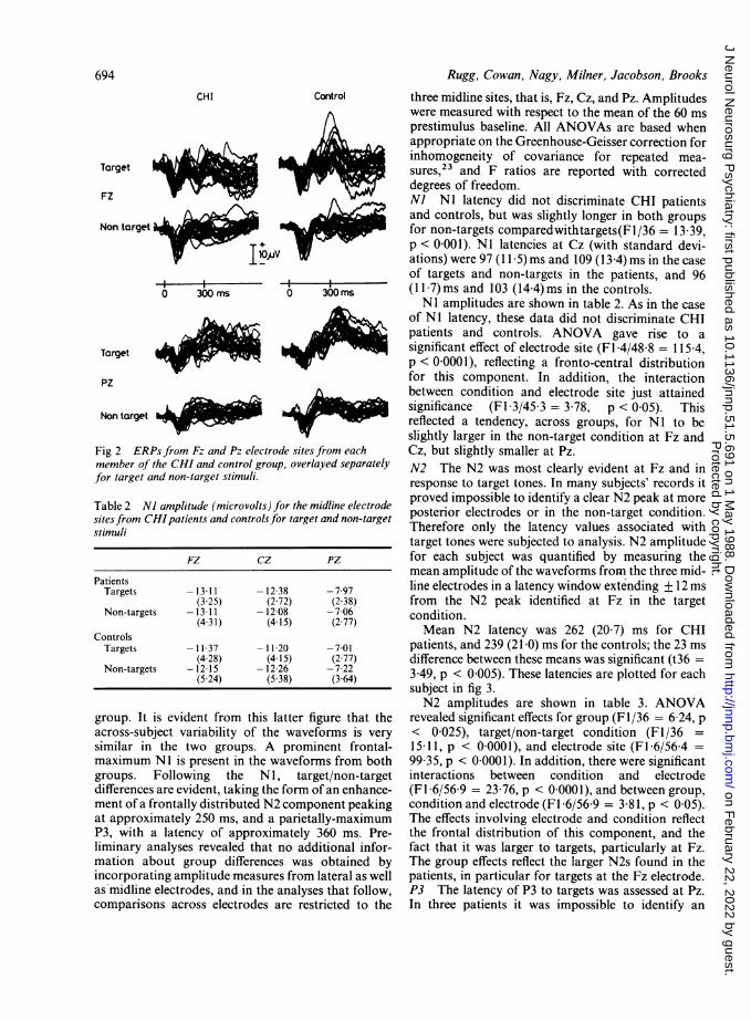
Fig 2 ERPs from Fz and Pz electrode sites from eachmember of the CHI and control group, overlayed separatelyfor target and non-target stimuli.
Table 2 NJ amplitude (microvolts) for the midline electrodesitesfrom CHIpatients and controlsfor target and non-targetstimuli
FZ CZ PZ
PatientsTargets -13-11 -12 38 -7-97
(325) (2-72) (2-38)Non-targets -13-11 -12 08 -7-06
(4-31) (4-15) (2 77)Controls
Targets -11-37 -11-20 -701(4-28) (4 15) (2-77)
Non-targets -12-15 -12 26 -7 22(5-24) (5-38) (3-64)
group. It is evident from this latter figure that theacross-subject variability of the waveforms is verysimilar in the two groups. A prominent frontal-maximum Ni is present in the waveforms from bothgroups. Following the N 1, target/non-targetdifferences are evident, taking the form of an enhance-ment of a frontally distributed N2 component peakingat approximately 250 ms, and a parietally-maximumP3, with a latency of approximately 360 ms. Pre-liminary analyses revealed that no additional infor-mation about group differences was obtained byincorporating amplitude measures from lateral as wellas midline electrodes, and in the analyses that follow,comparisons across electrodes are restricted to the
Rugg, Cowan, Nagy, Milner, Jacobson, Brooks
three midline sites, that is, Fz, Cz, and Pz. Amplitudeswere measured with respect to the mean of the 60 msprestimulus baseline. All ANOVAs are based whenappropriate on the Greenhouse-Geisser correction forinhomogeneity of covariance for repeated mea-sures,23 and F ratios are reported with correcteddegrees of freedom.NJ NI latency did not discriminate CHI patientsand controls, but was slightly longer in both groupsfor non-targets comparedwithtargets(Fl/36 = 13 39,p < 0-001). NI latencies at Cz (with standard devi-ations) were 97 (11 5) ms and 109 (13.4) ms in the caseof targets and non-targets in the patients, and 96(17)ms and 103 (14-4)ms in the controls.NI amplitudes are shown in table 2. As in the case
of NI latency, these data did not discriminate CHIpatients and controls. ANOVA gave rise to asignificant effect of electrode site (Fl 4/48 8 = 115 4,p < 00001), reflecting a fronto-central distributionfor this component. In addition, the interactionbetween condition and electrode site just attainedsignificance (FIl3/45 3 = 3.78, p < 0-05). Thisreflected a tendency, across groups, for NI to beslightly larger in the non-target condition at Fz andCz, but slightly smaller at Pz.N2 The N2 was most clearly evident at Fz and inresponse to target tones. In many subjects' records itproved impossible to identify a clear N2 peak at moreposterior electrodes or in the non-target condition.Therefore only the latency values associated withtarget tones were subjected to analysis. N2 amplitudefor each subject was quantified by measuring themean amplitude of the waveforms from the three mid-line electrodes in a latency window extending + 12 msfrom the N2 peak identified at Fz in the targetcondition.Mean N2 latency was 262 (20 7) ms for CHI
patients, and 239 (21 0) ms for the controls; the 23 msdifference between these means was significant (t36 =3 49, p < 0-005). These latencies are plotted for eachsubject in fig 3.N2 amplitudes are shown in table 3. ANOVA
revealed significant effects for group (F1/36 = 6 24, p< 0-025), target/non-target condition (F1/36 =15-11, p < 00001), and electrode site (FI 6/564 =99 35, p < 0 0001). In addition, there were significantinteractions between condition and electrode(Fl 6/56 9 = 23 76, p < 0-0001), and between group,condition and electrode (Fl 6/56 9 = 3-81, p < 0 05).The effects involving electrode and condition reflectthe frontal distribution of this component, and thefact that it was larger to targets, particularly at Fz.The group effects reflect the larger N2s found in thepatients, in particular for targets at the Fz electrode.P3 The latency of P3 to targets was assessed at Pz.In three patients it was impossible to identify an
Protected by copyright.
on February 22, 2022 by guest.
http://jnnp.bmj.com
/J N
eurol Neurosurg P
sychiatry: first published as 10.1136/jnnp.51.5.691 on 1 May 1988. D
ownloaded from

N2 and P3 in closed head injury
2 2110
16
500
9
1115
12 147 13 192094
35
6
a
1 13 450-
17 -
20
2 16
8
12 19
10
21
4 7 15
3
11
18
CHI ControlN2 Latency
400-
350-
300
2
4
17
122159
3 1019
8 1120
14
13
7
3
1621
6 159 1371
11122 18 194 10
8
20
I I
CHI ControlP3 Latency
750-
700-
650-
600-
550-
500-
450-
400:
4
15
10 16
38121 5
2011
921
132
39
16 13
14 21
1 6 7
2 8 11 12
6 4 15 1820
10 1719
CHI Control4-Choice RT
CHI PTA duration7 days or less : 7 8 9 128-14days :56101113141516192115-28days :12 3420
Fig 3 Values for each member of the CHI control groups on measures ofN2 latency, P3 latency and 4-choice RT.Members of each group are identified by number, and those patients belonging to each of three categories ofPTA durationare identified
unequivocal P3 peak, either because of multiplepeaks, or because of a flattened, plateau-like wave-form. These subjects were excluded from the analysisof the latency of this component.Mean P3 latency from the 16 CHI patients in whom
it could be measured was 371 (43-0) ms, compared to357 (22.8) ms in the controls. The 14 ms differencebetween the groups did not approach significance(t21l9 = 1-19). (The variances of the two samples diddiffer significantly (F15/18 = 3-56, p < 0-05); becauseof this inhomogeneity of variance t, and its associated
degrees of freedom, were estimated using unpooledestimates of population variance, as suggested by Fer-guson).24 The individual latency data are shown in fig3, where it can be seen that the data from the patientgroup contain an outlier (patient 2) whose P3 latencyexceeds that of all other subjects by some 50 ms.Exclusion of this patient's data markedly reduced thevariability of P3 latency among the patients (SD =
32-7).The amplitude of P3 was measured by computing
the mean amplitude of the region between 275-600 ms
3001
ms
250
200-
.1lV.*1
695
Protected by copyright.
on February 22, 2022 by guest.
http://jnnp.bmj.com
/J N
eurol Neurosurg P
sychiatry: first published as 10.1136/jnnp.51.5.691 on 1 May 1988. D
ownloaded from

696Table 3 N2 amplitude (microvolts) for the midline electrodesites for CHI patients and controls for target and non-targetstimuli (measured as the mean amplitude of a 24 ms windowstraddling the peak latency of the target N2 at FZ)
FZ CZ PZ
PatientsTargets -7 46 1-39 2-29
(4*44) (4-39) (4-94)Non-targets -2 58 1-85 2-68
(4-42) (5-29) (4 18)Controls
Targets -2 98 2-42 3 93(4 36) (4-49) (3 82)
Non-targets -0-46 4 17 4 52(3 13) (3 87) (3-16)
Table 4 Mean amplitude (microvolts) of the 275-600 mslatency region for CHI and control subjects, for target andnon-target stimuli
FZ CZ PZ
PatientsTargets 409 2-63 7 50
(4-73) (4-43) (4-52)Non-targets - 158 0 50 1-95
(2.89) (2 61) (2-56)Controls
Targets 0 76 7 21 11 00(6.36) (5 43) (3 96)
Non-targets 027 1 28 2-33(3-55) (2 89) (2-32)
post-stimulus, allowing those patients in whom no
peak could be identified to contribute to the analysis.These data are shown in table 4. ANOVA gave rise tosignificant effects for group (Fl/16 = 7-20, p <0 025), target/non-target condition (F1/36 = 36-17, p< 0-0001) and electrode site (Fl 2/42 6 = 109-09, p <0-0001). The interactions between group and condi-tion (F1/36 = 8-67, p < 0-01) and condition andelectrode site (F1I3/47 3 = 83 24, p < 0 0001) werealso significant. These effects reflect the increased pos-itivity of this area of the waveforms in response totargets in both patients and controls, thistarget/non-target difference having a parietal max-imum. The group by condition interaction reflects thesmaller values of this measure in the patients' target,but not non-target, waveforms. An analysis was alsoperformed on P3 peak amplitude data, obtained bymeasuring the amplitude at each midline site of alatency window extending ± 12 ms either side of thetarget P3 peak at Pz. This unequal N ANOVA (16patients vs 19 controls) yielded an identical set ofeffects to those described for the 275-600 ms measure.The mean number of targets counted by the CHI
patients (summed across the two halves of the experi-mental run) was 47 9 (2 9), compared with 47-0 (0 5)for the controls. These means did not differ
Rugg, Cowan, Nagy, Milner, Jacobson, Brooks
significantly.4-Choice reaction time (RT) One patient was unableto perform this task because of motor impairment(bilateral upper-limb weakness and ataxia). The meanRT of the remaining 18 patients was 551 (105 3) ms,as opposed to 435 (44 5) ms in the case of the controls.The 116 ms difference between these means wassignificant (t22-6 = 4-32, p < 0 0005), and the vari-ances between the two groups also differedsignificantly (F 17/18 = 5 60, p < 0-01). The mean RTof the 16 subjects in whom P3 latency was determinedwas 532 (96.5) ms. This mean also differed reliablyfrom that of the controls (t20 3 = 3 73, p < 0-005),as did its associated variance (Fl5/18 = 4 70, p <0 05). Inspection of the individual data (fig 3) revealedthat four patients (patients 4, 15, 10 and 16) appearedto form an outlying group. When these patients weredropped from the analysis the mean of the remainderwas reduced to 506 (65 2) ms; this was still reliablydifferent from the mean of the controls (t21-6 = 3 51,p < 0 005).
Errors on this task (as defined by depression of anunilluminated button) were too infrequent to subjectto analysis (mean of 0-33 for the patients, and 0 37 forthe controls).Relationship between measures For each group, cor-relations were computed between the three measuresof "processing speed": N2 and P3 latency, and4-Choice RT. In neither group was there anysignificant correlation between 4-choice RT and eitherN2 or P3 latency. The correlation between N2 and P3latencies was also non-significant in the control group,but did not attain significance in the patients (rl4 =0-565, p < 005).More informally, it is clear from fig 3 that there is
little correspondence even between those patientsbelonging to the extremes of the distributions of eachof the three latency measures, with the possible excep-tion of patient 6, who shows the shortest or secondshortest value on all three measures. It is also note-worthy that patients 10 and 16 feature in the extremesof both the N2 and 4-Choice RT distributions (neithdrcontributed a P3 latency). On the other hand patient2, who belongs to the extreme of the N2 and P3 distri-butions, was the fourth fastest performer among thepatients on the 4-choice RT task!Relationship to severity of injury Using post trau-matic amnesia (PTA) duration as an overall measureof severity of injury, the CHI patients were catego-rised into three groups according to the scheme ofBrooks.25 Four patients had PTA durations of lessthan 7 days, 10 had durations of 8-14 days, and 5 haddurations of 15-28 days. As can be seen from fig 3,there was no systematic relationship between thosecategories and any of the three measures of"efficiency"' of information-processing.
Protected by copyright.
on February 22, 2022 by guest.
http://jnnp.bmj.com
/J N
eurol Neurosurg P
sychiatry: first published as 10.1136/jnnp.51.5.691 on 1 May 1988. D
ownloaded from

N2 and P3 in closed head injuryDiscussion
The principal ERP findings in this study can be sum-marised as follows: NI latency and amplitude did notdifferentiate CHI patients from controls. N2 latencyto targets was longer in the patients, and N2 ampli-tude was larger, particularly in response to targetstimuli. Finally, although the amplitude of P3 fromthe patients was smaller, P3 latency was notsignificantly different from that of the controls.The similarity of the NI component in patients and
controls indicates that at least some of the cerebralmechanisms underlying the generation of ERP com-ponents were intact in the patient group. This isimportant for two reasons: firstly, it means that thosefeatures of the ERPs that did distinguish patients andcontrols were not simply indexing some aspecific dys-function of ERP generator systems occurring in CHIindividuals. Secondly, it has been suggested that theamplitude of the vertex NI reflects, at least in part, thegeneral level of "arousal".26 It this is so, then thepresent data suggest that no differences in arousalexisted between the patients and controls in this study,at least during the oddball task. This is consistent withdata from other sources suggesting that thebehavioural problems experienced by CHI patientsare not attributable to "under-arousal".7
In contrast to previous studies,18 20 we found noevidence of a delay in P3 latency in the CHI patients.This negative result may reflect the employment in thepresent study of less severely injured patients than inprevious work. Alternatively, it could reflectdifferences in task and procedure between the presentand previous studies. This second alternative seemsunlikely in view of the fact that the studies reportinglonger P3 latencies in CHI patients18 20 employedtasks which differed considerably from one another.The selective attention task employed by Curry20required subjects to make RT responses to low prob-ability targets presented in a designated ear. In con-trast, Campbell et al"8 employed an auditory oddballtask very similar to that used in the present study, withthe requirement to silently count target tones. In viewof the positive findings in relation to N2 latency (seebelow), it is arguable that P3 latency may not be themost sensitive ERP sign of "speed of processing" inthe oddball task. A similar dissociation between N2and P3 latency has recently been reported by Brecher,Porjesz and Begleiter,27 who compared ERPs fromschizophrenic patients and controls in a visual oddballtask. As in the present case, the latency of N2 (mea-sured in that experiment at Oz), but not P3, wasslower in the patient group. We do not wish to implythat this finding is suggestive of similar underlyingpathology in CHI and schizophrenic populations.However, it serves to emphasise the fact that it may be
697unwise to conclude, on the basis of negative findingswith respect to P3 latency, that a clinical group doesnot exhibit a deficit in stimulus categorisation time.As in previous studies, the CHI patients showed
smaller P3s than controls. This result must be inter-preted cautiously, however, as it could simply reflectincreased latency jitter among the individual trialsmaking up the patients' ERPs. Thus far, no study hasemployed a latency correction procedure toinvestigate this possibility, and until this is done theP3 amplitude data will remain equivocal.The findings concerning the N2 component are also
consistent with previous work. 19 20 As noted above, itseems likely that the latency of this component isassociated with the time required to categorise theeliciting stimulus. Thus, the delay in this componentin the head-injured patients implies a dysfunction inthis process in addition to "later" ones, such asresponse selection, etc. This supports previous sug-gestions along similar lines derived from behaviouralwork employing "additive-factors" logic to decom-pose the sources of variance contributing to RT inCHI patients.28 Such logic is, however, open to ques-tion,29 and the use of a "real-time" technique able toidentify an "early" information-processing deficitprovides valuable converging evidence. Thus, theERP evidence is inconsistent with the idea that theslowness exhibited by patients on RT tasks is entirelythe result of inefficiency in "late" processes such asresponse selection and execution.
In view of the fact that it was the patient group thatshowed the largest N2 amplitudes, it seems unlikelythat differential latency jitter played a part in pro-ducing group differences in this variable. However, itis possible that the larger N2 observed in the CHIpatients resulted simply from overlap with a smallerP3. This possibility seems unlikely because thedifferences in P3 amplitude between patients and con-trols do not differ significantly across the scalp, yet thegroup differences in N2 are maximal at Fz. If thesedifferences in N2 had resulted from the influence ofthe P3 component, they would have had the samescalp distribution as the group differences in theamplitude of P3, that is, they would have been ofequal magnitude at the three midline electrodes,rather than frontally distributed.
In a series of auditory oddball tasks, Fitzgerald andPicton12 observed changes in the amplitude of N2 asa function of the difficulty of the target/non-targetdiscrimination. They considered these data to behighly suggestive of an association between N2 ampli-tude and the allocation of "cognitive effort," such thatstimuli whose processing demanded greater effortelicited larger N2s. A similar interpretation of thefronto-central N2 has also been put forward byNaatanen and Picton, 13 who argued that it reflects, in
Protected by copyright.
on February 22, 2022 by guest.
http://jnnp.bmj.com
/J N
eurol Neurosurg P
sychiatry: first published as 10.1136/jnnp.51.5.691 on 1 May 1988. D
ownloaded from

698part, the conscious allocation of attentional resourcesto stimuli indicated as salient by pre-attentive pro-cesses. Within this framework, the larger N2s in thepatients might be indicative of the greater levels ofeffort required by this group to perform the task ade-quately. This would be predicted by the "coping-hypothesis" of Van Zomeren et al,7 which states thatas a result of deficient information-processing, CHIpatients can cope with task demands only by the allo-cation of excessive cognitive effort. The present datasuggest that this may be so even in a task asundemanding as auditory oddball detection.
Finally, it should be noted that although two of thethree measures of "processing speed" used in thisstudy clearly discriminated CHI from control subjects(that is, N2 latency and 4-choice RT), these wereuncorrelated in both groups, and neither measure wasobviously associated with severity of injury asassessed by PTA duration. The lack of associationbetween N2 and 4-choice RT must undoubtably havebeen contributed to by the (unknown) intrinsic level oferror associated with these measures. However, itprobably also reflects the fact that 4-choice RT isdetermined by a multiplicity of interacting processes(deficits in any one of which will contribute to delayedresponding), and only a limited number of these arereflected by N2 latency. CHI patients exhibit abnor-malities in these latter processes; it remains to bedetermined how important these abnormalities are inaccounting for the wide range of psychological deficitsfound in these patients.
We thank Mr J D Miller, Department of Neuro-surgery, Western General Hospital, Edinburgh, foraccess to patients under his care. This research wassupported by the Medical Research Council and theMental Health Foundation.
References
I Mandelberg IA. Cognitive recovery after severe head injury. 2.The Weschler Adult Intelligence Scale during post-traumaticamnesia. J Neurol Neurosurg Psychiatry 1975;38:1127-32.
2 Mandelberg IA. Cognitive recovery after severe head injury. 3.WAIS verbal and performance IQs as a function of post-traumatic amnesia duration and time from injury. J NeurolNeurosurg Psychiatry 1976;39:1001-7.
3 Mandelberg IA, Brooks DN. Cognitive recovery after severehead injury. 1. Serial testing on the Weschler Adult Intel-ligence Scale. J Neurol Neurosurg Psychiatry 1975;38:1121-6.
4 Brooks DN. Long- and short-term memory in head-injuredpatients. Cortex 1975;11:329-40.
5 Levin HS, Grossman RG, Rose JE, Teasdale G. The long-termneuropsychological outcome of closed head injury. JNeurosurg 1979;50:41 2-22.
6 Van Zomeren AH, Deelman BG. Differential effects of simpleand choice reaction after closed head injury. Clin NeurolNeurosurg 1976;14:64-70.
7 Van Zomeren AH, Brouwer WH, Deelman BG. Attentional
Rugg, Cowan, Nagy, Milner, Jacobson, Brooksdeficits: the riddles of selectivity, speed and alertness. In:Brooks N, ed. Closed Head Injury. Psychological, Social andFamily Consequences. Oxford: Oxford University Press,1984:74-107.
8 Gronwall D, Sampson H. The Psychological Effects of Concus-sion. Aukland: Aukland University Press, 1974.
9 Ewing R, McCarthy D, Gronwall D, Wrightson P. Persistingeffects of minor head-injury observable during hypoxic stress.J Clin Neuropsychol 1980;2:147-55.
10 Donchin E, Ritter W, McCallum WC. Cognitive Psycho-physiology: the endogenous components of the ERP. In: Cal-laway E, Tueting P, Koslow SH, eds. Event-Related BrainPotentials in Man. New York: Academic Press, 1978:349-441.
11 Pritchard WS. Psychophysiology of P300. Psych Bull1981;89:506-40.
12 Fitzgerald PG, Picton TW. Event-related potentials recordedduring the discrimination of improbable stimuli. Biol Psychol1983;17:241-76.
13 Naaitainen R, Picton TW. N2 and automatic versus controlledprocesses. In: McCallum WC, Zappoli R, Denoth F, eds.Cerebral Psychophysiology: Studies in Event-RelatedPotentials. Amsterdam: Elsevier, 1986:169-86.
14 Ritter W, Simson R, Vaughan HG. Event-related potential cor-relates of two stages of information-processing in physical andsemantic discrimination tasks. Psychophysiol 1983;20:168-79.
15 Johnson R. A triarchic model of P300 amplitude. Psychophysiol1986;23:367-84.
16 McCarthy G, Donchin E. A metric for thought: a comparison ofP300 latency and reaction time. Science 1981;211:77-80.
17 Magliero A, Bashore TR, Coles MGH, Donchin E. On thedependence of P300 latency on stimulus evaluation processes.Psychophysiol 1984;21:171-86.
18 Campbell, K, Houle S, Lorrain D, Deacon-Elliot D, Proulx G.Event-related potentials as an index of cognitive functioningin head-injured outpatients. In: McCallum WC, Zappoli R,Denoth F, eds. Cerebral Psychophysiology: Studies in Event-Related Potentials. Amsterdam: Elsevier, 1986;486-8.
19 Curry SH. Event-related potentials as indicants of structural andfunctional damage in closed head injury. In: Kornhuber HH,Deeke L, eds. Motivation, Motor and Sensory Processes of theBrain: Electrical Potentials, Behaviour and Clinical Use.Amsterdam: Elsevier 1980:507-15.
20 Curry SH. Event-related potentials as a tool for assessment offunctional disability and recovery following closed headinjury. Unpublished PhD Thesis, University of Bristol 1983.
21 Buschke H, Fuld PA. Evaluating storage, retention and retrievalin disordered memory and learning. Neurology 1974;24:1019-25.
22 Stephenson WA, Gibbs FA. A balanced non-cephalic referenceelectrode. Electroencephalogr Clin Neurophysiol 1951;3:237-40.
23 Keselman HJ, Rogan JC. Repeated measures F tests and psycho-physiological research: controlling the number of false posi-tives. Psychophysiol 1980;17:499-503.
24 Ferguson GA. Statistical Analysis in Psychology and Education.5th Edition. New York: McGraw Hill 1981.
25 Brooks DN. Wechsler memory scale performance and itsrelationship to brain damage after severe closed head injury. JNeurol Neurosurg Psychiatry 1976;39:593-601.
26 Naatinen R. Selective attention and evoked potentials inhumans-a critical review. Biol Psychol 1975;2:237-307.
27 Brecher M, Porjesz B, Begleiter H. The N2 component of theevent-related potential in schizophrenic patients. Electro-encephalogr Clin Neurophysiol 1987;66:369-75.
28 Stokx LC, Gaillard AWK. Task and driving performance ofpatients with a severe concussion of the brain. J Clin ExpNeuropsychol 1986;8:421-36.
29 McClelland JL. On the time relations of mental processes: anexamination of systems of processes in cascade. Psychol Rev1979;86:287-330.
Protected by copyright.
on February 22, 2022 by guest.
http://jnnp.bmj.com
/J N
eurol Neurosurg P
sychiatry: first published as 10.1136/jnnp.51.5.691 on 1 May 1988. D
ownloaded from