head and neck56-405314 and neck-2.pdf · • prognosis -- best with lip lesion and poorest in...
TRANSCRIPT

1
Pathology of Head and Neck
ผูชวยศาสตราจารย แพทยหญิง จุลินทร สําราญ
• Pathology of Oral cavity • Pathology of Nose, and Nasopharynx • Pathology of Larynx • Pathology of Neck • Pathology of Salivary gland

2
Pathology of Oral cavity • Inflammations and infections • Reactive lesions • Oral manifestations of systemic disease • Tumors and precancerous lesions
Inflammations and infections • Herpes simplex virus infections • Aphthous ulcer ( Canker sores ) • Oral candidiasis ( Thrush ) • Glossitis • Xerostomia

3
Herpes simplex virus infections • Etiology ; HSV1 • Clinical feature ;
– Acute herpetic gingivostomatitis in young children , severe diffuse involvement of the oral and pharyngeal mucosa, tongue and gingiva ,and spontaneously clear in 3-4 weeks

4
– Recurrent herpetic stomatitis in young adult , milder form involving lip, nasal orifices and buccal mucosa and spontaneously clear in 1-2 weeks
• Morphology – Gross ; Small vesicles to bullae painful, red-
rimmed, shallow ulceration – Histo ; Acantholysis and presence of intranuclear
inclusion with multinucleated giant cells

5
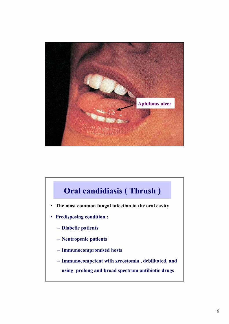
Aphthous ulcer ( Canker sores )
• Common superficial ulceration of oral mucosa • Most in first decade of life • Clinical feature; painful and recurrent ulceration
and clear within a week
• Morphology ; – Gross ; single or multiple, shallow, hyperemic
ulceration with red-rimmed and thin exudate covering
– Histo ; mainly mononuclear cell infiltration and neutrophilic infiltration when has secondary bacterial infection
6
Aphthous ulcer
Oral candidiasis ( Thrush ) • The most common fungal infection in the oral cavity • Predisposing condition ;
– Diabetic patients – Neutropenic patients – Immunocompromised hosts – Immunocompetent with xerostomia , debilitated, and
using prolong and broad spectrum antibiotic drugs

7
• Pseudomembranous candidiasis; a superficial, curdy, gray to white membrane composed of matted organisms enmeshed in a fibrinosuppurative exudate that can be readily scraped off to reveal an underlying erythematous inflammatory base
Glossitis • Inflammation of tongue • Etiology ;
– Deficiencies of vitamin B12 , riboflavin, niacin or pyridoxin, and iron
– Jagged carious teeth, ill-fitting dentures, syphilis,inhalation burn and ingestion of corrosive chemicals

8
• Morphology ; – Gross ; beefy-red
tongue with / without ulceration
– Histo ; atrophy of the papillae of tongue and thining of mucosa, exposing vasculature and shallow ulceration
Xerostomia • Dry mouth - lack of salivary secretion • Etiology ;
– Sjogren syndrome – Drug ( anticholinergic agents )
– A complication of radiation therapy

9
• Morphology ; – dry mucosa or atrophy of papillae of tongue
with fissuring and ulceration
Reactive lesions • Fibroma • Pyogenic granuloma • Peripheral giant cell granuloma

10
Fibroma • Occuring in bite line of buccal mucosa and
gingivodental margin • Etiology ; irritation • Morphology ;
– Nodular mass of fibrous tissue with scant inflammatory cell infiltration and covered by squamous mucosa

11

12
Pyogenic granuloma • Pregnancy tumor • Highly vascular, pedunculated tumor • Occuring in gingiva of children, young adult and
commonly pregnant woman

13

14
Peripheral giant cell granuloma • Inflammatory lesion sized > 1.5 cm. and
protruding from gingiva at some site of chronic inflammation
• Morphology ; – aggregation of multinucleate , foreign body -
type giant cells in fibroangiomatous stroma

15
Oral manifestations of systemic disease • Infection ;
– Scarlet fever - stawberry or rasberry tongue – Measle - Koplik spots – Infectious mononucleosis - acute pharyngitis
and tonsilitis
• Dermatologic condition ; – Lichen planus : reticulate, lacelike, white
keratotic lesion – Pemphigus and bullous pemphigoid : vesicle or
bullae – Erythema multiforme : maculopapula and
vesiculobullous eruption - Stevens-Johnson syndrome

16
• Hematologic disorder ; – Monocytic leukemia : leukemic infiltration
causing enlargement of gingiva • Miscellaneous ;
– Melanotic pigmentation : in Addison disease, Albright syndrome, Peutz-jegher syndrome .
– Dilantin : fibrous enlargement of the gingiva
Tumors and Precancerous lesions • Leukoplakia and erythroplakia
• Squamous cell carcinoma
• Odontogenic cysts and tumors

17
Leukoplakia • White plaque on the oral mucous membrane can not
remove and be classified in to another entities • Morphology ;
– Histo ; ranging from benign epithelial hyperplasia to dysplastic epithelium -- carcinoma in situ

18
Proliferation of squamous epithelium
Erythroplakia • Dysplastic leukoplakia • Morphology ;
– Gross : red, velvety, possible eroded area in oral cavity
– Histo : dysplastic squamous epithelium with / without surface erosion --- carcinoma in situ or invasive carcinoma at margin

19

20
• Clinical feature ; – Found between the age of 40-70 years – Male : Female = 2 : 1 – Associated with tobacco (highly association) ,
alcohol , ill-fitting denture, chronic exposure to irritants and HPV serotype 16
– Malignant transformation : Erythroplakia ( 50 % ) more than leukoplakia ( 5-6%)
Squamous cell carcinoma • 95 % of oral cancer • found in the age of 50-70 years • Etiology ;
– Environmental factors : use of tobacco , alcohol , marijuana , betel nut and pan
– HPV infection ( serotype 6 , 16 , and 18 )
– Genetic factor; deletion chromosome 3 , mutation in p53 and amplification of oncogene INT2 , and BCL1

21
(Irritation from jagged teeth , ill-fitting denture and chronic infection associated with leukoplakia ) • Clinical feature ;
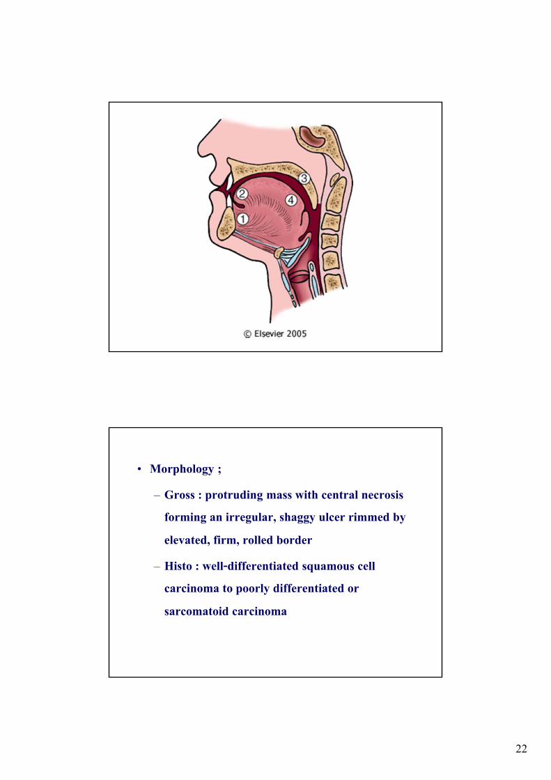
– Favored location : the floor of mouth, tongue , hard palate and base of tongue
– Favored sites of metastasis : mediastinal lymph nodes, lungs , liver , and bone
• Prognosis -- best with lip lesion and poorest in lesion of floor of mouth and base of tongue
22
• Morphology ; – Gross : protruding mass with central necrosis
forming an irregular, shaggy ulcer rimmed by elevated, firm, rolled border
– Histo : well-differentiated squamous cell carcinoma to poorly differentiated or sarcomatoid carcinoma

23

24

25
Pathology of Nose and Nasopharynx • Inflammation
– Infectious rhinitis and Allergic rhinitis
– Sinusitis
– Pharyngitis and Tonsilitis
• Nasal polyp and sinunasal papilloma
• Nasopharyngeal carcinoma

26
Infectious rhinitis (common cold) • Major offenders are adenoviruses, echoviruses, and
rhinoviruses. • Acute stages; the nasal mucosa is thickened, edematous,
and red; the nasal cavities are narrowed; and the turbinates are enlarged. These changes may extend, producing a concomitant pharyngotonsillitis.
• Secondary bacterial infection cause mucopurulent or sometimes frankly suppurative exudate.
Allergic rhinitis - hay fever • An immunoglobulin E-mediated immune reaction
with an early- and late-phase response • The allergic reaction is characterized by marked
mucosal edema, redness, and mucus secretion, accompanied by a leukocytic infiltration in which eosinophils are prominent
• The most common allergens: plant pollens, fungi, animal allergens, and dust mites.

27
Nasal Polyps • Recurrent attacks of rhinitis eventually lead to focal
protrusions of the mucosa, producing so-called nasal polyps, which may reach 3 to 4 cm in length.
• On histologic examination, these polyps consist of edematous mucosa having a loose stroma, often harboring hyperplastic or cystic mucous glands and infiltrated with a variety of inflammatory cells, including prominently neutrophils, eosinophils, and plasma cells with occasional clusters of lymphocytes .

28
Sinusitis • Acute sinusitis is most commonly preceded by acute or
chronic rhinitis, but maxillary sinusitis occasionally arises by extension of a periapical infection through the bony floor of the sinus.
• The offending agents are usually inhabitants of the oral cavity
• Impairment of drainage of the sinus by inflammatory edema of the mucosa – obstruction of outflow and accumulation of secretion or pus in sinuses
Pharyngitis and tonsillitis • Most common infectious agents; rhinoviruses, echoviruses,
and adenoviruses, and, less frequently, respiratory syncytial viruses and the various strains of influenza virus.
• Superimposed bacterial infections or Primary bacterial infection - the most common offenders are the β-hemolytic streptococci
• Reddening and slight edema of the nasopharyngeal mucosa, with reactive enlargement of the related lymphoid structures in most usual cases and in more severe case, inflamed nasopharyngeal mucosa may be covered by an exudative membrane (pseudomembrane), and the nasopalatine and palatine tonsils may be enlarged and covered by exudate.

29
Sinonasal Papillomas • Benign neoplasms arising from the sinonasal mucosa
and are composed of squamous or columnar epithelium. • Might related to HPV types 6 and 11 • Three forms: septal (most common), inverted (most
important biologically), and cylindrical. • Inverted papillomas are benign but locally aggressive
neoplasms occurring in both the nose and the paranasal sinuses - high rate of recurrence, with the potentially serious complication of invasion of the orbit or cranial vault; rarely, frank carcinoma

30
Nasopharyngeal Carcinomas • Association with EBV infection
• Three patterns: – keratinizing squamous cell carcinomas,
– nonkeratinizing squamous cell carcinomas, and
– undifferentiated carcinomas
• It is unresectable and radiotherapy is a standard treatment
• 50% to 70% 3-year survival rate.
Pathology of Larynx
• Laryngitis • Vocal cord polyp • Squamous papilloma • Laryngeal carcinoma

31
Laryngitis • May occur as the manifestation of allergic, viral, bacterial, or chemical
insult, but it is more commonly part of a generalized upper respiratory tract infection or the result of heavy exposure to tobacco smoke.
• May cause obstruction, especially laryngoepiglottitis, caused by Haemophilus influenzae or β-hemolytic streptococci in infants and young children
• The most common form of laryngitis, encountered in heavy smokers, constitutes an important predisposition to the development of squamous epithelial changes in the larynx and sometimes overt carcinoma
Vocal cord nodule or polyp • Reactive nodules, also called polyps, sometimes develop on the vocal
cords, most often in heavy smokers or in individuals who impose great strain on their vocal cords (singers' nodules).
• These nodules constitute smooth, rounded, sessile or pedunculated excrescences, generally only a few millimeters in greatest dimension, located usually on the true vocal cords.
• They are typically covered by squamous epithelium that may become keratotic, hyperplastic, or even slightly dysplastic. The core of the nodule is a loose myxoid connective tissue that may be variably fibrotic or punctuated by numerous vascular channels.

32
Squamous papilloma • Benign neoplasms, usually on the true vocal cords, that form
soft, raspberry-like excrescences rarely more than 1 cm in diameter
• Usually single in adults but are often multiple in children
• The lesions are caused by HPV types 6 and 11.
• They do not become malignant, but they frequently recur.
33
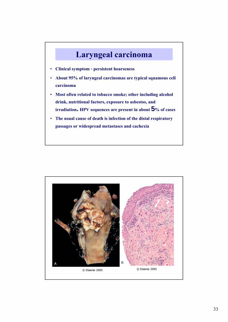
Laryngeal carcinoma • Clinical symptom - persistent hoarseness • About 95% of laryngeal carcinomas are typical squamous cell
carcinoma • Most often related to tobacco smoke; other including alcohol
drink, nutritional factors, exposure to asbestos, and irradiation. HPV sequences are present in about 5% of cases
• The usual cause of death is infection of the distal respiratory passages or widespread metastases and cachexia

34
Pathology of Neck • Branchial cyst ( Lymphoepithelial lesion ) • Thyroglossal tract cyst • Paraganglioma ( Carotid body tumor )
Branchial cyst ( Lymphoepithelial lesion ) • Benign cyst in anterolateral aspect of neck • Remnant of the branchial arches • Morphology ;
– Gross : circumscribed cyst , 2-5 cm. – Histo : cystic space lined by stratified squamous
and pseudostratified columnar epitheliums and presence of lymphoid tissue in fibrous wall

35
Cystic wall
Cystic space

36
Adjacent lymphoid tissue
Squmous epithelial lining
Thyroglossal tract cyst • Remnant of thyroglossal duct ( from foramen cecum ) • Morphology ;
– Gross : Cystic mass , 1-4 cm. at anterior of neck – Histo : cystic space lined by stratified squamous and
pseudostratified columnar epitheliums and presence of lymphoid tissue and thyroid follicles in fibrous wall

37
Cystic space
Thyroid tissue

38
Ciliated columnar epithelium
Squamous epithelium
Paraganglioma ( Carotid body tumor ) • Tumor of paraganglia ( clusters of neuroendocrine cells )
– Adrenal paraganglia -- Pheochromocytoma – Extraadrenal paraganglia :
• Paravertebral paraganglia ( sympathetic connection)
• Paraganglia related to great vessels of head and neck ( parasympathetic connection)

39
• Clinical feature ; – Arise in the age of 60 – Associated with MEN II --multiple and
bilateral lesions – frequency recurrent and local invasion
• Morphology ; – Gross : Reddish - brown mass not exceed 6
cm. at bifurcation of the common carotid body ( Chemodectoma)
– Histo : nests of polygonal chief cell ( uniform ) with abundant granular cytoplasm and vesicular nuclei surrounded by elongated sustenticular cells and fibrous tissue

40

41
Pathology of Salivary glands • Inflammation ( Sialadenitis )
• Mucocele
• Tumors
Inflammation ( Sialadenitis ) • Cause of sialadenitis ; infection ( virus and bacteria ) or
autoimmune origins eg. – Viral sialadenitis caused by mumps ( Epidemic
parotitis ) – Sjogren syndrome

42
Nonspecific bacterial sialadenitis • Involving major salivary gland • Etiology ;
– Secondary to duct obstruction caused by sialolithiasis – The most common organisms : Staphylococcus aureus
and streptococcus viridans • Morphology ; Acute and chronic inflammation

43
Mucocele • The most common lesion of the salivary glands.
• Results from either blockage or rupture of a salivary gland duct, with consequent leakage of saliva into the surrounding connective tissue stroma.
• Most often found on the lower lip

44
• The cyst-like space of mucocele is lined by inflammatory granulation tissue or by fibrous connective tissue
Salivary gland tumors • Salivary gland tumor - uncommon tumor • Site;
– 65 - 80 % in parotid gland – 10 % in submandibular glands – remaining arise in minor and sublingual gland

45
• Malignant tumor ; – 15 - 30 % of parotid gland tumor – 40 % of submandibular gland tumor – 50 % of minor salivary gland tumor – 70 % of sublingual gland tumor
• Age
– usually occur in adult
Pleomorphic adenoma • Mixed tumor • Most common tumor and benign tumor of
salivary glands • 60% in parotid gland • Epithelium-derived benign tumor with both
epithelial and mesenchymal differentiation

46
• Clinical feature ; – Painless, slow - growing mass in parotid,
submandibular glands and buccal mucosa • Carcinoma ex pleomorphic adenoma or malignant
mixed tumor - the most aggressive of all salivary gland malignant neoplasm , 30-50% mortality in 5 years
• Morphology ; – Gross : round, well-encapsulated masses ,not
exceeding 6 cm. – Histo : epithelial elements ( duct, irregular
tubules , strands, sheets ) and mesenchymal-like elements ( myxoid , hyaline , chondroid to bony matrix )

47

48

49

50
Warthin’s tumor • The second most common salivary gland neoplasm • present in parotid gland • found more commonly in male and fifth to seventh
decades of lifes • 10 % multifocal and bilateral
• Morphology ; – Gross : well-encapsulated masses , 2-5 cm. having
gray-brown surface with cleft-liked or cystic spaces – Histo : spaces lined by double layers of epithelial
cells ( abundant granular eosinophillic cytoplasm ) resting on lymphoid stroma

51

52
Mucoepidermoid carcinoma • The most common malignant tumor of salivary
gland ( 60-70% in parotid gland ) • radiation-induced neoplasm • Clinical course and prognosis -- grade of tumor • Grade of tumor ; three grades depend on amount
of mucus-secreting cells
• Morphology ; – Gross : nonencapsulated and circumscribed
masses , > 8 cmwith infiltrate margins . having gray-white cutsurfaces with small mucin containing cysts
– Histo : variable mixtures of squamous cells, mucus-secreting cells and intermediated cells

53
Mucin production

54
Adenoid cystic carcinoma • Malignant tumor • The most common neoplasm in minor salivary
gland • Clinical feature ;
– the most painful salivary gland neoplasm( perineural invasion of tumor )
• Morphology ; – Gross : poorly encapsulated gray-pink masses ,
with infiltrate margins . – Histo : tubular , solid and cribiform patterns of
small cells with dark, compact nuclei and scant cytoplasm

55

56
Perineural invasion

57
The end