head and neck - wolters kluwer · head and neck 213 ulcers or even some benign neoplastic lesions....
TRANSCRIPT

211
10
Head and neckAdriAnA Acurio And Jerome B. TAxy
IntroductIon
intraoperative consultation in head and neck cancer management con-cerns tumor diagnosis, margin evaluation, and nodal status, one or all of which help to determine the extent of surgery. These issues will be addressed here related to conventional mucosal squamous carcinoma and salivary gland tumors, the most common tumors in this anatomic region. The tissue artifacts commonly encountered on frozen sections and the effects of (neo) adjuvant therapy as possible pitfalls in interpretation will also be discussed. in addition, consideration will be given to the relatively recent application of frozen section to the management of acute invasive fungal rhinosinusitis as a guide for surgical debridement, which is an essential part of the therapy for this disease. Studies on the accuracy of frozen section in head and neck cancers suggest it is reliable, with an accuracy rate of 98% and a sensitivity and specificity of 89% and 99%, respectively (1).
Mucosal Lesions of the Head and Neck: Indications and Intraoperative Questions
an immediate diagnosis on a primary lesion. While most primary diagno-ses of squamous cell carcinoma rely on routinely processed endoscopic biopsies or excisions, a request for an immediate specimen evaluation reflects a set of clinical circumstances specific to each patient. An intraop-erative diagnosis of squamous cell carcinoma on an untreated primary mucosal lesion, or a posttreatment persistent/recurrent lesion, may be requested to ensure adequacy or to justify proceeding with an immediate wide resection (Fig. 10.1). if the frozen section is done for adequacy assess-ment, additional tissue should be requested not to be frozen. A frozen section may be helpful not only to establish a diagnosis but, if in situ car-cinoma is identified, to confirm that the lesion is primary (Fig. 10.2). The presence of carcinoma may also initiate an immediate neck dissection.
most difficulties in frozen section analysis of mucosal biopsies center on differentiating malignant from reactive lesions. Foremost among the latter are flat lesions simulating dysplasia but associated with infections or prior treatment. in addition, pseudoepitheliomatous hyperplasia may coexist with
LWBK1296-CH10_p211-233.indd 211 19/09/13 12:41 PM

212 Biopsy interpretation: tHe Frozen section
FIgure 10.2 Frozen section of the lesion in Figure 10.1, demonstrating easily discernible surface in situ squamous carcinoma and an infiltrating, keratinizing component. in situ disease helps to verify the lesion as a primary tumor.
FIgure 10.1 Gross photograph of a poorly circumscribed ulcerated white lesion of the retromolar trigone, excised and appropriately marked with sutures.
LWBK1296-CH10_p211-233.indd 212 19/09/13 12:41 PM
Head and neck 213
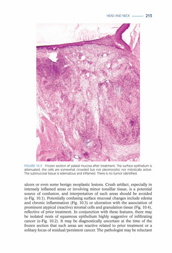
ulcers or even some benign neoplastic lesions. crush artifact, especially in intensely inflamed areas or involving minor tonsillar tissue, is a potential source of confusion, and interpretation of such areas should be avoided (e-Fig. 10.1). Potentially confusing surface mucosal changes include edema and chronic inflammation (Fig. 10.3) or ulceration with the association of prominent atypical (reactive) stromal cells and granulation tissue (Fig. 10.4), reflective of prior treatment. in conjunction with these features, there may be isolated nests of squamous epithelium highly suggestive of infiltrating cancer (e-Fig. 10.2). it may be diagnostically uncertain at the time of the frozen section that such areas are reactive related to prior treatment or a solitary focus of residual/persistent cancer. The pathologist may be reluctant
FIgure 10.3 Frozen section of palatal mucosa after treatment. the surface epithelium is attenuated, the cells are somewhat crowded but not pleomorphic nor mitotically active. the submucosal tissue is edematous and inflamed. there is no tumor identified.
LWBK1296-CH10_p211-233.indd 213 19/09/13 12:41 PM

214 Biopsy interpretation: tHe Frozen section
FIgure 10.4 Frozen section of a previously treated tonsil. the surface (top) is completely ulcerated. the tissue is replaced by abundant chronic inflammatory cells and active granulation tissue with the small vessels lined by fibrinous debris. this is early treatment effect. inset: treatment effect. Higher power view of the granulation tissue seen in Figure 10.4, vessels lined by fibrinous material, and intervening inflammatory infiltrate. note the scattered large cells.
to establish a malignant diagnosis based on such an isolated observation, especially if a radical procedure will be the result. These circumstances are common enough such that the frozen section might best be reported as deferred or descriptively as “atypical,” serving to alert the surgeon to obtain additional samples not to be frozen. The pathologist will also then have time to study the permanent material and arrive at a secure diagnosis.
in addition, related to the assessment of surface changes and the appreciation of changes in the underlying stroma is the presence of scar tissue and prominent blood vessels suggesting desmoplasia (e-Fig. 10.3).
LWBK1296-CH10_p211-233.indd 214 19/09/13 12:41 PM

Head and neck 215
FIgure 10.5 Frozen section of submucosal palatal tissue demonstrating minor salivary gland tissue with partial lobular atrophy. there is stromal edema and early fibrosis. the epithelial structures show occasional nuclear enlargement. this is early treatment-related sialometaplasia.
especially difficult in this regard are the changes in minor salivary gland tissue reflective of previous treatment. These changes histologically simu-late the changes of necrotizing sialometaplasia (Figs. 10.5 and 10.6, e-Figs. 10.4 and 10.5) and are principally characterized by the preservation of the lobular growth pattern, despite the nuclear atypia and squamoid features. Given the potential consequences, a frozen section diagnosis of infiltrat-ing cancer should be unequivocal, demonstrating irregular growth pat-terns and a desmoplastic reaction (Fig. 10.7, e-Fig. 10.6).
the status of a mucosal resection margin. While the histologic changes in the margins are similar to what has been described earlier for primary muco-sal lesions, at stake is whether additional tissue needs to be excised if the margin is involved. The more basic questions should be the following: What constitutes involvement, and what quantitative distance represents an ade-quate margin? That the answers to these questions are not readily apparent nor are likely to be adequately resolved by frozen section study does not prevent them from being frequently asked with a plethora of specimens (2).
The known multifocality (“field effect”) of squamous carcinoma and its dysplastic precursors in the upper aerodigestive tract, the uncertain predictive value of dysplasia related to the subsequent development of invasive cancer, and the lack of agreement as to what constitutes an ade-quate or involved (“positive”) margin makes it arguable whether preinva-sive dysplasia should be reported in an intraoperative setting. The grading of squamous dysplasia in the oral cavity traditionally utilizes the same
LWBK1296-CH10_p211-233.indd 215 19/09/13 12:41 PM

216 Biopsy interpretation: tHe Frozen section
A
B
FIgure 10.6 A: Frozen section after treatment. the lobular atrophy remains, the stroma is more fibrotic, and the epithelial atypia in the remaining ducts/acini is more pronounced with some squamous metaplasia. this should not be interpreted as pseudoglandular squa-mous carcinoma. B: Frozen section. a remaining salivary gland duct with marked surrounding inflammation and marked nuclear atypia with the suggestion of squamous differentiation.
system as in the uterine cervix but is without the predictable correlation with the subsequent development of or coexistence with invasive squa-mous cancer. experience has also shown that invasive squamous carci-noma in the head and neck is often not associated with typical in situ carcinoma. Being aware of the treatment algorithm, which may vary among surgeons, is critical in the interpretation of these samples.
LWBK1296-CH10_p211-233.indd 216 19/09/13 12:41 PM
Head and neck 217
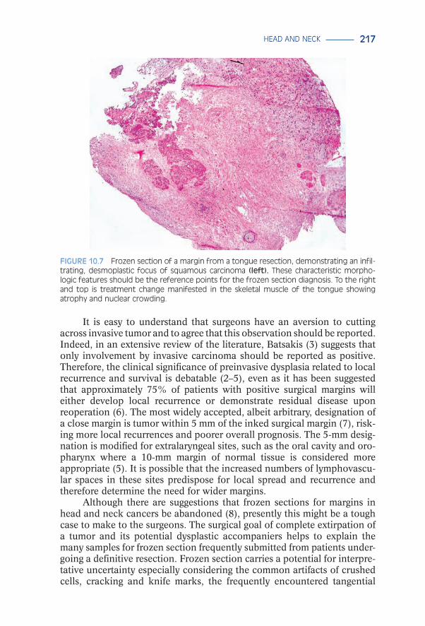
FIgure 10.7 Frozen section of a margin from a tongue resection, demonstrating an infil-trating, desmoplastic focus of squamous carcinoma (left). these characteristic morpho-logic features should be the reference points for the frozen section diagnosis. to the right and top is treatment change manifested in the skeletal muscle of the tongue showing atrophy and nuclear crowding.
it is easy to understand that surgeons have an aversion to cutting across invasive tumor and to agree that this observation should be reported. indeed, in an extensive review of the literature, Batsakis (3) suggests that only involvement by invasive carcinoma should be reported as positive. Therefore, the clinical significance of preinvasive dysplasia related to local recurrence and survival is debatable (2–5), even as it has been suggested that approximately 75% of patients with positive surgical margins will either develop local recurrence or demonstrate residual disease upon reoperation (6). The most widely accepted, albeit arbitrary, designation of a close margin is tumor within 5 mm of the inked surgical margin (7), risk-ing more local recurrences and poorer overall prognosis. The 5-mm desig-nation is modified for extralaryngeal sites, such as the oral cavity and oro-pharynx where a 10-mm margin of normal tissue is considered more appropriate (5). it is possible that the increased numbers of lymphovascu-lar spaces in these sites predispose for local spread and recurrence and therefore determine the need for wider margins.
Although there are suggestions that frozen sections for margins in head and neck cancers be abandoned (8), presently this might be a tough case to make to the surgeons. The surgical goal of complete extirpation of a tumor and its potential dysplastic accompaniers helps to explain the many samples for frozen section frequently submitted from patients under-going a definitive resection. Frozen section carries a potential for interpre-tative uncertainty especially considering the common artifacts of crushed cells, cracking and knife marks, the frequently encountered tangential
LWBK1296-CH10_p211-233.indd 217 19/09/13 12:41 PM

218 Biopsy interpretation: tHe Frozen section
sections and treatment effects which can easily complicate the tissue eval-uation. The morphologic challenge becomes more apparent when one considers that even with properly fixed and embedded samples under per-manent conditions, there is difficulty in differentiating ordinary squamous or pseudoepitheliomatous hyperplasias from intramucosal squamous dys-plasia, not to mention grading it. it is perhaps more than an understate-ment to say that margin assessment by frozen section can be uncertain.
in addition to awareness of the treatment algorithm, margin assess-ment should ensure that the pathologist has some knowledge of or actually is able to examine the gross specimen. Given the inherent complexity of head and neck anatomy, orientation of the resection specimen by the sur-geon in person, by diagram, or in the form of suture designations is essential. in addition, measurements, photographs, and/or specimen diagrams should be prepared before samples are taken for frozen section (Figs. 10.1, 10.8, and 10.9, e-Figs. 10.7 and 10.8). From large specimens, the manner in which the tissue is mounted and sectioned may vary. right angle or perpendicular
FIgure 10.8 Gross photograph of a total laryngectomy with bilateral neck dissections. a complicated specimen such as this needs to be oriented with the surgeon prior to any frozen section sampling. this specimen has been opened posteriorly, clearly demonstrating a supraglottic, ulcerated tumor extending on to the lower aspect of the epiglottis. the inferior tracheal margin is grossly free of tumor. Both contiguous neck dissections are spread out for easy recognition. this patient had been previously radiated, so the soft tis-sues of the neck are scant and lymph node recovery would be expected to be diminished.
LWBK1296-CH10_p211-233.indd 218 19/09/13 12:41 PM

Head and neck 219
gross sectioning best allows for thorough macroscopic analysis including measurement of the distance from the lesion to the nearest surgical margin (e-Fig. 10.8). The selected area for frozen section analysis should be visually closest to the margin. Parallel or en face sectioning does provide a larger area for microscopic examination, potentially the entire margin. However, this method does not allow for gross appreciation of the tumor or an accu-rate assessment of the distance between the lesion and the nearest margin.
Alternatively, the surgeon may submit separate samples from the resection bed resulting in the simultaneous arrival of large numbers of specimens to the surgical pathology laboratory for immediate examina-tion. in this event, the mucosal surface for these small pieces of tissue needs to be identified and the specimen mounted so that the microtome knife will cut perpendicular to this plane. Spiro et al. (6) reported an 89% diagnostic accuracy rate for frozen section of oral cancer irrespective of whether the margins were obtained from the patient or from the surgical specimen. A major limitation in margin analysis is the ability to relocate the sample site following frozen section diagnosis. one study assessing the correlation of frozen section specimens to the defect cavity showed a dif-ference of more than 1 cm in 32% of cases (9). These findings highlight the need to accurately trace the site of frozen sections if samples that sub-sequently prove positive are to be used to the greatest effect.
Assuming that the tissue sample has been properly embedded and the tissue block appropriately faced, there is no standard number of levels
FIgure 10.9 Bivalved superficial parotidectomy with a pleomorphic adenoma. note the edges that have been inked before sectioning. also, a single suture emanates from one end, designated by the surgeon as the superior and lateral edge. the tumor is generally well circumscribed except for the inferior aspect and does grossly extend to the edges of the sample. a gross examination in this case could justify taking additional inferior tissue.
LWBK1296-CH10_p211-233.indd 219 19/09/13 12:41 PM
220 Biopsy interpretation: tHe Frozen section
to examine. our practice is to examine two frozen sections at two different but relatively close levels from each block. Additional deeper sections can be cut as necessary. Two levels decrease the sampling error and ensure against loss of time incurred by a section inadvertently falling off the slide or being wiped off if the cover slip is erroneously placed on the wrong side. Sectioning the frozen sample to the depletion of the tissue block is occasionally justified but is not standard practice, as advocated by some (10). depletion of the sample could create a reconciliation problem with the subsequent paraffin embedding (“frozen control”), even though the diagnostic area seen on the frozen does not have to be present on the permanently embedded “control” for the diagnosis to be maintained.
the complicating effects of previous treatment. The histologic changes induced by radiation and/or chemotherapy may be misinterpreted as squamous carcinoma and therefore are potential pitfalls. Surface ulceration and regeneration, sialometaplasia, and stromal cell atypia have been par-tially illustrated earlier (Figs. 10.3–10.6). The major morphologic manifes-tations of treatment often encountered at frozen section are as follows:
a. Ulceration and epithelial hyperplasia. Although the ulcers are mor-phologically nonspecific, the adjacent epithelium may exhibit basal layer expansion with less definition of its palisading and some nuclear enlargement with nucleoli.
compensatory squamous hyperplasia, occasionally in the form of pseudoepitheliomatous hyperplasia, demonstrates expansion but maintenance of orderly maturation (Fig. 10.10). mitoses restricted to the basal layer may signify regeneration. The major diagnostic dilemma, even when examining such specimens which have not been frozen, is true dysplasia in which there is loss of maturation toward the surface and suprabasilar mitoses. uncertainty in this assessment should generate a deferred diagnosis and/or a request for more tissue. it is unlikely that dysplasia of low degree (mild) would generate additional margin samples or a wide resection.
b. Stromal changes manifested by varying degrees of cytologic atypia involving a single cell or groups of cells (Fig. 10.11). chiefly, these cells are fibroblasts (“radiation” fibroblasts), characterized by their large size and abundant basophilic cytoplasm with irregular stellate shapes. The nuclei are hyperchromatic with coarse chromatin distri-bution, irregular contours, and vacuolization. These changes are accompanied by hyalinized fibrosis and fibrin deposits.
c. Vascular changes seen earlier in radiation injury are characterized by proliferation of telangiectatic, asymmetric capillaries with intralu-minal fibrin (Fig. 10.4). intimal proliferation with luminal narrowing and the presence of foamy macrophages within the subintimal region are some of the most common delayed responses to radiation injury (Fig. 10.12). The latter changes are readily seen in frozen sections several weeks following treatment.
LWBK1296-CH10_p211-233.indd 220 19/09/13 12:41 PM

Head and neck 221
FIgure 10.10 a margin showing epithelial hyperplasia and pronounced elongation of the rete ridges simulating pseudoepitheliomatous hyperplasia. Underlying stromal edema and accentuation of the separation of the skeletal muscle fibers. no tumor.
A
FIgure 10.11 A: Frozen section of posttreatment stromal changes in soft tissue. in an inflamed and fibrotic background, there is edema, granulation tissue, and clusters of stellate- shaped markedly atypical cells with enlarged, hyperchromatic nuclei. the cells are noncohesive. B: a group of these atypical cells with stellate shape, hyperchromatic nuclei with internal vacuolization and the fibroinflammatory background. these cells could be misinterpreted as malignant. (continued )
LWBK1296-CH10_p211-233.indd 221 19/09/13 12:41 PM

222 Biopsy interpretation: tHe Frozen section
B
FIgure 10.11 (continued )
FIgure 10.12 soft tissue demonstrating later treatment changes affecting the vascular supply. note the marked arterial thickening with subintimal fibrous proliferation and lumen compromise. these factors contribute to the ischemic tissue changes seen clinically (ulceration and edema) and possibly to the development of sialometaplasia encountered histologically.
LWBK1296-CH10_p211-233.indd 222 19/09/13 12:41 PM

Head and neck 223
d. Treatment-related sialometaplasia (Figs. 10.5 and 10.6, e-Figs. 10.4 and 10.5). The characteristic histologic features are atrophic acini, ductal dilatation, and squamous metaplasia of the residual acinar and ductal elements. The lobular architecture is maintained but par-tially distorted by therapy-related inflammation and fibrosis. The metaplastic lobules vary in size and are often surrounded by granula-tion tissue and focally intense acute and chronic inflammation. With regeneration, mitotic activity, individual cell necrosis, enlarged nuclei, and prominent nucleoli may be present. The morphology of this injury to minor salivary glands is not necessarily specific to radi-ation or a particular cytotoxic drug. The mechanism, however, may be influenced by ischemia related to the vascular changes described above as well as to direct cytotoxic effect.
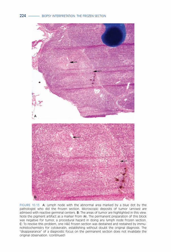
identification of nodal metastasis. The immediate identification of nodal metastases may initiate the performance or extent of a neck dissec-tion. The intraoperative diagnosis of carcinoma in lymph nodes is for the most part nonproblematic (11,12). However, finding small metastases, false positives, and false negatives are as important here as in the evalua-tion of lymph nodes from other organ systems. if a small lesion is identi-fied, calling attention to it by dotting the area is especially helpful to a subsequent examiner, especially in the event the focus disappears on the permanent sections (Fig. 10.13). To this must be added the possibility of treatment effects in the form of xanthogranulomatous and fibrotic reac-tions. in some studies, frozen section analysis of sentinel lymph nodes demonstrates a sensitivity of 93% and a negative predictive value of 94% (13). Frozen section of lymph nodes is contraindicated in cases where lymphoproliferative lesions are suspected, since no therapy is imminent and valuable tissue is wasted. However, the submission of fresh tissue is needed for triage purposes (flow cytometry, cytogenetics, and imprints).
Salivary gland Tumors: Indications and Intraoperative Questions
The major indications are similar to those involving mucosal lesions: Tumor identification and margin status. As in other settings, frozen sec-tion should not be initiated unless the reported diagnosis will directly influence surgical action. The detailed histopathologic features of the many benign and malignant salivary gland tumors are beyond the scope of this discussion and are best covered in standard texts of surgical pathology or in specialty texts on head and neck pathology. While the argument can be made that detailed knowledge of the histopathologic features of the major tumor types (both benign and malignant) is essential in intraopera-tive evaluation, it may be more productive to pay attention to patterns of growth and infiltration as well as cytologic features which are generically indicative of benign or malignant tumors. imprints in this setting are help-ful. Therefore, the intraoperative setting allows a certain relaxation in the
LWBK1296-CH10_p211-233.indd 223 19/09/13 12:41 PM
224 Biopsy interpretation: tHe Frozen section
A
B
FIgure 10.13 A: Lymph node with the abnormal area marked by a blue dot by the pathologist who did the frozen section. Microscopic deposits of tumor (arrows) are admixed with reactive germinal centers. B: the areas of tumor are highlighted in this view. note the pigment artifact as a marker from (A). the permanent preparation of this block was negative for tumor, a procedural hazard in doing any lymph node frozen section. C: to resolve this problem, one H&e frozen section was destained and restained by immu-nohistochemistry for cytokeratin, establishing without doubt the original diagnosis. the “disappearance” of a diagnostic focus on the permanent section does not invalidate the original observation. (continued )
LWBK1296-CH10_p211-233.indd 224 19/09/13 12:41 PM

Head and neck 225
C
FIgure 10.13 (continued )
exactness of classification of individual salivary gland tumors and relies foremost on the recognition of a benign or malignant lesion.
Primary tumors of the parotid gland comprise the largest number of salivary gland tumors and most of these are benign. eighty percent of parotid tumors, 50% of submandibular and sublingual tumors, and 20% of minor salivary gland tumors are benign. The most common tumor is pleomorphic adenoma followed by Warthin tumor. in the united States, salivary gland malignancies are uncommon, accounting for 6% of all head and neck cancers and 0.3% of all malignancies. common malignant tumors include mucoepidermoid carcinoma, adenoid cystic carcinoma, carcinoma ex-pleomorphic adenoma, and polymorphous low-grade ade-nocarcinoma. The successful frozen section recognition of salivary gland tumors varies and may be related to the relative infrequency of these enti-ties overall and their acknowledged innate histologic heterogeneity.
specimen handling. The gross examination of salivary gland tumors is critical, even if determinations of benign or malignant cannot be made. To this end, orientation of the specimen by the surgeon is an advantage, and if this is provided, the specimen should be inked appropriately. This exam-ination not only directs the sites of possible frozen section samplings, but also directs attention to those areas that must be studied on the routine permanent sections. Anatomically, the parotid gland lobes are delineated by being lateral (superficial) or medial (deep) to the facial nerve. Since most parotid gland tumors are located laterally, superficial lobectomies are the most common specimens. Tumors involving the sublingual or subman-dibular glands are usually treated by complete excision; intraoral minor salivary gland lesions are resected similarly to defined mucosal lesions.
LWBK1296-CH10_p211-233.indd 225 19/09/13 12:41 PM

226 Biopsy interpretation: tHe Frozen section
Benign and malignant tumors of the major salivary glands do not consistently follow rules of gross representation that indicate their biologic behavior especially regarding circumscription and local invasion. Figure 10.9 shows an oriented superficial lobectomy of a pleomorphic adenoma (mixed tumor) with multilobulation but without uniform circumscription, a somewhat worrisome feature for a benign tumor. Gross features such as this could be related to a recurrence of a previously incompletely or improperly resected tumor. in e-Figure 10.9, the circumscribed lesion is an acinic cell carcinoma (a diagnosis not established at frozen section), which extends to the inked edge of the specimen. Although this is a superficial lobectomy, there is gross involvement of the margin, easily communicated in an intraoperative setting. A frozen section is not required to advise the removal of additional subcutaneous tissue. The high-grade mucoepider-moid carcinoma grossly depicted in e-Figure 10.10 not only extends to the inked edge but is not well circumscribed, extending into the adjacent sali-vary gland and soft tissue. Since the diagnosis of malignancy had been established previously by fine needle aspiration (FnA), the surgeon was satisfied by a gross examination of the margins.
frozen section examination. masses in the head and neck, including salivary gland lesions, are easily accessible to FnA, which is widely accepted as a first-line diagnostic procedure because it is minimally inva-sive, virtually lacks complications, and is done in an outpatient setting. So, even prior to the receipt of the specimen in surgical pathology, the surgeon may have a specific diagnosis or at least a general idea of the diagnosis. The accuracy of FnA may be quite comparable to that of intraoperative frozen section (14); the use of both techniques may provide a useful redundancy in successful surgical management. it is, therefore, advantageous for the pathologist to at least know the FnA results; better to have the slides avail-able for review concurrent with the frozen section.
BenIgn tumors. The most common major or minor salivary gland neo-plasm is pleomorphic adenoma comprising 60% of all benign salivary gland tumors. A slowly growing, firm, painless mass, it most frequently occurs in the superficial lobe where a major hazard is compromise of the facial nerve by extrinsic compression. Grossly, tumors in the parotid often have a capsule while those originating in the minor salivary glands do not. The cut surface is tan white and uniform. Local recurrences are typically associated with previous attempts at enucleation. The recurrences, although histologically benign, are often multifocal, randomly scattered through remaining salivary gland and adjacent soft tissue (Fig. 10.9).
The cytomorphologic diversity of pleomorphic adenoma is well known and is related to the dual cellular components of epithelial and myoepithelial cells. The latter contributes to a fibrocollagenous, myxoid, and chondroid matrix. in the frozen section setting, the amount of each component is highly variable, not predictable, and the source of potential diagnostic problems. Frozen section sampling to demonstrate both cell components may not be consistently successful.
LWBK1296-CH10_p211-233.indd 226 19/09/13 12:41 PM

Head and neck 227
The major reason to establish the diagnosis of pleomorphic adenoma is to limit the operation to a superficial lobectomy in the case of a parotid tumor or a simple excision in the case of a minor salivary gland location. This is especially true in the absence of a preoperative FnA. A malignant diagnosis could result in a wider excision and sacrifice of the facial nerve. The diagnostic challenges are confronted in tumors more cellular than usual, raising the differential diagnosis of mucoepidermoid carcinoma or carcinoma ex-pleomorphic adenoma. occasional cribriform architecture raises the possibility of adenoid cystic carcinoma.
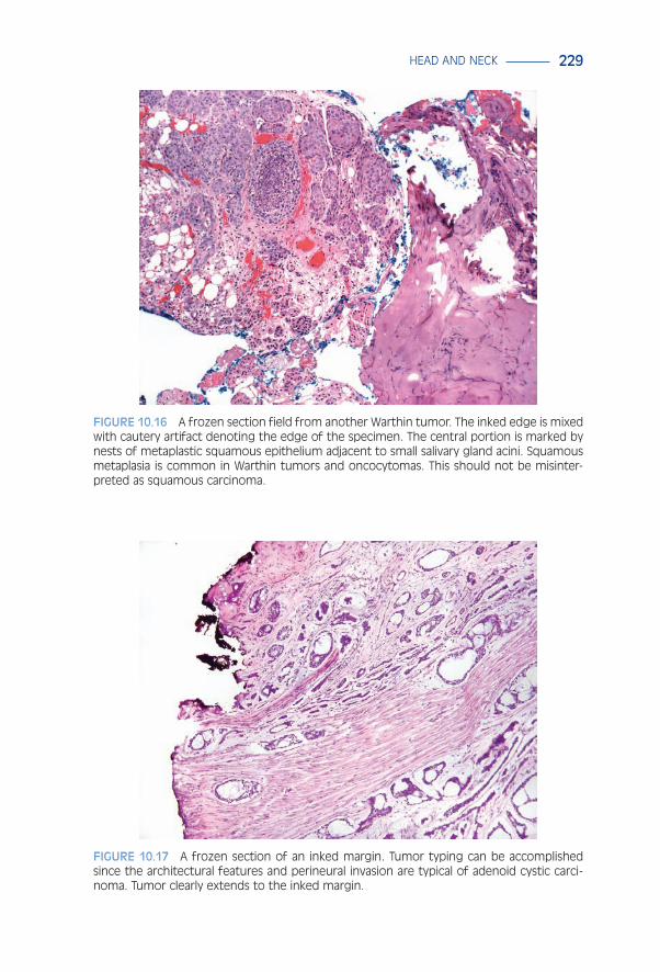
The second most common tumor in the major salivary glands is the Warthin tumor. most common in the parotid, the gross appearance con-sists of a poorly circumscribed, soft, and fluid-filled brown to tan tumor (Fig. 10.14). The characteristic admixture of mature lymphocytes and oncocytes is often easily appreciated on an imprint (Fig. 10.15A) and reflected histologically on the frozen section (Fig. 10.15B). difficulty may arise if the frozen section demonstrates the squamous metaplasia occa-sionally seen in these tumors (15). The metaplasia may be spontaneous, secondary to in vivo fluid extravasation or possibly to previous FnA attempts, and may be extensive but should not be misinterpreted as squa-mous carcinoma (Fig. 10.16).
malIgnant tumors. The critical role of frozen section in salivary gland tumor management may be in the assessment of both margin status and the distinction of a benign from a malignant tumor. Frozen sections for margin assessment should be taken from the closest margin as determined by gross examination through serial cuts. There is no magic quantitative
FIgure 10.14 Gross photograph of a Warthin tumor. a soft, circumscribed brown mass adjacent to lobulated, yellow-white salivary gland.
LWBK1296-CH10_p211-233.indd 227 19/09/13 12:41 PM

228 Biopsy interpretation: tHe Frozen section
distance agreed upon to constitute an adequate or close margin, so an actual measurement may be optimal. A positive margin should consist of tumor actually abutting the inked edge. Figure 10.17 illustrates an adenoid cystic carcinoma histologically extending to the inked margin. in this case, a good gross examination led to proper tissue selection. The tumor type was also easily recognized morphologically.
A
B
FIgure 10.15 A: imprint of a Warthin tumor demonstrating a dense background of small, mature lymphocytes. a solitary oncocyte is at the center of the field. B: Frozen section of a typical Warthin tumor. Uninvolved salivary gland tissue (left) is adjacent to a partially cystic tumor, the spaces of which are lined by oncocytes and abutted by dense lymphoid tissue. Blue ink denotes the specimen margin.
LWBK1296-CH10_p211-233.indd 228 19/09/13 12:41 PM
Head and neck 229
FIgure 10.16 a frozen section field from another Warthin tumor. the inked edge is mixed with cautery artifact denoting the edge of the specimen. the central portion is marked by nests of metaplastic squamous epithelium adjacent to small salivary gland acini. squamous metaplasia is common in Warthin tumors and oncocytomas. this should not be misinter-preted as squamous carcinoma.
FIgure 10.17 a frozen section of an inked margin. tumor typing can be accomplished since the architectural features and perineural invasion are typical of adenoid cystic carci-noma. tumor clearly extends to the inked margin.
LWBK1296-CH10_p211-233.indd 229 19/09/13 12:41 PM
230 Biopsy interpretation: tHe Frozen section
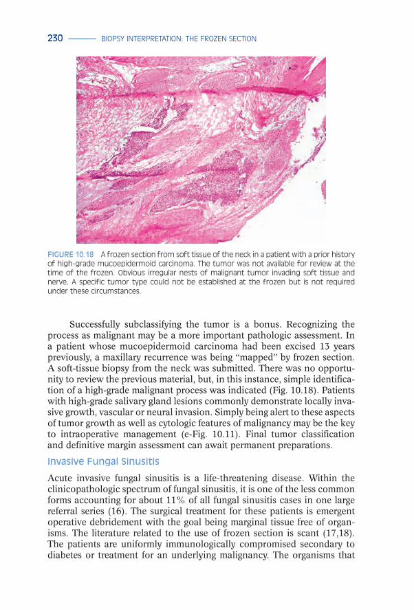
Successfully subclassifying the tumor is a bonus. recognizing the process as malignant may be a more important pathologic assessment. in a patient whose mucoepidermoid carcinoma had been excised 13 years previously, a maxillary recurrence was being “mapped” by frozen section. A soft-tissue biopsy from the neck was submitted. There was no opportu-nity to review the previous material, but, in this instance, simple identifica-tion of a high-grade malignant process was indicated (Fig. 10.18). Patients with high-grade salivary gland lesions commonly demonstrate locally inva-sive growth, vascular or neural invasion. Simply being alert to these aspects of tumor growth as well as cytologic features of malignancy may be the key to intraoperative management (e-Fig. 10.11). Final tumor classification and definitive margin assessment can await permanent preparations.
Invasive Fungal Sinusitis
Acute invasive fungal sinusitis is a life-threatening disease. Within the clinicopathologic spectrum of fungal sinusitis, it is one of the less common forms accounting for about 11% of all fungal sinusitis cases in one large referral series (16). The surgical treatment for these patients is emergent operative debridement with the goal being marginal tissue free of organ-isms. The literature related to the use of frozen section is scant (17,18). The patients are uniformly immunologically compromised secondary to diabetes or treatment for an underlying malignancy. The organisms that
FIgure 10.18 a frozen section from soft tissue of the neck in a patient with a prior history of high-grade mucoepidermoid carcinoma. the tumor was not available for review at the time of the frozen. obvious irregular nests of malignant tumor invading soft tissue and nerve. a specific tumor type could not be established at the frozen but is not required under these circumstances.
LWBK1296-CH10_p211-233.indd 230 19/09/13 12:41 PM

Head and neck 231
are typically associated with these infections, for example, Aspergillus and Mucor species, exhibit soft tissue, vascular and neural invasions. These features result in extensive necrosis, and purulence in the paranasal sinuses (Figs. 10.19 and 10.20). However, the organisms are easily recog-nized by standard hematoxylin and eosin stains on both frozen and per-manent sections. Tissue sections demonstrate organisms invading parana-sal sinus soft tissue, blood vessels, and nerves (e-Fig. 10.12). occasionally, tissue smears or sections stained by diff-Quik will be effective in demon-strating organisms (e-Fig. 10.13). The mortality rate in the short term from acute fungal sinusitis is approximately 80%.
Contraindications for Head and Neck Frozen Section
1. evaluation of small tissue samples when additional sampling for rou-tine processing is not planned.
2. evaluation of samples that do not directly impact the immediate sur-gical management of the patient.
3. evaluation of cutaneous lesions clinically suspicious for primary melanoma.
4. diagnostic lymph node biopsies, especially for hematopoietic dis-ease. Lymph nodes should, however, be submitted fresh for appropri-ate triage; for example, flow cytometry, imprints, microbiology.
5. evaluation of fat, heavily calcified, or ossified tissue.
FIgure 10.19 nasal sinus tissue from a patient with acute invasive fungal sinusitis. a por-tion of metaplastic squamous mucosa and underlying minor salivary gland tissue are sepa-rated by a focus of inflammation and a small colony of broad-based nonseptate hyphae, proven Mucor by culture.
LWBK1296-CH10_p211-233.indd 231 19/09/13 12:41 PM

232 Biopsy interpretation: tHe Frozen section
in the head and neck, as elsewhere, the potential for inappropriate utilization of the frozen section requires vigilance by the pathologist. Potential hazards include the misuse of limited material, the misconstru-ing of the frozen section as a shortcut to well-fixed and processed tissue, or the improper triage of diagnostic samples. The request for a frozen sec-tion does not mean it must always be honored. it is axiomatic that what we do as pathologists must have clinical relevance and the information we deliver must serve the patients’ best interests. Head and neck cancers are a challenge in this regard and optimally involve collaboration rather than contention with our surgical colleagues.
reFereNCeS
1. Jones AS, Bin Hanafi Z, nadapalan V, et al. do positive resection margins after ablative surgery for head and neck cancer adversely affect prognosis? A study of 352 patients with recurrent carcinoma following radiotherapy treated by salvage surgery. Br J Cancer. 1996;74:128–132.
2. Brandwein-Gensler m, Teixeira mS, Lewis cm, et al. oral squamous cell carcinoma: Histologic risk assessment, but not margin status, is strongly predictive of local disease-free and overall survival. Am J Surg Pathol. 2005;29:167–178.
3. Batsakis JG. Surgical excision margins: A pathologist’s perspective. Adv Anat Pathol. 1999;6:140–148.
4. ribeiro nF, Godden dr, Wilson Ge, et al. do frozen sections help achieve adequate surgical margins in the resection of oral carcinoma. Int J Oral Maxillofacial Surg. 2003;32:152–158.
FIgure 10.20 an abscess in the paranasal sinus soft tissue, exhibiting necrotic skeletal muscle and broad-based predominantly nonseptate fungi (arrows), proven Mucor by culture.
LWBK1296-CH10_p211-233.indd 232 19/09/13 12:41 PM

Head and neck 233
5. Sutton dn, Brown JS, rogers Sn, et al. The prognostic implications of the surgical margin in oral squamous cell carcinoma. Int J Oral Maxillofac Surg. 2003;32:30–34.
6. Spiro rH, Buillamondegui o, Paulino AF, et al. Pattern of invasion and margin assess-ment in patients with oral tongue cancer. Head Neck. 1999;21:408–413.
7. Vikram B, Strong eW, Shah JP, et al. Failure at primary site following multimodality treatment in advanced head and neck cancer. Head Neck. 1984;6:720–723.
8. ord rA, Aisner S. Accuracy of frozen sections in assessing margins in oral cancer resec-tion. J Oral Maxillofac Surg. 1997;55:663–669.
9. Kerawala cJ, ong TK. relocating the site of frozen sections—is there room for improve-ment? Head Neck. 2001;23:230–232.
10. cooley mL, Hoffman HT, robinson rA. discrepancies in frozen section mucosal margin tissue in laryngeal squamous cell carcinoma. Head Neck. 2002;24:262–267.
11. Luna mA. uses, abuse and pitfalls of frozen section diagnoses of diseases of the head and neck. in: Barnes L, ed. Surgical Pathology of the Head and Neck. new york, ny: marcel dekker; 2000:2–12.
12. Asthana S, deo SV, Shukla nK, et al. intraoperative neck staging using sentinel node biopsy and imprint cytology in oral cancer. Head Neck. 2003;25:368–372.
13. Tschopp L, nuyens m, Stauffer e, et al. The value of frozen section analysis of the sen-tinel lymph node in clinically n0 squamous cell carcinoma of the oral cavity and oro-pharynx. Otolaryngol Head Neck Surg. 2005;132(1):99–102.
14. Wong dS. Frozen section during parotid surgery revisited: efficacy of its applications and changing trend of indications. Head Neck. 2002;24:191–197.
15. Taxy JB. necrotizing squamous/mucinous metaplasia in oncocytic salivary gland tumors: A potential diagnostic problem. Am J Clin Pathol. 1992;97:40–45.
16. montone KT, Livolsi VA, Feldman md, et al. Fungal rhinosinusitis: A retrospective microbiologic and pathologic review of 400 patients at a single university medical center. Int J Otolaryngol. 2012;2012:1–9. doi:10.1155/2012/684835.
17. Hofman V, castillo L, Betis F, et al. usefulness of frozen section in rhinocerebral mucor-mycosis: diagnosis and management. Pathology. 2003;35:212–216.
18. Taxy JB, el-Zayaty S, Langerman A. Acute fungal sinusitis: natural history and the role of frozen section. Am J Clin Pathol. 2009;132:86–93.
LWBK1296-CH10_p211-233.indd 233 19/09/13 12:41 PM