he printed in u.s.a. the high resolution crystal structure ... · mans like hemolytic anemia (1, 2)...
TRANSCRIPT
The High Resolution Crystal Structure of Yeast Hexokinase PIIwith the Correct Primary Sequence Provides New Insights intoIts Mechanism of Action*
Received for publication, December 27, 1999, and in revised form, March 27, 2000Published, JBC Papers in Press, April 3, 2000, DOI 10.1074/jbc.M910412199
Paula R. Kuser‡, Sandra Krauchenco‡, Octavio A. C. Antunes§, and Igor Polikarpov‡¶
From the ‡Laboratorio Nacional de Luz Sıncrotron, Campinas, Sao Paulo, Brazil, Caixa Postal 6192-CEP 13083-970and §Departamento Quımica Inorganica, Instituto de Quımica, UFRJ, Rio de Janeiro, RJ, Brazil
Hexokinase is the first enzyme in the glycolytic path-way, catalyzing the transfer of a phosphoryl group fromATP to glucose to form glucose 6-phosphate and ADP.Two yeast hexokinase isozymes are known, namely PIand PII. The crystal structure of yeast hexokinase PIIfrom Saccharomyces cerevisiae without substrate orcompetitive inhibitor is determined and refined in atetragonal crystal form at 2.2-Å resolution. The foldingof the peptide chain is very similar to that of Schisto-soma mansoni and previous yeast hexokinase modelsdespite only 30% sequence identity between them. Dis-tinct differences in conformation are found that accountfor the absence of glucose in the binding site. Compari-son of the current model with S. mansoni and yeasthexokinase PI structures both complexed with glucoseshows in atomic detail the rigid body domain closureand specific loop movements as glucose binds. A hydro-phobic channel formed by strictly conserved hydropho-bic residues in the small domain of the hexokinase isidentified. The channel’s mouth is close to the active siteand passes through the small domain to its surface. Thepossible role of the observed channel in proton transferis discussed.
Hexokinase (HK),1 or ATP:D-hexose 6-phosphotransferase, isa member of the kinase family of tissue-specific isoenzymes(EC 2.7.1.1). Reduction in its activity causes illnesses in hu-mans like hemolytic anemia (1, 2) and cardiomyopathy (3).Mutations in glucokinase (hexokinase IV) are associated withearly onset non-insulin-dependent diabetes mellitus (4, 5).Hexokinase has been a target for the development of efficientinhibitors in the search for new drugs against diseases causedby trypanosomes (6) and has been used in the construction ofbiosensors capable of detecting glucose (7).
Hexokinase catalyzes the transfer of an ATP g-phosphategroup to the 6-position on the glucose (Glc) ring as follows.
Glucose 1 ATPO¡
Mg21 1 hexokinaseGlucose-6-P 1 ADP 1 H1
REACTION 1
Cloning the hexokinase genes from Saccharomyces cerevisiaehas shown that there are two isoenzymes of hexokinase inyeast: PI and PII, with an overall homology in their amino acidsequences of about 76% (8, 9). Hexokinase PII is the predomi-nant hexose kinase in S. cerevisiae grown on glucose (10) and isrequired for the catabolite repression, by glucose, of the expres-sion of other genes (11–13). The yeast HKs are known to existas phosphoproteins in vitro (14) and in vivo (15), with thedimer-monomer equilibrium affected by phosphorylation. Thein vivo phosphorylation site has been identified as Ser15 (16).The crystallographic structures of yeast hexokinase PI com-plexed with glucose (PI-Glc) refined at 3.5-Å resolution (17, 18)and the hexokinase PII from yeast complexed with the compet-itive inhibitor, ortho-toluoylglucosamine (PII-OTG), deter-mined at 2.1-Å resolution (19, 20), have already been reported.
The original structures of yeast hexokinases were deter-mined without a primary amino acid sequence, which did notbecome available until 1985 (8, 9). The identity of the sidechains was deduced from inspection of the electron density. Infact, only a 30% identity can be found between the primarysequence of the crystallographic models and the one obtainedfrom cDNA sequencing.
In an attempt to define the structure and function of thisenzyme more clearly, we solved the crystal structure of yeasthexokinase PII to 2.2-Å resolution, refining the model with thecorrect amino acid sequence. This is the first high resolutionhexokinase structure solved without a bound substrate or com-petitive inhibitor. We compare it with the structures of humanhexokinase determined to 2.8-Å resolution (21–23), Schisto-soma mansoni hexokinase also solved to 2.8-Å resolution (24),and previous structures of yeast hexokinase PI and PII (17–20).
The primary sequence of yeast hexokinase PII is comparedwith the amino acid sequences of other hexokinases, and apossible role for the conserved amino acids is proposed. Glucoseand ATP binding sites and a possible reaction mechanism arediscussed. We demonstrate the existence of a channel, formedby the conserved hydrophobic residues Ile85, Leu87, Leu127,Ile131, Leu135, Met139, Leu153, Phe155, Phe157, Phe178, Leu192,and Ile196, which passes from the active site of the enzymethrough the small domain to the exterior of the protein. Itspossible role as a proton sink is discussed.
EXPERIMENTAL PROCEDURES
Crystallization—The protein material used for crystallization waspurchased from Sigma and further purified using an anion exchangecolumn. Crystallization was carried out using the sitting-drop vapordiffusion method. The drops were composed of 3 ml of protein solutionand 3 ml of crystallization solution. The volume of the reservoir was 1ml. Crystal screenings were carried out at room temperature, varyingthe concentration of precipitant from 1.6 to 2.4 M ammonium sulfate in100 mM potassium phosphate buffer. Needles were observed underseveral conditions, and some data were collected for these crystals,
* This work was supported by Conselho Nacional de DesenvolvimentoCientıfico e Tecnologico Grant 150088-5 and Fundacao de Amparo aPesquisa do Estado de Sao Paulo Grant 99/03387-4. The costs of pub-lication of this article were defrayed in part by the payment of pagecharges. This article must therefore be hereby marked “advertisement”in accordance with 18 U.S.C. Section 1734 solely to indicate this fact.
¶ To whom correspondence should be addressed. Tel.: 55 19 287 4520;Fax: 55 19 287 4632; E-mail: [email protected].
1 The abbreviations used are: HK, hexokinase; OTG, ortho-toluoyl-glucosamine; r.m.s., root mean square.
THE JOURNAL OF BIOLOGICAL CHEMISTRY Vol. 275, No. 27, Issue of July 7, pp. 20814–20821, 2000© 2000 by The American Society for Biochemistry and Molecular Biology, Inc. Printed in U.S.A.
This paper is available on line at http://www.jbc.org20814
by guest on April 4, 2019
http://ww
w.jbc.org/
Dow
nloaded from

diffracting to 2.8-Å resolution. A single crystal, from which diffractiondata were collected, had an elongated bipiramidal shape, unlike themore usual thin needles. Its dimensions were 0.25 3 0.4 3 1.0 mm.
X-ray Diffraction Data—Diffraction data to 2.2 Å were collected atroom temperature (127 °C) on a MAR image plate system at the ded-icated protein crystallography beamline of the Brazilian synchrotronlight source (25, 26) and processed with the programs DENZO andSCALEPACK (27). Data were collected on frames of 1.0° rotation withthe same amount of x-ray dose per frame. The crystal-to-detector dis-tance was 200 mm. A total of 53,732 reflections were measured andmerged to 24,924 unique reflections, with a completeness of 94.9% forall data to 2.2 Å (Table I). The crystal belonged to tetragonal spacegroup I4, with cell constants of a 5 b 5 142.81 Å and c 5 58.46 Å.Estimation of the solvent content indicated that the crystal containedone molecule per asymmetric unit with Vm 5 2.7 Å Da21. The structurewas solved using Patterson search techniques. Rotational and transla-tional searches were performed with AMoRe (28) using data to 2.2 Åand a P152K mutant of yeast hexokinase (29) as the starting model. Aunique solution was found after rigid body refinement, with a correla-tion coefficient of 53.5% and an R-factor of 44.8%.
Refinement and Model Building—Repeated cycles of rebuilding in O(30) and refinement using REFMAC (62) with insertion of water mole-cules using ARPP (32) lowered the R-factor to a final value of 16.3% andR-free (33), calculated for 5% randomly chosen reflections that were notincluded in the refinement, to 24.6%. The root mean square (r.m.s.)deviation of bond lengths was 0.011 Å, and r.m.s. deviation of bondangles was 2.1°. A summary of the refinement statistics, together withother crystallographic and final model parameters, is given in Table I.Analysis of the final model using PROCHECK (40) indicates that 90.5%of the residues fall within “core” regions of the Ramachandran plot (34,35). No residues are found in disallowed regions. The G-factor of themodel is 20.06. The final model contains residues 18–486 and 442water molecules. No continuous density can be observed for the first 17residues of the N terminus. Yeast HK is susceptible to limited proteol-ysis by endogenous proteases, resulting in loss of the first 11 N-terminalamino acid residues (36, 37), which, together with the thermal disorder,is probably the reason for the lack of electron density at the N terminus.
The average thermal parameter for the model is 23.41 Å2, with thehighest B value of 72.44 Å2 for the side chain of residue Asp116, whichis located in a loop between b-strand b4 and a-helix a3. The final (2Fo
2 Fc) electron density, calculated using data to 2.2 Å, is well defined formost residues.
RESULTS AND DISCUSSION
Secondary and Tertiary Structure—The molecule has a palmshape, with approximate dimensions of 59 3 78 3 54 Å3, and isin its open conformation. It has the same a/b fold observed inother hexokinase structures (17–24). The polypeptide chain of486 residues is distinctly folded into two domains of unequalsize: the large and small domains. These are separated by adeep cleft containing the residues making up the enzyme activesite.
The secondary structure was analyzed using the PROMOTIF(39) program. In all, there are 14 a-helices, 13 b-strands, andthree 310-helices (Fig. 1). The large domain (residues 13–76 and212–457) comprises a six-stranded mixed b-sheet (b1, b9, b10,b11, b12, and b13) and a number of additional a-helices. Themixed sheet has 23,1,1,23x,21x topology in the Richardsonnotation (38). On one side, the sheet packs against the smalldomain, and on the other side it is shielded by several a-helices.The domain also contains a particularly long helix, a11 (36residues and 53.35 Å in length), which exhibits a slight bend.
The small domain comprises residues 77–211 and 458–486,and its dominant feature is a five-stranded mixed b-sheet (b2,b3, b4, b5, and b8), with a sheet topology of 21,21,3x,1x. Thesheet is flanked by two helices on one side and by one helix onthe other. The domain also has an additional b-sheet formed bytwo antiparallel strands (b6 and b7).
Two regions of distortion were identified in the b-sheets,both classified as classic parallel b-bulges, one involvingb-strands b9 and b12 and the other involving b5 and b8. Fiveb-hairpins are also present in the structure, between antipar-allel b-strands b2:b3, b3:b4, b6:b7, b9:b10, and b10:b11.
All these secondary structure elements match those presentin the human HK structure apart from three additional 310-helices (a59, 245–251, a50, 269–273; a129, 445–449) found in theyeast HK structure. The structure of S. mansoni HK shows thesame secondary structural pattern. Direct comparison with theprevious HK structures (17–20) can only be somewhat approx-imate due to the limited refinement and the uncertain aminoacid composition of those earlier models. However, such anapproximate comparison suggests that most of the secondarystructural elements in the current structure have their equiv-alents in the previous ones.
The superposition of the yeast and S. mansoni hexokinasestructures (Fig. 2) clearly shows the open conformation of thepolipeptide chain (17–20).
Conserved Amino Acid Residues—The primary sequencealignment of a selection of proteins from the hexokinase familyis shown in Fig. 3. This clearly demonstrates extensive simi-larity between the N- and C-terminal halves of type I humanhexokinase, rat hexokinase, and hexokinase from S. mansoniand between these and yeast hexokinase, consistent with thegene duplication-fusion concept proposed by Colowick (36). Ap-proximately 34% of the amino acid residues are conserved in allmembers of the family, and 13% are perfect matches. Thestrong conservation of these residues implies their relevance tobiological function. Various strictly conserved amino acid resi-dues are present in the binding site. A high number of glycineresidues are also conserved (Gly76, Gly80, Gly88, Gly89, Gly154,Gly233, Gly235, Gly297, Gly307, Gly418, and Gly461). These resi-dues are located at the ends of b-strands or a-helices, changingthe direction of the chain. Their conservation probably givesthe hexokinase molecule the flexibility necessary for bindingglucose and ATP. Some of them are directly involved in theactive site formation and are essential for enzymatic activity(see discussion below).
Comparison of the HK Structures and the ConformationalChanges—Fig. 2a shows a comparison of yeast hexokinase PII
TABLE IStatistics of data collection and refinement
Parameters Values
Data collectionResolution limit of data (Å) 2.2Number of measurements 53,732Number of measurements . 1s 48,332Number of unique reflections 28,523Completeness of data (last shell) (%) 95.1 (96.8)Rsym
a 11.3Refinement
Resolution range for refinement (Å) 13.00–2.2 ÅTotal number of atoms 3671Total number of solvent molecules 442R-factorb 16.27Rfree
c 24.60Average B factor
All atoms (Å2) 23.41Solvents (Å2) 43.41
r.m.s. deviations from ideal geometrybonds (Å) 0.011bond angles (degrees) 2.1
Ramachandran plot statisticsResidues in core Ramachandran (%) 90.5Residues in disallowed regions (%) 0.0a Rsym 5 (uI 2 ^I&u/(I 3 100.b Crystallographic R-factor 5 ( (iFobsu-uFcalci)/(Fobs where Fobs and
Fcalc are the observed and calculated structure factor amplitudes re-spectively.
c Rfree is the crystallographic R-Factor calculated for a subset ofrandomly selected reflections (5%) not used in the phasing process.
Crystal Structure of Yeast Hexokinase PII 20815
by guest on April 4, 2019
http://ww
w.jbc.org/
Dow
nloaded from

FIG. 1. Schematic diagram of the yeast hexokinase PII model (a) and stereo C-a trace (b). The chain is color-coded from blue (Nterminus) to red (C terminus). This model is in an open conformation, and the active site is located between the two domains. Secondary structureassignments are made in accordance with the method of Kabsh and Sander (60). This figure was generated using MOLSCRIPT (61).
Crystal Structure of Yeast Hexokinase PII20816
by guest on April 4, 2019
http://ww
w.jbc.org/
Dow
nloaded from
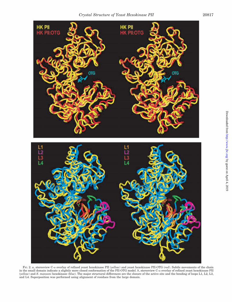
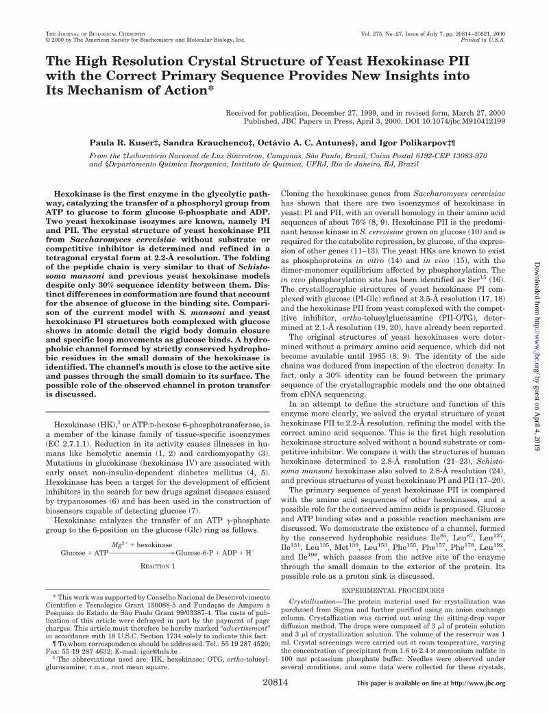
FIG. 2. a, stereoview C-a overlay of refined yeast hexokinase PII (yellow) and yeast hexokinase PII-OTG (red). Subtle movements of the chainin the small domain indicate a slightly more closed conformation of the PII-OTG model. b, stereoview C-a overlay of refined yeast hexokinase PII(yellow) and S. mansoni hexokinase (blue). The major structural differences are the closure of the active site and the bending of loops L1, L2, L3,and L4. Superposition was performed using alignment of residues from the large domain.
Crystal Structure of Yeast Hexokinase PII 20817
by guest on April 4, 2019
http://ww
w.jbc.org/
Dow
nloaded from
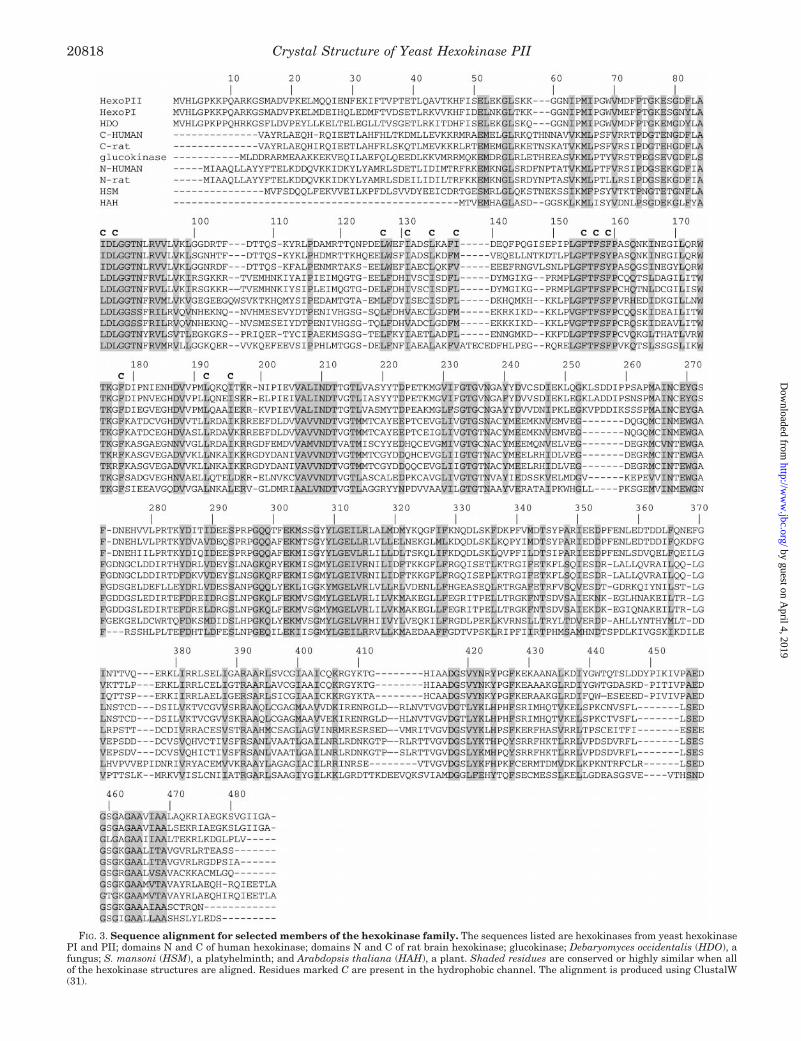
FIG. 3. Sequence alignment for selected members of the hexokinase family. The sequences listed are hexokinases from yeast hexokinasePI and PII; domains N and C of human hexokinase; domains N and C of rat brain hexokinase; glucokinase; Debaryomyces occidentalis (HDO), afungus; S. mansoni (HSM), a platyhelminth; and Arabdopsis thaliana (HAH), a plant. Shaded residues are conserved or highly similar when allof the hexokinase structures are aligned. Residues marked C are present in the hydrophobic channel. The alignment is produced using ClustalW(31).
Crystal Structure of Yeast Hexokinase PII20818
by guest on April 4, 2019
http://ww
w.jbc.org/
Dow
nloaded from

with the previously determined PII-OTG model. An r.m.s. de-viation calculated using the LSQMAN program (41) for 452 C-aatom pairs is 0.93 Å. The main difference is that the presentstructure adopts a slightly more open conformation, suggestingthat OTG binding induces a slight closure of the cleft betweentwo domains.
A comparison with hexokinase from S. mansoni, determinedas a complex with glucose, reveals a movement of the domainsclosing the cleft (Fig. 2b). The overall r.m.s. deviation, calcu-lated for 414 C-a atom pairs is 4.7 Å. However, if the twodomains are considered separately, the superposition of thelarge domains gives an r.m.s. deviation of 1.8 Å for 320 C-aatom pairs, while the superposition of the small domains givesan r.m.s. deviation of 2.2 Å for 261 C-a atom pairs. Similarchanges in conformation have been observed in the yeast PI-Glc complex (17) with an r.m.s. deviation of 8.1 Å for 409 C-aatom pairs.
When the large domains of these molecules are superposed,it becomes clear that, in addition to the rigid body closure of thesmall domain, four peptide segments, namely residues 87–92(L1), 115–124 (L2), 158–163 (L3), and 174–178 (L4) (yeasthexokinase PII numbering), move forward to embrace the bind-ing site. These loops move by as much as 8 Å measured at C-aatom positions. The movement of these loops seems to be func-tionally important to complete the formation of the glucosebinding site and to preform the nucleotide binding site. Two ofthe loops, namely L1 (87LGGTN91) and L4 (174WTKGF178), arecomposed of amino acid residues conserved in all but two of thehexokinase sequences listed in Fig. 3. In the N-terminal regu-latory domains of rat and human hexokinases, residues Thr90
and Asn91 of the loop L1 are substituted by serines, whereasthe residue Gly177 of the loop L4 is substituted by an arginine.These substitutions might be directly related to the lack ofenzymatic activity of these domains.
In addition to the significant conformational changes of theloops involved in glucose and ATP binding, differences in theconformations of the external loops are also observed, mainly inthe positions of insertions and deletions in respective aminoacid sequences.
Comparison of the current structure with the models of hex-okinase complexed with substrate provides precise informationabout the conformational changes this enzyme undergoes uponsubstrate binding and confirms the “induced fit” mechanism
theory (42) and previous observations that initial recognition ofglucose by HK is followed by closure of the cleft altering theenzyme-substrate interaction (43).
The Glucose Binding Site—The glucose binding site on ourmodel is in close agreement with the binding site as previouslyelucidated (17–20). Asp211, identified as a catalytic base, makesa hydrogen bond with the hydroxyl at position 6 of glucose inboth open and closed forms of HK, whereas side chains of theamino acid residues Asn237, Glu269, and Glu302 are at hydrogenbonding distances from the O-4, O-3, and O-1 atoms of glucose,respectively. Initial glucose recognition followed by the closureof the cleft between the two HK domains brings amino acidresidues of the small domain, notably Thr172 and Lys173, withinhydrogen interaction distances of the substrate.
Crystallographic structures clearly show that in the closedconformation Ser158 interacts with hydroxyl group 3 of theglucose molecule via a carboxyl oxygen (distance 3.22 Å in theS. mansoni hexokinase structure), which confirms the resultsof mutational studies (44); mutation of Ser158 to Ala impairsHK catalytic activity, implying functional significance of thehydrogen bonding via the side chain. Ser158 is also strictlyconserved among HK sequences. It has also been shown thatthe binding of certain types of glucose inhibitors, such as D-xylose or D-lyxose, promotes the short lived hydrolytic activityof the enzyme followed by inactivation of the enzyme via auto-phosphorylation (36, 45, 46). The autophosphorylation site hasrecently been identified as Ser158 (47). Taken together, theseobservations support the idea that additional conformationalchanges might occur upon binding of ATP (48, 49) and thatSer158 might play an important role in the process of thephosphate transfer.
As observed by Steitz et al. (17–20), in the open conformationOTG forms few hydrogen bonds or Van der Waals contacts withthe amino acid residues of the small domain. Projection of OTGinto the S. mansoni model shows that the glucosamine positionis very close to the site occupied by glucose, but its toluoylgroup prevents complete domain closure. However, there isroom for a significantly larger closure of the HK domains thanthe comparison of yeast hexokinase PII and PII-OTG modelsreveals. Taking this into account, we believe that the stericclashes of the OTG toluoyl group with the amino acid residuesof the small domain prevent formation of an energetically fa-vorable closed conformation of PII-OTG complex. Recent scan-
FIG. 4. The sulfate ion bound to theactive site of yeast hexokinase. A sul-fate molecule, hydrogen-bonded to resi-dues Thr234 and Ser419 and to water mol-ecule W149 is shown as a ball-and-stickmodel. 2Fo 2 Fc density for the regionaround the sulfate anion is shown in blue.The sulfate molecule is located at the en-trance to the active site.
Crystal Structure of Yeast Hexokinase PII 20819
by guest on April 4, 2019
http://ww
w.jbc.org/
Dow
nloaded from
ning calorimetry measurements of the D-glucose binding to HKhave demonstrated that this process is entropy-driven and thatthe binding enthalpy is zero (50). The OTG toluoyl group in-teractions may make it impossible to trap the hexokinase in theclosed conformational state, with the open conformation beingthermodynamically more favorable.
The ATP Binding Site—Crystallographic analysis of a binarycomplex of yeast HK with 8-bromo-AMP and modeling of ATPinto the active site of the enzyme (49) indicated that amino acidresidues 344–348 and 422–424 interact with the adenine ringof ATP. Comparison of the three-dimensional structures of the44-kDa ATPase fragment of 70-kDa heat shock cognate(HSC70) protein (51), of actin (52), and of HK led to the iden-tification of an ATPase domain common to prokaryotic cellcycle proteins, sugar kinases, actin, and HSC70 proteins (53).The ATP binding pattern was defined as consisting of fivesequence motifs: phosphate 1 (residues 82–103), connect 1 (res-idues 203–223), phosphate 2 (residues 229–248), adenosine(residues 411–439), and connect 2 (residues 453–473) with anumber of Gly, Ser, Thr, and Asp residues being strictly con-served between all of the compared sequences.
The present model of yeast hexokinase was determined inthe absence of any substrate in the active site. However, in thelast stages of refinement a (Fobs 2 Fcalc) difference map exhib-ited pronounced electron density within the catalytic cleft, andthis density was modeled as a sulfate molecule. Sulfate waspresent in 2 M concentration in the crystallization conditions.Its well defined density is located inside the cleft, close to theposition of a sulfate ion observed in the S. mansoni hexokinaseand PI-Glc models. The sulfate ion was inserted into the modeland refined with an occupancy of 1.0. The thermal parametersare comparable with the average thermal parameter for sol-vent molecules and similar to the B value of water 149 to whichthe sulfate ion is bound. The sulfate group makes a hydrogenbond to the main chain nitrogens of residues Ser419 and Thr234
(Fig. 4). Remarkably, these are the strictly conserved residuesfrom the phosphate 2 (Thr234) and adenosine (Ser419) ATPasepattern recognition motifs (54). Based on these motifs, the ATPposition was modeled, by superposing the actin-ATP modelonto the present HK structure. Contrary to the suggestion inRef. 19, the sulfate ion does not occupy the position of the b- org-phosphate. Rather, it occupies a position close to the a-phos-phate of ATP. If ATP does indeed bind to HK in the modeledposition and conformation, its g-phosphate will be 3.7 Å from
the hydroxyl-6 of glucose. Interestingly, the b-phosphate inter-acts only with amino acid residues of the small domain. Thismeans that after the enzymatic reaction has taken place andthe HK cleft opens, the small domain will drag ADP away fromthe active site, opening the way to release Glc-6-P. This isconsistent with the view that glucose binds first and then ATP,whereas ADP is released first, followed by Glc-6-P (36).
The presence of a sulfate ion in the open conformation ofhexokinase proves that it does not provoke significant confor-mational change upon binding to the enzyme. The fact that thesame sulfate/phosphate anion binding site was repeatedlyfound in yeast (17–20), human (21–23), and S. mansoni (24)hexokinase models shows that the HK ATP binding site is ableto bind monophosphates. This may have functional importance.It is known, for example, that glucose binding is strongly pro-moted in the presence of 0.05 M phosphate (54). Phosphatebinding might therefore somehow restrict the conformations ofthe amino acid residues in the glucose binding site, facilitatingthe binding of glucose.
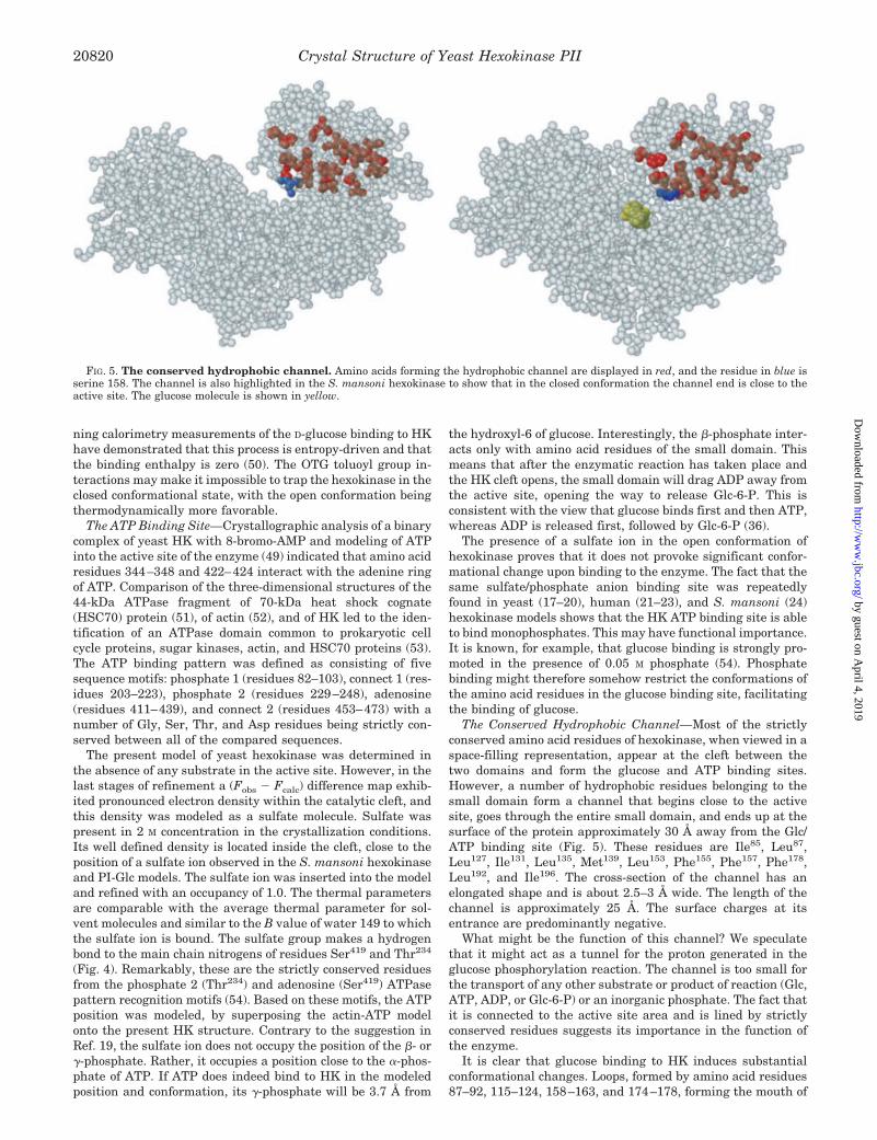
The Conserved Hydrophobic Channel—Most of the strictlyconserved amino acid residues of hexokinase, when viewed in aspace-filling representation, appear at the cleft between thetwo domains and form the glucose and ATP binding sites.However, a number of hydrophobic residues belonging to thesmall domain form a channel that begins close to the activesite, goes through the entire small domain, and ends up at thesurface of the protein approximately 30 Å away from the Glc/ATP binding site (Fig. 5). These residues are Ile85, Leu87,Leu127, Ile131, Leu135, Met139, Leu153, Phe155, Phe157, Phe178,Leu192, and Ile196. The cross-section of the channel has anelongated shape and is about 2.5–3 Å wide. The length of thechannel is approximately 25 Å. The surface charges at itsentrance are predominantly negative.
What might be the function of this channel? We speculatethat it might act as a tunnel for the proton generated in theglucose phosphorylation reaction. The channel is too small forthe transport of any other substrate or product of reaction (Glc,ATP, ADP, or Glc-6-P) or an inorganic phosphate. The fact thatit is connected to the active site area and is lined by strictlyconserved residues suggests its importance in the function ofthe enzyme.
It is clear that glucose binding to HK induces substantialconformational changes. Loops, formed by amino acid residues87–92, 115–124, 158–163, and 174–178, forming the mouth of
FIG. 5. The conserved hydrophobic channel. Amino acids forming the hydrophobic channel are displayed in red, and the residue in blue isserine 158. The channel is also highlighted in the S. mansoni hexokinase to show that in the closed conformation the channel end is close to theactive site. The glucose molecule is shown in yellow.
Crystal Structure of Yeast Hexokinase PII20820
by guest on April 4, 2019
http://ww
w.jbc.org/
Dow
nloaded from

the conserved hydrophobic channel, close up over the activesite, bringing the entrance to the channel into close proximitywith the ligand binding sites. The closed active site conforma-tion is probably completed after additional conformationalchanges that accompany ATP binding. Nucleophilic attack ofthe glucose’s 6-hydroxyl group by the g-phosphate of ATP ispromoted by Asp211 and Lys173 and is followed by “in-line”transfer of the g-phosphate group to glucose, presumablythrough the dissociative transitional state (55) and liberation ofa proton.
How does release of the reaction products occur? The drivingforce of product release is probably the repulsion between Glc-6-P and the b-phosphate of ADP. Since the b-phosphate isbound to residues of the small domain, its repulsion from theg-phosphate, now bound to glucose, would drive the HK open.Repulsion of these two phosphates will depend on the localproton concentration. Low active site local pH would shieldelectrostatic repulsion between the negative charges of thephosphates and impede release of ADP and Glc-6-P from theHK active site. Proton transfer through the hydrophobic chan-nel might provide the means for decreasing the concentrationof protons in the active site cavity, increasing ADP and Glc-6-Pmutual repulsion and facilitating the release of the reactionproducts. In a way, the putative channel function might besimilar and opposite to that of the ATPase synthase protonchannel (56–59).
If this hypothesis is valid, the disruption of the channelshould impede the reaction product release, whereas the inver-sion of the proton flux through the channel would change thereaction balance increasing the phosphate transferase activityof HK. In this situation, ATP and glucose should be producedfrom ADP and Glc-6-P by HK. Site-directed mutagenesis of thestrictly conserved hydrophobic amino acids forming the chan-nel will probably tell whether our hypothesis is correct and willshed light on the nature, purpose, and function of the hydro-phobic channel.
Acknowledgments—We thank Prof. Maria Lucia Bianconi and Prof.Jose Abrahao Neto for the kind gift of some of the enzyme and Alex-ander Golubev, Ricardo Aparıcio, and Elisabete de Souza for help withcrystallization. We are indebted to Dr. Roman Laskowski, Dr. Bill Boys,and Dr. Stefan Kycia for critically reading the manuscript and correct-ing the English.
REFERENCES
1. Valentine, W. N., Oski, F. A., Paglia, D. E., Baughan, M. A., Schneider, A. S.,and Naiman, J. L. (1967) N. Engl. J. Med. 276, 1–11
2. Magnani, M., Stocchi, V., Cucchiarini, L., Novelli, G., Lodi, S., Isa, L., andFornaini G. (1985) Blood 66, 690–697
3. Barrie, S. E., Saad, E. A., Ubatuba, S., Da Silva Lacaz, P., and Harris, P. (1979)Res. Commun. Chem. Pathol. Pharmacol. 23, 375–381
4. Vionnet, N., Stoffel, M., Takeda, J., Yasuda, K., Bell, G. I., Zouali, H., Lesage,S., Velho, G., Iris, F., Passa, P., Froguel, P., and Cohen, D. (1992) Nature356, 721–722
5. Gupta, B. L., Nehal, M., and Baquer, N. Z. (1997) Indian J. Exp. Biol. 35,792–795
6. Willson, M., Alric, I., Perie, J., and Sanejouand, Y. H. (1997) J. EnzymeInhibition 12, 101–121
7. Cheng, Q., and Stevens, R. C. (1997) Adv. Mater 9, 481–4838. Kopetzki, E., Entian, K., and Mecke, D. (1985) Gene (Amst.) 39, 95–1029. Frohlich, K., Entian, K., and Mecke, D. (1985) Gene (Amst.) 36, 105–111
10. Gancedo, J. M., Clifton, D., and Fraenkel, D. G. (1977) J. Biol. Chem. 252,4443–4444
11. Entian, K.-D. (1980) Mol. Gen. Genet. 178, 633–63712. Rose, M., Albig, W., and Entian, K.-D. (1991) Eur. J. Biochem. 199, 511–51813. De Winde, J. H., Crauwels, M., Hohmann, S., Thevelein, J. M., and
Winderickx, J. (1996) Eur. J. Biochem. 241, 633–64314. Fernandez, R., Herrero, P., Fernandez, M. T., and Moreno, F. (1986) J. Gen.
Microbiol. 132, 3467–347215. Vojtek, A. B., and Fraenkel, D. G. (1990) Eur. J. Biochem. 190, 371–37516. Behlke, J., Heidrich, K., Naumann, M., Muller, E.-C., Otto, A., Reuter, R., and
Kriegel, T. (1998) Biochemistry 37, 11989–1199917. Steitz, T. A., Fletterick, R. J., Anderson, W. F., and Anderson, Ch. M. (1976) J.
Mol. Biol. 104, 197–22218. Bennet, W. S., Jr., and Steitz, T. A. (1980) J. Mol. Biol. 140, 183–20919. Anderson, C. M., Stenkamp, R. E., McDonald, R. C., and Steitz, T. A. (1978) J.
Mol. Biol. 123, 15–3420. Anderson, C. M., Zucker, F. H., and Steitz, T. A. (1979) Science 204, 375–38021. Aleshin, A. E., Zeng, C., Bartunik, H. D., Fromm, H. J., and Honzatko, R. B.
(1998) J. Mol. Biol. 282, 345–35722. Aleshin, A. E., Zeng, C., Bourenkov, G. P., Bartunik, H. D., Fromm, H. J., and
Honzatko, R. B. (1998) Structure 6, 39–5023. Aleshin, A. E., Fromm, H. J., and Honzatko, R. B. (1998) FEBS Lett. 434,
42–4624. Mulichack, A. M., Wilson, J. E., Padmanabhan, K., and Garavito, R. M. (1998)
Nat. Struct. Biol. 5, 555–56025. Polikarpov, I., Perles, L. A., de Oliveira, R. T., Oliva, G., Castellano, E. E.,
Garrat, R. C., and Craievich A. (1998) J. Synchrotron Rad. 5, 72–7626. Polikarpov, I., Oliva, G., Castellano, E. E., Garrat, R. C., Arruda, P., Leite, A.,
and Craievich A. (1998) Nuclear Instruments and Methods 405, 159–16427. Otwinowski Z. (1993) in Proceedings of the CCP4 Study Weekend: Data collec-
tion and Processing (Sawyer, L., Isaacs, N., and Bailey, S., eds) pp. 56–62,Daresbury Laboratory, Warrington, United Kingdom
28. Navaza, J. (1994) Acta Crystallogr. Sec. A 50, 157–16329. Polikarpov, I., Reibner, C., Beecken, V., Jacob, L., Rose, M., Entian, K.-D., and
Bartunik, H. D. (1993) Hasylab Annual Report, pp. 777–784, Hasylab,Hamburg, Germany
30. Jones, T. A., Zou, J.-Y., Cowan, S. W., and Kjeldgaard, M. (1991) Acta Crys-tallogr. Sec. A 47, 110–119
31. Thompson, J. D., Higgins, D. G., and Gibson, T. J. (1994) Nucleic Acids Res. 22,4673–4680
32. Lamzin, V. S., and Wilson, K. S. (1997) Methods Enzymol. 277, 269–30533. Brunger, A. T. (1992) Nature 355, 472–47534. Laskowski, R. A., MacArthur, M. W., Moss, D. S., and Thornton, J. M. (1993)
J. Appl. Crystallogr. 26, 283–29135. Ramachandran, N. G., and Sasisekharen, V. (1968) Adv. Protein Chem. 23,
283–43736. Kleywegt, G. J., and Jones T. A. (1996) Structure 4, 1395–140037. Colowick, S. P. (1973) in The Enzymes (Boyer, P. D., ed) Vol. 9, pp. 1–48,
Academic Press, Inc., New York38. Schimdt, J. J., and Colowick, S. P. (1969) Arch. Biochem. Biophys. 158,
471–47739. Richardson, J. S. (1981) Adv. Protein. Chem. 34, 167–33940. Hutchinson, E. G., and Thornton J. M. (1996) Protein Sci. 5, 212–22041. Kleywegt, G. J. (1996) Acta Crystallogr. Sec. D 52, 842–85742. Koshland, D. E. (1959) in The Enzymes (Boyer, P. D., Lardy, H. and Myrback,
K., eds.) 2nd Ed., Vol. 1, pp. 305–346, Academic Press, Inc., New York43. Wilson, J. E. (1995) Rev. Physiol. Biochem. Pharmacol. 126, 65–19844. Arora, K. K., Filburn, C. R., Pedersen, P. L. (1991) J. Biol. Chem. 266,
5359–536245. DelaFuente, G., and Sols, A. (1970) Eur. J. Biochem. 16, 234–23946. DelaFuente, G. (1970) Eur. J. Biochem. 16, 240–24347. Heidrich, K., Otto, A., Behlke, J., Rush, J., Wenzel, K. W., and Kriegel, T.
(1997) Biochemistry 36, 1960–196448. Shoham, M., and Steitz, T. A. (1980) J. Mol. Biol. 140, 1–1449. Shoham, M., and Steitz, T. A. (1980) Biochim. Biophys. Acta 705, 380–38450. Catanzano, F., Ganbuti, A., Graziano, G., and Barobe, G. (1997) J. Biochem.
(Tokyo) 121, 568–57751. Flaherty, K. M., DeLuca-Flaherty, C., McKay, D. B. (1990) Nature 346,
623–62852. Kabsch, W., Mannherz, H. G., Suck, D., Pai, E. F., Holmes, K. C. (1990) Nature
347, 37–4453. Bork, P., Sander, C., and Valencia, A. (1992) Proc. Natl. Acad. Sci. U. S. A. 89,
7290–729454. Gazith, J., Schulze, I. T., Gooding, R. H., Womack, F. C., and Colowick, S. P.
(1968) Ann. N.Y. Acad. Sci. 151, 307–33155. Jones, J. P., Weiss, P. M., and Cleland, W. W. (1991) Biochemistry 30,
3634–363956. Futai, M., Noumi, T., and Maeda, M. (1989) Annu. Rev. Biochem. 58, 111–13657. Senior, A. E. (1988) Physiol. Rev. 68, 177–23158. Lewis, M. L., Chang, J. A., and Simoni R. D. (1990) J. Biol. Chem. 265,
10541–1055059. Cain, B. D., Simoni, R. D. (1989) J. Biol. Chem. 264, 3292–330060. Kabsch, W., and Sander, C. (1983) Biopolymers 22, 2577–263761. Kraulis, J. (1991) J. Appl. Crystallogr. 24, 946–95062. Murshudor, G. N., Vagin, A. A., Dodson, E. J. (1997). Acta Crystallogr. Sec. D
50, 240–255
Crystal Structure of Yeast Hexokinase PII 20821
by guest on April 4, 2019
http://ww
w.jbc.org/
Dow
nloaded from
Paula R. Kuser, Sandra Krauchenco, Octávio A. C. Antunes and Igor PolikarpovPrimary Sequence Provides New Insights into Its Mechanism of Action
The High Resolution Crystal Structure of Yeast Hexokinase PII with the Correct
doi: 10.1074/jbc.M910412199 originally published online April 3, 20002000, 275:20814-20821.J. Biol. Chem.
10.1074/jbc.M910412199Access the most updated version of this article at doi:
Alerts:
When a correction for this article is posted•
When this article is cited•
to choose from all of JBC's e-mail alertsClick here
http://www.jbc.org/content/275/27/20814.full.html#ref-list-1
This article cites 60 references, 7 of which can be accessed free at
by guest on April 4, 2019
http://ww
w.jbc.org/
Dow
nloaded from