harmonisation of endocrine dynamic testing -adult (hedta)
TRANSCRIPT

1 HEDT Version 1.9 2021
Harmonisation of Endocrine Dynamic Testing -Adult (HEDTA)
This manual is a joint initiative from ESA/AACB/RCPA and is freely available as a resource
for Endocrinologists and Biochemists.
Chiang C, Inder W, Grossmann M, Clifton-Bligh R, Coates P, Lim EM, Ward P, Stanford P,
Florkowski C, Doery J. Harmonisation of Endocrine Dynamic Testing - Adult (HEDTA).
The Endocrine Society of Australia and The Australasian Association of Clinical
Biochemists, Australia, 2021
Available at:
https://www.endocrinesociety.org.au/guidelines.asp
https://www.aacb.asn.au/documents/item/5429
The information provided is a guide only and needs to be verified by individual clinicians and
laboratories prior to use. Modifications might be required according to local procedures (e.g.,
patient consent, sample type, name of test set, collection, interpretation and reporting
procedures). Queries can be directed to the chair of the HEDTA working party. A separate
paediatric endocrine dynamic testing protocol is in progress with the HDET-P working party.

2 HEDT Version 1.9 2021
Table of Contents HEDT Working Group Members: .......................................................................................................... 4
Endocrine Analyte Reporting Unit and Sample Tube............................................................................. 5
1 Adrenal ............................................................................................................................................ 8
1.1 SHORT SYNACTHEN TEST ........................................................................................................ 8
1.2 PRIMARY ALDOSTERONISM INVESTIGATION ........................................................................ 12
1.3 SALINE SUPPRESSION TEST ................................................................................................... 14
1.4 FLUDROCORTISONE SUPPRESSION TEST .............................................................................. 16
1.5 ORAL SODIUM LOADING TEST .............................................................................................. 18
1.6 ADRENAL VENOUS SAMPLING .............................................................................................. 19
2 Cushing Overview ......................................................................................................................... 24
2.1 OVERNIGHT DEXAMETHASONE SUPPRESSION TEST (1 mg DST) .......................................... 25
2.2 LATE NIGHT SALIVARY CORTISOL .......................................................................................... 27
2.3 24 HOUR URINE FREE CORTISOL (UFC) ................................................................................. 29
2.4 2-DAY LOW DOSE DEXAMETHASONE SUPPRESSION TEST (LDDST) ...................................... 31
2.5 DEXAMETHASONE-CRH TEST ................................................................................................ 33
2.6 IV 4 mg DEXAMETHASONE SUPPRESSION TEST .................................................................... 35
2.7 ACTH Dependent Cushing’s Syndrome ................................................................................. 37
2.7.1 HIGH DOSE DEXAMETHASONE SUPPRESSION TEST (HDDST) ........................................... 37
2.7.2 PERIPHERAL CRH TEST ...................................................................................................... 38
2.7.3 INFERIOR PETROSAL SINUS SAMPLING (IPSS) .......................................................................... 39
3 Hypopituitarism ............................................................................................................................ 42
3.1 INSULIN TOLERANCE TEST .................................................................................................... 42
3.2 OVERNIGHT METYRAPONE TEST ........................................................................................... 46
3.3 GLUCAGON STIMULATION TEST ........................................................................................... 48
3.4 ARGININE STIMULATION TEST .............................................................................................. 51
3.5 GONADOTROPHIN RELEASING HORMONE STIMULATION TEST .......................................... 53
4 Acromegaly ................................................................................................................................... 55
4.1 GROWTH HORMONE SUPPRESSION TEST ............................................................................ 55
4.2 GROWTH HORMONE 5 POINT DAY CURVE ........................................................................... 57
5 Hyperglycaemia investigation....................................................................................................... 58
5.1 ORAL GLUCOSE TOLERANCE TEST (OGTT) ............................................................................ 58
6 Hypoglycaemia investigation ........................................................................................................ 62
6.1 MIXED MEAL TEST ................................................................................................................. 62

3 HEDT Version 1.9 2021
6.2 PROLONGED OGTT ................................................................................................................ 66
6.3 72 HOUR FAST ....................................................................................................................... 67
6.4 CALCIUM STIMULATION TEST FOR INSULINOMA ................................................................. 70
7 Diabetes Insipidus ......................................................................................................................... 73
7.1 WATER DEPRIVATION TEST ................................................................................................... 73
7.2 ARGININE STIMULATED COPEPTIN TEST ............................................................................... 78
7.3 HYPERTONIC SALINE STIMULATED COPEPTIN TEST .............................................................. 80
8 Thyroid .......................................................................................................................................... 83
8.1 TRH TEST ............................................................................................................................... 83
8.2 T3 SUPPRESSION TEST ........................................................................................................... 85
8.3 CALCIUM STIMULATION TEST FOR MEDULLARY THYROID CANCER ..................................... 87
9 Phaeochromocytoma ..................................................................................................................... 89
9.1 CLONIDINE SUPPRESSION TEST............................................................................................. 89
10 Appendix ................................................................................................................................... 91
10.1 URINE 5 HIAA PATIENT INSTRUCTIONS ................................................................................. 91
10.2 URINE 5 HIAA DOCTOR INSTRUCTIONS ................................................................................ 93
Acknowledgements: .............................................................................................................................. 94
Amendment history: .............................................................................................................................. 95

4 HEDT Version 1.9 2021
HEDT Working Group Members:
Members Site Affiliations Stream email
Assoc Prof Cherie Chiang (chair)
VIC ESA, AACB, RCPA Adult [email protected]
Assoc Prof Warrick Inder QLD ESA Adult [email protected]
Prof Mathis Grossmann VIC ESA Adult [email protected]
Assoc Prof Rory Clifton-Bligh NSW ESA Adult [email protected]
Dr Penny Coates SA ESA, AACB, RCPA Adult [email protected]
Dr Ee Mun Lim WA ESA, AACB, RCPA Adult [email protected]
Mr Peter Ward NSW AACB Adult [email protected]
Dr Phoebe Stanford NSW ESA, AACB, RCPA Adult [email protected]
Assoc Prof Chris Florkowski NZ AACB, RCPA Adult [email protected]
Assoc Prof James Doery VIC AACB, RCPA Paediatric [email protected]

5 HEDT Version 1.9 2021
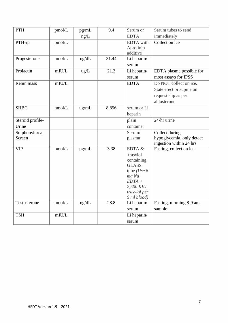
Endocrine Analyte Reporting Unit and Sample Tube The following table is provided as a guide, check with local laboratory for sample type.
Test Preferred
unit
Conventio
nal unit
Conversion
to
Convention
al unit
Collection
tube
Collection comment
5-HIAA umol/day 24 hr urine
acid bottle
See Appendix.
ACTH
pmol/L pg/mL
ng/L
4.54 EDTA Collect on ice
Aldosterone pmol/L ng/dL 0.036 EDTA/
serum
State erect (seated for 10
mins) or supine (for 30
mins) on request slip
Aldosterone-
Urine
pmol/L ng/dL 0.036 plain
container
Urine 24 h
Keep the container
refrigerated during the
collection period
AVP / ADH pmol/L pg/mL 0.923 2 x 4 ml
EDTA
Collect on ice
Androstenedione nmol/L ng/dL 28.65 Serum
βHCG IU/L mIU/mL 1 Li heparin/
serum
C-peptide nmol/L ng/mL 0.331 Li heparin/
serum
Fasting
Calcitonin pmol/L pg/mL 0.292 Li heparin
or serum
tube
NOT
EDTA.
Fasting specimen is
preferred.
Collect on ice.
Chromogranin A ug/L Serum Fasting specimen preferred.
Collect on ice.
Copeptin pmol/L Li heparin
Cortisol nmol/L ug/dL 27.588 Li heparin/
serum
Morning 8 – 9 am sample
Cortisol - Saliva nmol/L ug/dL 27.588 Salivette 11 pm to midnight
collection. Nil by mouth/ no
teeth brushing 30 mins
prior.
DHEAS umol/L ug/dL 0.027 Serum
Estradiol pmol/L pg/mL 0.272 Serum

6 HEDT Version 1.9 2021
Free T3 pmol/L pg/dL 64.9 Li heparin/
serum
Free T4 pmol/L ng/dL 0.0775 Li heparin/
serum
FSH IU/L mIU/mL 1 Li heparin/
serum
Gastrin pmol/L pg/mL 0.481 Serum Fasting, collect on ice.
Proton pump inhibitors
elevate result.
Glucagon ng/L pg/mL 1 EDTA &
trasylol
containing
GLASS
tube (Use 6
mg Na
EDTA +
2,500 KIU
trasylol per
5 ml blood)
Fasting. Collect on ice
Glucose mmol/L mg/dL 18 Fluoride
oxalate/ Na
F-EDTA-
citrate
Li heparin/ serum if rapid
transport to laboratory
GH ug/L mU/L 3 Li heparin
or Serum
tube
Insulin pmol/L mU/L 0.144 Serum Fasting. Collect on ice
Insulin Ab unit Serum
IGF-1 nmol/L ng/mL 0.131 Serum or Li
heparin
IGF BP3 nmol/L Serum Collect on ice
LH IU/L Li heparin/
serum
17-OHP nmol/L Serum
Metanephrines
(plasma)
pmol/L Li heparin Fasting, collect on ice,
supine for 30 mins.
3-methoxytyramine might
need to be specified on
request if required
Osteocalcin ug/L Serum, Li-
heparin or
K3 EDTA
Pancreatic
polypeptide
pmol/L Serum Fasting, morning sample
7 HEDT Version 1.9 2021
PTH pmol/L pg/mL
ng/L
9.4 Serum or
EDTA
Serum tubes to send
immediately
PTH-rp pmol/L EDTA with
Aprotinin
additive
Collect on ice
Progesterone nmol/L ng/dL 31.44 Li heparin/
serum
Prolactin mIU/L ug/L 21.3 Li heparin/
serum
EDTA plasma possible for
most assays for IPSS
Renin mass mIU/L EDTA Do NOT collect on ice.
State erect or supine on
request slip as per
aldosterone
SHBG nmol/L ug/mL 8.896 serum or Li
heparin
Steroid profile-
Urine
plain
container
24-hr urine
Sulphonylurea
Screen
Serum/
plasma
Collect during
hypoglycemia, only detect
ingestion within 24 hrs
VIP pmol/L pg/mL 3.38 EDTA &
trasylol
containing
GLASS
tube (Use 6
mg Na
EDTA +
2,500 KIU
trasylol per
5 ml blood)
Fasting, collect on ice
Testosterone nmol/L ng/dL 28.8 Li heparin/
serum
Fasting, morning 8-9 am
sample
TSH mIU/L Li heparin/
serum

8 HEDT Version 1.9 2021
1 Adrenal
1.1 SHORT SYNACTHEN TEST
RATIONALE:
The cortisol response to Synacthen (Tetracosactide) stimulation will be low or absent due to primary
adrenal pathology (e.g. Addison’s disease, bilateral adrenal infiltration) or adrenal atrophy secondary
to severe ACTH deficiency of at least 4 weeks' duration. (1) This test does not assess adequacy of
ACTH/ CRH response to stress if pathology was of short duration. This is assessed by the ITT or
overnight metyrapone test.
Also used for diagnosis of non-classical 21-hydroxylase deficiency, if a morning, screening follicular
phase 17 OH progesterone is > 6 nmol/L (lower level for mass spectroscopy assay). For other causes of
congenital adrenal hyperplasia, contact laboratory for required tests.
PREPARATION AND PROCEDURE:
1) Withhold any steroid treatment for 24 hours prior to the test (patients treated with dexamethasone
require at least 48 hours of steroid withdrawal) if appropriate.
2) Baseline blood is collected for cortisol and ACTH. Procedure should be performed between 8 -
9:30am when cortisol peak is present.
3) IM or IV Synacthen (Tetracosactide) 250 ug
4) Blood for cortisol collected at 30 and 60 minutes
Time Procedure/ Test Comment
Baseline ACTH, cortisol Also 17-OH progesterone if CAH queried
0 minute IV or IM Synacthen
(Tetracosactide) 250 ug
30 minutes cortisol Also 17-OH progesterone if CAH queried
60 minutes cortisol Also 17-OH progesterone if CAH queried
INTERPRETATION:
Normal SST requires a cortisol from at least one time point to exceed the minimum
peak cortisol cut-off specified for that assay. The concentration of peak cortisol cut-off
is assay dependent, and for female, OCP raises total cortisol level due to rise in CBG.
(1)
The use of historical peak cortisol of 550 nmol/L in newer cortisol-specific assays may
result in false positive results. (2) Previous requirement for a minimum cortisol
increment from baseline (e.g. 250 nmol/L) is also redundant as normal individuals with
high baseline cortisol will not achieve this increment.
Laboratories need to determine their own individual cut-off. The table below
describes the minimum cortisol level achieved post synacthen (Tetracosactide) at 30
minutes for different immunoassays. (3) The 60 minutes cortisol level was reported to
be around 15% higher than the 30 minutes level. (4)
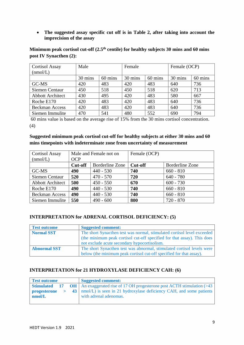
9 HEDT Version 1.9 2021
The suggested assay specific cut off is in Table 2, after taking into account the
imprecision of the assay
Minimum peak cortisol cut-off (2.5th centile) for healthy subjects 30 mins and 60 mins
post IV Synacthen (2):
Cortisol Assay
(nmol/L)
Male Female Female (OCP)
30 mins 60 mins 30 mins 60 mins 30 mins 60 mins
GC-MS 420 483 420 483 640 736
Siemen Centaur 450 518 450 518 620 713
Abbott Architect 430 495 420 483 580 667
Roche E170 420 483 420 483 640 736
Beckman Access 420 483 420 483 640 736
Siemen Immulite 470 541 480 552 690 794
60 mins value is based on the average rise of 15% from the 30 mins cortisol concentration.
(4)
Suggested minimum peak cortisol cut-off for healthy subjects at either 30 mins and 60
mins timepoints with indeterminate zone from uncertainty of measurement
Cortisol Assay
(nmol/L)
Male and Female not on
OCP
Female (OCP)
Cut-off Borderline Zone Cut-off Borderline Zone
GC-MS 490 440 - 530 740 660 - 810
Siemen Centaur 520 470 - 570 720 640 - 780
Abbott Architect 500 450 - 550 670 600 - 730
Roche E170 490 440 - 530 740 660 - 810
Beckman Access 490 440 - 530 740 660 - 810
Siemen Immulite 550 490 - 600 800 720 - 870
INTERPRETATION for ADRENAL CORTISOL DEFICIENCY: (5)
Test outcome Suggested comment:
Normal SST The short Synacthen test was normal, stimulated cortisol level exceeded
(the minimum peak cortisol cut-off specified for that assay). This does
not exclude acute secondary hypocortisolism.
Abnormal SST The short Synacthen test was abnormal, stimulated cortisol levels were
below (the minimum peak cortisol cut-off specified for that assay).
INTERPRETATION for 21 HYDROXYLASE DEFICIENCY CAH: (6)
Test outcome Suggested comment:
Stimulated 17 OH
progesterone > 43
nmol/L
An exaggerated rise of 17 OH progesterone post ACTH stimulation (>43
nmol/L) is seen in 21 hydroxylase deficiency CAH, and some patients
with adrenal adenomas.

10 HEDT Version 1.9 2021
Stimulated 17 OH
progesterone 30 - 43
nmol/L
A moderate rise of 17 OH progesterone post ACTH stimulation (30-43
nmol/L) constitutes a grey zone whereby genetic tests might be required
to confirm or exclude 21 hydroxylase deficiency CAH.
Stimulated 17 OH
progesterone < 30
nmol/L
A mild rise of 17 OH progesterone post ACTH stimulation (< 30 nmol/L)
is a normal physiological response and not consistent with 21
hydroxylase deficiency CAH.
The cut-offs for 17 OH progesterone in the table are based on radioimmunoassay, LCMS cut-
off for 17 OH progesterone is lower at 9 nmol/L. (7)
NOTES:
Nausea, palpitation, hot flushes or allergic reaction can rarely occur with synacthen
(Tetracosactide).
Although IV administration is preferred, IM administration is also valid, however
cortisol at 30 minutes is more variable. (8)
SST result is difficult to interpret in critically ill patients due to difficulties in
interpreting total cortisol results from immunoassays. (9)
REFERENCES:
1. Courtney CH, McAllister AS, Bell PM, McCance DR, Leslie H, Sheridan B, Atkinson AB.
Low- and standard-dose corticotropin and insulin hypoglycemia testing in the assessment of
hypothalamic-pituitary-adrenal function after pituitary surgery. The Journal of clinical
endocrinology and metabolism 2004; 89:1712-1717
2. El-Farhan et al. Method-specific serum cortisol responses to the adrenocorticotrophin test:
comparison of gas chromatography mass spectrometry and five automated immunoassays.
Clinical Endocrinology (2013) 78, 673–680
3. S Fletcher, EM Lim, R Wardrop, N Hadlow, J Joseph, D Henley. Time To Review
Synacthen Stimulation Tests: Caution on 550 nmol/LCut-off! Clin Biochem Rev 2012; Vol
33 Suppl; S21 Proceedings of the Australasian Association of Clinical Biochemists’ 50th
Annual Scientific Conference
4. Chitale A et al. Determining the utility of the 60 min cortisol measurement in the short
synacthen test. Clinical Endocrinology (2013) 79, 14 - 19
5. Bornstein, S.R., et al., Diagnosis and Treatment of Primary Adrenal Insufficiency: An
Endocrine Society Clinical Practice Guideline. J Clin Endocrinol Metab, 2016. 101(2): p. 364-
89.
6. El-Maouche D, Arlt W, Merke DP. Congenital adrenal hyperplasia. Lancet (London,
England) 2017
7. Costa-Barbosa FA, Carvalho VM, Oliveira KC, Vieira JGH, Kater CE. Reassessment of
predictive values of ACTH-stimulated serum 21-deoxycortisol and 17-hydroxyprogesterone

11 HEDT Version 1.9 2021
to identify CYP21A2 heterozygote carriers and nonclassic subjects. Clin Endocrinol (Oxf).
2021.
8. Longui CA, Vottero A, Harris AG, Chrousos GP. Plasma cortisol responses after
intramuscular corticotropin 1-24 in healthy men. Metabolism: clinical and experimental 1998;
47:1419-1422
9. Cooper MS et al. Corticosteroid insufficiency in acutely ill patients. NEJM. (2003); 348
(8):727.

12 HEDT Version 1.9 2021
1.2 PRIMARY ALDOSTERONISM INVESTIGATION
PATIENT PREPARATION
Algorithm for the detection, confirmation, subtype testing, and treatment of Primary hyperaldosteronism
(PA). Adapted from Management of Primary Aldosteronism: Case Detection, Diagnosis, and
Treatment: An Endocrine Society Clinical Practice Guideline. (1)
Interfering drugs which can affect renin, aldosterone or both should be stopped for at
least o 4 weeks: Spironolactone, eplerenone, amiloride, and triamterene, potassium-
wasting diuretics, licorice. o 2 weeks: Angiotensin-converting enzyme inhibitors, angiotensin receptor
blockers, renin inhibitors, and dihydropyridine calcium channel antagonists,
clonidine, methydopa, beta-blockers.
Drugs which do not affect renin, aldosterone for blood pressure control includes:
verapamil slow-release, prazosin, hydralazine, moxonidine.
Hypokalemia needs to be corrected.
Aldosterone:renin ratio (ARR) is the preferred screening test. Preferably two elevated
values should be obtained prior to confirmation testing. The ARR test is most sensitive
when samples are collected in the morning after patients have been out of bed for at
least 2 hours, usually after they have been seated for 5–15 minutes.

13 HEDT Version 1.9 2021
There is no gold standard for confirmation testing. Of the 4 testing procedures
available, captopril challenge test can have false negative equivocal results and
therefore not mentioned in this document. (1) Oral sodium loading test requires
sensitive and specific urinary aldosterone measurement (LC-MSMS) in patients
without renal impairment.
In florid Primary aldosteronism (hypokalemia, suppressed renin, elevated
aldosterone > 550 pmol/L), confirmation tests might not be required.
14 HEDT Version 1.9 2021
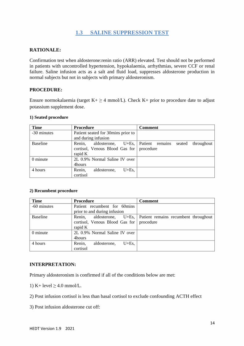
1.3 SALINE SUPPRESSION TEST
RATIONALE:
Confirmation test when aldosterone:renin ratio (ARR) elevated. Test should not be performed
in patients with uncontrolled hypertension, hypokalaemia, arrhythmias, severe CCF or renal
failure. Saline infusion acts as a salt and fluid load, suppresses aldosterone production in
normal subjects but not in subjects with primary aldosteronism.
PROCEDURE:
Ensure normokalaemia (target K+ ≥ 4 mmol/L). Check K+ prior to procedure date to adjust
potassium supplement dose.
1) Seated procedure
Time Procedure Comment
-30 minutes Patient seated for 30mins prior to
and during infusion
Baseline Renin, aldosterone, U+Es,
cortisol, Venous Blood Gas for
rapid K
Patient remains seated throughout
procedure
0 minute 2L 0.9% Normal Saline IV over
4hours
4 hours Renin, aldosterone, U+Es,
cortisol
2) Recumbent procedure
Time Procedure Comment
-60 minutes Patient recumbent for 60mins
prior to and during infusion
Baseline Renin, aldosterone, U+Es,
cortisol, Venous Blood Gas for
rapid K
Patient remains recumbent throughout
procedure
0 minute 2L 0.9% Normal Saline IV over
4hours
4 hours Renin, aldosterone, U+Es,
cortisol
INTERPRETATION:
Primary aldosteronism is confirmed if all of the conditions below are met:
1) K+ level ≥ 4.0 mmol/L.
2) Post infusion cortisol is less than basal cortisol to exclude confounding ACTH effect
3) Post infusion aldosterone cut off:

15 HEDT Version 1.9 2021
A) Seated procedure (6) (7)
Liaison immunoassay
o ≤ 170 pmol/L = PA unlikely (sensitivity 95%. specificity 80%)
o 171-220pmol/L = borderline zone
o > 220 pmol/L = PA likely (sensitivity 86%. specificity 87%) #
LCMS < 160 pmol/L = PA unlikely
IDS (iSYS) immunoassay
o < 140 pmol/L = PA unlikely
B) Recumbent procedure (cut-offs based on immunoassays)
o < 140 pmol/L = PA unlikely
o 140 – 280 pmol/L = borderline
o >280 pmol/L = PA likely
NOTES:
Fluid status check should take place during infusion, particularly for those prone to
fluid overload.
Seated normal saline suppression test was found to have higher sensitivity compared to
supine normal saline suppression and has good agreement with the more cumbersome
fludrocortisone suppression test. (2) (5)
Aldosterone cut offs are lower if measured using LCMS compared to immunoassays.
# 220 pmol/L rounded from 217 pmol/L in reference (6)

16 HEDT Version 1.9 2021
1.4 FLUDROCORTISONE SUPPRESSION TEST
RATIONALE:
Confirmation test when aldosterone:renin ratio (ARR) elevated. Test should not be performed
in patients with uncontrolled hypertension, hypokalaemia, arrhythmias, severe CCF or renal
failure. Fludrocortisone, a potent mineralocorticoid, suppresses aldosterone production in
normal subjects but not in subjects with primary aldosteronism.
PROCEDURE:
1) Most patients require admission to monitor BP and K+ status
2) Ensure normokalaemia throughout the procedure with oral potassium supplements (target
K+ at least 4 mmol/L)
3) Salt loading is required, e.g., a liberalized dietary sodium intake, supplemented by Slow-
Na 10 mmol three tablets TDS, target urine Na excretion > 3 mmol/kg/day
4) Collect 24 hr urine for aldosterone, sodium, potassium and creatinine levels 1 day prior to
fludrocortisone administration and again on the last 24h
5) Give Fludrocortisone 0.1 mg every 6 hours for 4 days (0400,1000,1600,2200)
6) Daily blood test for renin, aldosterone, U+Es, cortisol. Extra blood test might be required
for Slow K dosing
7) Blood test at 0700 and 1000 on Day 5 are required for interpretation
Time Procedure Comment
- 1 Day 24 hr urine aldosterone, sodium,
potassium and creatinine
Ensure normokalaemia (target K+ ≥ 4
mmol/L) and salt loading for the 4 days
of test. Check K+ to adjust potassium
supplement dose
Day 1: 0700 Recumbent: Renin, aldosterone,
U+Es, cortisol
Day 1: 1000 Upright: Renin, aldosterone,
U+Es, cortisol
Fludrocortisone 0.1 mg every 6 hours
(1000,1600,2200)
Day 1: 1600 Check K+ Optional to ensure K remains at target
Day 2: 0700 Recumbent: Renin, aldosterone,
U+Es, cortisol
Fludrocortisone 0.1 mg every 6 hours
(0400,1000,1600,2200)
Day 2: 1000 Upright: Renin, aldosterone,
U+Es, cortisol
Day 2: 1600 Check K+ Optional to ensure K remains at target
Day 3: 0700 Recumbent: Renin, aldosterone,
U+Es, cortisol
Fludrocortisone 0.1 mg every 6 hours
(0400,1000,1600,2200)
Day 3: 1000 Upright: Renin, aldosterone,
U+Es, cortisol
Day 3: 1600 Check K+ Optional to ensure K remains at target
Day 4: 0700 Recumbent: Renin, aldosterone,
U+Es, cortisol
Fludrocortisone 0.1 mg every 6 hours
(0400,1000,1600,2200)
Day 4: 1000 Upright: Renin, aldosterone,
U+Es, cortisol
24 hr urine aldosterone, sodium,
potassium and creatinine
Day 4: 1600 Check K+ Optional to ensure K remains at target

17 HEDT Version 1.9 2021
Day 5: 0700 Recumbent: Renin, aldosterone,
U+Es, cortisol
Last dose of fludrocortisone Day 5 at
0400.
Day 5: 1000 Upright: Renin, aldosterone,
U+Es, cortisol
INTERPRETATION:
Primary aldosteronism is confirmed if all of the conditions below are met:
1) upright aldosterone levels on Day 5 (4 days of fludrocortisone) are > 170 pmol/L (1)
2) upright renin on Day 5 is suppressed.
3) K+ level normal (at least 4.0 mmol/L) on Day 5
4) Plasma cortisol on Day 5 does not increase significantly from 0700h to 1000h (increase
may indicate ACTH stimulation of aldosterone production that may have prevented
suppression).
NOTES:
Blood pressure and fluid status check should take place during fludrocortisone and salt
loading.
Aldosterone cut off is lower (down to 130 pmol/L) if measured using LCMS rather than
immunoassay, consult laboratory for cut-off.

18 HEDT Version 1.9 2021
1.5 ORAL SODIUM LOADING TEST
RATIONALE:
Confirmation test when aldosterone:renin ratio (ARR) elevated. Test should not be performed
in patients with uncontrolled hypertension, hypokalaemia, arrhythmias, severe CCF or renal
insufficiency. Oral sodium suppresses aldosterone production in normal subjects but not in
subjects with primary aldosteronism.
PROCEDURE:
1) Ensure normokalaemia (target K+ ≥ 4 mmol/L). Check K+ prior to procedure date to adjust
potassium supplement dose.
2) Oral sodium 200 mmol or 6 g daily
3) 24 hr urine collection for aldosterone and Na starting on Day 3
4) Urine aldosterone needs analysis on a specific assay
Time Procedure Comment
Day 1 Oral sodium 200 mmol or 6 g
daily
Day 2 Oral sodium 200 mmol or 6 g
daily
Day 3 Oral sodium 200 mmol or 6 g
daily
Start 24 hr urine (aldosterone and Na)
collection 8am after discarding first void
urine
Day 4 Complete 24 hr urine (aldosterone and
Na) collection 8am
INTERPRETATION:
Elevated 24 hr urine aldosterone > 33 nmol/day by LCMS method makes primary
hyperaldosteronism likely, providing 24 hr urine Na is elevated (urine Na excretion >3
mmol/kg/day). Consult laboratory for local cut-off.
NOTES:
Non-specific aldosterone methods may blunt diagnostic accuracy due to cross-reactivity with
other metabolites in urine.

19 HEDT Version 1.9 2021
1.6 ADRENAL VENOUS SAMPLING
RATIONALE:
In patients with confirmed primary aldosteronism (PA) who are surgical candidates, adrenal
venous sampling (AVS) is the gold standard in lateralisation of the source of aldosterone excess
and differentiates between unilateral adrenal adenoma from bilateral adrenal hyperplasia. All
patients should have adrenal CT prior to AVS to exclude large (> 4 cm) adrenal masses.
CT and MRI can misdiagnose the cause of PA. Therefore, AVS is still required for
lateralisation with the exception of younger patients < 35 years with spontaneous
hypokalaemia, marked aldosterone excess, and unilateral adrenal cortical adenoma on CT who
might be able to proceed directly to unilateral adrenalectomy. (1)
AVS should be performed by experienced interventional radiologist. The use of ACTH
stimulation is used to improve successful cannulation rate by increasing the adrenal to
periphery gradient and to minimise stress induced fluctuation in sequential adrenal vein
sampling. Point of care cortisol kit during AVS also increased cannulation rates. (4)
PROCEDURE:
1) Book AVS with experienced interventional radiologist. Notify laboratory of test.
2) AVS can be
a) Unstimulated: only baseline AVS samples collected in early morning after
overnight recumbency.
b) Stimulated with Synacthen (Tetracosactide): baseline and post ACTH AVS
samples collected. Synacthen protocols include:
I. Bolus 250 ug Synacthen 15 mins before stimulated AVS collection
II. Continuous 50 ug/hr Synacthen (250ug in 500ml N saline, 100ml per
hr) 30 mins before stimulated AVS collection and continued until AVS
completion
III. Bolus 250 ug Synacthen followed by 50ug/hr Synacthen 15- 30 mins
before stimulated AVS collection and continued until AVS completion
3) Tubes should be pre-labelled with site, baseline/ stimulated samples and collected in
duplicate at each site for each timepoint.
Baseline AVS collection
Time Procedure Comment
Document time on
tube
Common femoral vein is punctured
and a 5 French sheath inserted.
Peripheral cubital fossa blood taken
for aldosterone and cortisol
Document time on
tube
Selective catheterization of left
adrenal vein and take blood for
aldosterone and cortisol
Document time on
tube
Selective catheterization of right
adrenal vein and take blood for
aldosterone and cortisol
Right adrenal vein is more
difficult to cannulate.
20 HEDT Version 1.9 2021
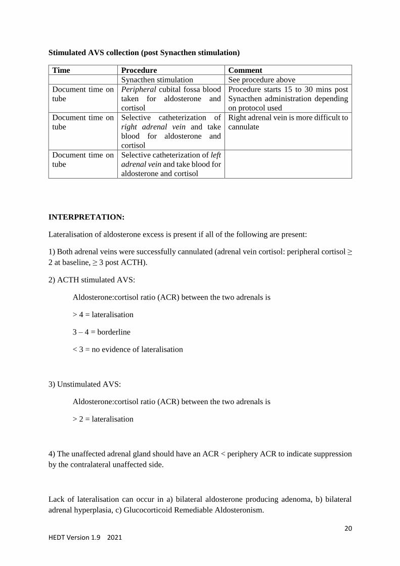
Stimulated AVS collection (post Synacthen stimulation)
Time Procedure Comment
Synacthen stimulation See procedure above
Document time on
tube
Peripheral cubital fossa blood
taken for aldosterone and
cortisol
Procedure starts 15 to 30 mins post
Synacthen administration depending
on protocol used
Document time on
tube
Selective catheterization of
right adrenal vein and take
blood for aldosterone and
cortisol
Right adrenal vein is more difficult to
cannulate
Document time on
tube
Selective catheterization of left
adrenal vein and take blood for
aldosterone and cortisol
INTERPRETATION:
Lateralisation of aldosterone excess is present if all of the following are present:
1) Both adrenal veins were successfully cannulated (adrenal vein cortisol: peripheral cortisol ≥
2 at baseline, ≥ 3 post ACTH).
2) ACTH stimulated AVS:
Aldosterone:cortisol ratio (ACR) between the two adrenals is
> 4 = lateralisation
3 – 4 = borderline
< 3 = no evidence of lateralisation
3) Unstimulated AVS:
Aldosterone:cortisol ratio (ACR) between the two adrenals is
> 2 = lateralisation
4) The unaffected adrenal gland should have an ACR < periphery ACR to indicate suppression
by the contralateral unaffected side.
Lack of lateralisation can occur in a) bilateral aldosterone producing adenoma, b) bilateral
adrenal hyperplasia, c) Glucocorticoid Remediable Aldosteronism.

21 HEDT Version 1.9 2021
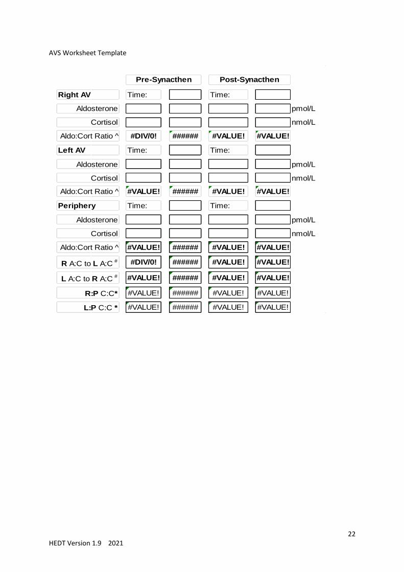
NOTES:
Sample worksheet on next page
Adrenal haemorrhage can occur in up to 2.5% of AVS procedures
In patients < 20 years old with confirmed PA or in those who have a family history of
PA or strokes < 40 years old, genetic testing for FH-I (Glucocorticoid Remediable
Aldosteronism, hybrid CYP11B1/CYP11B2 mutation) should be considered. (1)
In patients with confirmed PA presenting in childhood, germline mutations in KCNJ5
causing FH-III should be considered.
Glucocorticoid Remediable Aldosteronism mutation testing replaces indirect test such
as dexamethasone suppression test.
22 HEDT Version 1.9 2021
AVS Worksheet Template
Time: Time:
pmol/L
nmol/L
#DIV/0! ###### #VALUE! #VALUE!
Time: Time:
pmol/L
nmol/L
#VALUE! ###### #VALUE! #VALUE!
Time: Time:
pmol/L
nmol/L
#VALUE! ###### #VALUE! #VALUE!
#DIV/0! ###### #VALUE! #VALUE!
#VALUE! ###### #VALUE! #VALUE!
#VALUE! ###### #VALUE! #VALUE!
#VALUE! ###### #VALUE! #VALUE!
Cortisol
Left AV
Cortisol
Aldo:Cort Ratio ^
Periphery
Aldosterone
Aldo:Cort Ratio ^
Pre-Synacthen Post-Synacthen
Right AV
Aldosterone
Cortisol
Aldo:Cort Ratio ^
Aldosterone
R A:C to L A:C #
L A:C to R A:C #
R:P C:C*
L:P C:C *

23 HEDT Version 1.9 2021
REFERENCES:
1. Funder JW, Carey RM, Mantero F, Murad MH, Reincke M, Shibata H, Stowasser M, Young
WF, Jr. The Management of Primary Aldosteronism: Case Detection, Diagnosis, and
Treatment: An Endocrine Society Clinical Practice Guideline. The Journal of clinical
endocrinology and metabolism 2016; 101:1889-1916
2. Ahmed AH, Cowley D, Wolley M, Gordon RD, Xu S, Taylor PJ, Stowasser M. Seated saline
suppression testing for the diagnosis of primary aldosteronism: a preliminary study. The
Journal of clinical endocrinology and metabolism 2014; 99:2745-2753
3. Kempers MJ, Lenders JW, van Outheusden L, van der Wilt GJ, Schultze Kool LJ, Hermus
AR, Deinum J. Systematic review: diagnostic procedures to differentiate unilateral from
bilateral adrenal abnormality in primary aldosteronism. Annals of internal medicine 2009;
151:329-337
4. Page MM, Taranto M, Ramsay D, van Schie G, Glendenning P, Gillett MJ, et al. Improved
technical success and radiation safety of adrenal vein sampling using rapid, semi-quantitative
point-of-care cortisol measurement. Ann Clin Biochem. Jan 1 2018
5. Stowasser, M., et al., Comparison of Seated With Recumbent Saline Suppression Testing
for the Diagnosis of Primary Aldosteronism. J Clin Endocrinol Metab, 2018. 103(11): p. 4113-
4124.
6. Thuzar, M., et al., Diagnosis of Primary Aldosteronism by Seated Saline Suppression Test-
Variability Between Immunoassay and HPLC-MS/MS. J Clin Endocrinol Metab, 2020.
105(3).
7. Manolopoulou J, Fischer E, Dietz A, Diederich S, Holmes D, Junnila R, et al. Clinical
validation for the aldosterone-to-renin ratio and aldosterone suppression testing using
simultaneous fully automated chemiluminescence immunoassays. Journal of hypertension.
2015;33(12):2500-11

24 HEDT Version 1.9 2021
2 Cushing Overview
Screening investigations: (exclude exogenous glucocorticoids) (1)
1.1 Overnight 1 mg dexamethasone suppression test
1.2 Late night or 11pm salivary cortisol
1.3 24-hour UFC
Consider additional screening tests (if above results are equivocal or discrepant or to exclude
pseudo-Cushing’s)
1.4 2-day low dose oral dexamethasone suppression
1.5 Dexamethasone-CRH test
1.6 IV 4 mg dexamethasone suppression test
Cushing’s syndrome confirmed – measure plasma ACTH
Suppressed ACTH (< 10ng/L or 2 pmol/L) – adrenal imaging studies for ACTH
independent Cushing’s
Borderline ACTH (10-20ng/L or 2-4pmol/L) – consider peripheral CRH test
Normal or elevated ACTH (> 20ng/L or 4 pmol/L) – proceed to further differential
diagnostic tests for ACTH dependent Cushing’s
Differential diagnosis of ACTH-dependent Cushing’s syndrome
Differentiate between pituitary and ectopic source of ACTH
A. High Dose 8 mg Dexamethasone Suppression Test (not required if IV 4mg
dexamethasone test already performed)
B. Peripheral CRH Test
C. Bilateral Inferior Petrosal Sinus Sampling
25 HEDT Version 1.9 2021
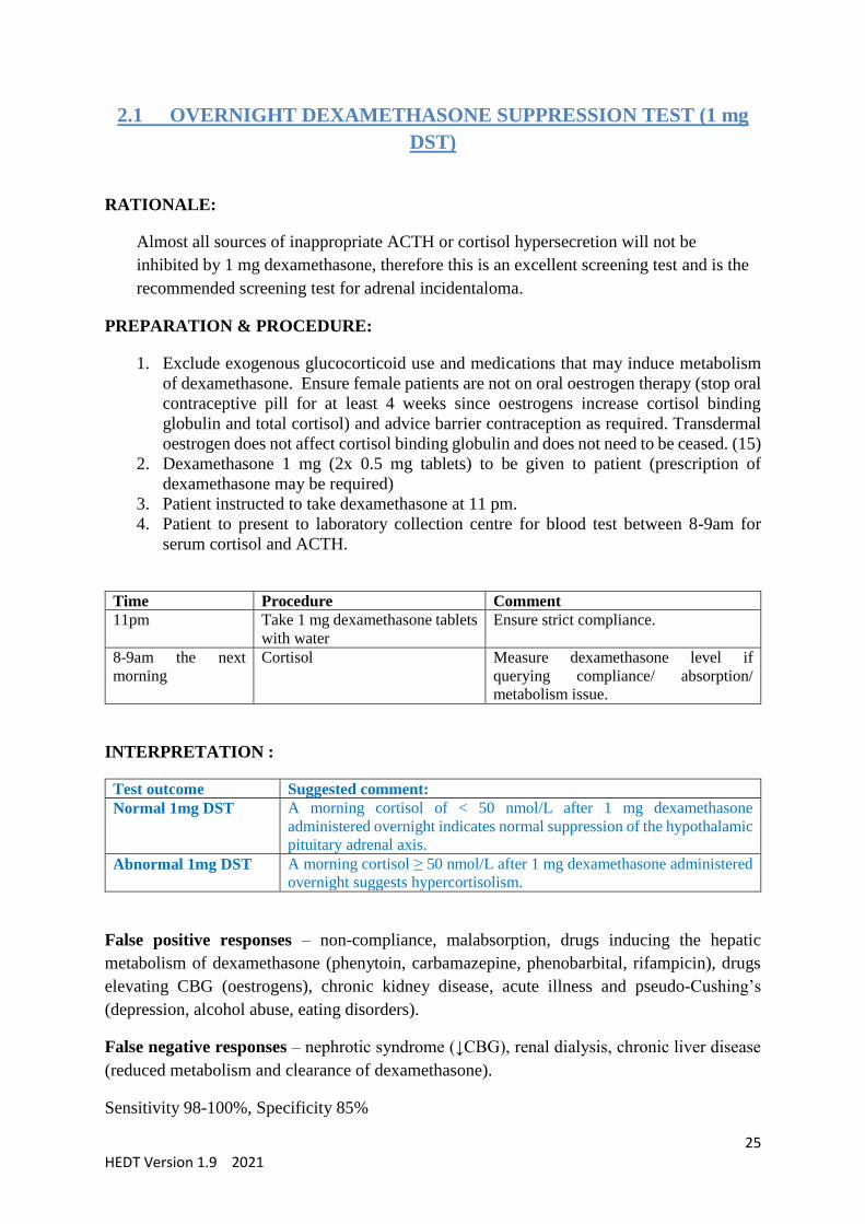
2.1 OVERNIGHT DEXAMETHASONE SUPPRESSION TEST (1 mg
DST)
RATIONALE:
Almost all sources of inappropriate ACTH or cortisol hypersecretion will not be
inhibited by 1 mg dexamethasone, therefore this is an excellent screening test and is the
recommended screening test for adrenal incidentaloma.
PREPARATION & PROCEDURE:
1. Exclude exogenous glucocorticoid use and medications that may induce metabolism
of dexamethasone. Ensure female patients are not on oral oestrogen therapy (stop oral
contraceptive pill for at least 4 weeks since oestrogens increase cortisol binding
globulin and total cortisol) and advice barrier contraception as required. Transdermal
oestrogen does not affect cortisol binding globulin and does not need to be ceased. (15)
2. Dexamethasone 1 mg (2x 0.5 mg tablets) to be given to patient (prescription of
dexamethasone may be required)
3. Patient instructed to take dexamethasone at 11 pm.
4. Patient to present to laboratory collection centre for blood test between 8-9am for
serum cortisol and ACTH.
Time Procedure Comment
11pm Take 1 mg dexamethasone tablets
with water
Ensure strict compliance.
8-9am the next
morning
Cortisol Measure dexamethasone level if
querying compliance/ absorption/
metabolism issue.
INTERPRETATION :
Test outcome Suggested comment:
Normal 1mg DST A morning cortisol of < 50 nmol/L after 1 mg dexamethasone
administered overnight indicates normal suppression of the hypothalamic
pituitary adrenal axis.
Abnormal 1mg DST A morning cortisol ≥ 50 nmol/L after 1 mg dexamethasone administered
overnight suggests hypercortisolism.
False positive responses – non-compliance, malabsorption, drugs inducing the hepatic
metabolism of dexamethasone (phenytoin, carbamazepine, phenobarbital, rifampicin), drugs
elevating CBG (oestrogens), chronic kidney disease, acute illness and pseudo-Cushing’s
(depression, alcohol abuse, eating disorders).
False negative responses – nephrotic syndrome (↓CBG), renal dialysis, chronic liver disease
(reduced metabolism and clearance of dexamethasone).
Sensitivity 98-100%, Specificity 85%

26 HEDT Version 1.9 2021
NOTES:
Hospitalised inpatients not infrequently have an abnormal 1 mg dexamethasone
suppression test response, if possible, investigations should be delayed until acute
illness has subsided.
To date, assay specific cut-offs for 1 mg DST are not readily available.
Concurrent quantification of dexamethasone may reduce false-positive rate and
improves specificity, this should be considered in patients with abnormal DST (13).

27 HEDT Version 1.9 2021
2.2 LATE NIGHT SALIVARY CORTISOL
RATIONALE:
A loss of diurnal variation is seen in patients with Cushing, whereas this is maintained in
pseudo-Cushing syndrome. Salivary cortisol is an ultra-filtrate of plasma and therefore
reflects free cortisol and is not affected by protein binding. This test is useful for patients with
suspected cyclical Cushing due to ease of repetitive testing.
PREPARATION & PROCEDURE:
1. Patient to collect Salivettes from clinic or laboratory collection centre.
2. Do not use steroid inhaler, relax and avoid vigorous activities for 1 hour prior to saliva
collection.
3. Do not eat/ drink/ brush or floss teeth for 30 minutes prior to saliva collection.
4. Ensure hands are clean (avoid topical glucocorticoid cream/lotion contamination) and
there is no bleeding inside the mouth just prior to collection.
5. Patient should chew on Salivette for 2 minutes (or until saturated with saliva) between
11pm to midnight.
6. Minimum of two separate samples should be collected on different nights.
7. Salivary cortisol is stable at room temperature. Patient should drop or post Salivettes
back to laboratory collection centre.
Time Procedure Comment
11pm Chew on salivette provided for 1-
2 minutes until saturated.
Collect prior to brushing teeth and nil by
mouth for at least 30 mins prior to
collection.
INTERPRETATION :
Normal response – Consult reference intervals provided by the laboratory.
False positive responses – smokers and especially patients who chew tobacco, contamination
of salivettes with corticosteroid, bleeding of the gum, shift workers.
False negative responses - non-compliance with collection procedure (drinking water dilutes
the sample), cyclical Cushing’s (off phase).
NOTES:
An excellent test to use in the investigation of cyclical Cushing’s syndrome where initial
screening tests are negative. Repeat frequently over expected cycle e.g. weekly for 1-2 months
as required.

28 HEDT Version 1.9 2021
29 HEDT Version 1.9 2021
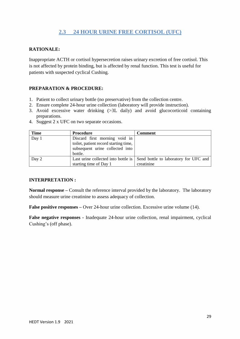
2.3 24 HOUR URINE FREE CORTISOL (UFC)
RATIONALE:
Inappropriate ACTH or cortisol hypersecretion raises urinary excretion of free cortisol. This
is not affected by protein binding, but is affected by renal function. This test is useful for
patients with suspected cyclical Cushing.
PREPARATION & PROCEDURE:
1. Patient to collect urinary bottle (no preservative) from the collection centre.
2. Ensure complete 24-hour urine collection (laboratory will provide instruction).
3. Avoid excessive water drinking (>3L daily) and avoid glucocorticoid containing
preparations.
4. Suggest 2 x UFC on two separate occasions.
Time Procedure Comment
Day 1 Discard first morning void in
toilet, patient record starting time,
subsequent urine collected into
bottle.
Day 2 Last urine collected into bottle is
starting time of Day 1
Send bottle to laboratory for UFC and
creatinine
INTERPRETATION :
Normal response – Consult the reference interval provided by the laboratory. The laboratory
should measure urine creatinine to assess adequacy of collection.
False positive responses – Over 24-hour urine collection. Excessive urine volume (14).
False negative responses - Inadequate 24-hour urine collection, renal impairment, cyclical
Cushing’s (off phase).

30 HEDT Version 1.9 2021

31 HEDT Version 1.9 2021
2.4 2-DAY LOW DOSE DEXAMETHASONE SUPPRESSION TEST
(LDDST)
RATIONALE:
Patients with pseudo-Cushing’s might not suppress their cortisol with 1 mg dexamethasone
suppression test and require the 2 day dexamethasone regimen.
PREPARATION & PROCEDURE:
1. Exclude exogenous glucocorticoid use and medications that may induce metabolism of
dexamethasone. Ensure female patients are not on oral oestrogen therapy.
2. Baseline serum cortisol and plasma ACTH to be taken prior to administration of
dexamethasone.
3. Dexamethasone 0.5 mg (require total of eight tablets of 0.5 mg tablets for this test) to be
given to patient (prescription of dexamethasone may be required)
4. Patient instructed to take dexamethasone 0.5 mg at exactly 6-hourly intervals.
Option A: 9am, 3pm, 9pm, 3am (patient to set alarm clock for 3am), 9am, 3pm, 9pm, 3am,
last blood test 9am (6 hrs after last dexamethasone dose)
Option B: 8am, 2pm, 8pm, 2am (patient to set alarm clock for 2am), 8am, 2pm, 8pm, 2am,
last blood test 8am (6 hrs after last dexamethasone dose)
5. Patient to present to laboratory collection centre for blood test at exactly 9am for serum
cortisol for two consecutive days. Two separate request forms should be given to patient.
1st request form “Baseline serum cortisol and plasma ACTH”.
2nd request form “2-day Dex Suppression – Day 2 serum cortisol”
Time Procedure Comment
Day 0 (Baseline) 8-
9 am
Blood test for cortisol and ACTH
(baseline)
Bring completed urine collection
to lab.
Day 1: 9 am Patient to take 0.5 mg
dexamethasone
Day 1: 3pm Patient to take 0.5 mg
dexamethasone
Day 1: 9pm Patient to take 0.5 mg
dexamethasone
Day 1: 3 am Patient to take 0.5 mg
dexamethasone
Patient to set alarm clock for 3am to take
the Dex tablet.
Day 2: 9am Patient to take 0.5 mg
dexamethasone.
Start second urine collection if
requested.
Day 2: 3pm Patient to take 0.5 mg
dexamethasone
Day 2: 9pm Patient to take 0.5 mg
dexamethasone

32 HEDT Version 1.9 2021
Day 2: 3 am Patient to take 0.5 mg
dexamethasone
Patient to set alarm clock for 3am to take
the Dex tablet.
Day 3: 9 am (end of
test)
Blood test for cortisol and ACTH
(day 2)
Measure dexamethasone level if
querying compliance/ absorption/
metabolism issue.
INTERPRETATION :
Normal response = Serum cortisol < 50 nmol/L
False positive responses – non-compliance, malabsorption, drugs inducing the hepatic
metabolism of dexamethasone (phenytoin, carbamazepine, phenobarbital, rifampicin), drugs
elevating CBG (oestrogens), chronic kidney disease.
False negative responses – nephrotic syndrome (↓CBG), renal dialysis, chronic liver disease
(reduced metabolism and clearance of dexamethasone).
Sensitivity 96%, Specificity 70%.

33 HEDT Version 1.9 2021
2.5 DEXAMETHASONE-CRH TEST
RATIONALE:
Patients with pseudo-Cushing's maintain sensitivity to negative feedback with
glucocorticoid and will be unable to mount a pituitary-adrenal response to CRH when pre-
treated with low dose dexamethasone. Conversely, patients with Cushing’s syndrome are
mostly insensitive to low-dose dexamethasone suppression and will display unsuppressed
cortisol levels and, in patients with a pituitary ACTH-secreting tumour, the pituitary will
respond to CRH stimulation.
PREPARATION & PROCEDURE:
1. Exclude exogenous glucocorticoid use and medications that may induce metabolism of
dexamethasone. Ensure female patients are not on oral oestrogen therapy.
2. Baseline serum cortisol and plasma ACTH to be taken prior to administration of
dexamethasone.
3. Dexamethasone 0.5 mg (require total of eight tablets of 0.5 mg tablets for this test) to
be given to patient (prescription of dexamethasone may be required)
4. Patient instructed to take dexamethasone 0.5 mg at exactly 6-hourly intervals.
(1200, 1800, 2400, 0600, 1200, 1800, 2400, 0600)
5. Blood test at 0800 for cortisol and ACTH
6. Inject CRH (1 ug/kg up to 100ug) at 0800 immediately after blood test (see peripheral
CRH test protocol for more details)
7. Blood test for cortisol 15 minutes after CRH
Time Procedure Comment
Day 1 (Baseline) 8-
9 am
Blood test for cortisol and ACTH
Day 1: 1200 Patient to take 0.5 mg dexamethasone
Day 1: 1800 Patient to take 0.5 mg dexamethasone
Day 1: 2400 Patient to take 0.5 mg dexamethasone
Day 2: 0600 Patient to take 0.5 mg dexamethasone
Day 2: 1200 Patient to take 0.5 mg dexamethasone
Day 2: 1800 Patient to take 0.5 mg dexamethasone.
Day 2: 2400 Patient to take 0.5 mg dexamethasone
Day 3: 0600 Patient to take 0.5 mg dexamethasone
Day 3: 0800 Blood test for cortisol and ACTH
Measure dexamethasone level if
querying compliance/ absorption/
metabolism issue.
Day 3: 0800 Inject 1 ug/kg CRH
Day 3: 0815 Blood test for cortisol
INTERPRETATION :
Normal or pseudo-Cushing :
Post dexamethasone : cortisol < 38 nmol/L AND

34 HEDT Version 1.9 2021
Post dex-CRH : cortisol < 38 nmol/L
Cushing’s Syndrome:
Post dexamethasone : cortisol > 38 nmol/L
Post dex-CRH : cortisol > 38 nmol/L
False positive responses – non-compliance, malabsorption, drugs inducing the hepatic
metabolism of dexamethasone (phenytoin, carbamazepine, phenobarbital, rifampicin), drugs
elevating CBG (oestrogens), chronic kidney disease
False negative responses – nephrotic syndrome (↓CBG), renal dialysis, chronic liver disease
(reduced metabolism and clearance of dexamethasone).
Note :
More recent studies have shown a lower specificity of the Dex-CRH test for
differentiating real Cushing’s from pseudo-Cushing’s, (10) the above cut-off is based
on Yanovski ‘s study. (11)

35 HEDT Version 1.9 2021
2.6 IV 4 mg DEXAMETHASONE SUPPRESSION TEST
RATIONALE:
Intravenous dexamethasone avoids potential issues of poor compliance, variable
gastrointestinal absorption and metabolism of oral dexamethasone.
PREPARATION & PROCEDURE:
Patient admitted for day procedure
1. Ensure female patients are not on oestrogen therapy
2. Insertion of IV cannula
3. Samples for baseline serum cortisol to be collected at two time-points (-60mins, -5
mins) prior to dexamethasone infusion. Commence -60 mins sampling at 0830h
4. Dexamethasone infusion 1 mg/h for 4 hours (1x4 mg ampoule of dexamethasone in 500
ml Normal Saline running at 125 ml/hr) commencing at 0930h
5. Samples for serum cortisol to be collected 3, 4 and 5 hours post infusion. Ensure blood
collection is taken opposite the infusion site.
6. Patient to present to the lab the following day for serum cortisol at 9:00 am and 9:30
am (unless patient remains an inpatient)
Time Procedure Comment
Day1 Insertion of IV cannula
-60 minutes
(Baseline, 08:30)
cortisol
-5 minutes cortisol
0 minutes (09:30) IV Dex infusion commences for 4
hours duration.
4mg dexamethasone in 500 ml Normal
Saline at 125 ml/hr
+ 3 hrs cortisol Ensure blood collection is taken opposite
the infusion site
+4 hrs cortisol Completion of the 4 hr Dex infusion
+5 hrs cortisol Patient sent home after blood test
Day 2
+23.5h (9:00)
cortisol Patient returns for blood test
+24h (9:30) cortisol
INTERPRETATION :
Diagnosis of Cushing’s syndrome: (2)
Day 2 serum cortisol level (mean of +23.5h and +24h cortisol values) >130 nmol/L or
>20% of baseline cortisol (Day 1 at -60 minutes)
Sensitivity 100%, Specificity 96%
Cushing’s disease tends to partially suppress on Day 1 with rebound increase on Day 2,
while ectopic ACTH patients rarely suppress during the infusion.

36 HEDT Version 1.9 2021

37 HEDT Version 1.9 2021
2.7 ACTH Dependent Cushing’s Syndrome
2.7.1 HIGH DOSE DEXAMETHASONE SUPPRESSION TEST (HDDST)
RATIONALE:
Once ACTH dependent Cushing’s syndrome has been established, overnight 8 mg
Dexamethasone Suppression Test assists to differentiate between Cushing’s Disease and
Ectopic ACTH.
PREPARATION & PROCEDURE:
1. Exclude exogenous glucocorticoid use and medications that may induce metabolism of
dexamethasone. Ensure female patients are not on oral oestrogen therapy.
2. Ensure patient already had baseline morning serum cortisol
3. Dexamethasone 8mg (2x 4 mg tablets) to be given to patient (prescription of
dexamethasone may be required)
4. Patient instructed to take dexamethasone at 11 pm.
5. Patient to present to laboratory collection centre for blood test between 8-9am for serum
cortisol and ACTH.
Time Procedure Comment
8 am cortisol Ensure patient already had baseline
morning cortisol.
11pm Take 8 mg dexamethasone tablets Ensure strict compliance.
8-9am the next
morning
cortisol Measure dexamethasone level if
querying compliance/ absorption/
metabolism
INTERPRETATION :
Decrease in serum cortisol >50% is suggestive of pituitary Cushing’s disease. Most Cushing’s
disease patients have a cortisol < 140 nmol/L, normal subjects have undetectable cortisol post
8mg dexamethasone suppression.
HOWEVER, 10% of patients with ectopic ACTH secretion suppress with high dose
dexamethasone and some patients with pituitary tumours fail to suppress. (Sensitivity 81%,
Specificity 79%)
38 HEDT Version 1.9 2021
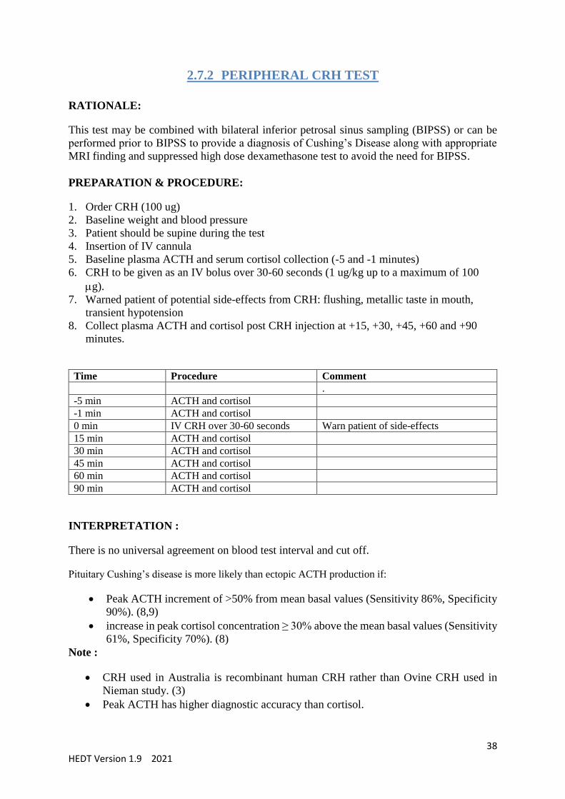
2.7.2 PERIPHERAL CRH TEST
RATIONALE:
This test may be combined with bilateral inferior petrosal sinus sampling (BIPSS) or can be
performed prior to BIPSS to provide a diagnosis of Cushing’s Disease along with appropriate
MRI finding and suppressed high dose dexamethasone test to avoid the need for BIPSS.
PREPARATION & PROCEDURE:
1. Order CRH (100 ug)
2. Baseline weight and blood pressure
3. Patient should be supine during the test
4. Insertion of IV cannula
5. Baseline plasma ACTH and serum cortisol collection (-5 and -1 minutes)
6. CRH to be given as an IV bolus over 30-60 seconds (1 ug/kg up to a maximum of 100
g).
7. Warned patient of potential side-effects from CRH: flushing, metallic taste in mouth,
transient hypotension
8. Collect plasma ACTH and cortisol post CRH injection at +15, +30, +45, +60 and +90
minutes.
Time Procedure Comment
.
-5 min ACTH and cortisol
-1 min ACTH and cortisol
0 min IV CRH over 30-60 seconds Warn patient of side-effects
15 min ACTH and cortisol
30 min ACTH and cortisol
45 min ACTH and cortisol
60 min ACTH and cortisol
90 min ACTH and cortisol
INTERPRETATION :
There is no universal agreement on blood test interval and cut off.
Pituitary Cushing’s disease is more likely than ectopic ACTH production if:
Peak ACTH increment of >50% from mean basal values (Sensitivity 86%, Specificity
90%). (8,9)
increase in peak cortisol concentration ≥ 30% above the mean basal values (Sensitivity
61%, Specificity 70%). (8)
Note :
CRH used in Australia is recombinant human CRH rather than Ovine CRH used in
Nieman study. (3)
Peak ACTH has higher diagnostic accuracy than cortisol.

39 HEDT Version 1.9 2021
2.7.3 INFERIOR PETROSAL SINUS SAMPLING (IPSS)
RATIONALE:
In established ACTH dependent Cushing’s syndrome, IPSS assists to differentiate whether the
source of excessive ACTH is central pituitary Cushing’s disease or peripheral ectopic ACTH
syndrome. IPSS might not be required if pituitary adenoma > 6mm AND cortisol suppressed
to high dose dexamethasone AND ACTH/ cortisol rose with peripheral CRH test.
PREPARATION & PROCEDURE:
1. Book experienced interventional radiologist, discuss if patient on blood thinning agents.
2. Confirm active phase of Cushing’s by late night saliva cortisol prior to the test, liaise with
laboratory regarding turn-around time and alert them to the IPSS booking. Metyrapone and
ketoconazole should be stopped 1 week prior to test.
3. Fast the patient from midnight for a morning procedure
4. Under radiological guidance in the Radiology Department, catheters are placed in the left
and right inferior petrosal sinuses.
5. Blood is collected at -5 min and -2 min before CRH administration, from left, right petrosal
catheters and peripheral vein to measure ACTH and prolactin.
6. CRH (1 ug/kg, maximum dose up to 100 ug) is given intravenously at 0 min.
7. Further samples to measure ACTH and Prolactin are collected from left and right petrosal
sinus and peripheral vein at + 2, 5, 10 and 15 minutes post CRH.
Time Procedure Comment
- 5 min ACTH and prolactin from left,
right petrosal sinus and peripheral
vein
1 x 4 ml EDTA tube from each site
- 2 min ACTH and prolactin from left,
right petrosal sinus and peripheral
vein
1 x 4 ml EDTA tube from each site
0 min IV CRH (1 ug/kg, up to 100 ug) Warn patient of side-effects of
flushing and hypotension
+ 2 min ACTH and prolactin from left,
right petrosal sinus and peripheral
vein
1 x 4 ml EDTA tube from each site
+ 5 min ACTH and prolactin from left,
right petrosal sinus and peripheral
vein
1 x 4 ml EDTA tube from each site
+10 min ACTH and prolactin from left,
right petrosal sinus and peripheral
vein
1 x 4 ml EDTA tube from each site
+15 min ACTH and prolactin from left,
right petrosal sinus and peripheral
vein
1 x 4 ml EDTA tube from each site
40 HEDT Version 1.9 2021
INTERPRETATION
A central to peripheral ACTH ratio of ≥ 2 pre CRH and / or a ratio of ≥ 3 post CRH is
consistent with Cushing’s disease. (5) Sensitivity and specificity 94%
IPSS has limited utility in localization of ACTH-secreting pituitary adenomas
Maximal IPS/ peripheral ratio is achieved at 5 minutes in 90% of Cushing’s Disease,
1% achieved the maximum ratio at 15 minutes. The 2 minutes time point was found to
have the best diagnostic accuracy. (5)
If the central to peripheral ACTH ratios were elevated on at least one side, then there is
no need to assess prolactin levels as petrosal sinus cannulation would have been
adequate on at least one side. De Sousa et.al. found co-lateralisation of prolactin and
ACTH during IPSS in Cushing’s disease subjects. (12) Therefore the prolactin inferior
petrosal sinus/ peripheral ratio on the ACTH non-dominant side can be low despite
adequate cannulation of IPS. The prolactin IPS/P ratio should NOT be used to 1)
differentiate Cushing’s Disease from ectopic ACTH, 2) correct for adequacy of IPSS,
3) lateralise ACTH producing pituitary adenoma.
If the central to peripheral ACTH ratios were not elevated on either side, the patient
might have ectopic source of ACTH, or petrosal sinus was not successfully cannulated.
A petrosal sinus/ peripheral prolactin ratio of ≥ 1.8 has been used to indicate adequate
petrosal sinus cannulation (7)
False results occur if patient was not in active phase of hypercortisolism at the time of
testing. Late night saliva cortisol before IPSS can assist in determining whether cyclical
Cushing patients are in active phase before proceeding with the test.
NOTES:
ACTH and prolactin can be analysed using a single 4 ml EDTA tube in most centres.
This vastly reduces the number of tubes and volume of blood required for IPSS and
need to be discussed with the laboratory.
Side effects of CRH includes flushing and hypotension. Rare complications during
IPSS include brain stem injury, (6) deep venous thrombosis, pulmonary embolism and
venous subarachnoid haemorrhage.
Anticoagulation with heparin can reduce prothrombotic complications.
REFERENCES:
1. Nieman LK, Biller BMK, Findling JW, Newell-Price J, Savage MO, Stewart PM and
Montori VM. J Clin Endocrinol Metab 2008;93(5):1526-1540.
2. Jung C, Alford FP, Topliss DJ, Burgess JR, Gome JJ, Stockigt JR and Inder WJ. The 4-
mg intravenous dexamethasone suppression test in the diagnosis of Cushing’s syndrome.
Clin Endocrinol 2010;73(1):78-84
3. Nieman LK, Oldfield EH, Wesley R, Chrousus GP, Loriaux DL and Cutler GB. A
simplified morning ovine corticotropin-releasing hormone stimulation test for the
differential diagnosis of adrenocorticotropin-dependent Cushing's syndrome. J Clin
Endocrinol Metab 1993;77:1308-12

41 HEDT Version 1.9 2021
4. Loriaux DL. Diagnosis and Differential Diagnosis of Cushing’s Syndrome. N Engl J Med
2017;376:14519.
5. Oldfield EH, Chrousos GP, Schulte HM, Schaaf M et al. Preoperative lateralization of
ACTH- secreting pituitary microadenomas by bilateral and simultaneous inferior
petrosal venous sinus sampling. N Engl J Med 1985; 312: 100–103.
6. Gandhi CD, Meyer SA, Patel AB, Johnson DM, Post KD. Neurologic Complications of
Inferior Petrosal Sinus Sampling. Am J Neuroradiol 2008; 29: 760–5.
7. Sharma ST, Raff H, Nieman LK. Prolactin as a marker of successful catheterization during
IPSS in patients with ACTH-dependent Cushing's syndrome. The Journal of clinical
endocrinology and metabolism 2011; 96:3687-3694
8. Reimondo G, Paccotti P, Minetto M, Termine A, Stura G, Bergui M, et al. The
corticotrophin-releasing hormone test is the most reliable noninvasive method to
differentiate pituitary from ectopic ACTH secretion in Cushing's syndrome. Clinical
endocrinology. 2003;58(6):718-24.
9. Invitti C, Pecori Giraldi F, de Martin M, Cavagnini F. Diagnosis and management of
Cushing's syndrome: results of an Italian multicentre study. Study Group of the Italian
Society of Endocrinology on the Pathophysiology of the Hypothalamic-Pituitary-Adrenal
Axis. The Journal of clinical endocrinology and metabolism. 1999;84(2):440-8
10. Guignat, L. and J. Bertherat, The diagnosis of Cushing's syndrome: an Endocrine Society
Clinical Practice Guideline: commentary from a European perspective. Eur J
Endocrinol, 2010. 163(1): p. 9-13.
11. Yanovski, J.A., et al., Corticotropin-releasing hormone stimulation following low-dose
dexamethasone administration. A new test to distinguish Cushing's syndrome from
pseudo-Cushing's states. Jama, 1993. 269(17): p. 2232-8.
12. De Sousa, S.M.C., et al., Prolactin correction for adequacy of petrosal sinus cannulation
may diminish diagnostic accuracy in Cushing's disease. Clin Endocrinol (Oxf), 2017.
87(5): p. 515-522.
13. Vogg N, Kurlbaum M, Deutschbein T, Gräsl B, Fassnacht M, Kroiss M. Method-
Specific Cortisol and Dexamethasone Thresholds Increase Clinical Specificity of the
Dexamethasone Suppression Test for Cushing Syndrome. Clinical Chemistry.
2021;67(7):998-1007.
14. Mericq MV, Cutler GB Jr: High fluid intake increases urine free cortisol excretion in
normal subjects. J Clin Endocrinol Metab 83:682–684, 1998
15. Qureshi AC, Bahri A, Breen LA, Barnes SC, Powrie JK, Thomas SM, et al. The
influence of the route of oestrogen administration on serum levels of cortisol-binding
globulin and total cortisol. Clin Endocrinol (Oxf). 2007;66(5):632-5.

42 HEDT Version 1.9 2021
3 Hypopituitarism
3.1 INSULIN TOLERANCE TEST
RATIONALE
To assess the integrity of the hypothalamic pituitary adrenal axis in patients with suspected
secondary adrenal insufficiency
To assess the integrity of the growth hormone axis in patients with suspected growth hormone
deficiency
PREPARATION AND PROCEDURE:
1. The test should not be undertaken in patients ischaemic heart disease, cerebrovascular disease,
cardiac arrhythmias or epilepsy. The test should only be done with caution in an experienced unit in
patients with morning cortisol < 100 nmol/L, or >70 years of age.
2. Patients should provide written informed consent prior to the procedure.
3. For HPA axis assessment, exclude exogenous glucocorticoid use. Ensure female patients are
not on oral oestrogen therapy.
4. Fast (water only) and no smoking from midnight the night before the test. Omit
glucocorticoids; hydrocortisone after 1600h the day before (at least 16h) and prednis(ol)one
from 0800h the day before (24h).
5. Baseline weight, pulse and BP.
6. ECG.
7. Insert an 18-20g cannula with a three-way tap into an antecubital vein. Secure venous
access is crucial prior to commencing the test. The cannula should both flush freely and draw
easily.
8. The dose of insulin analog should be determined by the requesting endocrinologist
prior to the procedure. The table below serves as a guide only.
Insulin doses for adults ≥18 years:
Dose Classification Condition Insulin dose
(units/kg)
Low dose High probability of hypopituitarism 0.1
Standard dose BMI <30 kg/m2, non-diabetic 0.15
Insulin resistant
dose
Obese (BMI >30 kg/m2) and/or metabolic syndrome
with fasting glucose >5.5 mmol/L
0.2
High dose Active acromegaly or Cushing’s syndrome, type 2
diabetes
0.3

43 HEDT Version 1.9 2021
9. The insulin analog should be diluted to 10 units/mL in 0.9% saline to ensure accurate
dosing.
10. Insulin analog (Novorapid, Humulin R) 100 units/ml. 0.5 mL (50 units) + 4.5 mL 0.9%
saline in a 5 ml syringe. The final dose should then be drawn up in a 1 mL or 2 mL syringe
according to whether the final dose is <10 units or >10 units.
12. Point of care (measured on the venous sample) and plasma glucose should be measured
throughout the test, but the final determination of adequate hypoglycaemia should be made on
the basis of the plasma glucose result.
13. Take baseline samples at -5 mins (Glucose, cortisol, GH) and 0 min (Glucose, cortisol,
GH, ACTH), then insulin iv over 1 minute immediately following blood sampling
14. Repeat samples at 20, 30 and 40 minutes
15. If glucose has not fallen to ≤2.2 mmol/L, administer second insulin dose 50% higher
than the initial dose
* Repeat insulin dosing in the event of inadequate hypoglycaemia:
If glucose remains >2.2 mmol/L and there are no hypoglycaemic symptoms at 40 min, a second
dose of iv insulin should be given at 50% higher than the initial bolus.
This should represent a new time 0 minutes, with sampling at 20, 30, 40, 60, 90 and 120 minutes
after the second injection.
If glucose remains >2.2 mmol/L after the second dose, a third dose of insulin double the initial
dose can be considered. However, by then both patient and investigator may be more willing
to abandon the procedure.
16. An oral carbohydrate solution (e.g. lemonade‡) and 50% glucose for intravenous use must
be available to treat hypoglycaemia if required, subsequent samples should still be taken
after hypoglycaemia treatment. Hydrocortisone 100 mg for intravenous use should also be
available if required, however further sampling is not possible after administration. (see notes
for glucose rescue)
17. A carbohydrate meal should be given at the end of the test. For patients at high risk of
hypopituitarism in whom the serum glucose has been slow to recover, consider hydrocortisone
50 mg IV at the completion of sampling.

44 HEDT Version 1.9 2021
PROCEDURE:
Time Procedure Comment
-15 minutes Glucose, cortisol, GH
0 minutes Glucose, ACTH, cortisol, GH,
then insulin iv over 1 minute
immediately following blood
sampling
20 minutes Glucose, cortisol, GH
30 minutes Glucose, cortisol, GH
40 minutes Glucose, ACTH, cortisol, GH If glucose has not fallen to ≤2.2
mmol/L, administer second
insulin dose 50% higher than the
initial dose *
60 minutes Glucose, cortisol, GH
90 minutes Glucose, cortisol, GH
120 minutes Glucose, cortisol, GH
INTERPRETATION:
Adequate hypoglycaemia defined as serum glucose ≤2.2 mmol/L
Cortisol cut-off is the same as the cut-off specified for short synacthen test for that
particular cortisol assay (refer Section 1.1 Short Synacthen Test).
Normal response for cortisol: peak cortisol ≥ 500 nmol/L (or the local cortisol cut-off
specified for short synacthen test) at any time of the test
Abnormal response for cortisol: peak cortisol < 500 nmol/L (or the local cortisol cut-
off specified for short synacthen test) at any time of the test
GH > 5 ug/L excludes GH deficiency, GH <3 ug/L consistent with GH deficiency. PBS
criteria for treatment of adult GH deficiency in Australia is a peak of < 2.5 ug/L.
NOTES:
Glucose rescue:
Intravenous glucose should be administered in the event of severe hypoglycaemia defined as
any of the following:
• Plasma glucose ≤1.5 mmol/L
• Altered level of consciousness
• Seizure
Initial dose recommended is 25 mL of IV 50% glucose.
If point of care glucose <3.0 mmol/L after 5 minutes, repeat IV dose (if patient is unable to
ingest oral liquid) OR administer lemonade‡ 200 mL orally.
For patients with mild-moderate hypoglycaemic symptoms, rescue with an oral carbohydrate
solution is unnecessary. Early rescue may blunt the stress response and result in a falsely
abnormal result.
If patient has definite hypoglycaemic symptoms for >10 minutes and point of care glucose
remains ≤2.2 mmol/L, administer lemonade‡ 200 mL orally.
45 HEDT Version 1.9 2021
‡ Concentration of sugars in Sprite is 10.1g/100 mL (sucrose), so approximately 5g glucose
and 5g fructose per 100 ml. Concentration in Schweppes lemonade is similar at 11g/100 ml.
REFERENCES:
1. Sarlos S and Inder WJ. Selective use of the insulin tolerance test to diagnose hypopituitarism.
Int Med J 2013; 43:89-93.
2. Lange M et al. An audit of the insulin-tolerance test in 255 patients with pituitary disease.
Eur J Endocrinol 2002; 147:41-7.
3. Fincuane FM et al. Clinical insights into the safety and utility of the insulin tolerance test
(ITT) in the assessment of the hypothalamo-pituitary-adrenal axis. Clin Endocrinol 2008;
69:603-7.

46 HEDT Version 1.9 2021
3.2 OVERNIGHT METYRAPONE TEST
RATIONALE:
Patients with suspected secondary adrenal insufficiency due to pituitary or hypothalamic
dysfunction may have a normal cortisol response to Synacthen (Tetracosactide) and require
a test of the entire HPA axis. The overnight metyrapone test provides a good alternative to
the ITT, particularly if there are contraindications to performing an ITT or assessment of
GH status is not required.
Metyrapone inhibits the last step (11-hydroxylation) in the synthesis of cortisol. The
negative feedback inhibition of cortisol on ACTH is thereby reduced, leading to elevated
ACTH and an increased 11-deoxycortisol in normal individuals.
PREPARATION & PROCEDURE:
If patient is taking glucocorticoid replacement, the morning glucocorticoid tablets should
be taken but the evening dose is NOT TO BE TAKEN.
Metyrapone comes as a 250 mg capsule
Between 11pm and midnight, patient is to have 30 mg/kg Metyrapone, rounded up to the
nearest 250 mg and maximum dose 3 g.
e.g. an 80 kg person would take 2.5 g (10 capsules)
Take with a glass of milk and a snack.
Remind patient that if they forget to have the metyrapone tablets, then not to present for a
blood test the following morning.
The patient is not to have their morning glucocorticoid tablets prior to blood test.
Between 0800h and 0900h, take blood sample for 11-deoxycortisol, cortisol and ACTH.
The patient should then have their usual morning glucocorticoid medication if prescribed.
PROCEDURE:
Time Procedure Comment
Day 0 Withhold evening dose of
glucocorticoid
Day 0
23:00-24:00
Metyrapone 30 mg/kg (Max 3 g) Take with a glass of milk and a
snack.
Day 1 Withhold morning dose of
glucocorticoid
Day 1
08:00 – 09:00
11-deoxycortisol, cortisol and
ACTH
After blood test, can take usual
morning glucocorticoid
medication if prescribed
INTERPRETATION:
If cortisol <200 nmol/L, metyrapone inhibition of cortisol and subsequent ACTH stimulation
has been adequate (i.e., test interpretable)
11-deoxycortisol: >200 nmol/L – normal.
<200 nmol/L – secondary adrenal insufficiency

47 HEDT Version 1.9 2021
NOTES
In a large series from Ireland1, side effects only occurred in 7/398 patients having 576 tests.
Side effects include nausea and vomiting, dizziness, nightmares.
The risk of adrenal crisis from acute cortisol deficiency is very low, but the test should not
be performed in patients with suspected primary adrenal insufficiency.
Some centres advocate giving the patients oral hydrocortisone or cortisone acetate to take
home in case of severe symptoms of acute cortisol deficiency
REFERENCES:
1. Fiad TM, Kirby JM, Cunningham SK, McKenna TJ. The overnight single-dose metyrapone
test is a simple and reliable index of the hypothalamic-pituitary-adrenal axis. Clin
Endocrinol 1994; 40:603-609
2. Soule S, van Zyl C, Parolis G, Attenborough S, Peter D, Kinvig S, Kinvig T, Coetzer E. The
low dose ACTH stimulation test is less sensitive than the overnight metyrapone test for the
diagnosis of secondary hypoadrenalism. Clin Endocrinol 2000; 53:221-227
3. English K, Inder WJ, Weedon Z, Dimeski G, Sorbello J, Russell AW, Duncan EL, Cuneo
R. Prospective evaluation of a week one overnight metyrapone test with subsequent
dynamic assessments of hypothalamic-pituitary-adrenal axis function after pituitary
surgery. Clin Endocrinol 2017; 87:35-43.

48 HEDT Version 1.9 2021
3.3 GLUCAGON STIMULATION TEST
RATIONALE:
Glucagon is a hormone that stimulates glycogenolysis in the liver as well as ACTH and growth
hormone release from the pituitary with peak GH response after 90-180 minutes. Glucagon
stimulation test is recommended as the preferred alternative to ITT for diagnosis of adult GH
deficiency based on its reproducibility and safety. Glucagon also stimulates adrenal production
of cortisol in subjects with adequate endogenous ACTH, however this is an inconsistent
stimulus and pituitary adrenal axis should be assessed using the ITT or SST instead. (1)
Glucagon may be associated with headaches or vomiting and there is a risk of late
hypoglycaemia at around 3 hours. This test should not be performed in malnourished subjects.
PREPARATION:
Test performed in the morning between 08:00 to 09:00 after fasting from midnight.
In view of duration and potential late hypoglycaemia best performed as a Day
Admission.
Patients with established secondary adrenal insufficiency should have a single dose of
hydrocortisone 20mg orally 30 minutes before commencement of the test in case of
side effects post glucagon.
Collect baseline GH, glucose.
Give Glucagon 1 mg (1.5 mg if weight > 90 kg) by intramuscular injection.
Take additional blood samples at 60, 90, 120, 150, 180, 210 and 240 minutes.
PROCEDURE:
Time Procedure Comment
Baseline GH, glucose, IGF-1 baseline serum IGF-1 measurement must
be < 12 weeks old for GH application
0 minute IM glucagon
60 minutes GH, glucose Monitor capillary glucose
90 minutes GH, glucose Monitor capillary glucose
120 minutes GH, glucose Monitor capillary glucose
150 minutes GH, glucose Monitor capillary glucose
180 minutes GH, glucose Monitor capillary glucose
210 minutes GH, glucose Monitor capillary glucose
240 minutes GH, glucose Monitor capillary glucose
In the event of symptomatic hypoglycaemia (glucose <3) following glucagon, obtain an
immediate blood sample for glucose and GH followed by 25 mL of 50% IV dextrose. Treat
with IV hydrocortisone if required. If point of care glucose <3.0 mmol/L after 5 minutes,
repeat IV dose (if patient is unable to ingest oral liquid) OR administer lemonade 200 mL
orally. All patients should have a small snack before discharge.

49 HEDT Version 1.9 2021
In the event of asymptomatic hypoglycaemia (glucose 2.5 mmol/L – 3 mmol/L), observe
closely for symptoms and treat with glucose rescue and hydrocortisone as required AFTER
obtaining an immediate blood sample for glucose and GH.
INTERPRETATION: (2,3,4)
Test outcomes: Result: Suggested comment:
Normal
glucagon
stimulation test
Any GH ≥ 3 ug/L. The glucagon stimulation test was normal,
the maximum stimulated growth hormone
was at least 3 ug/L therefore there was no
evidence of growth hormone deficiency.
Abnormal
glucagon
stimulation test
Maximum serum GH < 3 ug/L The glucagon stimulation test was
abnormal, the maximum stimulated growth
hormone was less than 3 ug/L, consistent
with growth hormone deficiency. This
result fulfils the PBS biochemical
requirement for growth hormone
replacement in adult.
NOTE:
The fixed dose glucagon regimen is provided above rather than the weight-based
glucagon regimen (0.03 mg/kg, to a maximum of 3 mg) because the fixed dose
regimen is consistent with the Endocrine Society and the AACE/ACE guidelines. (1,
5)
The sensitivity and specificity of the glucagon stimulation test was 100% in lean
subjects. (1) Recent studies suggest subjects with obesity have lower GH response
and the < 3 ug/L might over diagnose GH deficiency in this group. (1, 3) The above
cut-off is used to be consistent with PBS guideline.
(https://www.humanservices.gov.au/organisations/health-professionals/forms/pb248)
GH peak generally occur after120 minutes post glucagon, however some patients have
been reported to peak at 60 minutes.
REFERENCES:
1. Yuen KC, Tritos NA, Samson SL, Hoffman AR, Katznelson L. American Association of
Clinical Endocrinologists and American College of Endocrinology disease state clinical
review: update on growth hormone stimulation testing and proposed revised cut-point for the
glucagon stimulation test in the diagnosis of adult growth hormone deficiency. Endocrine
practice : official journal of the American College of Endocrinology and the American
Association of Clinical Endocrinologists. 2016;22(10):1235-44.
2. Gomez JM, Espadero RM, Escobar-Jimenez F, Hawkins F, Pico A, Herrera-Pombo JL, et al.
Growth hormone release after glucagon as a reliable test of growth hormone assessment in
adults. Clinical endocrinology. Mar 2002; 56(3):329-34.

50 HEDT Version 1.9 2021
3. Hamrahian AH et al. Revised GH and cortisol cut-points for the glucagon stimulation test in
the evaluation of GH and hypothalamic-pituitary-adrenal axes in adults. Pituitary. 2016;
19:332-341.
4. Holdaway, I.M., et al., Three-year experience with access to nationally funded growth
hormone (GH) replacement for GH-deficient adults. Clin Endocrinol (Oxf), 2015. 83(1): p. 85-
90.
5. Molitch ME, Clemmons DR, Malozowski S, Merriam GR, Vance ML. Evaluation and
treatment of adult growth hormone deficiency: an Endocrine Society clinical practice
guideline. J Clin Endocrinol Metab. 2011;96:1587-1609

51 HEDT Version 1.9 2021
3.4 ARGININE STIMULATION TEST
RATIONALE:
Arginine stimulates GH secretion via effects on α-receptors which influence GHRH and
somatostatin secretion from the hypothalamus. In this test, arginine is infused intravenously
and the response of GH is measured in peripheral blood. It is often a useful test in the
neonate or young infant when GH testing is required, since it is relatively free of adverse side
effects compared to other tests at this age. This test is contraindicated during acute illness,
severe cardiac or renal disease, electrolyte disturbances (acidosis) or hypothyroidism.
PREPARATION:
Test performed in the morning between 08:00 to 09:00 after fasting from midnight
and continue to fast during test.
Collect baseline GH, IGF-1 samples.
10% solution of L-Arginine hydrochloride in normal saline (10 g arginine per 100 ml
normal saline). IV infusion over 30 minutes. Dose = 0.5 g/kg body weight (to a
maximum of 30 g)
Take additional blood samples at 30, 45, 60, 75 minutes.
PROCEDURE:
Time Procedure Comment
Baseline GH, IGF-1 Monitor capillary glucose
0 minute Commence IV Arginine infusion Possible side effects: allergy,
hypotension, flushing, nausea, headache,
hypoglycemia.
Monitor capillary glucose
30 minutes GH, after cessation of infusion Monitor capillary glucose
45 minutes GH Monitor capillary glucose
60 minutes GH Monitor capillary glucose
Expected peak of GH
75 minutes GH Monitor capillary glucose
INTERPRETATION: (2)
Test outcomes: Result: Suggested comment:
Normal
Arginine
stimulation test
Any GH ≥ 0.4 ug/L. The Arginine stimulation test was normal,
the maximum stimulated growth hormone
was at least 0.4 ug/L therefore there was no
evidence of growth hormone deficiency.
Abnormal
Arginine
stimulation test
Maximum serum GH < 0.4 ug/L The Arginine stimulation test was
abnormal, the maximum stimulated growth
hormone was less than 0.4 ug/L, consistent
with growth hormone deficiency. This

52 HEDT Version 1.9 2021
result fulfils the PBS biochemical
requirement for growth hormone
replacement in adult.
NOTE:
The GH assay originally used to derive the 0.4 ug/L cut-off was established in 1998
and calibrated against the WHO 80/505 international GH standard. This assay is no
longer available.
Current commercial GH assays are calibrated against the 2nd International Standard for somatropin (a recombinant DNA-derived human GH, IS 98/574), therefore it is uncertain whether the same cut-off is applicable.
REFERENCES:
1. Cohen, P., et al., Consensus statement on the diagnosis and treatment of children with
idiopathic short stature: a summary of the Growth Hormone Research Society, the
Lawson Wilkins Pediatric Endocrine Society, and the European Society for Paediatric
Endocrinology Workshop. J Clin Endocrinol Metab, 2008. 93(11): p. 4210-7.
2. Biller, B.M., et al., Sensitivity and specificity of six tests for the diagnosis of adult GH
deficiency. J Clin Endocrinol Metab, 2002. 87(5): p. 2067-79.

53 HEDT Version 1.9 2021
3.5 GONADOTROPHIN RELEASING HORMONE STIMULATION
TEST
RATIONALE:
This test assists to differentiate severe delayed puberty vs hypogonadotrophic hypogonadism.
Gonadotrophin releasing hormone (GnRH, LHRH) is a hypothalamic decapeptide stimulating synthesis
and rapid release of LH and FSH in the anterior pituitary. Leuprorelin (Lucrin, Abbott) is a synthetic
GnRH analogue with similar action. An absent or sub-optimal response to its administration indicates
hypofunction of the hypothalamus or pituitary.
PREPARATION:
No specific preparation.
Generally performed in the morning but may be at any time of the day.
Leuprorelin (Lucrin, Abbott or alternative equivalent) 5,000 g/ml is available in a
multidose vial. Dose is 0.004 ml/kg given subcutaneously (e.g. 0.2 ml/50kg). Maximum
dose 0.2 ml (1,000 ug).
PROCEDURE:
Time Procedure Comment
Baseline LH, FSH plus
E2(F)/Testosterone (M)
0 minute Leuprorelin administration
30 minutes LH, FSH
60 minutes LH, FSH Typically peak response
120 minutes LH, FSH
INTERPRETATION:
Pre-pubertal peak LH response <5 mIU/L
Normal adult peak response 20-100 mIU/L
NOTES:
Patient should be observed for 5 minutes in case of allergic reaction although no such reaction has been
documented.
Some historic protocols have utilised more frequent samples with no obvious benefit.
REFERENCES:
1. Kletter GB. Commentary: How Should We Diagnose and Monitor Central Precocious
Puberty? Journal of Pediatric Endocrinology & Metabolism 2008; 21: 1105 - 6.
2. Ibanez L, Potau N, Zampolli M, Virdis R, Gussinye M, Carrascosa A, Saenger P &
Vicens-Calvet E. Use of Leuprolide acetate response patterns in the early diagnosis of pubertal

54 HEDT Version 1.9 2021
disorders: Comparison with the gonadotropin - releasing hormone test. JCEM 1994; 78 (1): 30-
5.
3. Resende EA, Lara BH, Reis JD, Ferreira BP, Pereira GA, & Borges MF. Assessment
of Basal and Gonadotropin-Releasing Hormone-Stimulated Gonadotropins by
Immunochemiluminometric and Immunofluorometric Assays in Normal Children. JCEM
2007; 92(4): 1424–9.
4. Neely EK, Hintz RL, Wilson DM, Lee PA, Gaultier T, Argente J, Stene M. Normal
ranges for immunochemiluminometric gonadotropin assays. J Pediatr 1995; 127: 40–6.
5. Eckert KL, Wilson DM, Bachrach LK, Anhalt H, Habiby RL, Olney RC, Hintz RL,
Neely EK. A Single-sample, Subcutaneous Gonadotropin-releasing Hormone Test for Central
Precocious Puberty. Pediatrics 1996; (4): 517-519.
6. Brito VN, Batista MC, Borges MF, Latronico AC, Kohek MB, Thirone AC, Jorge B,
Arnhold IV, Mendonc BB. Diagnostic value of fluorometric assays in the evaluation of
precocious puberty. J Clin Endocrinol Metab 1999; 84: 3539–44.
7. Street ME, Bandello MA, Terzi C, Ibanez L, Ghizzoni L, Volta C, Tripodi C, Virdis R.
Leuteinizing hormone responses to leuprolide acetate discriminate between hypogonadotropic
hypogonadism and constitutional delay of puberty. Fertility & Sterility, 2002; 77(3): 555-60.

55 HEDT Version 1.9 2021
4 Acromegaly
4.1 GROWTH HORMONE SUPPRESSION TEST
RATIONALE:
To diagnose acromegaly (growth hormone (GH) excess) when IGF-1 elevated or discordant
with clinical presentation. Normal subjects suppress GH secretion in response to a glucose load,
whereas acromegalic subjects fail to suppress GH secretion or show a paradoxical rise. Random GH
has no role for diagnosis.
In post-op acromegalic patient with elevated IGF-1 or random GH ≥ 1.0 ug/L more than 12
weeks post-surgery, OGTT suppression is used to assess residual disease. This test cannot be
used in patients already on somatostatin analogue. Pegvisomant interferes with many
conventional GH assays, please check with laboratory beforehand.
PREPARATION:
1. The patient should fast overnight prior to the test.
2. Control hyperglycaemia prior to testing.
PROCEDURE:
1. Patient attends for blood test after 10 hr fast
2. Collect baseline glucose, GH and IGF-1
3. Give 75g oral glucose load
4. Collect Glucose and GH every 30 minutes for 2 hours
Time Procedure/ Test Comment
0 minute Glucose, GH, IGF-1
0 minute 75g oral glucose load
30 minutes Glucose, GH
60 minutes Glucose, GH
90 minutes Glucose, GH
120 minutes Glucose, GH
INTERPRETATION:
Normal response: GH < 1.0 ug/L at any time point
A nadir GH post glucose load < 0.4 ug/L has been considered, but due to variable performance
of the commercial GH assays, the 2014 Endocrine Society Clinical Practice Guideline
recommended using a universal cut-off < 1.0 ug/L for acromegalic diagnosis. Nadir GH post
glucose load < 0.4 ug/Lis used as the definition for post-operative remission.

56 HEDT Version 1.9 2021
NOTES:
Poorly controlled diabetes mellitus, nutritional disorders (malnutrition, malabsorption,
anorexia nervosa), renal disease and liver failure are associated with acquired GH insensitivity
to glucose suppression, associated with risk of false-positive OGTT results.
REFERENCES:
1. Carmichael JD, Bonert VS, Mirocha JM, Melmed S. The Utility of Oral Glucose Tolerance
Testing for Diagnosis and Assessment of Treatment Outcomes in 166 Patients with
Acromegaly. J Clin Endocrinol Metab 2009; 94: 523–527.
2. Katznelson L, Laws ER, Jr., Melmed S, Molitch ME, Murad MH, Utz A, Wass JA,
Endocrine S. Acromegaly: an endocrine society clinical practice guideline. The Journal of
clinical endocrinology and metabolism 2014; 99:3933-3951
3. Manolopoulou J, Alami Y, Petersenn S, Schopohl J, Wu Z, Strasburger CJ, et al. Automated 22-
kD growth hormone-specific assay without interference from Pegvisomant. Clin Chem.
2012;58(10):1446-56.

57 HEDT Version 1.9 2021
4.2 GROWTH HORMONE 5 POINT DAY CURVE
RATIONALE:
The 75g oral glucose tolerance test (OGTT) is commonly used to confirm the diagnosis of acromegaly
after an initially elevated IGF1. However, the OGTT is not recommended in a patient with diabetes
mellitus, particularly if this is poorly controlled. Growth hormone (GH) is secreted in a pulsatile fashion
in normal individuals, with low-undetectable GH concentrations observed for much of the day. Day GH
profiles have been used to determine a measure of mean GH across a 24h period 1,2. The correlation
between a 13 point 24 hour day curve and a 5 point GH day curve is excellent ( r = 0.99) 1. Therefore,
a 5 point GH day curve is suggested as a feasible alternative to a 75g OGTT for patients with diabetes
under investigation for suspected acromegaly.
PREPARATION:
The patient should fast overnight (from 10pm) prior to the test.
PROCEDURE:
1. Patient attends for blood test after 10 hr fast
2. Collect baseline GH and IGF-1 at 0800
3. Patient have breakfast and usual medication.
4. Avoid vigorous exercise or food other than the designated time points.
5. Collect sample for GH at 1000, 1200, 1400 and 1600
6. Patient have lunch after 1200 blood test
Time Procedure/ Test Comment
0 minute GH, IGF-1
1 minute Breakfast After baseline blood collected
120 minutes GH
240 minutes GH
241 minutes Lunch After 240 minutes blood collected
360 minutes GH
480 minutes GH
INTERPRETATION:
Mean GH across the 5 point profile of >1 ug/L is suggestive of active acromegaly.
REFERENCES:
1. D’Arcy R, Courtney CH, Graham U et al. Twenty-four-hour growth hormone
profiling in the assessment of acromegaly. Endocrinol Diab Metab 2018; 1:e00007
2. Roelfsema F, Biermasz NR, Pereira AM et al. Optimizing blood sampling protocols
in patients with acromegaly for the estimation of growth hormone secretion. J Clin
Endocrinol Metab 2016; 101:2675-2682.

58 HEDT Version 1.9 2021
5 Hyperglycaemia investigation
5.1 ORAL GLUCOSE TOLERANCE TEST (OGTT)
RATIONALE:
To diagnose diabetes mellitus or gestational diabetes. Patients who have a history of bariatric
surgery (including gastric band (lap band), gastric sleeve and gastric bypass) should not be
booked for the OGTT. These patients are unlikely to tolerate the 300ml volume required within
5 minutes and have a high incidence of adverse events, including potentially serious
hypoglycaemia. Instead these patients should have alternate means of diabetes screening.
PREPARATION:
1. The oral carbohydrate intake should be normal for three days prior to testing.
2. The patient should fast from 10 pm the day prior to the morning OGTT test (water allowed).
3. For diagnosis of gestational diabetes, the test is conducted between 26-28 weeks, but can be
earlier for high risk patients.
PROCEDURE:
1. The test is performed in the morning, patient should rest during the test and may not eat or
smoke, drinking water is permitted.
2. At baseline, collect blood for fasting glucose measurement. Concurrently test glucose on a
glucometer if available. If results are >10.0 mmol/L, consider terminating the test after
discussion with requesting doctor as per local procedure.
3. Give glucose solution 75 g orally (75 gram of anhydrous glucose or 82.5 gram glucose
monohydrate), consume within 5 minutes.
4. Collect blood hourly for 2 hours. (Note that 1 hour glucose collection is unnecessary in non-
pregnant subjects)
5. If the patient has any abnormal symptoms during the test (vomiting, sweating, tremors,
unwell), discuss with medical personnel as per local procedure.
Time Procedure/ Test Comment
Baseline Glucose
(glucose testing on
glucometer if available)
Consider terminating test if point of care fasting
glucose >10 mmol/L as per local procedure
0 minute 75g oral glucose load within 5
minutes
60 minutes glucose Not required in non-pregnant subjects
120 minutes glucose

59 HEDT Version 1.9 2021

60 HEDT Version 1.9 2021
INTERPRETATION:
OGTT results in the non-pregnant patient are interpreted as below according to WHO
criteria (4):
Plasma venous glucose (mmol/L)
Test Outcome: Fasting 2 hrs post glucose load
Normal glucose tolerance ≤6.0 AND <7.8
Impaired fasting glycaemia 6.1 – 6.9 AND < 7.8
Impaired glucose tolerance < 7.0 AND 7.8 – 11.0
Diabetes Mellitus ≥7.0 OR ≥11.1
OGTT results in the pregnant patient are interpreted as below according to ADIPS criteria
(1):
Plasma venous glucose (mmol/L)
Test Outcome: Fasting 1 hr 2 hrs post glucose load
Normal ≤ 5.0 AND < 10.0 AND < 8.5
Gestational diabetes
mellitus
5.1–6.9 OR >10.0 OR 8.5–11.0
Diabetes mellitus in
pregnancy
≥ 7.0 OR * OR ≥11.1
NOTES:
*there are no established criteria for the diagnosis of diabetes based on the 1-h post-load value
The above ADIPS criteria are not used in New Zealand, gestational diabetes mellitus is
diagnosed if fasting ≥ 5.0 mmol/L and 2-h glucose ≥ 9 mmol/L.

61 HEDT Version 1.9 2021
REFERENCES:
1. Nankervis A, McIntyre HD, Moses R, Ross GP, Callaway L, Porter C, Jeffries W,
Boorman C, De Vries B, McElduff A for the Australasian Diabetes in Pregnancy
Society. ADIPS Consensus Guidelines for the Testing and Diagnosis of Gestational
Diabetes Mellitus in Australia.
2. ADPSG Consensus Panel International association of diabetes and pregnancy study
group’s recommendations on the diagnosis and classification of hyperglycemia in
pregnancy. Diabetes Care 2010; 33:676‐682
3. HAPO Collaborative Research Group. Hyperglycemia and adverse pregnancy outcomes.
New Eng J Med 2008; 358:1991-2002
4. World Health Organisation / International Diabetes Federation, Definition and
Diagnosis of Diabetes Mellitus and Intermediate Hyperglycaemia, 2006. (Web address:
http://www.who.int/diabetes/publications/en/).

62 HEDT Version 1.9 2021
6 Hypoglycaemia investigation
6.1 MIXED MEAL TEST
RATIONALE:
To trigger a hypoglycaemic episode which can be captured with plasma glucose and insulin in
patients with post prandial hypoglycaemia. This is preferred to the OGTT for capturing
hypoglycaemia.
PREPARATION:
Patient to fast (may drink water) from 2400 the night before test. Avoid smoking, undue
exercise and alcohol. Withhold all non-essential medications.
Patient brings the Test Meal (Similar to one which provokes hypoglycemic symptoms) or a
commercial meal can be provided (see Notes below).
PROCEDURE:
1. Insert intravenous cannula - usually into cubital fossa.
2. Take baseline blood for glucose, insulin, C-peptide, proinsulin
3. Give Test Meal (provided by patient, if possible). Test meal to be similar to that which the
patient reports has caused symptoms. Record test meal on work sheet.
4. Collect blood specimens for glucose, insulin, C-peptide at times 0, +30, +60, +90, +120,
+180, +210, +240, +270, +300 mins. Extra specimens taken if symptoms or signs of
hypoglycaemia.
5. Record any symptoms experienced by patient. If hypoglycaemic symptom occurs prior to
300 minutes, take samples for Glucose, insulin, C-peptide, proinsulin, confirm glucose < 3.0
mmol/L, then correct hypoglycaemia.
6. If Whipple’s triad is demonstrated a sample should also be collected for sulphonylurea
analysis and insulin antibodies. Add request for proinsulin in baseline sample and hypoglycaemic
sample only

63 HEDT Version 1.9 2021
Time Procedure/ Test Comment
Baseline Glucose, insulin, C-peptide Analyse proinsulin in baseline sample and
hypoglycaemic sample only
0 minute Mixed meal
30 minutes Glucose, insulin, C-peptide If hypoglycaemia occurs prior to 300
minutes, take samples for Glucose, insulin, c-
peptide, proinsulin, confirm glucose < 3.0
mmol/L, then correct hypoglycaemia.
60 minutes Glucose, insulin, C-peptide
90 minutes Glucose, insulin, C-peptide
120 minutes Glucose, insulin, C-peptide
180 minutes Glucose, insulin, C-peptide
210 minutes Glucose, insulin, C-peptide
240 minutes Glucose, insulin, C-peptide
270 minutes Glucose, insulin, C-peptide
300 minutes Glucose, insulin, C-peptide
INTERPRETATION:
Inappropriate endogenous hyperinsulinaemia findings are:
Plasma glucose < 3.0 mmol/L AND both of:
Insulin 20 pmol/L (≥ 3.0 IU/L)
C-peptide 200 pmol/L ( 0.2 nmol/L)
Ratios of insulin and glucose should not be used.
NOTES:
A standardised 470 kcal (1966 kJ) mixed meal (71 g carbohydrate, 8.5 g fat, 20 g protein) from
commercially available solid and liquid supplements was recently validated (2). For comparison, the
75-gram glucose drink in a standard oral glucose tolerance test provides 300 kcal (1255 kJ).

64 HEDT Version 1.9 2021
Sample work Sheet – MIXED MEAL TEST
NAME: UR: DOB:
DATE:
LOCATION:
MINS
ACTUAL
TIME
MEDS. GIVEN
BSL
BLOOD TESTS
COMMENTS
-5
Glucose Insulin CPep
0
Test meal (provided by patient)
Test Meal Contents:
Glucose Insulin CPep
Glucose Insulin CPep
Glucose Insulin CPep
Glucose Insulin CPep
Glucose Insulin CPep
Glucose Insulin CPep
Glucose Insulin CPep
Glucose Insulin CPep
Glucose Insulin CPep
Glucose Insulin CPep

65 HEDT Version 1.9 2021
Glucose Insulin CPep
Glucose Insulin CPep
Glucose Insulin CPep
Glucose Insulin CPep
REFERENCES:
1. Cryer PE, Axelrod L, Grossman AB et al. Evaluation and management of adult
hypoglycaemic disorders: an Endocrine Society clinical practice guideline. J Clin
Endocrinol Metab 2009; 94 (3):709-728.
2. Shankar SS, Vella A, Raymond RH et al. Foundation for the National Institutes of Health
beta-Cell Project T: standardized mixed-meal tolerance and arginine stimulation tests
provide reproducible and complementary measures of beta-cell function: results from the
foundation for the national institutes of health biomarkers consortium investigative series.
Diabetes Care. 2016;39:1602–13.
3. Berry SE, Valdes AM, Drew DA, Asnicar F, Mazidi M, Wolf J, et al. Human postprandial responses to food and potential for precision nutrition. Nature Medicine. 2020;26(6):964-73.

66 HEDT Version 1.9 2021
6.2 PROLONGED OGTT
RATIONALE:
To capture a hypoglycaemic episode in patients with post-prandial hypoglycaemia, however a
mixed meal test is preferable.
PREPARATION:
1. The oral carbohydrate intake should be normal for three days prior to testing.
2. The patient should fast from 10 pm the day prior to the morning OGTT test (water allowed).
PROCEDURE:
1. The test is performed in the morning, patient should rest during the test and may not eat or
smoke, drinking water is permitted.
2. At baseline, collect blood for fasting glucose measurement.
3. Give glucose solution 75 g orally.
4. Collect blood glucose, insulin and C-peptide hourly for 3 hours.
5. If the patient has any abnormal symptoms during the test (vomiting, sweating, tremors,
unwell), take blood sample immediately and discuss with medical personnel as per local
procedure.
Time Procedure/ Test Comment
Baseline Glucose, insulin, C-peptide
0 minute 75g oral glucose load Take blood samples immediately if
hypoglycaemic symptoms.
60 minutes Glucose, insulin, C-peptide
120 minutes Glucose, insulin, C-peptide
180 minutes Glucose, insulin, C-peptide
INTERPRETATION:
Inappropriate endogenous hyperinsulinaemia findings are the same for the mixed meal test
and 72 hr fast:
Plasma glucose < 3.0 mmol/L AND both of:
Insulin 20 pmol/L (≥ 3.0 IU/L)
C-peptide 200 pmol/L ( 0.2 nmol/L)
67 HEDT Version 1.9 2021
6.3 72 HOUR FAST
RATIONALE:
To trigger a fasting hypoglycaemic episode which can be captured with plasma glucose and
insulin in patients in whom samples could not be captured during spontaneous hypoglycaemia.
PREPARATION:
1. IV access inserted and patient admitted to hospital.
2. The patient is to fast (nothing to eat) and is allowed only water, black tea or black coffee
for the duration of the test. Patient is to remain active during waking hours.
3. Baseline blood tests for glucose, insulin, C-peptide (add proinsulin and beta-
hydroxybutyrate if hypoglycaemic episode occurs during fast)
4. Capillary blood glucose taken 3-hourly and recorded.
5. Throughout the investigation blood samples are taken every 6 hours for:
2 ml Fluoride EDTA - glucose
8 ml Serum - ON ICE for insulin and C-peptide.
If capillary glucose falls below 3.3 mmol/L, increase blood sampling frequency to
hourly.
6. If there are symptoms of hypoglycaemia during the test:
a. Notify the Endocrine Team
b. Record symptoms (eg. drowsiness, mood change, anxiety, hunger, sweating or
symptoms previously experienced by patient)
c. Take blood samples BEFORE treating hypoglycaemia for:
i. 2 ml Fluoride EDTA - glucose
ii. 2x 8 ml Serum - ON ICE for insulin, C-peptide, and beta-hydroxybutyrate.
Do not terminate the test before medical review.
The medical officer may terminate the test if one of these conditions is met.
A) Symptoms and/or signs of hypoglycaemia AND plasma glucose <2.5 mmol/L
measured by either plasma glucose or whole blood on blood gas analyser OR
B) If 72 hours have elapsed without symptoms
Once decision made to terminate fast.
7. Take blood samples for
Glucose
2 x 8 ml Serum - ON ICE for insulin and C-peptide and beta-hydroxybutyrate,
proinsulin, sulphonylurea screen and insulin antibody levels.
8. Administer 1mg glucagon IV before patient is fed and collect blood for glucose, beta-
hydroxybutyrate at 10, 20, 30 minutes after glucagon.
9. Feed patient and ensure normoglycaemia

68 HEDT Version 1.9 2021
Time Procedure/ Test Comment
0 minute Baseline glucose, insulin, C-
peptide, (add proinsulin
retrospectively only if positive
72 hr fast)
Commence Fast,
4-hourly capillary blood
glucose.
allowed only water, black tea or black
coffee
6 hours Glucose, insulin, C-peptide. If capillary glucose <3.3 mmol/L, check
plasma glucose
If plasma glucose <3.3 mmol/L, blood
sampling hourly
If symptomatic AND plasma glucose < 2.5
mmol/L, terminate fast
12 hours Glucose, insulin, C-peptide.
18 hours Glucose, insulin, C-peptide.
24 hours Glucose, insulin, C-peptide.
30 hours Glucose, insulin, C-peptide.
36 hours Glucose, insulin, C-peptide.
42 hours Glucose, insulin, C-peptide.
48 hours Glucose, insulin, C-peptide.
54 hours Glucose, insulin, C-peptide.
60 hours Glucose, insulin, C-peptide.
66 hours Glucose, insulin, C-peptide.
72 hours Glucose, insulin, C-peptide. If asymptomatic, plasma glucose ≥ 2.5
mmol/L, terminate fast at 72 hours
Termination of fast Glucose, insulin, C-peptide,
beta-hydroxybutyrate,
proinsulin, sulphonylurea
screen, and insulin antibody
levels.
After samples taken, IV 1 mg
glucagon
10 minutes Glucose, beta-hydroxybutyrate
20 minutes Glucose, beta-hydroxybutyrate
30 minutes Glucose, beta-hydroxybutyrate
Feed patient Ensure normoglycaemia before discharge
INTERPRETATION:
Inappropriate endogenous hyperinsulinaemia (insulinoma, nesidioblastosis, post gastric bypass
hypoglycaemia) findings are:
Plasma glucose < 3.0 mmol/L AND:
Insulin 20.8 pmol/L (≥ 3.0 IU/L)
C-peptide 200 pmol/L
Proinsulin ≥ 5 pmol/L
beta-hydroxybutyrate < 2.7 mmol/L
plasma glucose increased by 1.4 mmol/L post glucagon injection

69 HEDT Version 1.9 2021
Patterns of findings during prolonged fasting Symptoms
and/or
signs
Glucose
mmol/L
Insulin
mIU/L
C-peptide
pmol/L
Proinsulin
pmol/L
β-
Hydroxy-
butyrate
mmol/L
Glucose
increase
after
glucagon
mmol/L
Circulating
oral hypo-
glycaemic
Antibodies
to insulin
Diagnostic
interpretation
No < 3.0 < 3 <200 < 5 > 2.7 < 1.4 No Neg Normal
Yes < 3.0 >> 3 <200 < 5 ≤ 2.7 > 1.4 No Neg (Pos) Exogenous
insulin
Yes < 3.0 ≥ 3 ≥200 ≥ 5 ≤ 2.7 > 1.4 No Neg Insulinoma,
NIPHS, PGBH
Yes < 3.0 ≥ 3 ≥200 ≥ 5 ≤ 2.7 > 1.4 Yes Neg Oral hypo-
glycaemic agent
Yes < 3.0 >> 3 >>200 >> 5 ≤ 2.7 > 1.4 No Pos Insulin
autoimmune
Yes < 3.0 < 3 <200 < 5 ≤ 2.7 > 1.4 No Neg IGF
Yes < 3.0 < 3 <200 < 5 > 2.7 < 1.4 No Neg Not insulin (or
IGF)- mediated
Neg, negative; Pos, positive; NIPHS, noninsulinoma pancreatogenous hypoglycaemia syndrome ; PGBH, post gastric bypass hypoglycaemia.
Table copied from Journal of Clinical Endocrinology & Metabolism, March 2009, 94(3): 709-728 – “Evaluation and Management of Adult
Hypoglycemic Disorders: An Endocrine Society Clinical Practice Guideline”
NOTES:
Insulin, C-peptide and proinsulin clearance is reduced in renal failure, above cut-offs for
these analytes might not apply.
75% of insulinomas are diagnosed after 24 hours fast, 90% at 48 hours.
REFERENCE:
Cryer PE, Axelrod L, Grossman AB et al. Evaluation and management of adult hypoglycaemic
disorders: An Endocrine Society clinical practice guideline. J Clin Endocrinol Metab 2009; 94
(3):709-728.

70 HEDT Version 1.9 2021
6.4 CALCIUM STIMULATION TEST FOR INSULINOMA
RATIONALE:
Reserved for cases of suspected insulinoma that do not localize by conventional noninvasive
means or if confirmation is required. The calcium stimulation test may also be useful in
differentiating insulinoma from diffuse nesidioblastosis, especially if imaging is negative or
inconclusive1. The decision to proceed with calcium stimulation testing should be made in
conjunction with the operating Endocrine surgeon. The test is based on the observation that
exogenous intra-arterial calcium injection stimulates the release of insulin from tumor beta
cells, but not from normal beta cells2.
PREPARATION AND PROCEDURE:
1. Discontinue calcium channel blockers (may lead to severe hypoglycaemia during test) as
well as non-essential medications and those with the potential to interfere with insulin
secretion by insulinoma cells (such as diazoxide), 5 half-lives prior to the procedure.
2. Overnight fast – run dextrose infusion as required to avoid hypoglycaemia.
3. Visceral arteriography into
a. Superior mesenteric artery
b. Proximal splenic artery
c. Midsplenic artery
d. Gastroduodenal artery
e. Proper hepatic arteries
4. 10% calcium gluconate diluted to volume of 5 mL with normal saline is directly injected
as a bolus into individual artery at a dose of 0.0125 mmol Ca2+/kg body weight (maximum
total dose for entire procedure is 11.63 mmol or 93 mg Ca2+).
5. 5 mL blood samples are taken at 0, 20, 40 and 60 sec after calcium injection from the right
hepatic vein for insulin levels.
6. Double check samples are labeled correctly with the time and arterial site injected with
calcium.
Time Procedure
Baseline Insulin checked from right hepatic vein
0 Calcium bolus injected into selected artery, repeat
procedure for each artery.
1. Superior mesenteric Proximal artery
2. Splenic artery
3. Midsplenic artery
4. Gastroduodenal artery
5. Proper hepatic artery 20 seconds Insulin checked from right hepatic vein
40 seconds Insulin checked from right hepatic vein
60 seconds Insulin checked from right hepatic vein

71 HEDT Version 1.9 2021
INTERPRETATION:
A greater than 2-fold rise in right hepatic vein insulin levels from 0 at times 20, 40 and /or 60
sec is required to localize an insulin secreting tumor in that portion of the pancreas supplied by
the artery studied.
Artery from which positive response
achieved from calcium stimulation
Tumour Localisation Site
Gastroduodenal artery or superior mesenteric
artery
Pancreatic head and neck region
Proximal splenic or midsplenic artery Pancreatic body and tail region
Proper hepatic arteries Liver metastases
If a greater than 2-fold rise in insulin levels occurs with calcium injection into more than one
artery, the dominant site is used to predict tumour localization (may represent overlap in tumour
arterial supply). In a large (retrospective) study3, this correctly predicted tumour location in
84% (38 of 45 cases). False negative results occurred in 5 of 45 cases (11%) attributed to
arterial anomalies/technical flaws. False positive occurred in 2 of 45 cases (4%) attributed to
tumour necrosis or unexplained reasons. In a retrospective analysis of 240 patients with
insulinoma at the Mayo Clinic, calcium stimulation test was performed in 25% of patients, with
a sensitivity of 93% for the regionalization of functioning insulinomas4. Nesidioblastosis is
suggested by a >2-fold rise in right hepatic vein insulin level following Ca injection in the
gastroduodenal, superior mesenteric and splenic arteries but not the proper hepatic artery.
NOTES:
Procedure should be performed by experienced interventional radiologists. Hypoglycaemia can
occur during the procedure.
REFERENCES:
1. Thompson SM, Vella A, Thompson GB, Rumilla KM, Service FJ, Grant CS, et al.
Selective Arterial Calcium Stimulation With Hepatic Venous Sampling Differentiates
Insulinoma From Nesidioblastosis. The Journal of clinical endocrinology and
metabolism. 2015;100(11):4189-97.
2. Doppman JL, Chang R, Fraker DL, Norton JA, Alexander HR, Miller DL, et al.
Localization of insulinomas to regions of the pancreas by intra-arterial stimulation with
calcium. Annals of internal medicine. 1995;123(4):269-73.

72 HEDT Version 1.9 2021
3. Guettier JM, Kam A, Chang R, Skarulis MC, Cochran C, Alexander HR, et al.
Localization of insulinomas to regions of the pancreas by intraarterial calcium
stimulation: the NIH experience. The Journal of clinical endocrinology and
metabolism. 2009;94(4):1074-80
4. Morera J, Guillaume A, Courtheoux P, Palazzo L, Rod A, Joubert M, et al.
Preoperative localization of an insulinoma: selective arterial calcium stimulation test
performance. J Endocrinol Invest. 2016;39(4):455-63.

73 HEDT Version 1.9 2021
7 Diabetes Insipidus
7.1 WATER DEPRIVATION TEST
RATIONALE:
For the diagnosis of polyuria-polydipsia syndrome.
PREPARATION:
1. Document polyuria (i.e. > 3 L/24 hours or 40-50 ml/kg/24 hours)
2. Exclude other reasons for polyuria such as hyperglycaemia and hypercalcaemia
3. Check renal function and electrolytes and pituitary function including fT4, TSH and basal cortisol
4. Check complete electrolytes and serum osmolality, copeptin (if available) and urine osmolality (i.e.
2nd urine sample after one hour) after overnight water deprivation (> 8 hours).
- If urine osmolality is > 800 mOsm/kg, no further testing is required. In all other cases a classic
water deprivation test is indicated. Note maximal urine osmolality ~1200 mOsm/kg. * Depending
on age and renal impairment, urine osmolality of > 600 mOsm/kg can be acceptable according
to clinical judgment. (5)
Copeptin testing might be useful:
- If copeptin levels are < 2.6 pmol/L after overnight fluid deprivation diagnosis of central diabetes
insipidus is suggested
- If copeptin levels are ≥ 21.4 pmol/L without prior thirsting the diagnosis of nephrogenic diabetes
insipidus is likely and no further testing is required.
5. Patients should refrain from tobacco, alcohol and caffeine 24 hours prior to test.
6. Stop DDAVP 24 hours prior to test
PROCEDURE:
1. Start water deprivation at 8pm for mild polyuria (i.e. 3-5 L/24h), at 12am for moderate polyuria
(i.e. 5-8 L/24h) and at 8am for severe polyuria (i.e. >8 L/24h)
2. On admission to day unit, take baseline weight, blood pressure and pulse, baseline blood test for
serum osmolality, electrolytes, renal function, copeptin (if available) plus baseline urine osmolality
and urine sodium
3. Repeat weight, blood pressure, pulse, urine osmolality and urine sodium hourly from 8am onwards
4. Repeat blood test every 4 hours (i.e. 8am, 12pm and 4pm) and at termination of test immediately
prior to injection of DDAVP
5. Duration of water deprivation period will vary for different patients. Indication for termination of
water deprivation are any of the following:
a. - Urine osmolality plateaus (i.e. <30 mOsm/kg increase between two consecutive hourly
measurements). This indicates maximal urinary concentrating ability.
b. - Weight loss of more than 3% of baseline weight
c. - Serum sodium levels > 150 mmol/L
d. - Urine osmolality > 800 mOsm/kg
6. Intravenous injection of 2 g DDAVP if termination urine osmolality is < 800 mOsm/kg
7. 1 hour after DDAVP administration: repeat blood test with serum osmolality and electrolytes plus
repeat urine osmolality and urine sodium

74 HEDT Version 1.9 2021
Time Procedure Comment
Baseline (8am) Weight, BP, HR
serum osmolality, U+Es (± copeptin)
urine osmolality, sodium
Hourly weight, BP, HR
urine osmolality and urine sodium
12:00
serum osmolality, U+Es (± copeptin) In the setting of ongoing severe polyuria,
more frequent checks of serum osmolality,
electrolytes and renal function may be
required according to the treating
physician
16:00 serum osmolality, U+Es (± copeptin) In the setting of ongoing severe polyuria,
more frequent checks of serum osmolality,
electrolytes and renal function may be
required according to the treating
physician
Termination serum osmolality, U+Es (± copeptin) - Urine osmolality plateaus (i.e. <30
mOsm/kg increase between two
consecutive hourly measurements).
This indicates maximal urinary
concentrating ability
- Weight loss of more than 3% of
baseline weight
- Serum sodium levels > 150 mmol/L
- Urine osmolality > 800mOsm/kg
IV 2 g DDAVP
1 hour post
DDAVP
Weight, BP, HR
serum osmolality, U+Es (copeptin)
urine osmolality, sodium
Patient to limit fluid intake to 500-800ml
for next 24 hrs if urine concentrated post
DDAVP

75 HEDT Version 1.9 2021
INTERPRETATION:
Primary
polydipsia
Nephrogenic
Diabetes
Insipidus
Complete
Central Diabetes
Insipidus
Partial Central
Diabetes
Insipidus
Baseline
copeptin
(pmol/L)
5 ≥ 21.4 < 2.6 < 5
Serum osmol
(mOsm/kg)
< 300 > 300 > 300 > 300
Urine osmol
(mOsm/kg)
> 800
(300 – 800 if
chronic)
< 300 < 300 300-800
Post DDAVP
urine osmol
Rise (%)
< 10% < 50% > 50% 10-50%
Figure: Diagnostic algorithm for the differential diagnosis of the polyuria-polydipsia syndrome
(3)
NOTES:
In the setting of severe ongoing polyuria, more frequent checks of serum osmolality,
electrolytes and renal function may be required according to the treating physician.
Hyponatraemia can occur due to excess water retention after administration of
desmopressin, therefore patients should be instructed to restrict oral intake to 500-800
ml for the next 24 hours if urine is concentrated post DDAVP. (4)

76 HEDT Version 1.9 2021
Urine concentrating ability following water deprivation declined by 20% among
healthy older subjects (60-79 year vs 20 – 39 year of age). This age-related decline in
urine concentrating ability is not explained by ADH response or decrease in GFR. (5)
REFERENCES:
1. Miller M, Dalakos T, Moses AM, Fellerman H, Streeten DH. Recognition of partial defects
in antidiuretic hormone secretion. Annals of internal medicine. 1970;73(5):721-9.
2. Fenske W, Quinkler M, Lorenz D, Zopf K, Haagen U, Papassotiriou J, et al. Copeptin in the
differential diagnosis of the polydipsia-polyuria syndrome--revisiting the direct and indirect
water deprivation tests. The Journal of clinical endocrinology and metabolism.
2011;96(5):1506-15.
3. Nigro, N., M. Grossmann, C. Chiang and W. J. Inder (2018). "Polyuria-polydipsia syndrome:
a diagnostic challenge." Intern Med J 48(3): 244-253.
4. Fenske W, Refardt J, Chifu I, Schnyder I, Winzeler B, Drummond J, et al. A Copeptin-
Based Approach in the Diagnosis of Diabetes Insipidus. N Engl J Med. 2018;379(5):428-39.
5. Rosinger AY, Pontzer H, Raichlen DA, Wood BM, Tanner SN, Sands JM. Age-related
decline in urine concentration may not be universal: Comparative study from the U.S. and two
small-scale societies. American journal of physical anthropology. 2019;168(4):705-16.

77 HEDT Version 1.9 2021
WATER DEPRIVATION TEST – Worksheet
Patient Label:
Date of Test:
Patient thirsting since:
Time BP/HR Weight kg Urine Vol
mL/hr
Urine Na
mmol/L
Serum Na
mmol/L
Serum Osm
mosm/Kg
08:00
09:00
10:00
11:00
12:00
13:00
14:00
15:00
16:00
IV 2 g DDAVP
17:00

78 HEDT Version 1.9 2021
7.2 ARGININE STIMULATED COPEPTIN TEST
RATIONALE:
The infusion of the amino acid arginine is a non-osmotic stimulus of the posterior pituitary in healthy
adults and children. Arginine-stimulated copeptin concentrations can differentiate between patients
with central diabetes insipidus and primary polydipsia with high diagnostic accuracy. (1)
PREPARATION:
The requesting clinician must make an evaluation on the severity of the condition and likely risk to the patient of the test.
o High risk patients: may be allowed to drink freely until 8am on the day of the test. o Low risk patients: must fast with no food or fluids after midnight of the night before
the test. (E.g., if the test is on a Monday, patients must fast and thirst from midnight on Sunday night.)
Patients may have their usual glucocorticoid replacement (if any) before the test.
Patients on desmopressin should have discontinued this at least 24 hours before the test.
PROCEDURE:
1) Patient is fasted and thirsted from midnight.
2) At 8 am, obtain baseline weight, blood pressure, heart rate.
3) Take baseline blood for U+Es, plasma osmolality and copeptin
4) Set up an arginine (L-arginine-hydrochloride) infusion diluted in 500 mL of 0.9%
sodium chloride solution, at a dose of 0.5 g/kg bodyweight to infuse over 30 minutes.
5) Obtain weight, blood pressure, heart rate at 30,60,90,120 minutes post arginine
infusion. Call for medical review if weight loss of > 3%
6) At 60 mins after the start of infusion, take blood for U+Es, plasma osmolality and
copeptin, after which the patient can eat and drink freely.
Time Procedure Comment
Baseline (8am) Weight, BP, HR
plasma osmolality, U+Es, copeptin
Patient is fasted and thirsted from
midnight and throughout the duration of
the test
0 minute arginine infusion diluted in 500 mL
of 0.9% sodium chloride solution, at
a dose of 0.5 g/kg bodyweight to
infuse over 30 minutes
30 minutes Weight, BP, HR
60 minutes
Weight, BP, HR
plasma osmolality, U+Es, copeptin
Patient can eat and drink after blood test
taken
90 minutes Weight, BP, HR
120 minutes Weight, BP, HR
INTERPRETATION: (1 – 3)
Baseline copeptin level:

79 HEDT Version 1.9 2021
>21.4 pmol/L = nephrogenic diabetes insipidus (sensitivity = 100% , specificity =
100%).
< 2.6 pmol/L with prior fluid deprivation (> 8hrs) = complete central DI
Arginine-stimulated copeptin
< 3.8 pmol/L without hypernatremia or nausea or vomiting = central DI, (sensitivity
= 93%, specificity = 92%).
≥ 3.8 pmol/L to ≤ 4.9 pmol/L
o without hypernatremia or nausea or vomiting = central DI (sensitivity = 92% ,
specificity = 93%).
o with hypernatremia or nausea or vomiting, then uninterpretable, progress to
hypertonic saline stimulated test
> 4.9 pmol/L = primary polydipsia
REFERENCES:
1) Winzeler B, Cesana-Nigro N, Refardt J, Vogt DR, Imber C, Morin B, et al. Arginine-
stimulated copeptin measurements in the differential diagnosis of diabetes insipidus: a
prospective diagnostic study. Lancet (London, England). 2019;394(10198):587-95.
2) Reference Interval for fasting and water deprived adults ( > 8hours) was adopted from the
Mayo Clinic in house study, www. Mayoliniclabs.com.
3) Fenske W, Quinkler M, Lorenz D, Zopf K, Haagen U, Papassotiriou J, et al. Copeptin in the
differential diagnosis of the polydipsia-polyuria syndrome--revisiting the direct and indirect
water deprivation tests. The Journal of clinical endocrinology and metabolism.
2011;96(5):1506-15.

80 HEDT Version 1.9 2021
7.3 HYPERTONIC SALINE STIMULATED COPEPTIN TEST
RATIONALE:
For the diagnosis of polyuria-polydipsia syndrome to differentiate between primary
polydipsia from central diabetes insipidus. When an individual is subjected to an osmotic stimulus
through the intravenous infusion of hypertonic saline, AVP (and copeptin) secretion is stimulated in
subjects with intact posterior pituitary function. Therefore, in a person presenting with polyuria-
polydipsia syndrome, a low stimulated serum copeptin indicates the presence of central diabetes
insipidus. Nephrogenic diabetes insipidus can often be determined by a baseline copeptin (>21.4
pmol/L) and these patients should not undergo a hypertonic saline-stimulated copeptin test. (1)
PREPARATION:
1) Exclude the following conditions which are contraindicated for this test: Polyuria secondary to hyperglycemia or hypercalcemia, electrolyte disorders, untreated or
insufficiently replaced pituitary, adrenal or thyroid hormone deficiency, impaired renal
function, nephrogenic Diabetes Insipidus, hypervolemia states (heart failure, chronic liver
disease), uncontrolled hypertension, pregnancy, epilepsy, acute illness.
2) The following investigations will need to be checked a few days prior to the test to ensure the
patient is suitable to be booked: paired urine and serum measured osmolality, U+E, calcium,
LFTs, glucose, TFTs, 8 am cortisol
3) Withhold interfering medications as required for 24 hours prior to testing and advise patient
when to re-start taking these medications: Diuretics, SGLT-2 inhibitors, Desmopressin,
Carbamazepine, Non-steroidal anti-inflammatory drugs.
4) Refrain from tobacco, alcohol and caffeine 24 hours prior to the test as these substances can
stimulate AVP/copeptin release.
5) Fast from midnight, water permitted until 6 am prior to procedure. 6) If a patient has adrenal insufficiency, administer their usual dose of glucocorticoid on the
morning prior to the test. Stress doses of glucocorticoids may affect AVP/copeptin release. If
a patient with adrenal insufficiency becomes significantly unwell during the test, the test
should be terminated, and the patient administered stress dose hydrocortisone.
PROCEDURE:
1. Obtain baseline observations of: weight, blood pressure, heart rate, respiratory rate and
oxygen saturation
2. Insert a cannula (20 gauge) into the antecubital vein of each arm, one for infusion and the
other for blood sampling (hypertonic saline can cause thrombophlebitis and should be
administered via large peripheral vein only).
3. Collect at baseline for:
Venous blood gas for sodium, glucose
plasma U+Es, albumin, osmolality
Urine volume and osmolality
4. Collect urine whenever urine is passed for urine volume and osmolality 5. An initial 250 mL bolus infusion of 3% saline is administered over 15 minutes (check the
maximum rate as per local institution protocol), followed by continued infusion at a rate of

81 HEDT Version 1.9 2021
0.15 mL per kilogram per minute using actual body weight. Typically, allow for up to a
maximum of three hours of infusion.
6. Sodium levels are to be taken every 30 minutes by venous blood gas until the target sodium of
at least 150 mmol/L is achieved. Sodium levels may be monitored more frequently (i.e., 15
mins) if approaching 150 mmol/L to avoid excessive hypernatremia. Once this level is
achieved, a final blood sample for sodium, serum osmolality and copeptin will be drawn and
the 3% hypertonic saline infusion stopped.
7. Immediately after the hypertonic infusion being stopped, the patient is to be given water
orally (30 mL per kilogram body weight) to drink as needed to thirst.
8. Within 40 minutes of the patient initially receiving the water orally, an infusion of 500 mL
5% glucose infusion over one hour will be administered.
9. The plasma sodium will be measured one hour after the commencement of the glucose
infusion to ensure normalization of the sodium level (serum Na 135-145 mmol/L) prior to
discharge. If the serum sodium has not normalised, allow the patient to consume any
remaining oral water and repeat the plasma sodium level.
Time Procedure Comment
Baseline (8am) Weight, BP, HR, oxygen saturation
plasma osmolality, U+Es, albumin
copeptin
VBG for Na, glucose
urine osmolality
Patient is fasted and thirsted from
midnight and throughout the duration of
the test until termination
0 minute 250 mL bolus infusion 3%
hypertonic saline administered over
15 minutes (250 mL at 1000
mL/hour)
15 minutes
Infusion 3% hypertonic saline at rate
of 0.15 mL/kg/minute
VBG for Na every 15 to 30 minutes
Measure urine osmolality and
volume whenever urine is voided.
Frequency of VBG depends on how fast
and how close the previous Na approaches
150 mmol/L
Termination plasma osmolality, U+Es, copeptin Terminate and stop 3% saline infusion
when VBG Na ≥ 150 mmol/L
Immediately
post termination
Patient offered water orally (30 mL
per kilogram body weight) to drink
as needed to thirst
40 minutes post
termination
Start infusion of 500 mL 5% glucose
infusion over one hour
1 hour post
dextrose
infusion
Weight, BP, HR, oxygen saturation
VBG for sodium
VBG Na must be normal 135-145 mmol/L
prior to discharge. If Na > 145 mmol/L,
continue water orally and re-test in 30
minutes
INTERPRETATION:
Copeptin:

82 HEDT Version 1.9 2021
≤ 4.9 pmol/L = central diabetes insipidus
> 4.9 pmol/L = primary polydipsia
(sensitivity of 93.2% , and specificity of 100% to discriminate between primary polydipsia and central
diabetes insipidus)(1)
NOTES:
Please ensure the rate of 3% saline is compliant with local hospital protocols. Adverse effects that may occur are usually mild and transient and include
thirst, vertigo, headache, nausea, vomiting, malaise, shivering, and diarrhoea. If the patient is very thirsty during the test, ice-chips can be given.
Fluid overload can occur in patients who did not have appropriate clinical and biochemical screening.
Subjects who experience nausea and vomiting during the test have an increased copeptin response (2). This may cause a false positive elevation in copeptin in an individual with partial central diabetes insipidus because non-osmotic AVP/copeptin secretion can be preserved.
Anti-emetic medications can affect AVP/copeptin release and should not be administered during the test.
REFERENCES:
1. Fenske W, Rerardt J, Chifu I et al. A copeptin-based approach in the diagnosis of
diabetes insipidus. N Engl J Med 2018; 379:428-439
2. Brooks E, Bachmeier C, Vorster J et al. Copeptin is increased by nausea and vomiting
during hypertonic saline infusion in healthy individuals. Clin Endocrinol 2021;
94:820-826

83 HEDT Version 1.9 2021
8 Thyroid
8.1 TRH TEST
RATIONALE:
The sole indication for the TRH test is the evaluation of elevated thyroid hormones (free T4 and free
T3) in the setting of a normal or elevated TSH – the differential diagnosis of resistance to thyroid
hormone versus a TSH secreting pituitary adenoma (TSH-oma).
The following should be considered:
1. It is essential to exclude heterophil antibody interference within the discordant thyroid
function panel.
2. Once interference is excluded, and temporal pattern of discordant TFTs persists, other
helpful tests include:
a. SHBG and CTX are elevated in hyperthyroidism (TSH-oma)
b. Alpha subunit elevated in 70% of TSH-oma (not helpful in post-menopausal
women)
c. TFT testing in first degree relatives if available (thyroid hormone resistance)
d. THRB gene test is available for testing of thyroid hormone resistance and
is positive in 90% of RTHβ.
PREPARATION AND PROCEDURE:
1) The patient need not be fasting but should empty their bladder immediately prior to the test.
2) TSH is collected at baseline, 20 and 60 mins after IV bolus of 200 ug of TRH over 1 minute
Time Procedure Comment
Baseline TSH
0 minute IV bolus of 200 ug of TRH over
1 minute *
Side effects: nausea, flushing, headache,
micturition urgency
20 minutes TSH
60 minutes TSH
INTERPRETATION:
In normal individuals TSH increases 4-14 fold with a mean of 8.3 fold. 1
Autonomously secreted TSH from a TSH-oma rarely increases following TRH. 2, 3
In the setting of thyroid hormone resistance there is a normal to exaggerated TSH response. 2
Proposed diagnostic criteria: †
TSH-oma – < two fold elevation in serum TSH
Thyroid hormone resistance – > four fold increase in serum TSH

84 HEDT Version 1.9 2021
NOTES:
Some protocols have used 400-500 ug of TRH in adults 3
† Absolute incremental criteria proposed include “absent response” TSH rise <2 mU/L or
“reduced response” TSH rise <5 mU/L 3. Incremental TSH response to TRH may be normal in
TSH-oma patients with prior thyroid ablation 3

85 HEDT Version 1.9 2021
8.2 T3 SUPPRESSION TEST
RATIONALE:
For investigation of suspected cases of resistance to thyroid hormone (RTH) linked to thyroid
hormone receptor (THR) beta mutation. As per rationale in TRH stimulation test, analytical
interference needs to be excluded first. T3 suppression test might be useful in cases where other
available results are contradictory, for example a) TRH test is abnormal (TSH rise <2) and
MRI pituitary is normal, b) TRH test is normal and MRI revealed pituitary adenoma (adenoma
occurs in 20% of RTH), c) thyroidectomised or patients with thyroid ablation.4 However due
to cardiac side effects from T3, obtaining thyroid function tests in first degree relatives and
genetic testing for a mutation in the thyroid hormone receptor gene (identified in around
90% of RTH cases) may be considered preferential to confirm the diagnosis where
possible.
PROCEDURE:
Time Procedure Comment
Baseline TSH, FT4, FT3 (Cholesterol, CK, Ferritin,
SHBG)
Day 1 -3 Patient to take Tertroxin 30ug mane, 20ug
nocte (total 50 ug daily)
Day 4 TSH, FT4, FT3 (Cholesterol, CK, Ferritin,
SHBG, CTX)
If TSH suppressed, TSH-
oma excluded with high
certainty, no need to
proceed with test
Day 4-6 Patient to take Tertroxin 30ug mane, 20ug
midi, 30ug nocte (total 80 ug daily)
Day 7 TSH, FT4, FT3 (Cholesterol, CK, Ferritin,
SHBG, CTX)
If TSH suppressed, TSH-
oma excluded with high
certainty, no need to
proceed with test
Day 7-9 Patient to take Tertroxin 40 ug mane, 20 ug
midi, 40 ug nocte (total 100 ug daily)
Day 10 TSH, FT4, FT3 (Cholesterol, CK, Ferritin,
SHBG, CTX)

86 HEDT Version 1.9 2021
INTERPRETATION:
Patient with RTH typically display partial, dose dependant suppression of baseline TSH with
exogenous T3. Failure to suppress TSH to any degree is suggestive of a TSHoma. Partial,
dose dependant suppression of TSH with LT3 is consistent with RTH. With short term 100 ug
LT3 daily >80% reduction in TSH from baseline suggests RTH while <40% suggests
TSHoma.2
Additional analytes may be useful to assess the peripheral tissue effects of thyroid hormones.
A normal response to administration of T3 is an increase in SHBG, ferritin and CTX and a
decrease in cholesterol and CK. In patients with RTH these responses are reduced, or
paradoxical.
CONTRAINDICATIONS:
T3 suppression test should not be performed in patients with severe pulmonary/ cardiovascular
disease/ psychiatric illness/ potential to decompensate from a short period of hyperthyroidism.
REFERENCES:
1. Dare G et al. Hypothalamic-pituitary axis and peripheral tissue responses to TRH
stimulation and liothyronine suppression tests in normal subjects evaluated by current
methods. Thyroid 2008; 18:401-9.
2. Macchia E et al. Clinical and genetic characteristics of a large monocentric series of
patients affected by thyroid hormone (Th) resistance and suggestions for differential
diagnosis in patients without mutation of Th receptor. Clin Endocrinol 2014; 81:921-8.
3. Brucker-Davis F et al. Thyrotropin-secreting pituitary tumors: diagnostic criteria, thyroid
hormone sensitivity and treatment outcome in 25 patients followed at the National
Institutes of Health. J Clin Endocrinol Metab 1999; 84:476-86.
4. Beck-Peccoz P, Lania A, Beckers A, Chatterjee K, Wemeau JL. 2013 European thyroid
association guidelines for the diagnosis and treatment of thyrotropin-secreting pituitary
tumors. European thyroid journal 2013; 2:76-82
5. Beck-Peccoz P, Persani L, Lania A. Thyrotropin-Secreting Pituitary Adenomas. In: De
Groot LJ, Chrousos G, Dungan K, Feingold KR, Grossman A, Hershman JM, Koch C,
Korbonits M, McLachlan R, New M, Purnell J, Rebar R, Singer F, Vinik A, eds. Endotext.
South Dartmouth (MA): MDText.com, Inc.; 2000. Last updated 2015.
6. Sarne D, Sobieszczyk S, Ain K et al. Serum Thyrotropin and Prolactin in the Syndrome of
Generalized Resistance to Thyroid Hormone: Responses to Thyrotropin-Releasing
Hormone Stimulation and Short Term Triiodothyronine Suppression*. J Clin Endocrinol
Metab 1990; 70: 1305-1311

87 HEDT Version 1.9 2021
8.3 CALCIUM STIMULATION TEST FOR MEDULLARY THYROID
CANCER
RATIONALE:
The test is used in patients with mildly raised basal calcitonin levels and remaining diagnostic
uncertainty to distinguish medullary thyroid carcinoma (MTC) from C-cell hyperplasia (CCH)
and from (for example in MEN-1 patients) co-existing calcitonin-producing neuroendocrine
tumours (NET). C-cells (like parathyroid cells) express the Ca-sensing receptor and acute
increases in ionized calcium will lead to a greater increase of calcitonin from MTC vs. CCH
patients (1). In patients with NET or those who have elevated calcitonin due to interfering
antibodies there will be limited calcitonin increase.
PREPARATION:
1. Patient fast overnight.
2. Check electrolytes and serum calcium at baseline.
3. i.v. Cannula, 18-20g.
4. Saline flush.
5. Calcium gluconate 10% (10 - 20 ml required = 93 to 186 mg of elemental calcium).
PROCEDURE:
1. Insert cannula and flush.
2. Take baseline sample for serum calcitonin and calcium.
3. Give elemental calcium 2.3 mg/kg (0.06 mmol/kg) body weight using 10% calcium
gluconate at 10 ml per minute*. (4)
4. Flush cannula.
5. Take samples at 1, 2, 3, 5 and 10 and 15 minutes for calcitonin and calcium.
6. Send immediately on ice to the lab for centrifugation and freezing.
*70 kg patient should be infused with 161 mg or 4.2 mmol of elemental calcium. This amount
corresponds to 18.4 ml of the 10% Ca gluconate solution
INTERPRETATION:
In a study of healthy volunteers (95th percentile of basal calcitonin values 5.0 pg/ml in
males and 5.7 pg/ml in females) 95th percentile maximally stimulated calcitonin values
were 131 pg/ml in men and 90 pg/ml in women (2).
In a study of >100 patients with MTC (n=42), RET gene mutation carriers (n=14),
multinodular goitre (n=69) and healthy volunteers (n=16), basal calcitonin values >68
pg/ml in males and >18.7 pg/ml in females and stimulated calcitonin values >1,620
pg/ml in males and >184 pg/ml in females had the highest accuracy to distinguish MTC
cases from CCH and normal (4).

88 HEDT Version 1.9 2021
NOTES:
Rapid calcium infusion can lead to vasodilatation and arrhythmias - cardiac arrest after iv
calcium stimulation in a healthy young man without known cardiac disease has been reported
(3). While larger case series have not reported serious adverse effects, use with caution and
consider cardiac monitoring, and avoid in patients with significant cardiac disease.
More transient side effects lasting up to 15 minutes include flushing sensation/ feeling of
warmth (98%), facial/ extremity paraesthesia, altered gustatory sensation (20%).
REFERENCES:
1. Fudge NJ et al, Physiological studies in heterozygous calcium sensing receptor (CaSR)
gene-ablated mice confirm that the CaSR regulates calcitonin release in vivo. BMC
physiology, 2004;4:5
2. Doyle P, Duren C, Nerlich K, Verburg FA, Grelle I, Jahn H, et al. Potency and
tolerance of calcitonin stimulation with high-dose calcium versus pentagastrin in
normal adults. The Journal of clinical endocrinology and metabolism.
2009;94(8):2970-4.
3. Russo M, Scollo C, Padova G, Vigneri R, Pellegriti G. Cardiac arrest after intravenous
calcium administration for calcitonin stimulation test. Thyroid : official journal of the
American Thyroid Association. 2014;24(3):606-7
4. Colombo C, Verga U, Mian C, Ferrero S, Perrino M, Vicentini L, et al. Comparison of
calcium and pentagastrin tests for the diagnosis and follow-up of medullary thyroid
cancer. The Journal of clinical endocrinology and metabolism. 2012;97(3):905-13.

89 HEDT Version 1.9 2021
9 Phaeochromocytoma
9.1 CLONIDINE SUPPRESSION TEST
RATIONALE:
To exclude the diagnosis of phaeochromocytoma / paraganglioma (PPGL) in patients with hypertension
and borderline raised catecholamines or catecholamine metabolites. The test intends to discriminate
patients with mildly elevated test results for plasma normetanephrine due to increased sympathetic
activity from patients with elevated test results due to a PPGL. Alternate causes for elevated plasma
metanephrines (e.g. medications, collection procedure) must be excluded prior to clonidine suppression
test. The clonidine suppression test is only recommended in patients with equivocal biochemistry on
initial testing and in situations where the diagnosis remains uncertain. Clonidine can cause hypotension
and is contraindicated in frail patients with a history of hypotensive episodes/ severe coronary/ carotid
disease.
PREPARATION:
1. Obtain the clonidine from pharmacy:
2. Clonidine hydrochloride 100 ug or 150 ug tablets for oral administration
3. Test Dose = 300 ug orally (4.3 ug / kg) (1, 2)
4. Stop sympatholytic medications (e.g. beta blockers) for at least 48 hours before the test.
5. No paracetamol, diuretics or tricyclics anti-depressants for 5 days. Medications including
diuretics, tricyclic antidepressants and β-blockers are known to interfere with noradrenaline
responses to the clonidine suppression test
6. No smoking or caffeine for 24h
7. Fast overnight.
8. Quiet environment.
9. Cancel test if baseline blood pressure is <110/60 mmHg or in volume-depleted patients.
Profound hypotensive responses to clonidine can also occur in patients taking other
antihypertensive medications.
PROCEDURE:
Time Procedure Comment
-30 minutes Insert cannula Patient to lie supine during the test where
possible and must rest in the supine position
rest for 30 minutes prior to baseline measures.
Baseline Collect 2 samples for plasma
metanephrines at 5 minutes apart.
Record blood pressure and pulse.
Two baseline blood samples taken on ice.
0 minute Give 300ug clonidine orally.
+60 minutes Record blood pressure and pulse.
+120 minutes Record blood pressure and pulse.
+180 minutes Collect blood for plasma
metanephrines.
Record blood pressure and pulse.
Blood needs to be taken ice.

90 HEDT Version 1.9 2021
INTERPRETATION:
Normally, by activating α2-adrenoceptors in the brain and on sympathetic nerve endings,
clonidine (an α2-adrenoceptors agonist) suppresses catecholamine release by sympathetic
nerves in patients without PPGL. However, due to autonomous tumoral secretion of
catecholamines, PPGL are not influenced by clonidine-induced suppression of the sympathetic
nervous system (1).
An abnormal test result indicating a PPGL includes an elevation of plasma normetanephrine at
3 h after clonidine administration and a less than 40% decrease in levels compared with
baseline (2). In this retrospective analysis (2) of 48 patients with and 49 patients without PPGL
who underwent clonidine suppression testing, a positive result with normetanephrine (both
elevated plasma concentration after clonidine and lack of suppression) had a sensitivity of 96%
and a specificity of 100%. If there was either only an elevated plasma concentration after
clonidine or a lack of suppression of normetanephrine, sensitivity remained 96% but specificity
dropped to 67% to 96%. Noradrenaline response to clonidine suppression was less sensitive
and specific (2).
NOTES:
Hypotension and sedation can occur with clonidine.
REFERENCES:
1. Lenders et al., Pheochromocytoma and Paraganglioma: An Endocrine Society Clinical
Practice Guideline. Journal of Clinical Endocrinology & Metabolism 2014; 99:1915–1942.
2. Eisenhofer G, Goldstein DS, Walther MM, Friberg P et al. Biochemical diagnosis of
pheochromocytoma: how to distinguish true- from false-positive test results. Journal of Clinical
Endocrinology & Metabolism 2003; 88: 2656-66.

91 HEDT Version 1.9 2021
10 Appendix
10.1 URINE 5 HIAA PATIENT INSTRUCTIONS 5-hydroxyindoleacetic acid (5-HIAA) is the primary breakdown product of serotonin, a chemical
substance that transmits messages between nerve cells. Serotonin helps to constrict blood vessels,
participates in the wake-sleep cycle, and affects mood. Your doctor may requests urine 5-HIAA
and/or serotonin when you have symptoms such as flushing of the face, diarrhoea, and/or wheezing.
Before the test
Discuss with your doctor
Some medications interfere with the measurement of 5-HIAA. If you are taking any medications,
including non-prescription over-the-counter medications, cough syrups, nutritional supplements
and complimentary medicines, you should check with your doctor to see if any need to be
temporarily withheld or changed to alternatives prior to the test. In particular, drugs used for
treatment of depression, psychosis, anxiety, nausea, and migraines, might affect your test result.
DO NOT STOP ANY MEDICATIONS UNLESS INSTRUCTED TO DO SO BY A MEDICAL
PROFESSIONAL.
What food should I avoid?
It is important that you AVOID certain foods or drinks that will interfere with the testing process.
For 48-HOURS (2 days) BEFORE AND DURING the urine collection of a 24-hour urine
specimen, DO NOT eat the following foods or any food or drinks containing:
Fruit Vegetables Nuts Supplements
– Bananas
– Kiwifruit
– Pineapple
– Plums
– Avocado
– Tomato
(including sauces
and soups)
– All nuts
– Nut containing
products such as
peanut butter and
biscuits or snacks
containing nuts
– Supplements
containing
serotonin
For 24-HOURS (1 day) BEFORE AND DURING the urine collection of a 24-hour urine
specimen, DO NOT drink beverages or eat foods containing alcohol.
You will be given one sterile specimen collection container by your doctor or pathology
collection centre.
Write your full name and date of birth on the container.
The bottle contains a preservative solution which ensures accurate test results.
DO NOT discard the solution in the specimen collection container, tip the bottle or allow the
solution to come into contact with your clothing, skin or eyes and during the test.
DO NOT urinate directly into the container as the preservatives used may cause injury.
If you come into contact with the solution, rinse well with cool water.
During the test
Day 1
Empty your bladder first thing when get up in the morning but DO NOT collect this first urine.
The first urine should be passed into the toilet and flushed as usual.
Write the time of your first urination on the specimen collection container as Date and Time
Commenced.
All urine passed in the next 24 hours needs to be collected in the specimen collection container.

92 HEDT Version 1.9 2021
To make collection easier, please use a clean disposable plastic container and once collected,
pour the urine into the specimen collection container. Rinse the disposable plastic container in
plain water after use.
Swirl the container to mix the urine with preservative after each collection.
Keep the specimen collection container in the refrigerator or a cool, dark place during collection.
Day 2
Collect all urine up until 24-hours after the collection began. For example, if you commenced at
8:00 am Monday, the collection should continue to 8:00 am Tuesday.
It is important to completely empty your bladder exactly 24 hours after the collection began and
add this last urine to the collection bottle, even if you do not need to pass urine at this time.
Write the time of your final urination on the specimen collection container as Date and Time
Completed.
Screw on the lid tightly and make sure that the container does not leak.
Return the test request and specimen collection container as soon as possible after the last
collection to your pathology collection centre. Collection centre locations are on the back of your
request forms.
IF ANY URINE IS LOST OR CONTAMINATED WITH FAECES DURING THE TIMED
COLLECTION, STOP THE COLLECTION. A NEW SPECIMEN COLLECTION CONTAINER
WILL BE REQUIRED AS IT IS IMPORTANT THAT ALL URINE IS COLLECTED FOR
EXACTLY 24 HOURS.

93 HEDT Version 1.9 2021
10.2 URINE 5 HIAA DOCTOR INSTRUCTIONS Patients should have their diet and medication review by the test requester before undergoing 5-HIAA
urine collection.
Diet The following foods contain quantities of serotonin which may significantly elevate the results of 24
hour urine collections for 5-HIAA.
Patients should avoid alcohol for 1 day before and 24 hours during the collection. Patients should
avoid eating the foods below 2 days before and 24 hours during the collection:
Plantain, Banana, Walnuts, Hickory Nuts, Pineapple, Plums, Tomatoes, Kiwifruits, Avocadoes.
Patients should be advised not to restrict their salt intake. A low salt diet (1.2 g/day) raises 5-HIAA
levels ~ 50% above the levels seen in a high salt (14 g/day) diet. Australian adults typically eat ~ 5.5 g
of salt per day.
Medications
Encourage patients to disclose all medications as well as supplements, syrups, ointments and other
substances which they might not normally consider medications.
5-HTP (5-Hydoxytryptophan) supplements are widely available over the counter as a pre-cursor to
serotonin commonly advertised to improve mood, sleep and brain function. The recommended dose
varies widely, as does the effect on 5-HIAA levels in individual patients. Patients taking 5-HTP
100mg nocte had an up to 22-fold increase in 5-HIAA levels. We recommend discontinuing 5-HTP
supplements for at least 14 days prior to testing.
Consult testing laboratory for list of interfering medications. Paracetamol can interfere with some
assay.
Many drugs including antidepressants, antipsychotics, anxiolytics, antiemetics, antimigraine drugs are
known to affect the serotonin system. In particular, Monoamine oxidase inhibitors and serotonin
reuptake inhibitors may theoretically alter 5-HIAA secretion. However, in practice there is little
available data to show false elevations caused by these agents. As these agents are often necessary
treatments and may be difficult to rapidly and/or easily stop, we recommend initial testing be
performed on the patient’s usual psychiatric medications. If 5-HIAA results while on therapy are
above the reference interval, consideration for weaning the agents and recollection can be considered

94 HEDT Version 1.9 2021
Acknowledgements:
The working party acknowledge the following institutions for their generosity in sharing their
departmental protocols to improve the harmonisation process. In addition, the 5HIAA
working group who provided the harmonised collection protocol.
Austin Hospital
Mater Children’s Hospital
Monash Medical Centre
New South Wales Pathology
Pathology Queensland
PathWest
Prince of Wales Hospital
Royal Children Hospital
Royal Melbourne Hospital
Royal North Shore Hospital
St Vincent’s Hospital (Sydney)
St Vincent’s Hospital (Melbourne)
Westmead Hospital

95 HEDT Version 1.9 2021
Amendment history:
Version Date Author/s Comments
1.0 2017 HEDT Working group
1.5 9/2018 HEDT Working group Open for comments on ESA and AACB
website
1.6 3/2020 HEDT Working group Modified after feedback from members
1.8 8/2021 HEDT Working group Inclusion of borderline zone for short
synacthen test, hypertonic saline
stimulated copeptin test, arginine
stimulated copeptin test. GH 5 point curve.
5 HIAA collection instruction.
1.9 12/2021 HEDT Working group Modified after ESA feedback.