harmful effects of exercise intensity and exercise
TRANSCRIPT

J A C C : C L I N I C A L E L E C T R O P H Y S I O L O G Y VO L . 4 , N O . 6 , 2 0 1 8
ª 2 0 1 8 T H E A U T H O R S . P U B L I S H E D B Y E L S E V I E R O N B E H A L F O F T H E AM E R I C A N
C O L L E G E O F C A R D I O L O G Y F O U N DA T I O N . T H I S I S A N O P E N A C C E S S A R T I C L E U N D E R
T H E C C B Y - N C - N D L I C E N S E ( h t t p : / / c r e a t i v e c o mm o n s . o r g / l i c e n s e s / b y - n c - n d / 4 . 0 / ) .
Harmful Effects of Exercise Intensity andExercise Duration in Patients WithArrhythmogenic Cardiomyopathy
Øyvind H. Lie, MD,a,b,c Lars A. Dejgaard, MD,a,b,c Jørg Saberniak, MD,a,b Christine Rootwelt, MD,a,bMathis K. Stokke, MD, PHD,a,b,c,d Thor Edvardsen, MD, PHD,a,b,c,e Kristina H. Haugaa, MD, PHDa,b,c,e
ABSTRACT
ISS
Fro
Os
Os
Un
He
au
All
ins
vis
Ma
OBJECTIVES The goal of this study was to explore the association between exercise duration versus exercise intensity
and adverse outcome in patients with arrhythmogenic cardiomyopathy (AC).
BACKGROUND Vigorous exercise aggravates and accelerates AC, but there are no data assessing the harmful effects of
exercise intensity and duration in these patients.
METHODS Exercise habits at time of diagnosis were recorded by standardized interviews in consecutive AC patients.
Exercise >6 metabolic equivalents was defined as high intensity, and exercise duration was categorized as long if above
median. Life-threatening ventricular arrhythmia (VA) was defined as aborted cardiac arrest, documented sustained
ventricular tachycardia, ventricular fibrillation, or appropriate implantable cardioverter-defibrillator therapy.
RESULTS We included 173 AC patients (53% probands; 44% female; 41 � 16 years of age). Median weekly exercise
duration was 2.5 h (interquartile range: 2.0 to 5.5 h), and 91 patients (52%) reported high-intensity exercise. VA had
occurred in 83 patients (48%) and was more prevalent in patients with high-intensity exercise than low-intensity exercise
(74% vs. 20%, p < 0.001), and more prevalent in long-duration than short-duration exercise (65% vs. 31%, p < 0.001).
High-intensity exercise was a strong and independent marker of VA, even when adjusted for the interaction with
long-duration exercise (odds ratio: 3.8; 95% confidence interval: 1.3 to 11.0, p < 0.001), whereas long-duration exercise
was not.
CONCLUSIONS High-intensity exercise was a strong and independent marker of life-threatening VA in AC patients,
independent of exercise duration. AC patients could be advised to restrict their exercise intensity. (J Am Coll Cardiol EP
2018;4:744–53) © 2018 The Authors. Published by Elsevier on behalf of the American College of Cardiology Foundation.
This is an open access article under the CC BY-NC-ND license (http://creativecommons.org/licenses/by-nc-nd/4.0/).
A rrhythmogenic cardiomyopathy (AC), alsoknown as arrhythmogenic right ventricularcardiomyopathy, is an inheritable heart dis-
ease associated with a high risk of life-threateningventricular arrhythmia (VA) and sudden cardiacdeath in apparently healthy young individuals. It is
N 2405-500X
m the aDepartment of Cardiology, Oslo University Hospital, Rikshospitalet
lo University Hospital, Rikshospitalet, Oslo, Norway; cInstitute of Clinica
lo, Norway; dCenter for Heart Failure Research, University of Oslo, Oslo
iversity of Oslo, Oslo, Norway. This work was supported by a public rese
alth Authority, Oslo, Norway, and the Center for Cardiological Innovati
thors have reported that they have no relationships relevant to the conte
authors attest they are in compliance with human studies committe
titutions and Food and Drug Administration guidelines, including patien
it the JACC: Clinical Electrophysiology author instructions page.
nuscript received December 4, 2017; revised manuscript received Januar
a disease of the cardiac desmosomes (1), with an esti-mated mutation prevalence of up to 1 in 1,000 (2).Dysfunctional desmosomes result in reduced wall-stress tolerance, which leads to cardiac remodelingwith an early predilection of the right ventricle (RV),as well as involvement of the left ventricle (LV) (3).
https://doi.org/10.1016/j.jacep.2018.01.010
, Oslo, Norway; bCenter for Cardiological Innovation,
l Medicine, Faculty of Medicine, University of Oslo,
, Norway; and the eInstitute for Surgical Research,
arch grant from the South-Eastern Norway Regional
on funded by the Norwegian Research Council. All
nts of this paper to disclose.
es and animal welfare regulations of the authors’
t consent where appropriate. For more information,
y 18, 2018, accepted January 19, 2018.
AB BR E V I A T I O N S
AND ACRONYM S
AC = arrhythmogenic
cardiomyopathy
CMR = cardiac magnetic
resonance imaging
ECG = electrocardiogram
ICD = implantable
cardioverter-defibrillator
IQR = interquartile range
LV = left ventricle
MET = metabolic equivalent
RV = right ventricle
ventricular arrhythmia
J A C C : C L I N I C A L E L E C T R O P H Y S I O L O G Y V O L . 4 , N O . 6 , 2 0 1 8 Lie et al.J U N E 2 0 1 8 : 7 4 4 – 5 3 Exercise Intensity and Duration in AC
745
Regular physical activity is a cornerstone of ahealthy lifestyle and is recommended to healthyadults (4). Athletes commonly exercise in vast excessof the recommended minimum doses (5) for healthyindividuals. Vigorous exercise aggravates and accel-erates AC disease, and athletes are overrepresented inAC patient cohorts (6–8). Athletes are accustomed toan active lifestyle and are often concerned about theinactivity and exercise restrictions recommendedin the treatment guidelines during the past decade(9–11). There is no established exercise threshold asso-ciated with adverse outcome in AC, and data consid-ering harmful effects of different types of exercise arelacking. Previous studies have reported the total exer-cise dose or perceived activity level of AC patients (6,12)without separating the impact of exercise intensity andexercise duration. We aimed to explore the impact ofexercise intensity, exercise duration, and exercise doseon outcome in AC patients.
SEE PAGE 754
METHODS
STUDY POPULATION. We included consecutive pa-tients diagnosed with AC at the Department of Cardi-ology, Oslo University Hospital, Rikshospitalet, Oslo,Norway, between January 2008 and November 2016 inan observational cohort study. Probands fulfilling ACdiagnosis by the current Task Force Criteria (1) under-went genetic testing, and family members of probandswith pathogenic mutations were screened andincluded ifmutation positive (1).We defined a probandas the first person in a family to exhibit clinical symp-toms or signs that triggered an evaluation of AC. Pa-tients with heart or lung comorbidities were excluded.We performed a clinical examination and recorded anyuse of AC-related medication. VA, the primaryoutcome, was defined as a history of 1 or more of thefollowing events: aborted cardiac arrest, documentedsustained ventricular tachycardia (>100 beats/min,>30 s) (13) on electrocardiogramorHolter recordings orappropriate implantable cardioverter-defibrillator(ICD) therapy at last clinical follow-up. AppropriateICD therapy was defined as antitachycardia pacing orshock therapy for documented ventricular tachycardiaor ventricular fibrillation. VA and age at the firstdocumented VA were recorded retrospectively by anindependent observer blinded to exercise data. Car-diac function and dimensions, the secondary out-comes, were assessed by echocardiography andcardiac magnetic resonance imaging (CMR). Writteninformed consent was given by all patients, and thestudy complied with the Declaration of Helsinki and
was approved by the Regional Committee forMedical Research Ethics in Norway.
EXERCISE. At time of diagnosis, we advisedour AC patients to abstain from competitivesports (11). We interviewed all patients abouttheir exercise habits immediately before theirAC diagnosis, either by direct interview duringtheir clinical visit or by telephone calls. Exer-cise was defined as physical activity per-formed on a regular basis during the past 3years (14). Duration of exercise was defined asthe actual time in motion and was expressedas average hours per week, which allowed forseasonal variation. Median duration of exer-
cise in our cohort served as the cutoff to categorizeexercise duration as short or long. Exercise intensitywas assigned from the main reported exercise activityusing the 2011 Compendium of Physical Activities (15)and was expressed as metabolic equivalents (METs).Patients were classified as regularly engaging in high-intensity exercise (>6 METs), for example, running,aerobics, fast swimming, or competitive sports (16), orlow-intensity exercise (3 to 6 METs), for example,walking, dancing, or weight lifting. Regular physicalactivity <3 METs was not recorded as exercise. On thebasis of exercise intensity and duration, we catego-rized patients into 4 groups:1. Low-intensity and short-duration exercise(low/short);
2. Low-intensity and long-duration exercise(low/long);
3. High-intensity and short-duration exercise(high/short);
4. High-intensity and long-duration exercise(high/long)
The exercise dose was calculated by multiplyingthe exercise intensity in METs by the weekly exer-cise duration in hours and was expressed as MET-h/week (14).
ELECTROCARDIOGRAM. All patients underwent a12-lead electrocardiogram (ECG) at the time of diag-nosis, and major and minor criteria according to the2010 AC Task Force Criteria (1) were assessed. Asignal-averaged ECG was also obtained at the time ofdiagnosis (MAC 5000, GE Medical Systems, Milwau-kee, Wisconsin) in a subgroup of patients. A patho-logical signal-averaged ECG was defined according tothe 2010 AC Task Force Criteria (1).
CARDIAC MAGNETIC RESONANCE IMAGING. A sub-group of patients without contraindications,including noncompatible ICD leads, underwent CMR
VA =

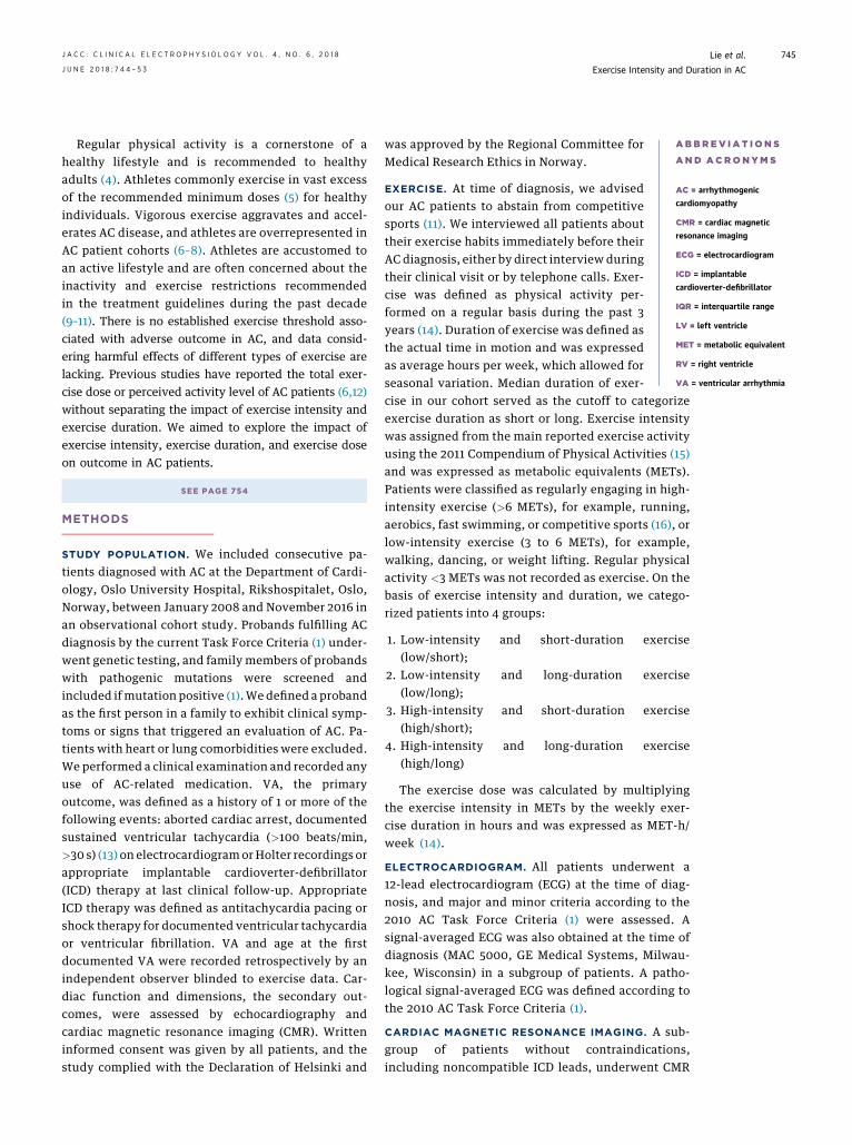
FIGURE 1 Cardiac Structure and Function Outcomes
By cardiac imaging, we defined the secondary outcomes as left ventricular (LV) dysfunction (top), right ventricular (RV) dilation (bottom left),
and RV dysfunction (bottom right). (Top) LV dysfunction was defined as (a) ejection fraction <54% in female patients and <52% in male
patients or (b) global longitudinal strain (GLS) worse than –18%. (Bottom left) RV dilation was defined as (a) proximal RV outflow tract (RVOT)
diameter $32 mm in parasternal short-axis view, (b) basal RV diameter (RVD) >41 mm in RV focused apical 4-chamber view (blue arrows), or
(c) major criterion indexed RV end-diastolic volume (RVEDVi) by cardiac magnetic resonance imaging. (Bottom right) RV dysfunction was
defined as (a) fractional area change #40%, (b) tricuspid annular systolic excursion <17 mm, or (c) major criterion RV ejection fraction by
cardiac magnetic resonance imaging. Blue arrows and areas are end-diastolic measures, and red are end-systolic measures.
Lie et al. J A C C : C L I N I C A L E L E C T R O P H Y S I O L O G Y V O L . 4 , N O . 6 , 2 0 1 8
Exercise Intensity and Duration in AC J U N E 2 0 1 8 : 7 4 4 – 5 3
746

TABLE 1 Clinical Characteristics of 173 AC Patients, Comparing Mutation-Positive Family
Members and Probands
All Patients(n ¼ 173)
Family Members(n ¼ 82)
Probands(n ¼ 91) p Value
Age, yrs 41 � 16 38 � 17 43 � 15 0.02
Female 76 (44) 46 (56) 30 (33) 0.002
BSA, m2 1.9 � 0.2 1.9 � 0.2 2.0 � 0.2 0.09
Heart rate, beats/min 62 � 12 67 � 14 58 � 10 <0.001
VA 83 (48) 5 (6) 78 (86) <0.001
Exercise characteristics
Duration, h/week 2.5 (2.0–5.5) 2.0 (2.0–4.1) 5.0 (2.0–6.3) <0.001
Long duration 86 (50) 29 (35) 57 (63) <0.001
Intensity, METs 6.7 � 1.9 5.9 � 1.5 7.4 � 1.8 <0.001
High intensity 91 (53) 22 (27) 69 (76) <0.001
Dose, MET-h/week 18 (12–41) 12 (10–21) 38 (14–56) <0.001
Electrocardiogram
Epsilon wave 18 (10) 2 (2) 16 (18) 0.001
TWI, major 51 (30) 11 (13) 40 (46) <0.001
TWI, minor 17 (10) 9 (11) 8 (9) 0.68
TAD >55 ms 17 (10) 5 (6) 12 (14) 0.10
SAECG pathology (n ¼ 139) 83 (48) 30 (42) 53 (78) <0.001
Medication
Beta blocker 106 (62) 39 (48) 67 (75) <0.001
Flecainide 14 (8) 5 (6) 9 (10) 0.33
Amiodarone 36 (21) 11 (13) 25 (28) 0.02
Values are mean � SD, n (%), or median (interquartile range). Unpaired Student’s t-test, chi-squared, orMann-Whitney U test as appropriate.
AC ¼ arrhythmogenic cardiomyopathy; BSA ¼ body surface area; METs ¼ metabolic equivalents; SAECG ¼signal-averaged electrocardiogram; TAD ¼ terminal activation duration; TWI ¼ T-wave inversion; VA ¼ ven-tricular arrhythmia.
J A C C : C L I N I C A L E L E C T R O P H Y S I O L O G Y V O L . 4 , N O . 6 , 2 0 1 8 Lie et al.J U N E 2 0 1 8 : 7 4 4 – 5 3 Exercise Intensity and Duration in AC
747
on clinical indication in a 1.6-T unit (MagnetomSonata, Vision Plus or Avanto Siemens, Erlangen,Germany) using a phased-array body coil as reportedpreviously (7). The presence of RV contractionabnormalities was recorded, and the major Task ForceCriteria (1) were included in the imaging endpoints ofRV dilation and dysfunction.
ECHOCARDIOGRAPHY. All participants underwentechocardiography at the time of AC diagnosis (Vivid 7,E9 or E95, GE, Vingmed, Horten, Norway), and data-sets were analyzed offline (EchoPac 201, GE,Vingmed) by 2 independent observers blinded toclinical and exercise information. LV ejection fractionwas assessed by the Simpson biplane method. LVglobal longitudinal strain was assessed by the speckletracking technique and calculated as the average peaknegative longitudinal strain in a 16-segment model(17). LV dysfunction was defined as ejectionfraction <54% in females and 52% in males orglobal longitudinal strain worse than �18% (Figure 1)(18–20).
RV function and dimensions were assessed byechocardiographic RV fractional area change,tricuspid annular plane systolic excursion, proximaldiameter of RV outflow tract, RV basal diameter (21),and CMR parameters. RV dilation was defined as RVbasal diameter >41 mm, RV outflow tract $32 mm(Figure 1), or indexed RV end-diastolic volume$100 ml/m2 in females and $110 ml/m2 in males withconcomitant RV akinesia/dyskinesia by CMR (1). RVdysfunction was defined as fractional area change#40%, tricuspid annular plane systolic excursion<17 mm (Figure 1) (22), or RV ejection fraction #40%with concomitant RV akinesia/dyskinesia by CMR (1).
GENETIC ANALYSES. Genomic DNAwas isolated fromperipheral blood, and testing was performed as a partof the diagnostic workup as described previously (7).
STATISTICAL ANALYSIS. Values are expressed asmean � SD, frequencies with percentages, or medianwith interquartile range (IQR) and were compared byStudent’s t-test, chi-square test, Fisher exact test, orMann-Whitney U test as appropriate (SPSS version21.0, SPSS Inc., Chicago, Illinois). Receiver operatingcharacteristic (ROC) curves were computed for exer-cise parameters to identify patients with adverseoutcome, and the curve coordinate closest to theupper left corner was defined as the statisticaloptimal threshold value. Logistical regressionanalyses were performed in patients with long-and short-duration exercise, in patients with high-and low-intensity exercise, and in patients with andwithout VA. Possible confounders (p < 0.10) were
added to multivariable logistical regression for theprimary endpoint, including an interaction term forhigh-intensity and long-duration exercise. Multi-collinearity was defined by correlation coefficients>0.7 or variance inflation factor >5 (23). The distri-butions of outcomes across the 4 groups of high- orlow-intensity and short- or long-duration exercisewere presented as modified radar plots that reflectedthe prevalence of outcomes as a percentage of thelength from the center of the diagram to the respec-tive corner. Distributions of outcomes around theaxes were compared by chi-squared test. The p valueswere 2-sided, and values <0.05 were consideredsignificant.
RESULTS
Of 191 patients diagnosed with AC at our center, 11had heart or lung comorbidities and 7 were unavail-able for exercise interview, which left 173 includedpatients (53% probands; 44% female; 41 � 16 years ofage) (Table 1) with detailed exercise information anddata on the primary outcome. An ECG was available inall 173 patients, and a satisfactory signal-averaged

TABLE 2 Clinical and Cardiac Imaging Characteristics of 173 AC Patients Divided by
Short and Long Exercise Duration
#2.5 h/week(n ¼ 87)
>2.5 h/week(n ¼ 86) p Value
Adjusted* OR(95% CI) p Value
Age, yrs 42 � 18 39 � 14 0.32
BSA, m2 1.9 � 0.2 1.9 � 0.2 0.69
Female 52 (60) 24 (28) <0.001 0.5 (0.2–1.1) 0.08
Probands 34 (39) 57 (84) <0.001†
High intensity 25 (29) 66 (77) <0.001 4.6 (2.0–11) <0.001
VA
Total VA 27 (31) 56 (65) <0.001 1.6 (0.6–3.9) 0.35
ACA 4 (5) 13 (16) 0.02‡
VT 19 (22) 51 (61) <0.001‡
ICD therapy 10 (12) 39 (46) <0.001‡
Age at VA, yrs 46 � 16 38 � 15 0.03§ 1.0 (0.9–1.0) 0.06
Cardiac imaging
LV dysfunction 24 (29) 42 (49) 0.008 1.3 (0.6–2.8) 0.54
EF, % 58 � 5 54 � 9 0.002‡
GLS, % �19.5 � 3.0 �18.3 � 3.5 0.02‡
RV dilation 53 (63) 75 (87) <0.001 2.5 (0.9–6.4) 0.07
RVD, mm 40 � 7 44 � 9 0.001‡
RVOT, mm 33 � 7 38 � 9 0.001‡
CMR RVEDVi, ml/m2 103 � 41 109 � 32 0.51
RV dysfunction 47 (53) 62 (71) 0.02 0.9 (0.4–2.1) 0.81
FAC, % 41 � 10 37 � 10 0.01‡
TAPSE, mm 20 � 5 19 � 6 0.36
CMR RV EF, % 49 � 12 46 � 11 0.30
Values are mean � SD, or n (%). Unpaired Student’s t-test, chi-squared, Fisher exact test, and multivariablelogistical regression. *Multivariable logistical regression including significant variables from univariable analysesand age. †Proband status was not retained in multivariable analyses because of multicollinearity with VA.‡Parameters used to define VA, LV dysfunction, RV dilation, and RV dysfunction were not retained in multi-variable analysis. §Age at VA was adjusted in a separate model without age, because of multicollinearity.
ACA ¼ aborted cardiac arrest; CI ¼ confidence interval; CMR ¼ cardiac magnetic resonance imaging; EF ¼ejection fraction; FAC ¼ fractional area change; GLS ¼ global longitudinal strain; ICD ¼ implantable cardioverter-defibrillator; LV ¼ left ventricle; OR ¼ odds ratio; RV ¼ right ventricle; RVD ¼ RV diameter; RVEDVi ¼ RV end-diastolic volume indexed; RVOT ¼ RV outflow tract; TAPSE ¼ tricuspid annular plane systolic excursion; VT ¼ventricular tachycardia; other abbreviations as in Table 1.
Lie et al. J A C C : C L I N I C A L E L E C T R O P H Y S I O L O G Y V O L . 4 , N O . 6 , 2 0 1 8
Exercise Intensity and Duration in AC J U N E 2 0 1 8 : 7 4 4 – 5 3
748
ECG was available in 139 (80%). Five patients hadunsatisfactory echocardiographic acquisitions ofeither LV or RV, and thus, 168 patients (97%) wereanalyzed for all the secondary outcomes (LV and RVdysfunction and RV dilation). CMR was performed in134 patients (79%). Genetic analyses had been per-formed in 170 patients, of whom 125 (72%) had path-ogenic mutations (109 [87%] in plakophilin-2, 8 [6%]in desmoplakin, 7 [6%] in desmoglein-2, and 2 [2%] indesmocollin-2 genes). Forty-five patients weremutation-negative probands with definite AC, and 3probands with definite AC were not geneticallytested. As expected, probands had more severe dis-ease than family members (Table 1).
Seventeen patients had experienced aborted car-diac arrest, 70 had documented sustained ventriculartachycardia, and 49 had received appropriate ICDtherapy (Online Table). In total, VA had occurred in83 patients (34% female; age 40 � 16 years). Patientswith VA were more frequently probands (86% vs. 6%,
p < 0.001) and had more prevalent LV dysfunction(52% vs. 27%; p ¼ 0.001), RV dysfunction (83% vs.47%, p < 0.001), and RV dilation (93% vs. 59%;p < 0.001) than patients without VA.
EXERCISE DURATION: LONG VERSUS SHORT. Medianweekly exercise duration was 2.5 h (IQR: 2.0 to 5.5 h).According to the predefined cutoff, 86 patients wereclassified as regularly engaging in long-durationexercise (>2.5 h/week), and 87 as participating inshort-duration exercise (#2.5 h/week) (Table 2).Ventr i cu lar a r rhythmia . Long-duration exercisewas more common in males and in probands and wasassociated with VA, but none of these markersremained when adjusted for confounders (Table 2).Exercise duration $2.3 h/week was the statisticalthreshold associated with VA (C-statistic, 0.69; 95%CI: 0.61 to 0.77).Funct iona l and structura l a l terat ions . Patientswith long-duration exercise had worse LV and RVfunction, and more RV dilation, than patientswith short exercise duration. There was a trendtowards an association between long-durationexercise and RV dilation in multivariable analysis(p¼ 0.07). The statistical threshold associated with RVdilation was $2.8 h of exercise per week, $3.2 h/weekwith RV dysfunction, and $3.4 h/week with LVdysfunction.
EXERCISE INTENSITY: HIGH VERSUS LOW. The meanexercise intensity was 6.7 � 1.9 METs. High-intensityexercise (>6 METs) was reported by 91 patients, and82 reported low-intensity exercise (3 to 6 METs)(Table 3). High-intensity exercise was more commonin males, probands, and patients reporting long-duration exercise.Ventr i cu lar ar rhythmia . Patients with high-intensity exercise had more prevalent VA than pa-tients with low-intensity exercise, an association thatremained strong in multivariable analysis (Table 3).Male sex, long-duration exercise, and high-intensityexercise were markers of VA in univariable logisticalregression, and higher age was a possible confounder.High-intensity exercise and the interaction betweenhigh-intensity and long-duration exercise were in-dependent markers of VA in multivariable analysis(Table 4). In a subgroup analysis of mutation-positivefamily members (n ¼ 82), VA occurred in 5 (6%), andall of them reported high-intensity exercise (23% vs.0%, p ¼ 0.001).Funct iona l and structura l a l terat ions . Patientsreporting high-intensity exercise had worse LV andRV function and more RV dilation than patientsreporting low-intensity exercise, but not whenadjusted for confounders (Table 3). ROC analyses

TABLE 3 Clinical and Cardiac Imaging Characteristics of 173 AC Patients and Mutation
Carriers With History of Low-Intensity and High-Intensity Exercise
#6 METs(n ¼ 82)
>6 METs(n ¼ 91)
pValue
Adjusted* OR(95% CI)
pValue
Age, yrs 41 � 17 40 � 15 0.54
BSA, m2 1.9 � 0.2 1.9 � 0.2 0.61
Female 52 (63) 24 (26) <0.001 0.3 (0.1–0.6) 0.003
Probands 22 (27) 69 (76) <0.001†
Long duration 20 (24) 66 (73) <0.001 4.7 (2.0–11) <0.001
VA
Total VA 16 (20) 67 (74) <0.001 14.1 (5.1–39) <0.001
ACA 4 (5) 13 (15) 0.03‡
VT 12 (15) 58 (66) <0.001‡
ICD therapy 6 (7) 43 (49) <0.001‡
Age at VA, yrs 45 � 14 39 � 16 0.20
Cardiac imaging
LV dysfunction 21 (27) 45 (50) 0.002 1.4 (0.6–3.5) 0.45
EF, % 58 � 5 55 � 9 0.01‡
GLS, % �19.9 � 2.6 �18.0 � 3.5 <0.001‡
RV dilation 52 (64) 76 (85) 0.001 0.7 (0.2–1.8) 0.42
RVD, mm 39 � 7 45 � 9 <0.001‡
RVOT, mm 34 � 6 37 � 9 0.004‡
CMR RVEDVi, ml/m2 96 � 30 117 � 39 0.02‡
RV dysfunction 42 (52) 67 (76) 0.001 0.8 (0.3–2.2) 0.70
FAC, % 42 � 9 36 � 10 <0.001‡
TAPSE, mm 21 � 5 18 � 6 0.005‡
CMR RV EF, % 50 � 11 45 � 12 0.07
Values are mean � SD, or n (%). Unpaired Student’s t-test, chi-squared, Fisher exact test, and multivariablelogistical regression. *Multivariable logistical regression including significant variables from univariableanalyses and age. †Proband status was not retained in multivariable analyses because of multicollinearity withVA. ‡Parameters used to define VA, LV dysfunction, RV dilation, and RV dysfunction were not retainedin multivariable analysis.
Abbreviations as in Tables 1 and 2.
TABLE 4 Clinical Markers of VA (n ¼ 83) in 173 AC Patients
Univariable Multivariable
OR (95% CI) p Value OR (95% CI) p Value
Age, 1 yr 1.02 (1.00–1.04) 0.05 1.04 (1.01–1.06) 0.004
BSA, 0.1 m2 1.06 (0.92–1.22) 0.45
Female 0.37 (0.20–0.68) 0.002 0.79 (0.35–1.77) 0.56
High-intensity exercise 11.52 (5.62–23.61) <0.001 3.84 (1.33–11.04) 0.01
Long-duration exercise 4.15 (2.20–7.83) <0.001 0.37 (0.07–1.88) 0.23
Interaction of high-intensityand long-duration exercise
12.75 (1.86–87.29) 0.009
Logistic regression of clinical and exercise variables. High-intensity and long-duration exercise correlatedmoderately and were retained in multivariable analysis with other potential confounders (p < 0.10) from uni-variable analysis and an interaction term of high-intensity and long-duration exercise.
Abbreviations as in Tables 1 and 2.
J A C C : C L I N I C A L E L E C T R O P H Y S I O L O G Y V O L . 4 , N O . 6 , 2 0 1 8 Lie et al.J U N E 2 0 1 8 : 7 4 4 – 5 3 Exercise Intensity and Duration in AC
749
suggested exercise intensities between 6 and 7 METsas the optimal statistical threshold values for detect-ing VA (C-statistic, 0.78; 95% CI: 0.70 to 0.85) and forall secondary outcomes (LV and RV dysfunction andRV dilation).
COMBINATIONS OF EXERCISE DURATION AND
INTENSITY. The distributions of outcomes in the 4groups stratified by high or low intensity and long orshort duration of exercise shown on the modifiedradar plots (Figure 2) revealed that VA was mostprevalent in high/long exercise (Figure 2A). Interest-ingly, VA was more prevalent among patients withhigh/short exercise than patients with low/long ex-ercise (Figure 3). LV dysfunction was most prevalentin high/long exercise and significantly more preva-lent than in low/short exercise (Figure 2B). RVdysfunction was also more prevalent in high/longexercise than in both groups of low-intensity exercise(Figure 2C). RV dilatation was more prevalent in high/long exercise than in both groups of short durationbut not different from patients with low/long exer-cise (Figure 2D).
EXERCISE DOSE. Median exercise dose was 18 MET-h/week (IQR: 12 to 41 MET-h/week) and was higherin probands (Table 1) and men (36 MET-h/week [IQR:14 to 55 MET-h/week] vs. 12 MET-h/week [IQR: 9 to 22MET-h/week] in women, p < 0.001).Ventr i cu la r ar rhythmia . Patients with exercisedoses above median had more prevalent VA (67% vs.30%, p < 0.001) than patients with lower exercisedoses. ROC analysis suggested $21 MET-h/week to beassociated with VA (C-statistic, 0.74 [95% CI: 0.67 to0.82]).Funct iona l and structura l a l terat ions . Patientswith exercise doses above median had more preva-lent LV dysfunction (49% vs. 29%, p ¼ 0.01), RVdysfunction (75% vs. 55%, p ¼ 0.006), and RV dilation(88% vs. 63%, p < 0.001) than patients with lowerexercise doses. A total of $13 MET-h/week wasassociated with RV dilation, $17 MET-h/week withRV dysfunction, and $25 MET-h/week with LVdysfunction.
DISCUSSION
This study supports the previously reported rela-tionship between exercise and AC disease and showsfor the first time that high-intensity exercise has astronger association with adverse outcome than longexercise duration. These findings suggest that high-intensity exercise is the main premise for harmfuleffects of exercise in AC and could be helpful whengiving exercise advice to AC patients.
EXERCISE DURATION. Long-duration exercise wasassociated with adverse outcome; however, themajority of patients with a history of long-durationexercise also had a history of high-intensity exer-cise. Adjusted for high-intensity exercise and otherpotential confounders, long-duration exercise was

FIGURE 2 Distribution of Outcomes
Modified radar plots of the distributions of life-threatening ventricular arrhythmia (VA), LV dysfunction, RV dysfunction, and RV dilation across
4 groups of high- and low-intensity exercise (x-axis) and long- and short-duration exercise (y-axis). The p value by chi-squared test for the
distribution of outcomes across 4 groups of intensity/duration as stated. (A) VAs (red): VA was more prevalent in high-intensity/long-duration
exercise than in high-intensity/short-duration (82% vs. 52%, p ¼ 0.004), low-intensity/long-duration (82% vs. 10%, p < 0.001), or low-
intensity/short-duration exercise (82% vs. 23%, p < 0.001) and more prevalent in patients reporting high-intensity/short-duration exercise
than in low-intensity/long-duration (52% vs. 10%, p ¼ 0.003) or low-intensity/short-duration exercise (52% vs. 23%, p ¼ 0.007). (B) LV
dysfunction (blue): LV dysfunction was more prevalent in high-intensity/long-duration exercise than in low-intensity/short-duration exercise
(55% vs. 25%, p ¼ 0.001). (C) RV dysfunction (green): RV dysfunction was more prevalent in patients reporting high-intensity/long-duration
exercise than in low-intensity/long-duration (83% vs. 40%, p < 0.001) and low-intensity/short-duration exercise (83% vs. 56%, p ¼ 0.001).
(D) RV dilatation (orange): RV dilatation was more prevalent in patients with high-intensity/long-duration exercise than in high-intensity/
short-duration exercise (91% vs. 70%, p ¼ 0.01) and low-intensity/short-duration exercise (91% vs. 61%; p < 0.001). METs ¼ metabolic
equivalents.
Lie et al. J A C C : C L I N I C A L E L E C T R O P H Y S I O L O G Y V O L . 4 , N O . 6 , 2 0 1 8
Exercise Intensity and Duration in AC J U N E 2 0 1 8 : 7 4 4 – 5 3
750

FIGURE 3 Prevalence of Life-Threatening Ventricular
Arrhythmia in 173 Patients With Arrhythmogenic
Cardiomyopathy Divided Into 4 Groups Defined by Exercise
Intensity and Duration
METs ¼ metabolic equivalents.
J A C C : C L I N I C A L E L E C T R O P H Y S I O L O G Y V O L . 4 , N O . 6 , 2 0 1 8 Lie et al.J U N E 2 0 1 8 : 7 4 4 – 5 3 Exercise Intensity and Duration in AC
751
not a marker of VA. In the adjusted model, there wasonly a trend towards an association between long-duration exercise and RV dilation. Increased RVdimensions are acknowledged effects of endurancetraining in healthy athletes (24) but are also associatedwith disease progression in AC (25). Therefore, RVdilation was considered a soft secondary outcome.This was further supported by the finding that theexercise duration thresholds associated with VAand RV dilation were shorter than for RV and LVdysfunction. This is well in line with the progressivenature of AC, in which VA and RV dilation areearly phenomena that could be followed by laterdevelopment of myocardial dysfunction (26).
EXERCISE INTENSITY. High-intensity exercise wasstrongly associated with adverse outcome. AC pa-tients reporting high-intensity exercise had aggra-vated phenotypes according to echocardiography andCMR, indicative of more severe disease. Combininghigh-intensity with long-duration exercise was thestrongest marker of VA, which implies that high-intensity exercise performed for long durations wasthe most harmful form of exercise. The majority ofpatients reporting high-intensity exercise also re-ported long-duration exercise. High-intensity exer-cise remained a strong and independent marker of VAwhen adjusted for long-duration exercise and other
potential confounders. Interestingly, high-intensityexercise of short duration was a strong and inde-pendent marker of adverse outcome, whereas low-intensity exercise of long duration was not.
The more harmful effects of high intensitycompared with long duration could have severalpossible explanations. During high-intensity exer-cise, the increase in RV wall stress exceeds LV wallstress because of the thinner wall of the RV (27). Thisincremental increase in wall stress has been proposedas an explanation of the harmful effects of vigorousexercise and the RV predilection observed in AC(7,28). The wall stress increase is less pronouncedduring low-intensity exercise, which might thereforebe tolerated, even for longer durations. Our resultsare in line with previous reports of harmful effects ofexercise in AC (6,28,29) and add important informa-tion by highlighting the role of exercise intensity inAC. Therefore, the advice to AC patients to abstainfrom competitive sport should be more specific. Thethreshold for harmful exercise intensity in our studywas around 6 to 7 METs for all adverse outcomes,which suggests that AC patients should not performactivities such as soccer, aerobics, or fast swimmingon a regular basis, even noncompetitively.
Although VA occurred rarely in mutation-positivefamily members, it occurred exclusively in familymembers reporting high-intensity exercise. Thisfinding adds to previous studies reporting moreprevalent VA in an AC mutation–positive family withhigh exercise doses (7,29) and indicates that high-intensity exercise has also an impact on disease pro-gression in family members with no or mild diseasemanifestation (7).
Low-intensity exercise, reflecting an activeeveryday lifestyle (e.g., walking, gardening), wasassociated with myocardial function and RV di-mensions within the normal range. Low-intensityexercise seemed to be tolerated better, even whenperformed for long durations. However, one-fifth ofpatients reporting low-intensity exercise had expe-rienced VA, which might reflect the high prevalenceof VA in AC independently of exercise (6,28). Dataon the safety of moderate-intensity exercise issparse in AC. There were no results in our study thatsuggested that low-intensity exercise had an adverseimpact in AC; however, further studies shouldexplore safe exercise thresholds in AC, preferably inexperimental studies, because randomized exercisetraining trials in AC patients are unfeasible forethical reasons.
EXERCISE DOSE. Patients with high exercise doseshad more prevalent VA, LV dysfunction, RV

PERSPECTIVES
COMPETENCY IN MEDICAL KNOWLEDGE: Pa-
tients with arrhythmogenic cardiomyopathy reporting
exercise above 6 metabolic equivalents at the time of
diagnosis had higher prevalence of life-threatening
ventricular arrhythmia and impaired myocardial func-
tion. These patients could be advised to restrict ex-
ercise to lower intensities in order to reduce the odds
of adverse outcome.
TRANSLATIONAL OUTLOOK: The safety of low
intensity exercise for long durations, and the effect of
exercise restriction after arrhythmogenic cardiomy-
opathy diagnosis, should be explored in further,
preferably experimental, studies. Due to convincing
evidence supporting the link between exercise and
outcome in these patients, clinical trials are unfeasible
for ethical reasons.
Lie et al. J A C C : C L I N I C A L E L E C T R O P H Y S I O L O G Y V O L . 4 , N O . 6 , 2 0 1 8
Exercise Intensity and Duration in AC J U N E 2 0 1 8 : 7 4 4 – 5 3
752
dysfunction, and RV dilation than patients with lowerdoses. The threshold of exercise doses ranged fromapproximately 13 MET h/week for RV dilation,equivalent to the upper American Heart Association–recommended level for healthy adults of 30 min oflow-intensity exercise 5 days per week (4), to almostdouble that dose (25 MET h/week) for LV dysfunction.We demonstrated that exercise intensity had astronger association with outcome in AC patientsthan exercise duration, which implies that sheermultiplication of the two might not give an appro-priate estimation of the exercise load in these pa-tients. Interestingly, female patients in our study hadlower exercise doses than male patients, which mightbe one of several explanations of the lower diseasepenetrance in females.
CLINICAL IMPLICATIONS. Our study showed thatexercise at >6 METs was associated with VA and amore severe AC phenotype than low-intensity exer-cise and underscores the advice that AC patientsshould avoid high-intensity exercise. Low-intensityexercise, even for long durations, was not associ-ated with an unfavorable outcome and might be anacceptable alternative for patients diagnosed with ACwho wish to maintain an active lifestyle.
STUDY LIMITATIONS. The observational single-center study design limited external validity. Exer-cise duration was self-reported, and intensity wasassigned on the basis of the reported exercise activ-ity. The categorization of exercise duration by medianwas arbitrary but coincidentally reflected the mini-mum exercise duration recommended for healthyadults (4). We reported exercise habits during theimmediate 3 years before AC diagnosis and did notinclude lifelong exercise data in analyses becauseexercise habits can change over time and are subjectto recall bias. At diagnosis, patients were advised torestrict their exercise (11), and exercise after diagnosiswas not assessed. High-intensity exercise was oftencombined with long-duration exercise, but the 2 pa-rameters were only moderately correlated. The highodds of an adverse outcome in high-intensity exercisewas independent of the interaction between the two,
which further strengthens the impact of high-intensity exercise.
CONCLUSIONS
High-intensity exercise and long-duration exercisewere both associated with unfavorable outcome in ACpatients, but high-intensity exercise was a strongmarker of VA independent of exercise duration. Low-intensity exercise, even for long durations, wasassociated with a milder phenotype and can beadvised for patients with AC. Further research shouldexplore the safety of low-intensity exercise and theeffects of exercise intensity restriction on ACdiagnosis.
ACKNOWLEDGMENTS The authors are grateful to thestudy participants for their valuable contribution.
ADDRESS FOR CORRESPONDENCE: Dr. Kristina H.Haugaa, Department of Cardiology, Oslo UniversityHospital, Rikshospitalet, PO Box 4950 Nydalen, 0424Oslo, Norway. E-mail: [email protected].
RE F E RENCE S
1. Marcus FI, McKenna WJ, Sherrill D, et al. Diag-nosis of arrhythmogenic right ventricular cardio-myopathy/dysplasia: proposed modification of theTask Force criteria. Circulation 2010;121:1533–41.
2. Corrado D, Basso C, Judge DP. Arrhythmogeniccardiomyopathy. Circ Res 2017;121:784–802.
3. Thiene G, Nava A, Corrado D, Rossi L,Pennelli N. Right ventricular cardiomyopathy and
sudden death in young people. N Engl J Med1988;318:129–33.
4. Haskell WL, Lee IM, Pate RR, et al.Physical activity and public health: updatedrecommendation for adults from the Amer-ican College of Sports Medicine and theAmerican Heart Association. Circulation 2007;116:1081–93.
5. Sharma S, Merghani A, Mont L. Exercise and theheart: the good, the bad, and the ugly. Eur Heart J2015;36:1445–53.
6. Ruwald AC, Marcus F, Estes NA 3rd, et al.Association of competitive and recreational sportparticipation with cardiac events in patients witharrhythmogenic right ventricular cardiomyopathy:results from the North American multidisciplinary

J A C C : C L I N I C A L E L E C T R O P H Y S I O L O G Y V O L . 4 , N O . 6 , 2 0 1 8 Lie et al.J U N E 2 0 1 8 : 7 4 4 – 5 3 Exercise Intensity and Duration in AC
753
study of arrhythmogenic right ventricular cardio-myopathy. Eur Heart J 2015;36:1735–43.
7. Saberniak J, Hasselberg NE, Borgquist R, et al.Vigorous physical activity impairs myocardialfunction in patients with arrhythmogenic rightventricular cardiomyopathy and in mutation posi-tive family members. Eur J Heart Fail 2014;16:1337–44.
8. Corrado D, Basso C, Rizzoli G, Schiavon M,Thiene G. Does sports activity enhance the risk ofsudden death in adolescents and young adults?J Am Coll Cardiol 2003;42:1959–63.
9. Maron BJ, Udelson JE, Bonow RO, et al. Eligi-bility and disqualification recommendations forcompetitive athletes with cardiovascular abnor-malities: Task Force 3: Hypertrophic Cardiomyop-athy, Arrhythmogenic Right VentricularCardiomyopathy and Other Cardiomyopathies, andMyocarditis: a scientific statement from theAmerican Heart Association and American Collegeof Cardiology. J Am Coll Cardiol 2015;66:2362–71.
10. Corrado D, Wichter T, Link MS, et al. Treat-ment of arrhythmogenic right ventricular cardio-myopathy/dysplasia: an international task forceconsensus statement. Eur Heart J 2015;36:3227–37.
11. Maron BJ, Zipes DP. 36th Bethesda Confer-ence: eligibility recommendations for competitiveathletes with cardiovascular abnormalities. J AmColl Cardiol 2005;45:2–64.
12. Mazzanti A, Ng K, Faragli A, et al. Arrhyth-mogenic right ventricular cardiomyopathy: clinicalcourse and predictors of arrhythmic risk. J Am CollCardiol 2016;68:2540–50.
13. Pedersen CT, Kay GN, Kalman J, et al. EHRA/HRS/APHRS expert consensus on ventricular ar-rhythmias. Europace 2014;16:1257–83.
14. Wasfy MM, Baggish AL. Exercise dose in clin-ical practice. Circulation 2016;133:2297–313.
15. Ainsworth BE, Haskell WL, Herrmann SD, et al.2011 Compendium of Physical Activities: a secondupdate of codes and MET values. Med Sci SportsExerc 2011;43:1575–81.
16. World Health Organization. Intensity of phys-ical activity. Available at: http://www.who.int/dietphysicalactivity/physical_activity_intensity/en/.Accessed February 13, 2017.
17. Edvardsen T, Haugaa KH. Imaging assessmentof ventricular mechanics. Heart 2011;97:1349–56.
18. Haugaa KH, Edvardsen T. Global longitudinalstrain: the best biomarker for predicting prognosisin heart failure? Eur J Heart Fail 2016;18:1340–1.
19. Lang RM, Badano LP, Mor-Avi V, et al. Rec-ommendations for cardiac chamber quantificationby echocardiography in adults: an update from theAmerican Society of Echocardiography and theEuropean Association of Cardiovascular Imaging[published correction appears in Eur Heart J Car-diovasc Imaging 2016;17:412]. Eur Heart J Car-diovasc Imaging 2015;16:233–70.
20. Yingchoncharoen T, Agarwal S, Popovic ZB,Marwick TH. Normal ranges of left ventricularstrain: a meta-analysis. J Am Soc Echocardiogr2013;26:185–91.
21. Leren IS, Saberniak J, Haland TF, Edvardsen T,Haugaa KH. Combination of ECG and echocardi-ography for identification of arrhythmic events inearly ARVC. J Am Coll Cardiol Img 2017;10:503–13.
22. Haugaa KH, Basso C, Badano LP, et al. EACVIScientific Documents Committee, EACVI Boardmembers and external reviewers. Comprehensivemulti-modality imaging approach in arrhythmo-genic cardiomyopathy: an expert consensusdocument of the European Association of
Cardiovascular Imaging. Eur Heart J CardiovascImaging 2017;18:237–53.
23. Vatcheva KP, LeeM,McCormick JB, RahbarMH.Multicollinearity in regression analyses conductedin epidemiologic studies. Epidemiology (Sunnyvale)2016;6:227.
24. D’Ascenzi F, Pisicchio C, Caselli S, Di Paolo FM,Spataro A, Pelliccia A. RV remodeling in Olympicathletes. J Am Coll Cardiol Img 2016;10:385–93.
25. Mast TP, James CA, Calkins H, et al. Evaluationof structural progression in arrhythmogenic rightventricular dysplasia/cardiomyopathy. JAMA Car-diol 2017;2:293–302.
26. Basso C, Corrado D, Marcus FI, Nava A,Thiene G. Arrhythmogenic right ventricular car-diomyopathy. Lancet 2009;373:1289–300.
27. La Gerche A, Heidbuchel H, Burns AT, et al.Disproportionate exercise load and remodeling ofthe athlete’s right ventricle. Med Sci Sports Exerc2011;43:974–81.
28. James CA, BhonsaleA, Tichnell C, et al. Exerciseincreases age-related penetrance and arrhythmicrisk in arrhythmogenic right ventricular dysplasia/cardiomyopathy-associated desmosomal mutationcarriers. J Am Coll Cardiol 2013;62:1290–7.
29. Sawant AC, Te Riele AS, Tichnell C, et al.Safety of American Heart Association-recommended minimum exercise for desmosomalmutation carriers. Heart Rhythm 2016;13:199–207.
KEY WORDS arrhythmia, arrhythmogenicright ventricular cardiomyopathy, athletesheart, exercise intolerance, ventriculararrhythmia, myocardial function
APPENDIX For a supplemental table, pleasesee the online version of this paper.