articlelabs.bio.unc.edu/goldstein/leemarston2006.pdfjeff hardin, 2 ,3 ari halberstadt, 1and bob...
TRANSCRIPT


Current Biology 16, 1986–1997, October 24, 2006 ª2006 Elsevier Ltd All rights reserved DOI 10.1016/j.cub.2006.08.090
ArticleWnt/Frizzled SignalingControls C. elegans Gastrulationby Activating Actomyosin Contractility
Jen-Yi Lee,1,4,5 Daniel J. Marston,1,4 TimothyWalston,2
Jeff Hardin,2,3 Ari Halberstadt,1 and Bob Goldstein1,*1Department of BiologyUniversity of North Carolina, Chapel HillChapel Hill, North Carolina 27599-32802Laboratory of Genetics3Program in Cellular and Molecular Biologyand Department of ZoologyUniversity of Wisconsin-MadisonMadison, Wisconsin 53706
Summary
Background: Embryonic patterning mechanisms regu-late the cytoskeletal machinery that drives morphogen-esis, but there are few cases where links between pat-terning mechanisms and morphogenesis are wellunderstood. We have used a combination of genetics,in vivo imaging, and cell manipulations to identify suchlinks in C. elegans gastrulation. Gastrulation in C. ele-gans begins with the internalization of endodermal pre-cursor cells in a process that depends on apical con-striction of ingressing cells.Results: We show that ingression of the endodermalprecursor cells is regulated by pathways, includingaWnt-Frizzled signaling pathway, that specify endoder-mal cell fate.We find thatWnt signaling has a role in gas-trulation in addition to its earlier roles in regulating endo-dermal cell fate and cell-cycle timing. In the absence ofWnt signaling, endodermal precursor cells polarize andenrich myosin II apically but fail to contract their apicalsurfaces. We show that a regulatory myosin light chainnormally becomes phosphorylated on the apical sideof ingressing cells at a conserved site that can lead tomyosin-filament formation and contraction of actomyo-sin networks and that this phosphorylation depends onWnt signaling.Conclusions:We conclude that Wnt signaling regulatesC. elegans gastrulation through regulatory myosin light-chain phosphorylation, which results in the contractionof the apical surface of ingressing cells. These findingsforge new links between cell-fate specification andmor-phogenesis, and they represent a novel mechanism bywhich Wnt signaling can regulate morphogenesis.
Introduction
Themorphogenetic events that shape embryonic devel-opment rely on the movements and shape changes ofindividual cells. Because the cellular cytoarchitectureprovides the driving forces for these cellular events,
one of the keys to understanding the molecular basisof morphogenetic movements is determining how well-studied developmental pathways specifying cell fatelead to modulation of the cytoskeleton in individual cellsin ways that can produce forces capable of moving cellsor deforming tissues. Toward this goal, there has beenidentification of many genes that function upstream ofmorphogenetic movements, including many essentialfor cell-fate specification and extracellular signaling,and there is some understanding of the cytoskeletalmechanics that drive these movements. However, therehas been more limited progress in tying these two endstogether to provide a coherent thread from cell fate andsignaling molecules to the cytoskeletal dynamics re-sponsible for morphogenesis [1, 2].One of the earliest morphogenetic events in animal
development is gastrulation, the process by which theembryo reorganizes itself into three germ layers. Gastru-lation in C. elegans begins at the 26-cell stage when thetwo endodermal founder cells, Ea and Ep, begin to mi-grate from the outer, ventral surface of the embryo tothe embryonic interior [3] (Figure 1). The mechanismsthat specify endodermal fate in these cells are well stud-ied. SKN-1, an endomesodermal determinant, is segre-gated to two cells (P2 and EMS) at the four-cell stage.In P2, SKN-1 activity is repressed in the P2 cell, whereasSKN-1 activity persists in EMS to promote endomeso-dermal fate [4]. A Wnt interaction at the four-cell stagethen specifies endodermal fate on one side of the EMScell. Differential regulation of transcription factor activityin this cell’s two daughters results in a single endoder-mal precursor cell (E) at the eight-cell stage [5].After E is born, its daughter cells (Ea and Ep) ingress
during a cell cycle that is extended by the introductionof a gap phase [6]. The space left on the ventral side ofthe embryo is filled by neighboring cells, a total of sixcells from the MS, AB, and P4 lineages [7]. After ingres-sion, Ea and Ep divide (Figure 2A). For simplicity, we usethe term ‘‘gastrulation’’ here solely to refer to Ea-Ep in-gression, the internalization of the endoderm. Gastrula-tion in C. elegans continues later with the internalizationof other cells including mesoderm and germline progen-itors [8].Recent work has begun to shed light on the mecha-
nisms required for C. elegans gastrulation [9]. One ofthe driving forces for Ea-Ep ingression is apical constric-tion, which is likely powered by an actomyosin contrac-tion [7]. Consistent with this finding, NMY-2, a non-muscle myosin II heavy chain, accumulates at theapical (ventral) surfaces of Ea and Ep, and this polarizedaccumulation requires the activity of the PAR proteins[8, 10]. The PARproteinswere first identified as essentialfor polarity in the 1-cell C. elegans embryo [11] and havesince been found to be required for polarity in organismsas diverse as Drosophila and humans [12]. In gastrula-tion-stage embryos, the PAR proteins have polarizeddistributions that are established by cell-cell contacts,with PAR-3, PAR-6, and PKC-3 localized apically, at
*Correspondence: [email protected] authors contributed equally to this work.5Present address: Department of Molecular and Cell Biology,Mail Code #3200, University of California, Berkeley, California94720-3200

contact-free areas, and PAR-1 and PAR-2 localized ba-solaterally where cells contact neighboring cells [8, 10].Depleting the embryo of specific PAR proteins justbefore gastrulation compromises gastrulation move-ments. The timing of ingression is delayed significantlycompared to that in wild-type cells, but the E lineagecells still internalize [10]. This suggests that there mustbe additional and essential gastrulation regulators thatremain to be identified.The Wnt pathway has been implicated in cell-fate
specification, cell polarization, and morphogenesisacross the animal kingdom [13]. Wnt ligands or theirFrizzled (Fz) receptors, or both, are required in manyprocesses including establishment of Drosophila seg-ment polarity [14, 15], zebrafish and Xenopus gastrula-tion [16–18], and Xenopus neural tube closure [19]. Wntand Fz are known to act through various signal transduc-tion pathways, generally categorized as canonical andnoncanonical pathways. Canonical signaling results intranslocation of b-catenin from the cytoplasm to thenucleus, where it participates in the transcriptional acti-vation of downstream targets. In C. elegans, mom-2,the Wnt gene that functions during the four-cell stageto specify endoderm, acts in a variant of the canonicalmanner by activating WRM-1, a C. elegans b-catenin
homolog, which results in downregulation instead ofupregulation of POP-1, a TCF/LEF transcription factor[20]. In contrast, noncanonical pathways act throughcytoplasmic factors that ultimately regulate cytoskeletalcomponents but can also act to regulate transcription in-dependently of b-catenin [5]. Further understanding ofthe mechanisms by which Wnt signaling can regulatecellmovementswill be important for understandingmor-phogenesis during normal development and Wnt path-way function in tumor invasion and metastasis in humancancers [21].We have used a candidate approach to begin to iden-
tify genes required forC. elegans gastrulation. We foundthat the pathways that specify the endodermal precur-sors also regulate ingression of these cells. We showthatWnt/Fz signaling hasa role in gastrulation in additionto its function in regulating endodermal cell fate andcell-cycle timing. Although Wnt signaling functions incell polarization in many contexts [22], Wnt signalingdoes not affect C. elegans gastrulation by establishingpolarity in the ingressing cells or by affecting the rateof myosin accumulation at the apical cortex of thesecells. Instead, we found that Wnt/Fz signaling functionsin gastrulation by causing regulatory myosin light chainin the apical cortex of Ea and Ep to be phosphorylatedat a contraction-activating serine residue. Our resultsforge new links among cell fate, cell signaling, and cellform and suggest a novel role for intercellular signalingby a Wnt protein—the regulation of morphogeneticmovements through activation of myosin contractileactivity.
Results
Endodermal-Fate-Specification Pathways ActUpstream of C. elegans GastrulationTo determine whether endodermal fate specification isnecessary for Ea and Ep morphogenetic behavior, wefirst determined whether C. elegans embryos defectivein each of the endodermal-fate-specification genes alsohave gastrulation defects. The endodermal founder cellsare specified by two intersecting pathways: A GATA-factor transcriptional cascade initially restricts mesen-doderm fate to the appropriate endoderm and meso-derm precursors, and the Wnt signaling pathway thenacts to repress nuclear POP-1 and thus allow endodermdevelopment in the progeny of the E cell [4]. The endo-derm-specification pathways also affect cell-cycle tim-ing in endodermal precursors. The Ea and Ep cells arethe first cells in the C. elegans embryo to introducea gap phase, a G2 phase that results in the Ea and Epcells dividing approximately 20 min later than MSa andMSp cells (20.2 6 2.4 min SD; Figure 2B) [6]. This delaydoes not occur in endoderm-specification mutants [4].We speculated that either endoderm specification orcell-cycle delay, or both, might be required for gastrula-tion. Gastrulation defects have been found in embryosdefective in some of these genes, e.g., mom-2 [23],skn-1 [24], end-1 [25], and end-3 [26], but many of therelevant genetic backgrounds have not been analyzedfor gastrulation movements.Mutant embryoswere imaged by 4D time-lapse video-
microscopy and were subsequently analyzed for gas-trulation movements. For the experiments described
Figure 1. Three-Dimensional Illustrations of Embryos prior to andduring Gastrulation
Ea and Ep (green) become completely enveloped by neighboringcells as gastrulation proceeds. These 3D renderings are based ontracings of optical sections of 24-cell (A) and 28-cell (B) embryosthat were stained with labeled phalloidin to mark the cell cortex ineach cell. Six cells extend into the gap vacated by the ingressingEa and Ep cells. Two of these—one granddaughter of MS, left, andthe P4 cell, right—are shown here in opaque blue. Illustrations byJanet Iwasa ([email protected]).
Wnt Signaling and Myosin in Gastrulation1987
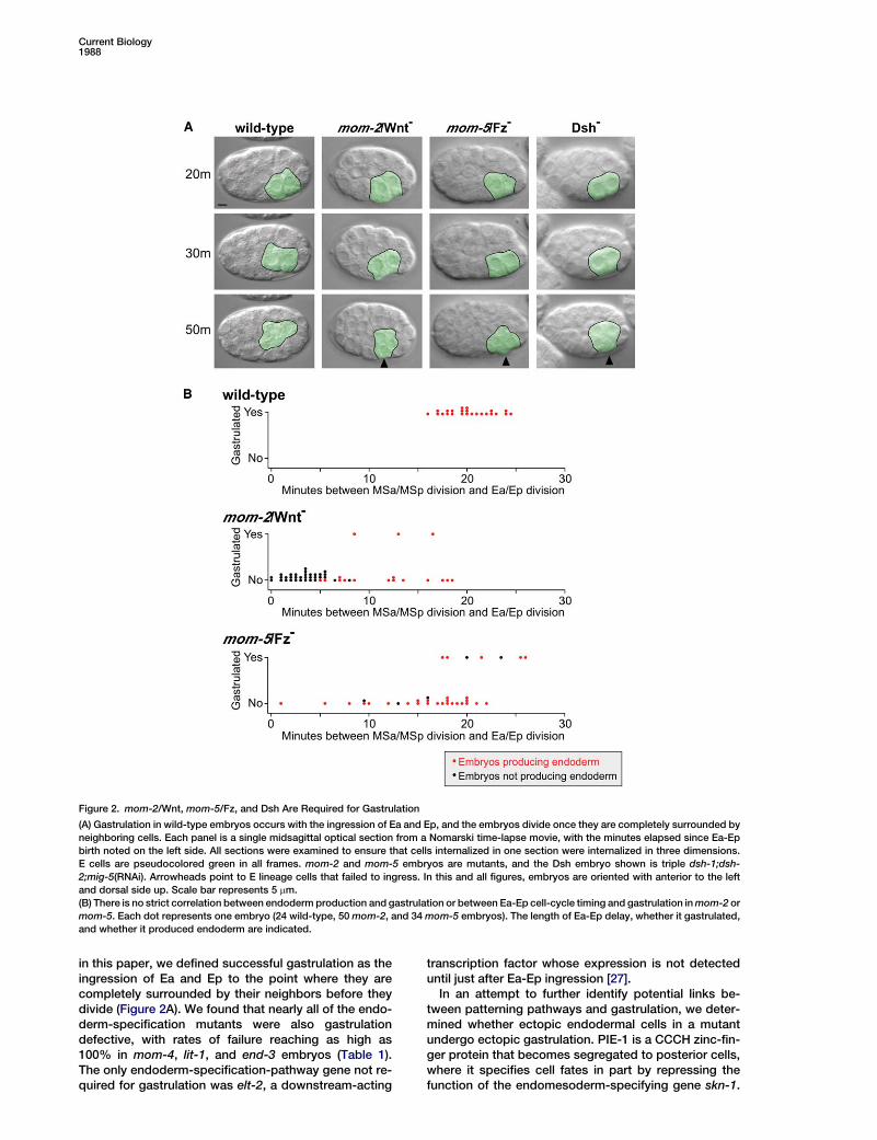
in this paper, we defined successful gastrulation as theingression of Ea and Ep to the point where they arecompletely surrounded by their neighbors before theydivide (Figure 2A). We found that nearly all of the endo-derm-specification mutants were also gastrulationdefective, with rates of failure reaching as high as100% in mom-4, lit-1, and end-3 embryos (Table 1).The only endoderm-specification-pathway gene not re-quired for gastrulation was elt-2, a downstream-acting
transcription factor whose expression is not detecteduntil just after Ea-Ep ingression [27].In an attempt to further identify potential links be-
tween patterning pathways and gastrulation, we deter-mined whether ectopic endodermal cells in a mutantundergo ectopic gastrulation. PIE-1 is a CCCH zinc-fin-ger protein that becomes segregated to posterior cells,where it specifies cell fates in part by repressing thefunction of the endomesoderm-specifying gene skn-1.
Figure 2. mom-2/Wnt, mom-5/Fz, and Dsh Are Required for Gastrulation
(A) Gastrulation in wild-type embryos occurs with the ingression of Ea and Ep, and the embryos divide once they are completely surrounded byneighboring cells. Each panel is a single midsagittal optical section from a Nomarski time-lapse movie, with the minutes elapsed since Ea-Epbirth noted on the left side. All sections were examined to ensure that cells internalized in one section were internalized in three dimensions.E cells are pseudocolored green in all frames. mom-2 and mom-5 embryos are mutants, and the Dsh embryo shown is triple dsh-1;dsh-2;mig-5(RNAi). Arrowheads point to E lineage cells that failed to ingress. In this and all figures, embryos are oriented with anterior to the leftand dorsal side up. Scale bar represents 5 mm.(B) There is no strict correlation between endoderm production and gastrulation or between Ea-Ep cell-cycle timing and gastrulation inmom-2 ormom-5. Each dot represents one embryo (24 wild-type, 50mom-2, and 34mom-5 embryos). The length of Ea-Ep delay, whether it gastrulated,and whether it produced endoderm are indicated.
Current Biology1988
Embryos from pie-1 loss-of-function mothers produceectopic endoderm from E’s posterior neighbor, P3,a cell that normally produces muscle and germlinefounder cells [25]. Time-lapse recordings of pie-1 mu-tant embryos showed ectopic gastrulation: The twodaughters of P3 invariably ingressed soon after Ea-Ep(17.5 6 3.3 min SD; Table 1). Because loss of functionof endoderm-specifying genes interferes with gastrula-tion and loss of function of pie-1, a gene that preventsectopic endoderm from forming, results in ectopic gas-trulation, we conclude that the genes that specify endo-dermal cell fate in Ea and Ep function upstream of thecytoskeletal mechanisms that drive gastrulation. Themost downstream player we found, the transcriptionalactivator END-1 [4], suggests that some molecularplayer(s) in gastrulation may be regulated transcription-ally, consistent with a known requirement for transcrip-tion in gastrulation [9].
Wnt/Frizzled Signaling Functions in Gastrulationas Well as Endoderm SpecificationNext, we asked whether any of these endoderm-specifi-cation genes played a direct role in determining gastru-lation behavior. Wnt ligands, Frizzled receptors, andFrizzled’s downstream effectors function upstream ofmorphogenetic events in diverse organisms [15–18,28]. We asked whether Wnt/Fz signaling functions dur-ing C. elegans gastrulation independently of its role inspecifying endoderm by using rhabditin granules asa terminal-differentiation marker for endoderm develop-ment (see the Supplemental Experimental Proceduresavailable with this article online). If Wnt/Fz signalingaffects gastrulation solely through its role in endodermspecification, then mom-2/Wnt or mom-5/Fz embryosthat produce endoderm should gastrulate and thoseembryos that fail to make endoderm should fail to gas-trulate. As expected, we found that all mom-2 deletionallele embryos that failed to produce endoderm alsofailed to gastrulate (n = 33); however, we were surprisedto find that most of the escapers—the endoderm-producing mom-2 embryos—also failed to gastrulate(82%; n = 17) (Figure 2B). Similarly, most mom-5 em-bryos that producedendoderm failed togastrulate (85%;n = 34) (Figure 2B). We also examined the expression of
two endoderm-specific molecular markers, END-1,which is expressed during gastrulation, and ELT-2,which is expressed after gastrulation. mom-5(RNAi)embryos exhibited similar gastrulation defects to mom-5(zu193) embryos, and the END-1 and ELT-2 expres-sion was comparable to that in wild-type cells in allembryos examined (n = 17 for END-1 and n = 9 forELT-2; Movies S1 and S2 and Figure S1). Because Eaand Ep in mom-5 embryos generally fail to gastrulatedespite producing END-1 and producing ELT-2 andrhabditin granules later, we propose that Wnt/Fz signal-ing has two functions: regulating endoderm specifica-tion and also, at least partially independently, regulatinggastrulation.The multidomain protein Dishevelled (Dsh) functions
downstream of the Frizzled receptor in several systems[29].C. elegans has three Dsh homologs: DSH-1, DSH-2,and MIG-5. Although null alleles of Dsh homologs affectendoderm specification, RNA interference (RNAi) ofdsh-1, dsh-2, and mig-5 individually or together has not[30]. This facilitated the determination of whether Dsh-mediated signaling is required for C. elegans gastrula-tion independently of a potential role in endodermspecification. We carried out RNAi of these three genesindividually and in combination. With the exception ofdsh-1 RNAi, all of these treatments resulted in someembryos that failed to gastrulate despite producing en-doderm, andRNAi of all three genes simultaneously pro-duced a more penetrant gastrulation-defective pheno-type than RNAi of any single gene alone (Figure 2 andFigure S2), suggesting that multiple Dsh proteins actredundantly to regulate gastrulation (we refer to thesethree proteins collectively as Dsh below). Our attemptsto determine whether canonical or noncanonical signal-ing is involved in gastrulation downstream of Dsh havenot yet resolved this issue. We conclude that MOM-2/Wnt, MOM-5/Fz, and multiple Dishevelled homologsfunction in the ingression of endodermal precursors inaddition to their roles in endoderm specification. Be-cause a mom-2 null allele does not abolish gastrulationmovements completely (Table 1), we conclude fromthese data that Wnt signaling acts partially redundantlyas direct or indirect regulators of gastrulationwith one ormore additional pathways.
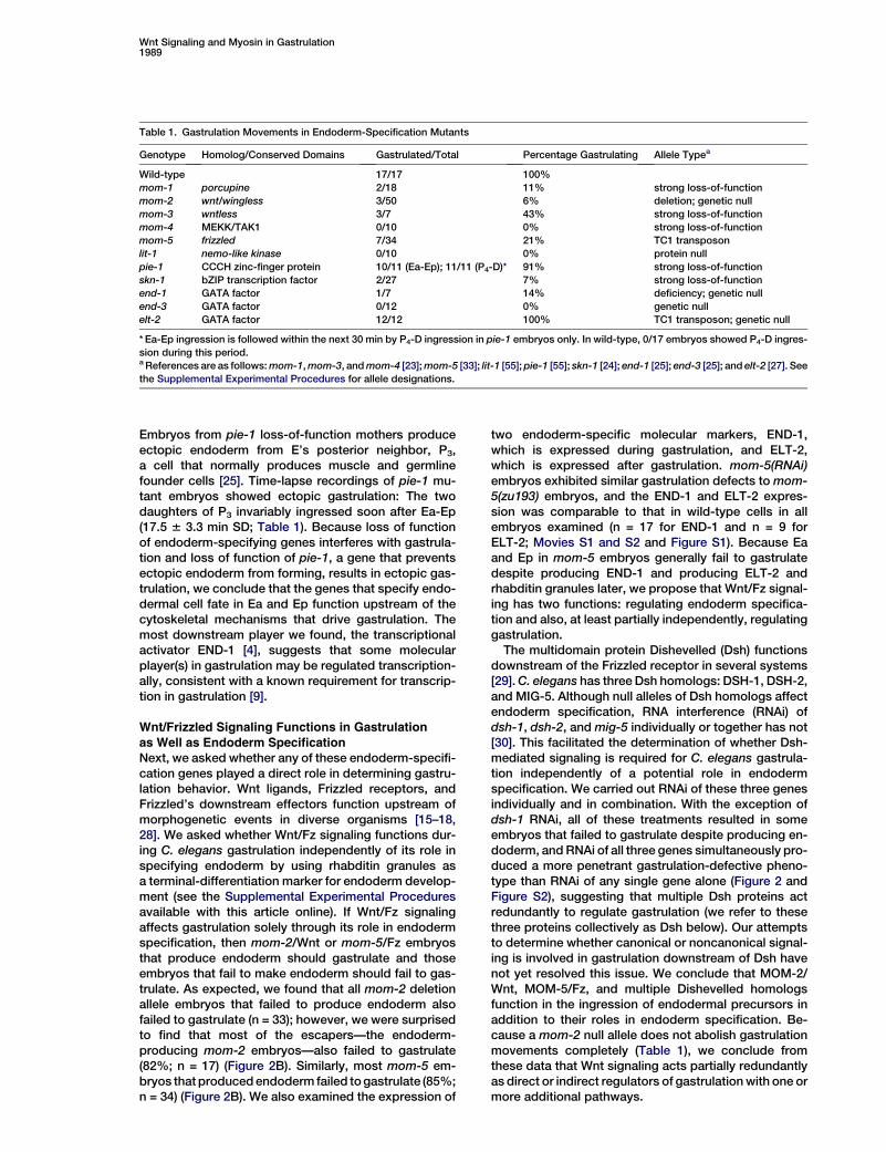
Table 1. Gastrulation Movements in Endoderm-Specification Mutants
Genotype Homolog/Conserved Domains Gastrulated/Total Percentage Gastrulating Allele Typea
Wild-type 17/17 100%mom-1 porcupine 2/18 11% strong loss-of-functionmom-2 wnt/wingless 3/50 6% deletion; genetic nullmom-3 wntless 3/7 43% strong loss-of-functionmom-4 MEKK/TAK1 0/10 0% strong loss-of-functionmom-5 frizzled 7/34 21% TC1 transposonlit-1 nemo-like kinase 0/10 0% protein nullpie-1 CCCH zinc-finger protein 10/11 (Ea-Ep); 11/11 (P4-D)* 91% strong loss-of-functionskn-1 bZIP transcription factor 2/27 7% strong loss-of-functionend-1 GATA factor 1/7 14% deficiency; genetic nullend-3 GATA factor 0/12 0% genetic nullelt-2 GATA factor 12/12 100% TC1 transposon; genetic null
* Ea-Ep ingression is followed within the next 30 min by P4-D ingression in pie-1 embryos only. In wild-type, 0/17 embryos showed P4-D ingres-sion during this period.aReferences are as follows:mom-1,mom-3, andmom-4 [23];mom-5 [33]; lit-1 [55];pie-1 [55]; skn-1 [24]; end-1 [25]; end-3 [25]; and elt-2 [27]. Seethe Supplemental Experimental Procedures for allele designations.
Wnt Signaling and Myosin in Gastrulation1989
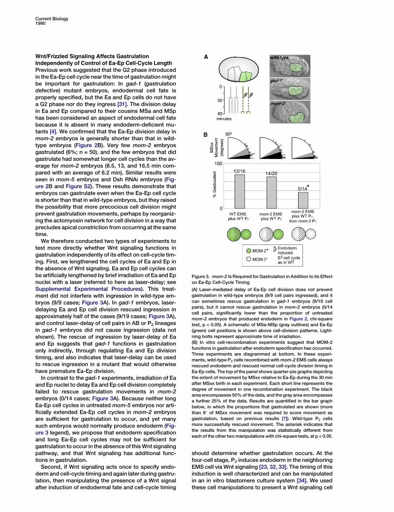
Wnt/Frizzled Signaling Affects GastrulationIndependently of Control of Ea-Ep Cell-Cycle LengthPrevious work suggested that the G2 phase introducedin the Ea-Ep cell cycle near the time of gastrulationmightbe important for gastrulation: In gad-1 (gastrulationdefective) mutant embryos, endodermal cell fate isproperly specified, but the Ea and Ep cells do not havea G2 phase nor do they ingress [31]. The division delayin Ea and Ep compared to their cousins MSa and MSphas been considered an aspect of endodermal cell fatebecause it is absent in many endoderm-deficient mu-tants [4]. We confirmed that the Ea-Ep division delay inmom-2 embryos is generally shorter than that in wild-type embryos (Figure 2B). Very few mom-2 embryosgastrulated (6%; n = 50), and the few embryos that didgastrulate had somewhat longer cell cycles than the av-erage for mom-2 embryos (8.5, 13, and 16.5 min com-pared with an average of 6.2 min). Similar results wereseen in mom-5 embryos and Dsh RNAi embryos (Fig-ure 2B and Figure S2). These results demonstrate thatembryos can gastrulate even when the Ea-Ep cell cycleis shorter than that in wild-type embryos, but they raisedthe possibility that more precocious cell division mightprevent gastrulation movements, perhaps by reorganiz-ing the actomyosin network for cell division in a way thatprecludes apical constriction fromoccurring at the sametime.We therefore conducted two types of experiments to
test more directly whether Wnt signaling functions ingastrulation independently of its effect on cell-cycle tim-ing. First, we lengthened the cell cycles of Ea and Ep inthe absence of Wnt signaling. Ea and Ep cell cycles canbe artificially lengthened by brief irradiation of Ea and Epnuclei with a laser (referred to here as laser-delay; seeSupplemental Experimental Procedures). This treat-ment did not interfere with ingression in wild-type em-bryos (9/9 cases; Figure 3A). In gad-1 embryos, laser-delaying Ea and Ep cell division rescued ingression inapproximately half of the cases (9/19 cases; Figure 3A),and control laser-delay of cell pairs in AB or P2 lineagesin gad-1 embryos did not cause ingression (data notshown). The rescue of ingression by laser-delay of Eaand Ep suggests that gad-1 functions in gastrulationonly indirectly, through regulating Ea and Ep divisiontiming, and also indicates that laser-delay can be usedto rescue ingression in a mutant that would otherwisehave premature Ea-Ep division.In contrast to the gad-1 experiments, irradiation of Ea
and Ep nuclei to delay Ea and Ep cell division completelyfailed to rescue gastrulation movements in mom-2embryos (0/14 cases; Figure 3A). Because neither longEa-Ep cell cycles in untreated mom-5 embryos nor arti-ficially extended Ea-Ep cell cycles in mom-2 embryosare sufficient for gastrulation to occur, and yet manysuch embryos would normally produce endoderm (Fig-ure 3 legend), we propose that endoderm specificationand long Ea-Ep cell cycles may not be sufficient forgastrulation to occur in the absence of thisWnt signalingpathway, and that Wnt signaling has additional func-tions in gastrulation.Second, if Wnt signaling acts once to specify endo-
derm and cell-cycle timing and again later during gastru-lation, then manipulating the presence of a Wnt signalafter induction of endodermal fate and cell-cycle timing
should determine whether gastrulation occurs. At thefour-cell stage, P2 induces endoderm in the neighboringEMS cell via Wnt signaling [23, 32, 33]. The timing of thisinduction is well characterized and can be manipulatedin an in vitro blastomere culture system [34]. We usedthese cell manipulations to present a Wnt signaling cell
Figure 3. mom-2 Is Required for Gastrulation in Addition to its Effecton Ea-Ep Cell-Cycle Timing
(A) Laser-mediated delay of Ea-Ep cell division does not preventgastrulation in wild-type embryos (9/9 cell pairs ingressed), and itcan sometimes rescue gastrulation in gad-1 embryos (9/19 cellpairs), but it cannot rescue gastrulation in mom-2 embryos (0/14cell pairs, significantly lower than the proportion of untreatedmom-2 embryos that produced endoderm in Figure 2, chi-squaretest, p < 0.05). A schematic of MSa-MSp (gray outlines) and Ea-Ep(green) cell positions is shown above cell-division patterns. Light-ning bolts represent approximate time of irradiation.(B) In vitro cell-recombination experiments suggest that MOM-2functions in gastrulation after endoderm specification has occurred.Three experiments are diagrammed at bottom. In these experi-ments, wild-type P2 cells recombined withmom-2 EMS cells alwaysrescued endoderm and rescued normal cell-cycle division timing inEa-Ep cells. The top of the panel shows quarter-pie graphs depictingthe extent of movement by MSxx relative to Ea-Ep during the 30 minafter MSxx birth in each experiment. Each short line represents thedegree of movement in one recombination experiment. The blackarea encompasses 50%of the data, and the gray area encompassesa further 25% of the data. Results are quantified in the bar graphbelow, in which the proportions that gastrulated are shown (morethan 8! of MSxx movement was required to score movement asgastrulation, based on previous results [7]). Wild-type P2 cellsmore successfully rescued movement. The asterisk indicates thatthe results from this manipulation was statistically different fromeach of the other twomanipulations with chi-square tests, at p < 0.05.
Current Biology1990

to specify E cell fate, and we then replaced it with eitherWnt-minus or Wnt-plus signaling cells to look for spe-cific effects on gastrulation. First, a number of controlexperiments were performed. When we separatedwild-type P2 from wild-type EMS cells and recombinedthese cells with wild-type partners that were at thesame developmental time point, most recombinantsexhibited gastrulation movements (75%, n = 16; Fig-ure 3B). In contrast,mom-2P2 andmom-2 EMSnegativecontrol recombinations resulted in little or no movement(Figure S3), as seen previously in related experimentswith mom-2 [7]. We then confirmed that gastrulationdefects could be rescued by recombining mom-2 EMScells with wild-type P2 cells (70%, n = 20; Figure 3B).Next, we asked whether a Wnt signaling cell during en-doderm specification was sufficient for gastrulationmovements by replacing aWnt-plus P2 cell withWnt-mi-nus cells after endoderm induction. We found that afterendoderm induction had occurred, a Wnt-plus P2 cell isable to rescue gastrulation movements (70%, n = 20)significantlymore effectively than aWnt-minus signalingcell (36%, n = 14; Figure 3B), suggesting that the P2 cellor its descendants, or both, are likely to be a source ofWnt signaling for gastrulation.Together, these results suggest that although Wnt
signaling can affect gastrulation indirectly by regulatingendodermal cell fate and division timing, Wnt signalingalso has a second role during gastrulation. This couldbe a second, independent Wnt-Fz interaction, or pos-sibly a higher threshold response to the interactionthat establishes endoderm cell fate. These resultsprompted us to look for more direct cellular effects ofWnt/Fz signaling during gastrulation by examining theeffect of Wnt/Fz signaling on the polarization and cyto-skeletal motility of ingressing cells. Below, we show thatin the absence of Wnt signaling, some cell biologicalevents implicated in ingression occur normally in Eaand Ep, but others do not.
Ea and Ep Apicobasal Polarization ProceedsIndependently of Wnt/Frizzled SignalingBecause Wnt and Fz-dependent signaling are known toaffect cell polarity in several systems [35], we examinedwhether Ea-Ep apicobasal polarity was disrupted in theabsence of Wnt/Fz signaling. During gastrulation, thePAR proteins are localized in apicobasally polarizedpatterns and are required for apical myosin enrichmentand efficient ingression movements [8, 10]. We exam-ined wild-type embryos expressing PAR-2::GFP andcompared them to PAR-2::GFP embryos from mothersthat were either fed with a bacterial strain expressingmom-2 dsRNA or injected with mom-5 dsRNA. PAR-2::GFP;mom-2(RNAi) embryos and PAR-2::GFP;mom-5(RNAi) embryos exhibited similar gastrulation defectsto mom-2(or309) and mom-5(zu193) embryos respec-tively (see Supplemental Experimental Procedures). Weconfirmed that GFP is detected in PAR-2::GFP embryosin a basolateral pattern in Ea-Ep (11/11 embryos; Fig-ure 4A). We found that PAR-2::GFP;mom-2(RNAi) em-bryos also display PAR-2::GFP in a basolateral pattern(16/16 embryos; Figure 4B) as do PAR-2::GFP;mom-5(RNAi) embryos (8/8 embryos; Figure 4C), indicatingthat Wnt-Fz signaling does not regulate PAR-2 basolat-eral distribution at this stage.
A second step in cell polarization occurs when NMY-2, a nonmuscle myosin II heavy chain, accumulates atthe apical surfaces of Ea and Ep, where it contributesto gastrulation movements [7, 8]. It is possible thatWnt/Fz signaling may effect gastrulation by acting inparallel with PAR proteins to enrich NMY-2 apically.The majority of cortical NMY-2 accumulation in Ea andEp occurs during the G2 phase of the Ea and Ep cells[8]. We used mom-5 mutants as a source of Wnt signal-ing-deficient embryos because Ea-Ep division timing inmom-5 mutant embryos more closely resembles thatin wild-type embryos than it does in mom-2 mutants(Figure 2B). We found that NMY-2::GFP accumulatedin the apical cortex of Ea and Ep in mom-5 embryos asmuch as in wild-type embryos and did not accumulateapically in other cells at this stage (Figure 5A). Quantifi-cation of cortical to cytoplasmic NMY-2::GFP ratiosconfirmed this: Wild-type and mom-5 Ea-Ep cells accu-mulated NMY-2::GFP at similar rates, and both accumu-lated significantlymoreNMY-2::GFP than did cells not ofE lineage in either background (Figure 5B). It is thereforeunlikely that Wnt/Fz signaling affects gastrulation byregulating the accumulation of NMY-2. These results
Figure 4. GFP::PAR-2 Distribution Shows that Apicobasal Polarity IsEstablished Normally in the Absence of mom-2/Wnt or mom-5/Fz
GFP::PAR-2 is enriched basolaterally in Ea and Ep in wild-type (A),mom-2 (B), and mom-5 (C) embryos. Ea-Ep are labeled with aster-isks. Arrows mark the basal sides of Ea-Ep cells, where GFP::PAR-2 is enriched, and arrowheads mark the apical sides. P4, on theposterior side of the embryo, exhibits nonlocalized GFP::PAR-2because the GFP construct is driven by a PIE-1 promoter that ismost active in the P lineage. Confocal images are shown; scalebars represent 5 mm.
Wnt Signaling and Myosin in Gastrulation1991
suggest that Wnt/Fz signaling does not appear to affectgastrulation through regulation of apicobasal polarity inthe ingressing cells.
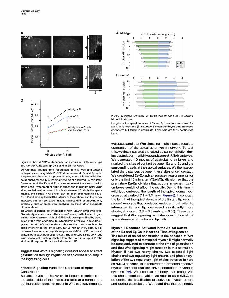
Frizzled Signaling Functions Upstream of ApicalConstrictionBecause myosin II heavy chain becomes enriched onthe apical side of the ingressing cells at a normal ratebut ingression does not occur in Wnt-pathway mutants,
we speculated thatWnt signaling might instead regulatecontraction of the apical actomyosin network. To testthis,wefirstmeasured the rate of apical constriction dur-ing gastrulation inwild-type andmom-5 (RNAi) embryos.We generated 4D movies of gastrulating embryos andmarked the sites of contact between Ea and Ep and thesurrounding cells at their apical surfaces.We then calcu-lated the distances between these sites of cell contact.We considered Ea-Ep apical-surface measurements foronly the first 10 min after MSa-MSp division so that thepremature Ea-Ep division that occurs in some mom-5embryos could not affect the results. During this time inwild-type embryos, the length of the apical domain de-creased at a rate of 7.16 1.3 nm/s (Figure 6). In contrast,the length of the apical domain of the Ea and Ep cells inmom-5 embryos that produced endoderm but failed tointernalize Ea and Ep decreased significantly moreslowly, at a rate of 2.3 6 3.6 nm/s (p < 0.05). These datasuggest that Wnt signaling regulates constriction of theapical domains of the Ea and Ep cells.
Myosin II Becomes Activated in the Apical Cortexof the Ea and Ep Cells Near the Time of IngressionThe failure of apical constriction in the absence of Wntsignaling suggested that apical myosin II might normallybecome activated to contract at the time of gastrulationand that Wnt signaling might function in this activation.Myosin II has two heavy chains, two essential lightchains and two regulatory light chains, and phosphory-lation of the two regulatory light chains (referred to hereas rMLC) at serine 19 is required for formation of activemyosin filaments that can drive contraction in varioussystems [36]. We used an antibody that recognizesthis phosphoepitope, which we refer to as p-rMLC, todetermine the localization of activated myosin beforeand during gastrulation. We found first that commonly
Figure 5. Apical NMY-2 Accumulation Occurs in Both Wild-Typeand mom-5/Fz Ea and Ep Cells and at Similar Rates
(A) Confocal images from recordings of wild-type and mom-5embryos expressing NMY-2::GFP. Asterisks mark Ea and Ep cells.d represents distance, t represents time, where ti is the initial timepoint analyzed and tf is the final time point analyzed 25 min later.Boxes around the Ea and Ep cortex represent the areas used tomake each kymograph at right, in which the maximum pixel valuealong each d position in each box is shown over 25min. In the kymo-graphs, the cortex in wild-type can be seen accumulating NMY-2::GFP andmoving toward the interior of the embryo, and the cortexin mom-5 can be seen accumulating NMY-2::GFP but moving onlyerratically. Similar areas were analyzed on three other quadrantsof the embryo.(B) Graph of cortical to cytoplasmic NMY-2::GFP level over time.Five wild-type embryos, and fourmom-5 embryos that failed to gas-trulate, were analyzed. NMY-2::GFP levels were quantified by calcu-lation of the ratio of cortical to cytoplasmic pixel level above back-ground. A ratio of one therefore indicates that the cortex is at thesame intensity as the cytoplasm. By 25 min after P4 birth, E cellcortexes have enriched significantly more NMY-2::GFP than non-Ecells, in both backgrounds (p < 0.05). The wild-type Ea-Ep GFP ratiois not statistically distinguishable from the mom-5 Ea-Ep GFP ratioat either time point. Error bars indicate 6 1 SD.
Figure 6. Apical Domains of Ea-Ep Fail to Constrict in mom-5Mutant Embryos
Lengths of the apical domains of Ea and Ep over time are shown for(A) 13 wild-type and (B) six mom-5 mutant embryos that producedendoderm but failed to gastrulate. Error bars are 95% confidencebars.
Current Biology1992
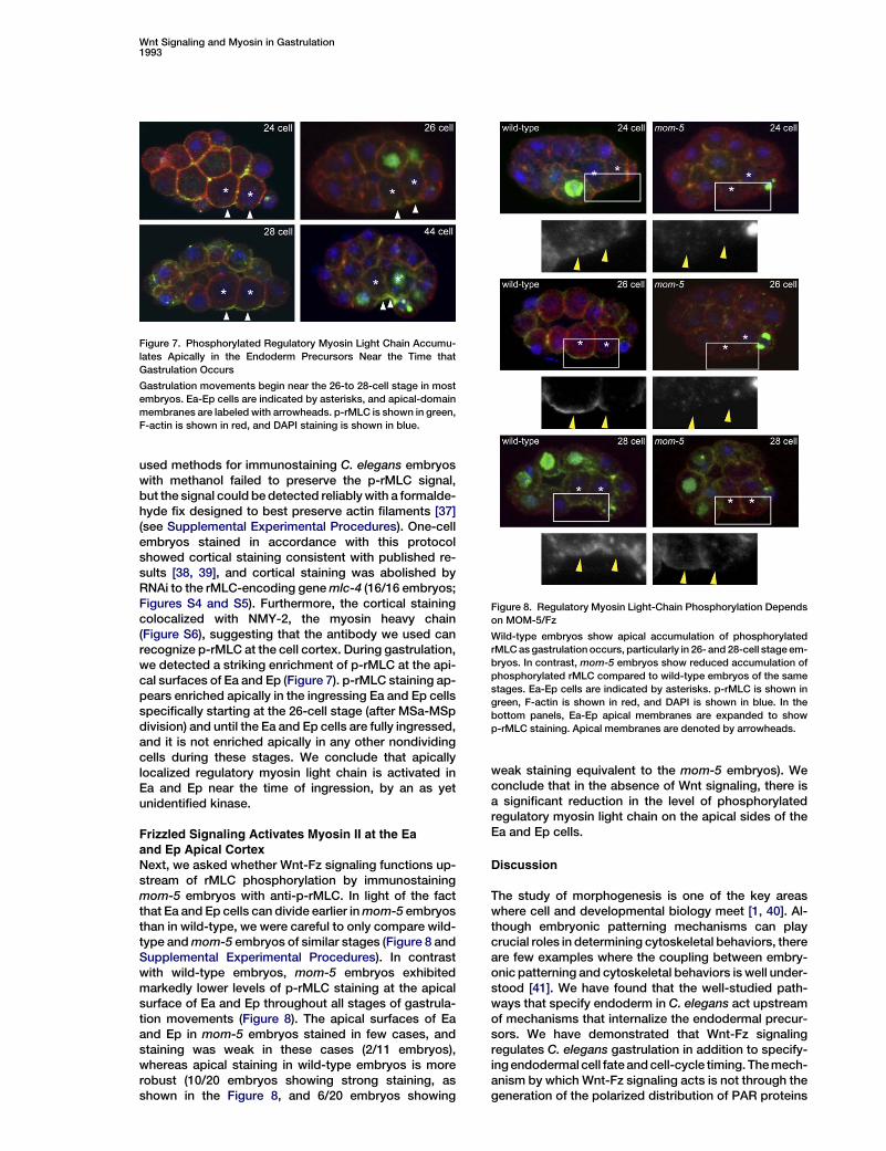
used methods for immunostaining C. elegans embryoswith methanol failed to preserve the p-rMLC signal,but the signal could bedetected reliablywith a formalde-hyde fix designed to best preserve actin filaments [37](see Supplemental Experimental Procedures). One-cellembryos stained in accordance with this protocolshowed cortical staining consistent with published re-sults [38, 39], and cortical staining was abolished byRNAi to the rMLC-encoding genemlc-4 (16/16 embryos;Figures S4 and S5). Furthermore, the cortical stainingcolocalized with NMY-2, the myosin heavy chain(Figure S6), suggesting that the antibody we used canrecognize p-rMLC at the cell cortex. During gastrulation,we detected a striking enrichment of p-rMLC at the api-cal surfaces of Ea and Ep (Figure 7). p-rMLC staining ap-pears enriched apically in the ingressing Ea and Ep cellsspecifically starting at the 26-cell stage (after MSa-MSpdivision) and until the Ea and Ep cells are fully ingressed,and it is not enriched apically in any other nondividingcells during these stages. We conclude that apicallylocalized regulatory myosin light chain is activated inEa and Ep near the time of ingression, by an as yetunidentified kinase.
Frizzled Signaling Activates Myosin II at the Eaand Ep Apical CortexNext, we asked whether Wnt-Fz signaling functions up-stream of rMLC phosphorylation by immunostainingmom-5 embryos with anti-p-rMLC. In light of the factthat Ea and Ep cells can divide earlier inmom-5 embryosthan in wild-type, we were careful to only compare wild-type andmom-5 embryos of similar stages (Figure 8 andSupplemental Experimental Procedures). In contrastwith wild-type embryos, mom-5 embryos exhibitedmarkedly lower levels of p-rMLC staining at the apicalsurface of Ea and Ep throughout all stages of gastrula-tion movements (Figure 8). The apical surfaces of Eaand Ep in mom-5 embryos stained in few cases, andstaining was weak in these cases (2/11 embryos),whereas apical staining in wild-type embryos is morerobust (10/20 embryos showing strong staining, asshown in the Figure 8, and 6/20 embryos showing
weak staining equivalent to the mom-5 embryos). Weconclude that in the absence of Wnt signaling, there isa significant reduction in the level of phosphorylatedregulatory myosin light chain on the apical sides of theEa and Ep cells.
Discussion
The study of morphogenesis is one of the key areaswhere cell and developmental biology meet [1, 40]. Al-though embryonic patterning mechanisms can playcrucial roles in determining cytoskeletal behaviors, thereare few examples where the coupling between embry-onic patterning and cytoskeletal behaviors is well under-stood [41]. We have found that the well-studied path-ways that specify endoderm in C. elegans act upstreamof mechanisms that internalize the endodermal precur-sors. We have demonstrated that Wnt-Fz signalingregulates C. elegans gastrulation in addition to specify-ingendodermal cell fateandcell-cycle timing. Themech-anism by which Wnt-Fz signaling acts is not through thegeneration of the polarized distribution of PAR proteins
Figure 7. Phosphorylated Regulatory Myosin Light Chain Accumu-lates Apically in the Endoderm Precursors Near the Time thatGastrulation Occurs
Gastrulation movements begin near the 26-to 28-cell stage in mostembryos. Ea-Ep cells are indicated by asterisks, and apical-domainmembranes are labeled with arrowheads. p-rMLC is shown in green,F-actin is shown in red, and DAPI staining is shown in blue.
Figure 8. Regulatory Myosin Light-Chain Phosphorylation Dependson MOM-5/Fz
Wild-type embryos show apical accumulation of phosphorylatedrMLCasgastrulation occurs, particularly in 26- and 28-cell stage em-bryos. In contrast, mom-5 embryos show reduced accumulation ofphosphorylated rMLC compared to wild-type embryos of the samestages. Ea-Ep cells are indicated by asterisks. p-rMLC is shown ingreen, F-actin is shown in red, and DAPI is shown in blue. In thebottom panels, Ea-Ep apical membranes are expanded to showp-rMLC staining. Apical membranes are denoted by arrowheads.
Wnt Signaling and Myosin in Gastrulation1993

or apical-myosin accumulation, suggesting that Wnt-Fzsignaling might directly affect apical constriction down-stream of myosin accumulation. In support of this, wefound an enrichment of an activated form of myosin atthe apical cortex of wild-type Ea and Ep during gastrula-tion, and we found that this activation of myosin isdependent on signaling through Fz.Our results rule out the simple possibility that Wnt sig-
naling affects C. elegans gastrulation solely through itswell-documented role in endoderm specification be-causewe found that Ea and Ep often produce endodermby multiple measures, but fail to gastrulate, in the ab-sence of Wnt signaling. However, it remains possiblethat rMLC phosphorylation could be a higher thresholdresponse to Wnt-Fz signaling at the four-cell stage. Al-ternatively, rMLC phosphorylation could depend on anindependent, later Wnt-Fz interaction. Signaling torMLC downstream of Dsh in either way could occur bya transcriptional mechanism or by signaling that ismore direct to rMLC. It will be of interest to identifymore members of this signaling pathway and to deter-mine when they function so that these questions canbe answered.Our results, together with previous results from us and
others [7, 9, 10] outline a molecular and mechanicalmodel for C. elegans gastrulation (Figure 9). The apico-basal polarity of all or most cells is determined by thepositions of cell-cell contacts and is reflected in the lo-calization of PAR proteins. The pathways that specifyendodermal fate in C. elegans, involving many of thegenes listed in Table 1, determine which cells will enrichmyosin heavy chain apically in response to PAR-proteinlocalization. Having myosin enriched at the apical, con-tact-free surfaces likely primes these cells for internali-zation. Wnt-Fz signaling leads to phosphorylation ofa conserved residue on rMLC, and this phosphorylationcan result in the formation of active myosin filaments.The contraction of the apical actomyosin machinerythen shrinks the contact-free apical areas, pulls neigh-boring cells under the Ea and Ep cells, and results inthe ingression of Ea and Ep into the center of the em-bryo. In this way, ingression appears to depend on thecombinatorial information from cell-fate specification,apico-basal polarity, and reception of a cell signal thatresults in contraction of the actomyosin network ona specific side of specific cells.
The role of cell-fate-specification mechanisms andWnt signaling in activating ingression of specific cellsin C. elegans gastrulation has both striking parallelsand critical differences with Drosophila gastrulation. InDrosophila, the mesoderm-specification protein Twistactivates apical secretion of a different intercellular sig-naling protein, Folded-gastrulation (Fog) [41, 42]. Fogacts through a presumed but unidentified cell-surfacereceptor and a G a protein, Concertina, to cause myosinlocalization to the apical surface of cells. Loss of the Fogdownstream targets, DRhoGEF or the Rho kinase Drok,causes much more severe defects in ventral furrow for-mation than does loss of Concertina, suggesting thatFog must act redundantly with another, unidentifiedpathway to drive apical constriction [41]. Myosin motoractivity is required for its apical localization in Drosoph-ila gastrulation [41]. Whether Fog signaling regulatesmyosin activity by rMLC phosphorylation in Drosophilagastrulation like theWnt pathway does inC. elegans hasnot been examined, but Rho kinase is known to causephosphorylation of rMLC or myosin activation in othersettings in Drosophila [43–45] and other organisms [36].In vertebrates, the actin-binding protein Shroom func-
tions to localize both actin filaments andmyosin and cancause apical constriction when expressed in MDCKcells and during normal neural-tube closure [46–48].Shroom functions in apical constriction by restrictingmyosin localization, and it is not known to affect myosinactivity. Shroom acts through a small GTPase like Fog,but does so through a different GTPase, Rap1. Fog,Shroom, and Wnt-dependent control of apical constric-tion appear to work independently rather than as partsof a single pathway because Fog and Shroom do nothave homologs in the C. elegans genome, and removalof the function of the Rap1 homolog in C. elegans hasno effect on gastrulation (T. Grana and J.H., unpublisheddata). Also, Shroom is required for apical constriction ofonly some cells in the organisms where it functions; forexample, Xenopus bottle cells do not require Shroomduring gastrulation [48]. These data suggest that apicalconstriction is regulated during animal development bymultiple, independentmechanisms that can affect eithermyosin distribution or activity, or both.Although ours is the first report of intercellular Wnt
signaling regulating morphogenesis through rMLCphosphorylation, there is precedent for Frizzled acting
Figure 9. Model of the Mechanisms Control-ling C. elegans Gastrulation
At left, genetic and cellular pathway regulat-ing apical constriction.At bottom right, diagrams of the events at leftwithout (top) and with (bottom) a Wnt signal.See Discussion for explanation. MOM-5 dis-tribution is not detected asymmetrically incells during gastrulation [54], andMOM-2 dis-tribution is not known. All other proteins areknown to localize as diagrammed.
Current Biology1994

in a similar but cell-autonomous fashion in Drosophila,suggesting that this might be an ancient mechanism ofmorphogenetic regulation. Frizzled can affect ommatid-ial polarity and actin bundle number in Drosophilaplanar-cell polarity (PCP) signaling pathways via Dsh,RhoA, Drok, and rMLC phosphorylation [45]. This path-way suggests some molecular players that might actbetween Dsh and rMLC phosphorylation in C. elegansgastrulation. The Drosophila PCP pathways differ fromwhat we have outlined in this report in that Frizzled isprobably not responding to intercellular Wnt signals inPCP signaling pathways [49, 50] and in that PCP signal-ing is not known to drive apical constriction. The mech-anism by which myosin activation regulates ommatidialpolarity and actin bundle number in PCP pathways isunknown.In zebrafish and frog embryos, PCP signaling is
required during gastrulation for convergence and exten-sion movements [17, 18]. Vertebrate PCP genes areknown to regulate the activity of the cytoskeletal modi-fiers Rho and Rac [51–53], but the direct effect of PCPsignaling on the cytoskeleton has not yet been analyzedin detail. Both the vertebrate PCP pathway and C. ele-gans Wnt/Fz signaling result in modification of cellshape and cell behavior during gastrulation. These path-ways differ in that vertebrate PCP signaling affects cellpolarity [17, 18], whereas cell polarity is undisturbed inmom-2/Wnt andmom-5/Fzmutant embryos (this report)and RNAi experiments targeting C. elegans homologsof the PCP genes fat and flamingo (D.M., unpublished)and van gogh/strabismus (T.W. and J.H., unpublisheddata) have not resulted in any gastrulation defects, sug-gesting that C. elegans Wnt/Fz signaling in gastrulationthrough conventional PCP signaling is unlikely.Why would myosin activity in C. elegans gastrulation
be regulated by a cell-cell signal? It seems plausiblethat constitutively activating myosin could achieve thesame goal because myosin is enriched in the apicalcortex of the Ea and Ep cells, and hence, activation ofmyosin throughout these cells might cause an imbal-ance of forces that could drive apical constrictionreliably. One possible explanation is that signaling en-sures that apical constriction occurs at the right time.For example, signaling could ensure that myosin activa-tion occurs at a time when the actomyosin network isapically enriched and is not being used for cell divisionor at a time when adhesion to neighboring cells is suffi-ciently strong for constriction to pull neighboring cellsunder the Ea and Ep cells. Signaling might also contrib-ute spatial specificity. For example, it is possible thatthere are stages in development when several cellshave the potential to ingress but only the appropriateones do so because they contact a Wnt signaling cell.Others have speculated that morphogenesis may
more often depend on redundant pathways than embry-onic patterning does [2]. Very few backgrounds thatwe examined prevented gastrulation in all embryos,suggesting that C. elegans gastrulation is regulated bymultiple, partially redundant mechanisms. Morpho-genesis depends on diverse mechanisms that are ofinterest in the field of cell and developmental biology,including spatial and temporal gene regulation, cell sig-naling, cell polarization, cell adhesion, and cytoskeletaldynamics. It will be of interest to explore how such
mechanisms work together in morphogenesis in C. ele-gans gastrulation.
Conclusions
We have used a combination of genetics, cell manipula-tions, and in vivo imaging to investigate the regulation ofthe cytoskeleton by cell-fate-specification genes and byintercellular signaling pathways during the morphoge-netic movements of C. elegans gastrulation. We haveshown that the pathways required for endoderm specifi-cation, including a Wnt/Frizzled pathway, are requiredfor gastrulation to occur. Furthermore, we have shownthat this Wnt/Frizzled pathway functions in gastrulationin addition to specifying endodermal cell fate. In the ab-sence of Frizzled, apical constriction of the endodermprecursors fails to occur. Additionally, embryos lackingFrizzled show reduced levels of phosphorylated myosinin the apical domains of the endodermal precursorscompared to wild-type embryos. Because this phos-phorylation of myosin is likely to drive actomyosin con-traction, and thus the ingression in these cells at thissite, we hypothesize that this failure to phosphorylatemyosin underpins the gastrulation defect inWnt/Frizzledsignaling defective embryos. Thus, we have demon-strated a novel role forWnt signaling duringmorphogen-esis through understanding its modulation of the cyto-skeleton and how this impacts upon cell movements.
Supplemental DataSupplemental Data include six figures and two movies and can befound with this article online at http://www.current-biology.com/cgi/content/full/16/20/1986/DC1/.
Acknowledgments
We thank Mark Peifer, Steve Rogers, John Wallingford and mem-bers of the Goldstein lab for useful discussions and encouragementand Jeremy Nance, Ed Munro, Jim Priess, Morris Maduro, and theCaenorhabditis Genetics Center for worm strains. The Caenorhabdi-tis Genetics Center is supported by the National Institutes of HealthNational Center for Research Resources. This work was supportedby National Science Foundation grant IOB0518081 to J.H. andNational Institutes of Health R01-GM68966 to B.G.
Received: February 21, 2006Revised: August 21, 2006Accepted: August 25, 2006Published: October 23, 2006
References
1. Dawes-Hoang, R.E., and Wieschaus, E.F. (2001). Cell and devel-opmental biology–a shared past, an intertwined future. Dev. Cell1, 27–36.
2. Wieschaus, E. (1995). From molecular patterns to morphogene-sis: The lessons from Drosophila. In Nobel Lectures, Physiologyor Medicine 1991–1995, N. Ringertz, ed. (Singapore: World Sci-entific Publishing Co.)
3. Sulston, J.E., Schierenberg, E., White, J.G., and Thomson, J.N.(1983). The embryonic cell lineage of the nematode Caenorhab-ditis elegans. Dev. Biol. 100, 64–119.
4. Maduro, M.F., and Rothman, J.H. (2002). Makingworm guts: Thegene regulatory network of the Caenorhabditis elegans endo-derm. Dev. Biol. 246, 68–85.
5. Thorpe, C.J., Schlesinger, A., and Bowerman, B. (2000).Wnt sig-nalling in Caenorhabditis elegans: Regulating repressors andpolarizing the cytoskeleton. Trends Cell Biol. 10, 10–17.
Wnt Signaling and Myosin in Gastrulation1995

6. Edgar, L.G., and McGhee, J.D. (1988). DNA synthesis and thecontrol of embryonic gene expression in C. elegans. Cell 53,589–599.
7. Lee, J.Y., and Goldstein, B. (2003). Mechanisms of cell position-ing during C. elegans gastrulation. Development 130, 307–320.
8. Nance, J., and Priess, J.R. (2002). Cell polarity and gastrulationin C. elegans. Development 129, 387–397.
9. Nance, J., Lee, J.-Y., and Goldstein, B. Gastrulation in C. ele-gans, in WormBook (The C. elegans Research Community,http://www.wormbook.org). 2005.
10. Nance, J., Munro, E.M., and Priess, J.R. (2003). C. elegans PAR-3 and PAR-6 are required for apicobasal asymmetries associ-ated with cell adhesion and gastrulation. Development 130,5339–5350.
11. Kemphues, K. (2000). PARsing embryonic polarity. Cell 101,345–348.
12. Wodarz, A. (2002). Establishing cell polarity in development. Nat.Cell Biol. 4, E39–E44.
13. Logan, C.Y., and Nusse, R. (2004). The Wnt signaling pathwayin development and disease. Annu. Rev. Cell Dev. Biol. 20,781–810.
14. Peifer, M., Rauskolb, C., Williams, M., Riggleman, B., and Wie-schaus, E. (1991). The segment polarity gene armadillo interactswith thewingless signaling pathway in both embryonic and adultpattern formation. Development 111, 1029–1043.
15. Wieschaus, E., and Riggleman, R. (1987). Autonomous require-ments for the segment polarity gene armadillo during Drosophilaembryogenesis. Cell 49, 177–184.
16. Gong, Y., Mo, C., and Fraser, S.E. (2004). Planar cell polarity sig-nalling controls cell division orientation during zebrafish gastru-lation. Nature 430, 689–693.
17. Heisenberg, C.P., Tada, M., Rauch, G.J., Saude, L., Concha,M.L., Geisler, R., Stemple, D.L., Smith, J.C., and Wilson, S.W.(2000). Silberblick/Wnt11mediates convergent extensionmove-ments during zebrafish gastrulation. Nature 405, 76–81.
18. Wallingford, J.B., Rowning, B.A., Vogeli, K.M., Rothbacher, U.,Fraser, S.E., and Harland, R.M. (2000). Dishevelled controlscell polarity during Xenopus gastrulation. Nature 405, 81–85.
19. Wallingford, J.B., and Harland, R.M. (2002). Neural tube closurerequires Dishevelled-dependent convergent extension of themidline. Development 129, 5815–5825.
20. Herman, M.A., and Wu, M. (2004). Noncanonical Wnt signalingpathways in C. elegans converge on POP-1/TCF and controlcell polarity. Front. Biosci. 9, 1530–1539.
21. Vincan, E. (2004). Frizzled/WNT signalling: The insidious pro-moter of tumour growth and progression. Front. Biosci. 9,1023–1034.
22. Sancho, E., Batlle, E., and Clevers, H. (2004). Signaling pathwaysin intestinal development and cancer. Annu. Rev. Cell Dev. Biol.20, 695–723.
23. Thorpe, C.J., Schlesinger, A., Carter, J.C., and Bowerman, B.(1997). Wnt signaling polarizes an early C. elegans blastomereto distinguish endoderm from mesoderm. Cell 90, 695–705.
24. Bowerman, B., Eaton, B.A., and Priess, J.R. (1992). skn-1, amaternally expressed gene required to specify the fate of ventralblastomeres in the early C. elegans embryo. Cell 68, 1061–1075.
25. Zhu, J., Hill, R.J., Heid, P.J., Fukuyama, M., Sugimoto, A., Priess,J.R., and Rothman, J.H. (1997). end-1 encodes an apparentGATA factor that specifies the endoderm precursor in Caeno-rhabditis elegans embryos. Genes Dev. 11, 2883–2896.
26. Maduro, M.F., Hill, R.J., Heid, P.J., Newman-Smith, E.D., Zhu, J.,Priess, J.R., and Rothman, J.H. (2005). Genetic redundancy inendoderm specification within the genus Caenorhabditis. Dev.Biol. 284, 509–522.
27. Fukushige, T., Hawkins, M.G., and McGhee, J.D. (1998). TheGATA-factor elt-2 is essential for formation of the Caenorhabdi-tis elegans intestine. Dev. Biol. 198, 286–302.
28. McEwen, D.G., Cox, R.T., and Peifer, M. (2000). The canonicalWg and JNK signaling cascades collaborate to promote bothdorsal closure and ventral patterning. Development 127, 3607–3617.
29. Wallingford, J.B., and Habas, R. (2005). The developmental biol-ogy of Dishevelled: An enigmatic protein governing cell fate andcell polarity. Development 132, 4421–4436.
30. Walston, T., et al. (2004). Multiple Wnt signaling pathways con-verge to orient the mitotic spindle in early C. elegans embryos.Dev. Cell 7, 831–841.
31. Knight, J.K., and Wood, W.B. (1998). Gastrulation initiation inCaenorhabditis elegans requires the function of gad-1, whichencodes a protein with WD repeats. Dev. Biol. 198, 253–265.
32. Goldstein, B. (1992). Induction of gut in Caenorhabditis elegansembryos. Nature 357, 255–257.
33. Rocheleau, C.E., Downs, W.D., Lin, R., Wittmann, C., Bei, Y.,Cha, Y.H., Ali, M., Priess, J.R., andMello, C.C. (1997).Wnt signal-ing and an APC-related gene specify endoderm in early C. ele-gans embryos. Cell 90, 707–716.
34. Goldstein, B. (1993). Establishment of gut fate in the E lineage ofC. elegans: The roles of lineage-dependent mechanisms andcell interactions. Development 118, 1267–1277.
35. Axelrod, J.D., and McNeill, H. (2002). Coupling planar cell polar-ity signaling to morphogenesis. ScientificWorldJournal 2, 434–454.
36. Somlyo, A.P., and Somlyo, A.V. (2003). Ca2+ sensitivity ofsmooth muscle and nonmuscle myosin II: Modulated by Gproteins, kinases, and myosin phosphatase. Physiol. Rev. 83,1325–1358.
37. Costa, M., Draper, B.W., and Priess, J.R. (1997). The role of actinfilaments in patterning the Caenorhabditis elegans cuticle. Dev.Biol. 184, 373–384.
38. Jenkins, N., Saam, J.R., and Mango, S.E. (2006). CYK-4/GAPprovides a localized cue to initiate anteroposterior polarityupon fertilization. Science 313, 1298–1301.
39. Piekny, A.J., and Mains, P.E. (2002). Rho-binding kinase (LET-502) and myosin phosphatase (MEL-11) regulate cytokinesis inthe early Caenorhabditis elegans embryo. J. Cell Sci. 115,2271–2282.
40. Keller, R. (2002). Shaping the vertebrate body plan by polarizedembryonic cell movements. Science 298, 1950–1954.
41. Dawes-Hoang, R.E., Parmar, K.M., Christiansen, A.E., Phelps,C.B., Brand, A.H., and Wieschaus, E.F. (2005). Folded gastrula-tion, cell shape change and the control of myosin localization.Development 132, 4165–4178.
42. Costa, M., Wilson, E.T., andWieschaus, E. (1994). A putative cellsignal encoded by the folded gastrulation gene coordinates cellshape changes during Drosophila gastrulation. Cell 76, 1075–1089.
43. Mizuno, T., Tsutsui, K., and Nishida, Y. (2002). Drosophila myo-sin phosphatase and its role in dorsal closure. Development 129,1215–1223.
44. Rogers, S.L., Wiedemann, U., Hacker, U., Turck, C., and Vale,R.D. (2004). Drosophila RhoGEF2 associates with microtubuleplus ends in an EB1-dependent manner. Curr. Biol. 14, 1827–1833.
45. Winter, C.G., Wang, B., Ballew, A., Royou, A., Karess, R., Axel-rod, J.D., and Luo, L. (2001). Drosophila Rho-associated kinase(Drok) links Frizzled-mediated planar cell polarity signaling tothe actin cytoskeleton. Cell 105, 81–91.
46. Hildebrand, J.D., andSoriano, P. (1999). Shroom, a PDZ domain-containing actin-binding protein, is required for neural tubemorphogenesis in mice. Cell 99, 485–497.
47. Hildebrand, J.D. (2005). Shroom regulates epithelial cell shapevia the apical positioning of an actomyosin network. J. CellSci. 118, 5191–5203.
48. Haigo, S.L., Hildebrand, J.D., Harland, R.M., and Wallingford,J.B. (2003). Shroom induces apical constriction and is requiredfor hingepoint formation during neural tube closure. Curr. Biol.13, 2125–2137.
49. Strutt, D. (2003). Frizzled signalling and cell polarisation inDrosophila and vertebrates. Development 130, 4501–4513.
50. Lawrence, P.A., Casal, J., andStruhl, G. (2002). Towards amodelof the organisation of planar polarity and pattern in the Drosoph-ila abdomen. Development 129, 2749–2760.
51. Habas, R., Kato, Y., and He, X. (2001). Wnt/Frizzled activation ofRho regulates vertebrate gastrulation and requires a novel For-min homology protein Daam1. Cell 107, 843–854.
52. Habas, R., Dawid, I.B., and He, X. (2003). Coactivation of Racand Rho by Wnt/Frizzled signaling is required for vertebrategastrulation. Genes Dev. 17, 295–309.
Current Biology1996

53. Marlow, F., Topczewski, J., Sepich, D., and Solnica-Krezel, L.(2002). Zebrafish Rho kinase 2 acts downstream of Wnt11 tomediate cell polarity and effective convergence and extensionmovements. Curr. Biol. 12, 876–884.
54. Park, F.D., Tenlen, J.R., and Priess, J.R. (2004). C. elegansMOM-5/frizzled functions in MOM-2/Wnt-independent cell po-larity and is localized asymmetrically prior to cell division.Curr. Biol. 14, 2252–2258.
55. Meneghini, M.D., Ishitani, T., Carter, J.C., Hisamoto, N., Nino-miya-Tsuji, J., Thorpe, C.J., Hamill, D.R., Matsumoto, K., andBowerman, B. (1999). MAP kinase and Wnt pathways convergeto downregulate an HMG-domain repressor in Caenorhabditiselegans. Nature 399, 793–797.
Wnt Signaling and Myosin in Gastrulation1997

Supplemental Data S1
Wnt/Frizzled SignalingControls C. elegans Gastrulationby Activating Actomyosin Contractility
Jen-Yi Lee, Daniel Marston, Timothy Walston,Jeff Hardin, Ari Halberstadt, and Bob Goldstein
Supplemental Experimental Procedures
Strains and Worm MaintenanceNematodes were cultured and handled as described [S1]. Unless in-dicated, experiments were performed with the wild-type N2 (Bristol)strain. The following mutant and reporter strains were used: EU361mom-1 (or10), EU855 mom-2 (or309), EU404 mom-3 (or78), EU414mom-4 (or39), EU452 mom-5 (zu193), EU1 skn-1 (zu67), JR728end-1 (wDf4), JM69 elt-2 (ca15), EU603 lit-1 (or131), BW1943 gad-1 (ct226), KK866 GFP: PAR-2 [S2], JJ1317 zuIs3 [end-1::GFP] [S3],JJ532 pie-1 (zu154), JR1838 wIs84 [elt-2::GFP], RB1331 end-3(ok1448), and JJ1473 unc-119 (ed3) III; zuIs45 [nmy-2::NMY-2::GFP; unc-119 (+)] [S3]; referred to here as NMY-2::GFP, LP25mom-5(zu193);NMY-2::GFP. LP25 was constructed by crossingJJ1473 NMY-2::GFP males with EU452 (zu193) hermaphrodites. Allstrains were maintained at 20!C, except for the following strains:KK866 PAR-2::GFP was maintained at room temperature (22!C–23!C) and EU603 lit-1 was maintained at 15!C. Imaging was per-formed at 20!C–23!C for all strains except EU603 lit-1, which wasfilmed at 25!C. In experiments where endoderm differentiationwas scored, this was assayed by examining of embryos or partialembryos the next day for the presence of birefringent rhabditingranules under polarized light [S4, S5].
DIC and Confocal Time-Lapse MicroscopyIntact and devitellinized embryos were mounted for DIC and confo-cal imaging as described [S6]. DIC images were acquired onaC2400-07HamamatsuNewviconvideocameramountedonaNikonEclipse 800 microscope. Time-lapse images were acquired at 1 mmsections every 30 s with 4D Grabber and subsequently analyzedwith 4D Viewer (both programs from LOCI, University of Wisconsin,Madison). Cell-cycle timings were scored from and to the time thata cleavage furrow could first be seen. Gastrulation was scored byexamination ofwhether the Ea andEp cells left no surface on the out-side of the embryo and were instead surrounded by neighboringcells in three dimensions at the time that Ea and Ep divided, asoccurs in wild-type embryos. If Ea and Ep divided before being com-pletely surrounded by neighboring cells, thenwe scored gastrulationas having failed. For measuring apical membranes, the longestventral surface was measured from the Ea-Ep ventral border toboth the Ep-P4 ventral border and the Ea-MSxx ventral border.Only embryos that produced endoderm, failed to gastrulate, had suf-ficiently long Ea-Ep cell cycles (>10min) and normal E-division orien-tation were analyzed. Confocal images were acquired with Meta-morph software (Molecular Devices) on a Nikon Inverted EclipseTE2000-U, equippedwith aYokogawaSpinningDiskConfocal Scan-ner Unit CSU10. Image processing was performed with MetamorphorAdobe Photoshop CS 8.0, or both.
Laser-Delay ExperimentsCells were irradiated with a VSL-337 nitrogen laser (2 mW, LaserScience Inc.) mounted on a Nikon Eclipse 800 microscope. Cellstargeted for delay at a sublethal laser dose by irradiation with several10–30 s durations of 3 nanosecond pulses at 10 Hz. The laser wasfocused primarily in the nucleus and on the nuclear envelope. Cellswere laser-irradiated until intracellular damage such as excessBrownian motion or an irregularly shaped nuclear envelope was vis-ible. Experiments were only included in analyses if the nucleus couldbe clearly seen after laser irradiation and the cell did not appear tolyse (approximately 90% of all experiments were included in analy-sis). In more than 85% of the experiments, targeted cells dividedagainwithin 120min. Nontargeted cells did not appear to be affectedby the laser because division rates measured in selected experi-ments were normal. Embryos in which the targeted cells were com-pletely surrounded by other cells in three dimensions by 120 minafter laser-delay were scored as being successfully ingressed.
Cell Recombination Experiments andMeasurement of MSxxCellMovements In VitroEmbryos were devitellinized, and P1 cells were isolated and culturedas described [S6]. Five min after formation of the cleavage furrow ofthe dividing P1 cell, P2 and EMS cells were separated from eachother. Cells of the same developmental time point were recombinedwithin approximately onemin of separation, mounted on a slide, andfilmed by 4D videomicroscopy. In experiments where the P lineagecell was replaced after induction, P2 was allowed to divide oncefor ensuring that cells were placed back in a way that P4 would bein contact with Ep during gastrulation. Analysis of MSxx movementwas performed as previously described [S6]. Except where other-wise noted, endoderm differentiation was assayed by examiningof embryos or partial embryos the next day for the presence of bire-fringent rhabditin granules under polarized light [S4, S5]. Rare caseswhere cell divisions stopped early were excluded from analysisbecause these were likely to be damaged.
RNA Interference (RNAi)RNAi by feeding was performed according to a standard protocol[S7], except that the experiments were performed at room tempera-ture (w22!C). Feeding constructs were obtained from adsRNA feed-ing library, MRC Geneservice [S8]. PAR-2::GFP worms were shiftedto 25!C the night before imaging so that theGFPsignal-to-noise ratiocould be improved. Control experiments with gad-1 RNAi, known toresult in embryonic lethality, were performed simultaneously withmom-2 RNAi experiments to ensure that the RNAi feeding protocolwas working. PAR-2::GFP; mom-2(RNAi) embryos that were ana-lyzed usually failed to gastrulate (13/16 embryos), had precociousdivision, and lacked endoderm (10/14 embryos; two were notanalyzed for endoderm), similar to the phenotypes seen in mom-2(or309) embryos. RNAi by injection was performed as previouslydescribed [S9]. For mom-5, the target region included residues 15–170 and was injected at a concentration of 100 ng/ml. Embryoswere dissected out for experiments 24–36 hr later and exhibitedmom-5phenotypes. Formlc-4, the targeted region included residues3–163 and was injected at a concentration of 100 ng/ml. Theseembryos failed to undergo cytokinesis. Embryos were stained 24 hrafter injection.
Analysis of NMY-2::GFP AccumulationNMY-2::GFP and mom-5;NMY-2::GFP embryos were imaged ona spinning disk confocal microscope (see above). Images were cap-tured once each min after the birth of P4 for 25 min. A line was drawnperpendicular to the cortex of four regions of the embryo, where theE, MS, C, and AB lineage cells were located. With Metamorph soft-ware, these lineswere converted into kymographs ofmaximumpixelintensity over time.
Immunostaining and Confocal MicroscopyImmunostaining of embryos was performed according to a protocolmodified from Costa and colleagues [S10]. In brief, embryos wereisolated by dissection of gravid adults in egg buffer (118 mM NaCl,40 mM KCl, 3.4 mM CaCl2, 3.4 mM MgCl2, and 5 mM HEPES [pH7.2]) and were allowed to develop to the desired stage. Embryoswere treated with 10% NaOCl solution (Sigma) in egg buffer for3–4 min and washed four times in egg buffer. The egg shells weredigested for 3 min with a combination of chitinase (3 U/ml; Sigma)and chymotrypsin (10 U/ml; Sigma) in egg buffer. Embryos werefixed in situ by excess addition of 4% formaldehyde (ElectronMicroscopy Sciences) in fix buffer (10 mM MES [pH 6.1], 125 mMKCl, 3 mM MgCl, 2 mM EGTA, and 10% sucrose) containing 0.1mg/ml L-a-lysolecithin (Sigma; to aid in permeabilization) for 2min. The embryos were then transferred to fresh 4% formaldehydein fix buffer for a further 10 min. The embryos were washed in PBSandmounted on poly-L-lysine coated coverslips then permeabilized

with 0.2% Triton-X in PBS for 5 min. Embryos were subsequently in-cubated in TSA block (Molecular Probes) for 1 hr, and then the blockwas replaced with primary antibody diluted in block solution. Theprimary antibody used was anti-Phospho-Ser19-MLC (1/100,Abcam), detected with the TSA (tyramide signal amplification) anti-body detection kit (Molecular Probes). F-actin was detected withTRITC conjugated phalloidin (Sigma). NMY-2::GFP was detectedwith GFP antibodies (1/100, Molecular Probes). Embryos wereimaged with a Zeiss LSM510 confocal microscope and LSM soft-ware, and images were processed with Metamorph software.Embryos were staged by counting nuclei, and only embryos fromthe 26- or 28-cell stages were counted when comparing mom-5with wild-type.
Supplemental References
S1. Brenner, S. (1974). The genetics of Caenorhabditis elegans.Genetics 77, 71–94.
S2. Cuenca, A.A., Schetter, A., Aceto, D., Kemphues, K., and Sey-doux, G. (2003). Polarization of the C. elegans zygote proceedsvia distinct establishment and maintenance phases. Develop-ment 130, 1255–1265.
S3. Nance, J., Munro, E.M., and Priess, J.R. (2003). C. elegansPAR-3 and PAR-6 are required for apicobasal asymmetriesassociated with cell adhesion and gastrulation. Development130, 5339–5350.
S4. Laufer, J.S., Bazzicalupo, P., andWood, W.B. (1980). Segrega-tion of developmental potential in early embryos of Caeno-rhabditis elegans. Cell 19, 569–577.
S5. Babu, P. (1974). Biochemical genetics of Caenorhabditis ele-gans. Mol. Genet. Genomics 135, 39–44.
S6. Lee, J.Y., and Goldstein, B. (2003). Mechanisms of cell posi-tioning during C. elegans gastrulation. Development 130,307–320.
S7. Timmons, L., and Fire, A. (1998). Specific interference by in-gested dsRNA. Nature 395, 854.
S8. Kamath, R.S., and Ahringer, J. (2003). Genome-wide RNAiscreening in Caenorhabditis elegans. Methods 30, 313–321.
S9. Dudley, N.R., Labbe, J.C., and Goldstein, B. (2002). Using RNAinterference to identify genes required for RNA interference.Proc. Natl. Acad. Sci. USA 99, 4191–4196.
S10. Costa, M., Draper, B.W., and Priess, J.R. (1997). The role ofactin filaments in patterning the Caenorhabditis eleganscuticle. Dev. Biol. 184, 373–384.
S11. Jenkins, N., Saam, J.R., and Mango, S.E. (2006). CYK-4/GAPprovides a localized cue to initiate anteroposterior polarityupon fertilization. Science 313, 1298–1301.
S12. Munro, E., Nance, J., and Priess, J.R. (2004). Cortical flowspowered by asymmetrical contraction transport PAR proteinsto establish andmaintain anterior-posterior polarity in the earlyC. elegans embryo. Dev. Cell 7, 413–424.
S13. Piekny, A.J., and Mains, P.E. (2002). Rho-binding kinase (LET-502) and myosin phosphatase (MEL-11) regulate cytokinesisin the early Caenorhabditis elegans embryo. J. Cell Sci. 115,2271–2282.
Figure S1. ELT-2::GFP Expression in Wild-Type and mom-5(RNAi)
(A) ELT-2::GFP is expressed in the endoderm cells in wild-type em-bryos well after these cells are internalized.(B) Comparable expression is seen in mom-5(RNAi) embryos ata similar stage but in cells that are not completely internalized.Both embryos have eight endoderm cells; some out of the opticalsection are shown.
S2

Figure S2. Dishevelled Phenotype
Gastrulation and Ea-Ep division delay as inFigure 2B for Dishevelled RNAi embryos. Allembryos produced endoderm.
Figure S3. mom-2 EMS and mom-2 P2
Placed in Contact Results in Little Movementof MSxx
Movement was assayed as in Figure 3B.(A) EMS granddaughter positions at birth and30 min later.(B) 12/12 cases resulted in movement of 8! orless.
S3

Figure S4. p-rMLC Accumulation after Fertilization
(A) p-rMLC (top panels) accumulates cortically after ferilization of the one cell embryo as previously published [S11].(B) During polarization, myosin accumulates in the anterior side of the embryo [S12].(C) During anaphase, p-rMLC is detected cortically.(D) p-rMLC accumulates in the cytokinetic furrow as the cell divides [S13]. Additional staining is seen at the nuclei prior to nuclear-envelopebreakdown and at centrosomes throughout the cell. Merged images (bottom panels) show DAPI staining (blue) and phospho-myosin (green).
S4
Figure S5. Cortical Anti-Phospho-rMLCStaining Depends on MLC-4
Phosphorylated myosin (top four panels) isdetected at the cortex in wild-type embryos(left panels) at pronuclearmeeting (upper em-bryo) and during anaphase (lower embryo).Additional staining is detected at the nucleusprior to nuclear-envelope breakdown and atthe centrosomes. In mlc-4(RNAi) embryos ofcomparable stages (right panels), no corticalstaining is detected, whereas the nuclear andcentrosomal staining is still present. Mergedimages (bottom four panels) show DAPIstaining (blue) and p-rMLC staining (green).
S5
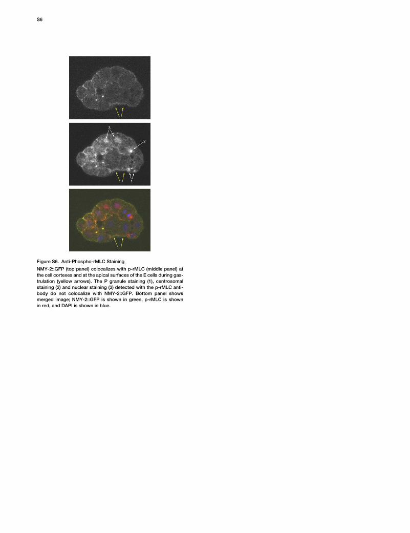
Figure S6. Anti-Phospho-rMLC Staining
NMY-2::GFP (top panel) colocalizes with p-rMLC (middle panel) atthe cell cortexes and at the apical surfaces of the E cells during gas-trulation (yellow arrows). The P granule staining (1), centrosomalstaining (2) and nuclear staining (3) detected with the p-rMLC anti-body do not colocalize with NMY-2::GFP. Bottom panel showsmerged image; NMY-2::GFP is shown in green, p-rMLC is shownin red, and DAPI is shown in blue.
S6