hand surgery
DESCRIPTION
how to remember hand musclesTRANSCRIPT

HAND SURGERY Dr. Diniega
Function of the hand: Prime function of
the hand is feeling and motion
Maintenance and restoration of basic function: Pinch and Grasp
“Position of Function” – should facilitate feeding and toilet
Primarily: pinch, grasp, sensation, emotions
Carpal bones mnemonic: Scare lover try position
that they can’t handle Scaphoid, lunate, triquetrum, pisiform, trapezium, trapezoid, capitate and hamate
X-ray of the Normal Hand Lateral view
(picture) AP view (picture)Due to its positions:
Capitates – largest carpal bone
Lunate – most dislocated
Scaphoid – most fractured (easily)Palmar fat pad – predetermined
- Not hypertrophy

- But can be subtracted (atrophy) in e.g. nerve injuries
Dorsal fat pad – same as the rest of the body
- Also grows fatPalmar fascia + numerous blood vessels dupuytren’s contracture(thick): constricts bv excisedThenar – thumbHypothenar – small fingerMuscles of the Hand
I. Extrinsic- does not originated
from the hand but acts on the hand
- more than 40 in number
- includes: flexor digitorum superficialis, flexor digitorum profundus, extensor digitorum, extensor indicis, extensor digiti minimi (act on the 2nd to 4th digits); flexor pollicis longus, extensor pollicis longus, extensor pollicis brevis, abductor pollicis longus (extrinsic thumb muscles)

- attach on the proximal interphalangeal joint
- innervated by median nerve
II. Intrinsic - muscles that
originate from the hand
- includes: lumbricales, thenal muscles and adductor pollicis, hypothenar muscles, dorsal interossei and volar interossei (acts only the digits)
- innervated by ulnar nerve
(Study actions of the hand muscles)
Thenar Muscles:Abductor pollicis brevis
- grasping- contributes to IP extension
Flexor pollicis brevis
- have ulnar and radial head- grasping
Opponens pollicis
- Opposition of thumb to each digit. Rotates 1st metacarpal so the thumbnail faces the

ceiling when the hand is resting palm up.
Hypothenar Muscles Abductor digiti minimi
-acts to abduct the fifth MP joint- contributes to PIP/DIP extension
Flexor digiti minimiOpponens digiti
minimi
4 dorsal interossei spread the fingers apart (2 on the middle MCP and no volar on the middle mcp???)3 volar interossei closes or puts the fingers togetherUlnar arch
- main vascular supply of the hand
- if compromise will result to Dupuytren contracture
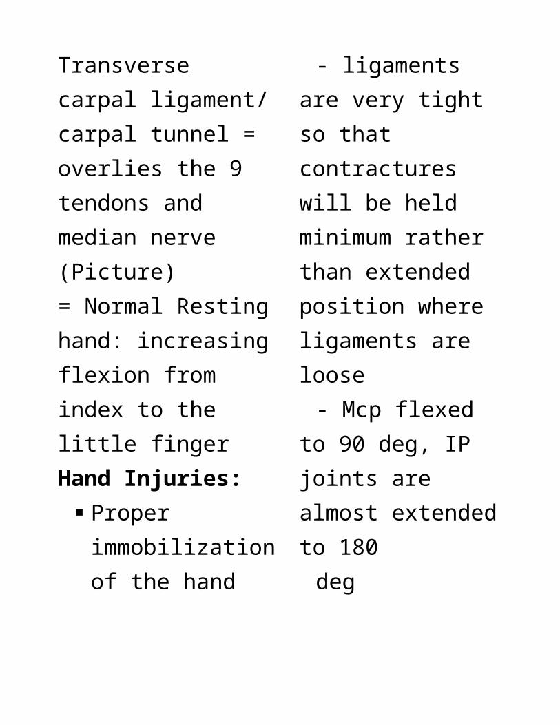
Transverse carpal ligament/ carpal tunnel = overlies the 9 tendons and median nerve (Picture)

= Normal Resting hand: increasing flexion from index to the little finger Hand Injuries: Proper
immobilization of the hand- ligaments are very
tight so that contractures will be held minimum rather than extended position where ligaments are loose
- Mcp flexed to 90 deg, IP joints are almost extended to 180
deg
- strap the injured finger to the uninjured finger (serve as a
splint); Finger can still move which prevents contractures “Boxing glove”
splint Incorrect way of
splinting of the fingers = using popsicle sticks
Proper splinting = garter strapping; use of a padded metal splint
General Principles Elevating the injured
hand Edema control

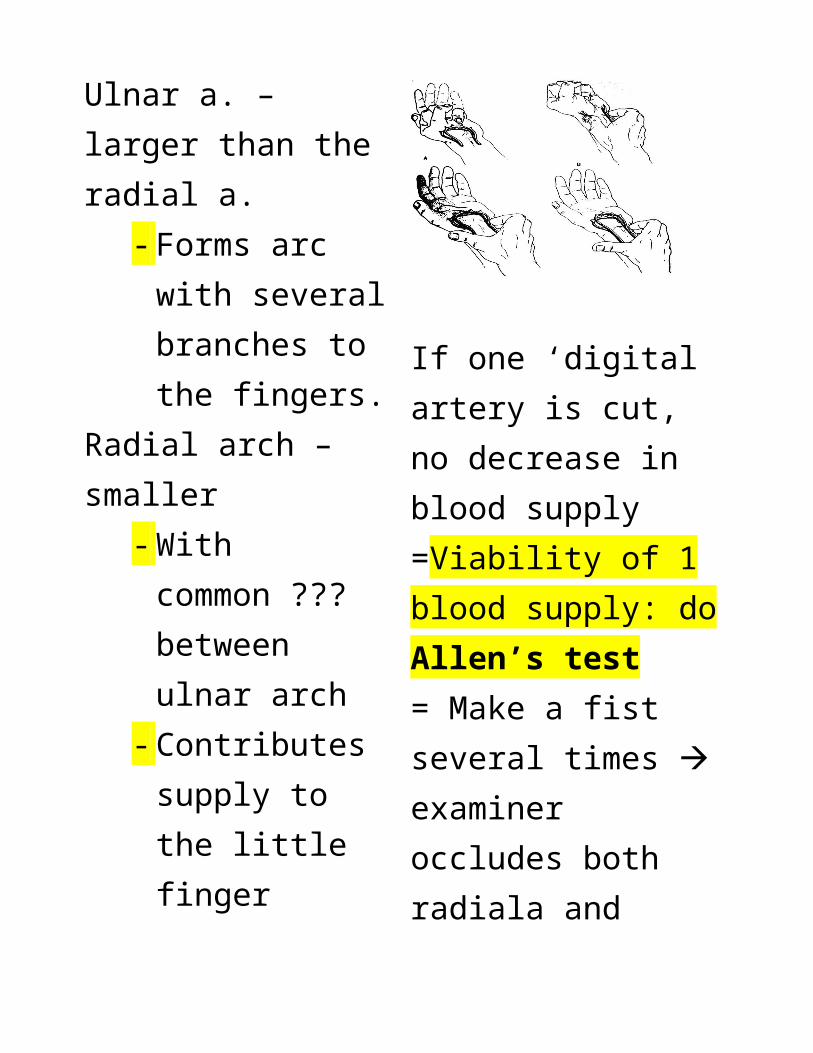
(Picture) Main blood supply of the hand – ulnar a. Ulnar a. – larger than the radial a.
- Forms arc with several branches to the fingers.
Radial arch – smaller- With common ???
between ulnar arch- Contributes supply
to the little finger
If one ‘digital artery is cut, no decrease in blood supply=Viability of 1 blood supply: do Allen’s test= Make a fist several times examiner occludes both radiala and ulnar arteries note the blanching of hand and palm’ (pale) release one artery of hand take the time when the color returns to normal then do it on the other side
Det. Vascularity of the hand
Injuries of the hand

Soft tissue transfers
Before surgery (gangrenous)
Tendons= Synergestic and antagonistic principle: If one is transected the other will predominate or exaggerate 1) Flexor tendon
transaction a.extensor (+)b. flexors (-) hand extends
Components of the Carpal Tunnel:
4 FDS – flexes the pip
4 FDP – flexes the dip 1 thumb flexor1 median nerve
Total: 10
How do you know the tendon of profundus is sever or intact?
= stabilize other fingers and let the px move the injured finger
How do you know the tendon of superficialis is sever or intact?
= stabilize pip then let the dip move
2) Extensor tendons- from one muscle- most cannot act on
their own except little finger and index finger
- can’t extend fingers
Extensor tendons:ext digitorum
communis – 4 fingers ext. dig. Minimi
- 1ext. hallucis
proprius?? – 1can extend the
thumb: EPL, EPB, APL Rupture of
the central hood
Boutonniere: “button hole” deformity
- central tendon is ruptured – cant extend dip
Mallet finger – severed tendon ends/ rupture of the extensor tendon inserting the distal phalanx bring the ends closer and bridge the scar tx: splintingo Tendon
injury or avulsion fracture
o Healing and complications:

Ruptures of the attachment of the long extensor tendon of the fingers heal in 6 weeks. If the injury is not treated, the injured person will have a permanent “mallet finger” deformity.
Flexor tendon avulsion
(Picture)= Difference between subluxation and dislocation:
- Subluxation: incomplete dislocation
- Dislocation: complete severance of the ligaments that support the joints
Fractures- interossei contracts
in hand fracture bent position of hand/ finger
- shortened length of the finger or rotated because of 2d x-ray cannot be noticed
- normally, all fingers should point towards the scaphoid if deviated –
malunion or malrotated
Boxers fracture – injured the little finger, mcp and thumb
Bennett’s fracture – is the fracture of the first metacarpal bone
Dislocations(Picture)(+) swelling – injuryExamine under anesthesia – bend the finger L to R ulnar collateral ligament is ruptured (Picture)- Complication:
= Avascular necrosis – loss of blood supply
= 15% of individual have no blood supply of the proximal half of the scaphoid (supplied by = Keinbocks disease – avascular necrosis of the lunate
Nerve Supply to the Hand
Motor Nerves of the Hand

Median nerve Flexor
digitorum superficialis, flexor carpi radialis, radial 2 flexor digitorum profundus, radial 2 lumbricals, flexor pollicis longus, abductor, opponens and superficial head of flexor pollicis brevis
= Median nerve - supplied the palmar
aspect of the thumb, index and middle
- Supply most extrinsic mm.
- More disabling if severed because of
prime digits are supplied by this
Ulnar Flexor carpi
ulnaris, ulnar 2 flexor digitorum profundus, ulnar 2 lumbricals all of the hypothenar muscles, abductor, flexor and opponens digiti quinti, deep head of the flexor pollicis brevis, adductor pollicis and all of the interossei
Radial Nerve Supplies all
extrinsic extensors of the wrist and fingers:

Extensor carpi radialis longus and brevis, extensor carpi ulnaris, abductor pollicis longus, extensor pollicis longus and brevis, extensor indicis propius, extensor digiti minimi, extensor digitorum communis
= no supply to the intrinsic muscles of the hand
Nerve Injuries Neuropraxia – no
loss of continuity, all axons are in continuity although functionless; usually results from
compression or ischemia; recovery is spontaneous or permanent= most common= resulting from closed injury
Axonotmesis – sheath is intact but some axons are physically interrupted; usually results from stretching, compression or concussion= recovery is incomplete = resulting from closed injury
Neurotmesis – all nerve continuity is

lost; due to crashing, laceration injury; requires surgery = results from open injury= treated by surgery
Median Nerve (Picture)- test strength of the
mm – APL (abduction)
- injury – ‘atrophy of the thenar musclesoFPB (2 nerve
supply, 1 ulnar and 1 median)
oHollow thenar prominence
o (-) opposition of thumb
oClawing of the radial 2 fingers Nerve
supply (radial nerve) above is still intact hence unopposed action of the FDS and FDP
Flexion of IP and hyperextension of mcp
P.E. Findings:

oHollow thenar prominence
o Inability to oppose thum to the other fingers
oClawing of the radial 2 fingers
o (+) Phalen’s test - test for integrity of
the median nerve at the wrist area
- done by acute flexing the wrist
- it compress the contents of the carpal tunnel
- (+) finding is paresthesia/numbness of the radial 3 fingers: thumb,
index and long finger
o Froment’s sign = tests for palsy of the ulnar nerve, specifically, the action of adductor pollicis. Patient will flex the thumb instead of adducting it (intact median damaged ulnar)
o Another test: letting the patient hold a paper using his/her thumb and index finger and the examiner will pull gently the paper
Ulnar Nerve

P.E. Findings:o Interossei
paralysis = no spreading of the fingers
o Hollow hypothenar eminence
o Clawing of the ulnar 2 fingers due to paralysis of proximal and distal interphalangeal jonts and hyperextension of the MCP’s
=Important, PE to know which nerve is
damaged test sensory of each nerve=Mostly intrinsic mm – loss dexterity of the hand= (Picture) Wasting of the first dorsal interosseous muscle and clawing of the ulnar 2 fingers in the left hand
Radial nerve Sensory –
dorsum of the hand and forearm
Motor – wrist drop if radial nerve is affected, paralysis of finger

extensors if PIN is affected
- No nerve supply to intrinsic
- Common in fractures of the middle shaft of humerus
- Wrist and finger extensors are supplied by radial nerve
- Posterior interosseous nerve injury: no wrist drop because it gives off branches at this level
- Wrist drop and inability to extend the MCPs
- Do not rely on wrist extension, also check finger extension.
- Wrist drop is not always synonymous with radial nerve injury
Hand Infections:Bursa = normally tendon slide smoothly= downside: can be a reservoir for pus or infxn can go all the way to the wrist area

Felon – pulp on the finger
Suppurative tenosynovitis– infection of the tendon sheath
Paronychia – nail Eponychia – nail
fold Horseshoe abscess –
whole hand Septic arthritis –
from human bite – dirtiest wound – treat aggressively, do not close
Tenosynovitis – emergency, tendon is bathe with pus; complication cause tendon rupture
Tumors Ganglion cysts – synovium of the joint- contain mucin or synovial fluid - connected to a tendon through a stalk - Must be excised properly can recuro Most common
soft tissue tumor of the hand
Bony tumors :1) Enchondroma –
bulging/expansion of bone + ‘xray signs
2) Ollier’s disease – multiple enchondroma

3) Maffucci’s Syndrome – enchondroma + skin hemangiomas
Non Specific Inflammatory and Constrictive Conditions
1) Trigger finger – thickened pulley – tendon cant glide through it – A1 mostly affected- tend to bow string - tenosynovium is
inflamed 2) de Quervain’s
disease – dorsum – first compartment – EPB and APL
a.narrowed canal – (+) Finkelstein’s test (abduction produce pain)
3) Carpal Tunnel syndrome
Compartments of the Extensors (Picture)Congenital Anomalies Syndactyly Polydactyly
Amputations
-END-