haemodynamic morphine in patients acute …heart.bmj.com/content/heartjnl/27/6/863.full.pdfthe...
TRANSCRIPT

Brit. Heart J., 1965, 27, 863.
HAEMODYNAMIC EFFECTS OF MORPHINE IN PATIENTS WITHACUTE MYOCARDIAL INFARCTION
BY
MICHAEL THOMAS, RAOUL MALMCRONA*, SIDNEY FILLMOREt, ANDJOHN SHILLINGFORD
From the Medical Research Council Cardiovascular Research Group, Department ofMedicine, Postgraduate MedicalSchool ofLondon W.12, and Medical Department 1, Sahlgrenska Sjukhuset, University of Gothenburg, Sweden
Received March 24, 1965
The administration of analgesic drugs to patients with acute myocardial infarction is routineclinical practice for the alleviation of pain and anxiety or simply to ensure sleep. Because of thepotent combination of analgesic and sedative properties, morphine is frequently given. Althoughthe associated depressant effects on the circulatory and respiratory systems have been documentedfrom animal experiments and work in normal man, the haemodynamic consequences of the adminis-tration of the drug to patients with acute myocardial infarction have previously not been investigated.The institution of a special unit for the intensive care and study of patients with acute myocardial
infarction has enabled hemodynamic measurements to be made throughout the acute illness andbefore and after the administration of drugs without disturbing the patients.
The object of this work has been to measure the changes in cardiac output, stroke volume, heartrate, and arterial blood pressure following the administration of morphine to a variety of patientswith acute myocardial infarction and at the same time to observe the clinical effects.
SUBJECTS AND METHODSFifteen studies were made in 13 patients. All had clinical histories typical of acute myocardial infarction.
In 12, the acute illness was accompanied by chest pain. Patient 3 presented following sudden loss of con-sciousness. All patients had electrocardiographic signs of myocardial infarction at some time during theacute illness: at the time of investigation S-T segment elevation was present in 11 and pathological Q wavesin 3. Serum glutamic oxalo-acetic transaminase (SGOT) or lactic dehydrogenase (LDH) was raised in 9of the 12 patients in whom it was measured.
Eight patients had previously had symptoms attributable to ischiemic heart disease. Patients 3 and 10had had a myocardial infarction in the past. Patients 5, 11, and 12 were known to have been hypertensivebefore the acute illness. Patient 11 was receiving guanethidine until 24 hours before the investigation. Twopatients (7 and 8) had chronic respiratory disease. The patient's body temperature was above 990 F.(37-20 C.) during seven investigations. Clinical details are given in Table I.
With one exception (Patient 8) the first investigation was undertaken within 24 hours of the onset of symp-toms. Patients 2 and 5 were studied a second time within 48 hours of the onset of symptoms. Patient 8was studied on the 2nd day only. All patients recovered and left hospital with the exception of Patient 10who died in the 4th week of admission.
* In receipt of grants from the Swedish National Association against Heart and Lung Diseases and from theWellcome Research Foundation.
t In receipt of Training Grant 5T1 HE5603-03 from the National Institutes of Health, U.S. Public Health Service.863
on 13 June 2018 by guest. Protected by copyright.
http://heart.bmj.com
/B
r Heart J: first published as 10.1136/hrt.27.6.863 on 1 N
ovember 1965. D
ownloaded from

THOMAS, MALMCRONA, FILLMORE, AND SHILLINGFORDTABLI
CLINICAJ
Patient Age Weight'D f Body General condition on
No. (yr.) (kg.) infarct. tempera- Past history Present symptoms admissiontture* ('C.)
I~~~~~IIIIPeriph. isch. dis.
Diab. mellit. 15 yr.
Myocard. infarct. 4 yr.prev.
Effort angina 6 mth.
Hypertension 5 yr.;diab. mellit.; 6 mth.angina
Nil relevant
Cough +sputum manyyr.; dyspn. on effort
Winter bronchitis;effortangina 1i yr.
Chest pain 6 yr. prev.
Effort angina 9 mth.;effort dyspn. 5 yr.
Hypertension noted1959; controlled byguanethidine
Renal tuberc.; L, neph-rectomy 1962; BP pre-viously 190/110
4 yr. occas. min. chestpain; 1 Wk. incr. chestpain
R. arm ache 2 wk.sudden sev. painarms chest
Sudden pain frontchest; felt weak &faint; vomited
Sudden loss ofconsci-ousness; recoveredconsc.; vomited; nopain
Awoke morning withsev. chest pain &down both arms;weak and nauseated
Acute sev. chest painextending to neck;weak feeling in arms
Sudden chest painwhile walking, armsfelt heavy, slightlyfaint
Sudden sev. chestpain
Incr. frequency ofangina 24 hr.; sud-den sev. chest pain
Sudden sev. chestpain
Sev. chest pain
Sev. chest pain
Sev. chest & R. armpain
Sev. chest pain
* Body temperature at time of investigation, M, mouth; R, rectal.t JVP, jugular venous pressure; HR, heart rate.
In pain; moist warm skin; HR 90;JVP not raised; BP170/90 mm.Hg; presyst. heart sound; fewrales lung bases
Pale & sweating; HR 100; JVPnot raised; BP 115/70 mm. Hg;normal heart sounds
Pale; HR 88; JVP not raised;BP 145/75 mm. Hg; apex beatdisplaced laterally; normal hearsounds; rales at lung bases
Pale; in pain; HR 50; JVP+3cm.; BP 140/80 mm. Hg; apexbeat displ. lat.; presyst. heartsound
Sev. pain; HR 60; JVP+4 cm.;BP 200/120; presyst. heartsound
Mod. sev. pain, HR 80; JVP notraised, BP 145/100 mm. Hg;presyst. heart sound
Mod. sev. pain; HR 80; BP 150/90 mm. Hg; JVP not raised;presyst. heart sound; reducedbreath sounds at both lungbases with coarse creps.
In pain; HR 68; JVP not raised;BP 115/70 mm. Hg; heartsounds normal
In pain; HR 90; JVP not raised;BP 134/90; presyst. heart sound
Sev. pain; distressed; HR 118;JVP not raised; BP 125/90 mm.Hg; 3rd heart sound
In pain; HR 70; JVP not raised;BP 170/115 mm. Hg; presyst.heart sound
Sev. pain; very pale; HR 90;JVP not raised; BP 150/100 mm.Hg; heart sounds normal
Pain-free; HR 80; BP 120/80;JVP not raised; heart soundsnormal
1
2
2'
3
4
5
5'
6
7
8
9
10
11
12
13
67
64
72
73
60
49
62
56
58
67
55
61
55
63
72
65
64
70
67
78
77
62
82
72
68
1
1
2
1
1
1
2
1
1
2
1
1
1
1
1
36(7(M)
37-3(M)
37-8(M)37.5(M)
36-9(M)
37-2(M)369(M)37(R)
37(M)
36-9(M)37.3(M)
38 5(R)
36-7(M)
37-6(R)37-1(R)
l~~~~~~~.
L
864
on 13 June 2018 by guest. Protected by copyright.
http://heart.bmj.com
/B
r Heart J: first published as 10.1136/hrt.27.6.863 on 1 N
ovember 1965. D
ownloaded from

MORPHINE IN ACUTE MYOCARDIAL INFARCTION
I
DATA
Severity of Breathing.Highest Morphine pain at time air orenzyme X-ray sulphate of morphine oxygen Response tovalue ECGt appearances (mg.)§ administration morphine
_ !INot SR; S-T elevation V2-measured V4
SR; S-T segm. elevationAVF, & III
SR; S-T segm. elevationV2-V7
SR; S-T segm. elevationAVF & IlI
SR; path. Q with S-Tsegm. elevation & Tinversion III, AVF
SR; path. Q II, III,
AVF; T inversion 11,
III, AVF
SR with ventric. ectopicbeats; S-T segm. ele-vation II, III, AVF,V4, V5, V6
SR; S-T segm. eleva-tion III, AVF
SR; S-T elevation I,
AVL, V4-7
SR; S-T segm. eleva-tion V2-V4
SR; S-T segnm. eleva-tion V4R-V7
S-T segm. elevationCR2, CR4
Path. Q in AVF, 11, III
Heart slightly en-larged; lung fieldsnormal
Heart enlarged; lungfields normal
Heart enlarged; slightcongestion; changesat bases
Heart enlarged;slightly full hilarshadows
LV enlargement; lungfields normal
Heart normal; min.congestive changesat lung bases
Mod. cardiac enlarge-ment; some collapseboth lower zoneswith patchy consol-idation
Min. cardiac enlarge-ment; segm. col-lapse at R. base
Heart slightly en-larged; lungs normal
Mod. cardiac enlarge-ment; subsequentLV aneurysm; verymarked venous en-gorgement; pulmon.cedema
Mod. cardiac enlarge-ment; lung fieldsnormal
LV enlargement; lungfields normal
Normal
5
7.5
7.5
3
5
10
10
5
3
10
3
10
10
5
5
Pain
Pain
Discomfort
No pain
Pain
Sev. pain
Discomfort
Pain
Pain
Pain
Pain
Sev. pain
Sev. pain
Sev. pain
Pain
02
Air
Air
Air
Air
02
02
02
02
02
02
02
02
02
Air
$ SR, sinus rhythm.§ Quantity of morphine sulphate injected intravenously.
Loss of pain; remainedawake
Loss of pain; remainedawake
Loss of pain
Became sleepy
Loss of pain; asleep1 hr. later
Pain diminished; lessrestless; remainedawake
Less discomfort
Loss of pain; returned2 hr. later
Minor effect on painbut mod. sedation
Mod. sedation
Vomited once after 3mg. morphine sul-phate; no other clinicalchange
Sleep induced soon aftermorphine admin.;slept several hr.
Rapid induction ofsleep; slept 30 min.;pain relieved; lookedpale after waking
Pain subsided duringinvestig.; patient re-mained awake
Sleep almost induced;patient felt improved
SGOT102 I.U.
SGOT6 I.U.
SGOT22 I.U.
SGOT56 I.U.
LDH 410
LDH 100
LDH 150
LDH 1050
LDH 1200
LDH 7J5
SGOT385 I.U.
SGOT74 I.U.
865
on 13 June 2018 by guest. Protected by copyright.
http://heart.bmj.com
/B
r Heart J: first published as 10.1136/hrt.27.6.863 on 1 N
ovember 1965. D
ownloaded from
THOMAS, MALMCRONA, FILLMORE, AND SHILLINGFORD
Eleven patients were investigated in a special intensive care and study unit with recording equipment per-manently installed (Shillingford and Thomas, 1964). Two (12, 13) were studied in a general ward. Afterclinical assessment, electrocardiograph, and chest radiographs, fine polythene catheters (PE60 IntramedicU.S.A.) were introduced percutaneously into a brachial artery and an antecubital vein, using local anes-thesia. The tip of the arterial catheter was advanced 10 cm. from the point of insertion and the tip of thevenous catheter was advanced until it lay in the region of the great veins.
Intravascular pressures were measured by use of P23Gb Statham transducers and were recorded on adirect writer (Devices Ltd). Measurements were made with respect to a point 5 cm. below the sternal angle.With one exception the patients lay horizontally with head and shoulders comfortably supported on one ortwo pillows. Patient 7 was supported from the waist at an angle of 450 to the horizontal. In 11 patients thecardiac output was measured by a dye dilution technique using the photoelectric earpiece (Cambridge Instru-ment Co. Ltd.) with Coomassie Blue dye (I.C.I.) as indicator. The first curve was calibrated by equatingthe height of the tail of the curve three minutes after injection of dye with the concentration of dye in a centralvenous blood sample taken at this time. Dye was extracted from the plasma and measured by spectrophoto-metry. Subsequent cardiac outputs were calculated according to the relative areas of the curves (Gabe,Tuckman, and Shillingford, 1962). In Patients 12 and 13, cardiac output was measured by intermittentarterial sampling using bromsulphthalein as indicator.
Heart rate was measured over 30-second intervals from an electrocardiographic tracing: in Fig. 5 and 6heart rate was measured over shorter periods. Peripheral resistance was calculated as the mean pressure inthe brachial artery, expressed in mm. Hg, divided by the cardiac output expressed in litres/min. In Fig. 8values are given to the nearest whole number.
After insertion of catheters and preliminary preparations for the investigation, repeated measurements ofarterial blood pressure were made for approximately 30 minutes or until a steady level of blood pressure wasrecorded. In one patient (5) this was not achieved. Two, or in some cases three, dye dilution curves werethen drawn at five-minute intervals, the first one being calibrated. Intravascular pressure measurementswere made immediately before and immediately after each cardiac output measurement. Morphine sulphate10 mg. in 10 ml. of saline was then infused unknown to the patient into the central venous system via thevenous catheter at a rate of approximately 1 mg./minute. Brachial arterial blood pressure was continuouslyrecorded during injection. The infusion was terminated if a persistent fall in blood pressure was seen. Ifthe blood pressure did not fall during injection, the maximum dose given was 10 mg. Measurements ofheart rate and arterial blood pressure were then made at frequent intervals. Cardiac output was measuredat intervals of 5-10 minutes over a period of 30-40 minutes.
Arterial blood samples were taken for measurement of Po2 and Pco2 before the administration of mor-phine and at 10 and 20 minutes after completion of injection. Arterial Po2 was measured by means of aBeckman electrode and arterial Pco2 by means of a Severinghaus electrode. pH was calculated from Pco2and serum bicarbonate values.
RESULTSResults of the haemodynamic investigations are illustrated in Fig. 1-8. In the diagrams the left-
hand circled number identifies patients; the right-hand circled number represents in addition anobserved value, except in Fig. 4 and 7 in which it is only an identification. Control measurementsshown are those obtained five minutes before and immediately before the injection of morphine.The times of subsequent measurements are given as the time after completion of the morphineinjection.
Heart Rate (Fig. 1). In 7 instances (1, 2', 7, 8, 10, 11, 13) there was no change in heart rate afterthe injection of morphine. In 6 instances (2, 4, 5, 5', 6, 9) heart rate increased, and in 2 (3, 12) theheart slowed. In those cases in which a change occurred, it was apparent at the first measurementafter the completion of injection.
Heart rate increase was related to vomiting in 2 patients (5 and 9). In Patient 9 this occurredsoon after beginning morphine injection. In 3 patients with an increased heart rate the increasewas sustained over the period of observation. The other patients who showed a heart rate increasereturned to control levels within 15 minutes.
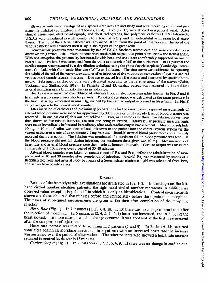
Cardiac Output (Fig. 2). In 7 instances (1, 2, 2', 5, 6, 9, 11) there was no change in cardiac out-
866
on 13 June 2018 by guest. Protected by copyright.
http://heart.bmj.com
/B
r Heart J: first published as 10.1136/hrt.27.6.863 on 1 N
ovember 1965. D
ownloaded from

MORPHINE IN ACUTE MYOCARDIAL INFARCTION
, 8)1-o
w 60-
30 _ L1.,-'Control 0 iO 20 30 40 50
TIME in minutesFIG. 1.-Heart rates measured before and after the injection of morphine.
put after injection of morphine. In 8 instances (3, 4, 5, 7, 8, 10, 12, 13) cardiac output was increasedfor part or the whole of the period of observation. In no patient did a convincing fall of cardiacoutput occur, though in one (Patient 5) with a high cardiac output and tachycardia the measurementshowed considerable fluctuation.
Stroke Volume (Fig. 3). In 7 instances (1, 2', 6, 9, 10, 11, 13) the stroke volume remained un-changed after injection of morphine. In 4 instances (3, 7, 8, 12) stroke volume rose after the injec-tion: in 2 of these (7 and 8) the times of the measurements were such that it was possible to see thatthe rise in stroke volume occurred more than ten minutes after the injection was complete. In fourinstances (2, 4, 5, 5') there was a decrease in stroke volume. The decrease in one of these (4) waspreceded by a slight rise, while in the others an early decrease was followed by a return to the controlvalue.
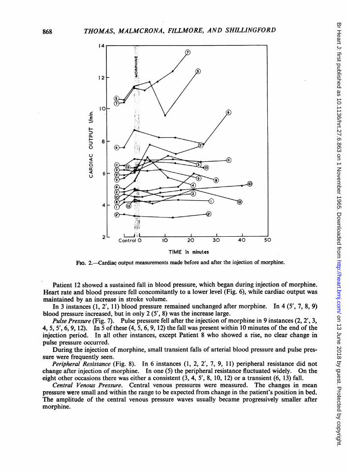
Arterial Blood Pressure (Fig. 4). In 7 instances (2, 3, 4, 5, 6, 10, 13) arterial mean blood pressuredecreased transiently, and in an eighth instance (12) there was a persistent fall.
The injection of morphine was stopped at an early stage (3 mg.) in one patient (3) on account ofa fall in blood pressure which continued progressively to very low levels (Fig. 5). When the patient'slegs were raised arterial blood pressure rapidly increased and returned in three minutes to the controllevel after an overshoot. Systolic blood pressure fell from 140 to 70 mm. Hg, while the heart rateremained constant. A further fall of 30 mm. Hg was then accompanied by a fall in heart rate from85 to 20 a minute. When the patient's legs were raised, heart rate increased to 100 a minute beforebecoming stable at 70 a minute. A few minutes after this event, cardiac output exceeded controllevels.
867
on 13 June 2018 by guest. Protected by copyright.
http://heart.bmj.com
/B
r Heart J: first published as 10.1136/hrt.27.6.863 on 1 N
ovember 1965. D
ownloaded from
868 THOMAS, MALMCRONA, FILLMORE, AND SHILLINGFORD
14
10
I oto 0 10 2 0 0 5
E
a.
4-
2
Control 0 IC 20 30 40 50
TIME in minutes
FIG. 2.-Cardiac output measurements made before and after the injection of morphine.
Patient 12 showed a sustained fall in blood pressure, which began during injection of morphine.Heart rate and blood pressure fell concomitantly to a lower level (Fig. 6), while cardiac output wasmaintained by an increase in stroke volume.
In 3 instances (1, 2', 11) blood pressure remained unchanged after morphine. In 4 (5', 7, 8, 9)blood pressure increased, but in only 2 (5', 8) was the increase large.
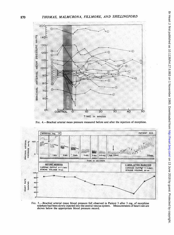
Pulse Pressure (Fig. 7). Pulse pressure fell after the injection of morphine in 9 instances (2, 2', 3,4, 5, 5', 6, 9, 12). In 5 of these (4, 5, 6, 9, 12) the fall was present within 10 minutes of the end of theinjection period. In all other instances, except Patient 8 who showed a rise, no clear change inpulse pressure occurred.
During the injection of morphine, small transient falls of arterial blood pressure and pulse pres-sure were frequently seen.
Peripheral Resistance (Fig. 8). In 6 instances (1, 2, 2', 7, 9, 11) peripheral resistance did notchange after injection of morphine. In one (5) the peripheral resistance fluctuated widely. On theeight other occasions there was either a consistent (3, 4, 5', 8, 10, 12) or a transient (6, 13) fall.
Central Venous Pressure. Central venous pressures were measured. The changes in meanpressure were small and within the range to be expected from change in the patient's position in bed.The amplitude of the central venous pressure waves usually became progressively smaller aftermorphine.
on 13 June 2018 by guest. Protected by copyright.
http://heart.bmj.com
/B
r Heart J: first published as 10.1136/hrt.27.6.863 on 1 N
ovember 1965. D
ownloaded from

MORPHINE IN ACUTE MYOCARDIAL INFARCTION
E10(
w
.20
8(
Oz 6(LO
TIME in minutesFIG. 3.-Stroke volume calculated from cardiac output and heart rate measurements made before and
after the injection of morphine.
TABLE IIHEMODYNAMIC STUDIES BEFORE AND AFTER MORPHINE
Before morphine 5-10 minutes 20-30 minutesBreathing after morphine after morphine
Patient oxygen orNo. air pH Po2 Pco2 pH Po2 Pco2 pH Po2 Pco2
3 Air 7.34 60 35-5 7-31 58 38 5 -4 Air 7-41 74 37 7-36 65 402 Air 68 38 69 42-5 - -2' Air 63 44 _ 65 47.58 Oxygen - 204 46 255 51 - 245 509 Oxygen 272 44*5 260 39 281 4110 Oxygen 157 41-5 164 45 149 45 51 1 Oxygen - 212 40 - 220 43 - - -
869
on 13 June 2018 by guest. Protected by copyright.
http://heart.bmj.com
/B
r Heart J: first published as 10.1136/hrt.27.6.863 on 1 N
ovember 1965. D
ownloaded from
THOMAS, MALMCRONA, FILLMORE, AND SHILLINGFORD
200
_40..101
130r
120)
I10f-
100
901_
80
70
60
O0
TIME in minutes
FIG. 4.-Brachial arterial mean pressure measured before and after the injection of morphine.
MORPHIA 3mg. IX]V PATIENT E.G.~~~~~~~~~~ 1
___kMi&MVh} ~ 4laIn. '2 in. 3mi. 5un., 6wui 65m sgiaa.hed 75fain.m.. ...._. . . ........U.I..,"J!^d11tr'Ls'-';|1,d=Itt1 U 5!t
¢-----.,, ,, .,,,---,,,,,",,,TIME IN SECONDS ____m____n_._ _ _ _ _ _'
BEFORE MORPHIIA I1 SINS.AFTER INJECTIONCARDIAC OUTPUT 6 1 L/min. CARDIAC OUTP%T 7-1 L/mIn.STROKE VOLUME 79ml STROKE VOLUME 99 ml
to ,1
870
aC.2
E
cr
U)CDQ.
z4aL
I-c4
LU
e--4
ccm
-Ac o I:C4
aD
I
'C
4c W
U W c
j0
a
I- xi
I-.4.Sl
FIG. 5.-Brachial arterial mean blood pressure fall observed in Patient 3 after 3 mg. of morphinesulphate had been slowly injected into the central venous system. Measurements of heart rate areshown below the appropriate blood pressure record.
6
41
2i
on 13 June 2018 by guest. Protected by copyright.
http://heart.bmj.com
/B
r Heart J: first published as 10.1136/hrt.27.6.863 on 1 N
ovember 1965. D
ownloaded from

MORPHINE IN ACUTE MYOCARDIAL INFARCTION
200*0"
1601-
=
Pr:E
cs
c:mw
00--Ji
1201-
80k_
40 -
InriControls
c
-E
tn-
TIME in minutesFIG. 6.-Diagrammatic representation of brachial arterial blood pressure and heart rate fall which
occurred simultaneously in Patient 12 after the injection of morphine.
Respiration. A fall in respiration rate and depth was observed clinically in most cases aftermorphine injection. In 8 patients in whom arterial blood samples were taken before and aftermorphine (2, 2', 3, 4, 8, 9, 10, 11) arterial Po2 did not show any consistent change (Table II), but allexcept one (9) showed a small rise in Pco2.
Clinical Response to Morphine. The severity of pain experienced by the patients at the time ofmorphine injection varied widely and is graded in Table I as discomfort, pain, and very severepain. In all but one of the patients with symptoms, some improvement occurred during the courseof the investigation. Five patients (3, 4, 10, 11, 13) became drowsy after morphine and in 2 (10, 11)sleep occurred for a period of 30 minutes and several hours respectively.
DIscussIoNThe use of opiates for the alleviation of pain has been practised for many years and the clinical
consequences of their use have been recognized (Comroe and Dripps, 1948; Denton and Beecher,1949). At an early stage in the study of acute myocardial infarction the powerful analgesic and hyp-notic effect of morphine was reported as beneficial (Moor, 1930). Since then the use of the drugin this condition has become widespread.
Although much has been written (Eckenhoff and Oech, 1960) on the effects of the opiate groupof drugs, there has been no attempt to measure the circulatory changes following morphine adminis-tration under the conditions in which it is used in the treatment of acute myocardial infarction.Without such information a critical evaluation of the use of the drug in these patients would beincomplete.
Previous work using ballistocardiography and sphygmomanometry, in normal subjects and in3J
871
OL_
on 13 June 2018 by guest. Protected by copyright.
http://heart.bmj.com
/B
r Heart J: first published as 10.1136/hrt.27.6.863 on 1 N
ovember 1965. D
ownloaded from
872 THOMAS, MALMCRONA, FILLMORE, AND SHILLINGFORD
130
12C
HO~~~~~~~~~~~~~~~~~~"r!00|
280 -
0.
TIME nm6nutes
FIG. 7.-Brachial arterial pulse pressure measurements made before and after the injection ofmorphmne
patients, has shown only small changes in heart rate, cardiac output, and blood pressure after dosesof morphine within the therapeutic range (Starr et at., 1937; Papper and Bradley, 1942; Wange-man and Hawk, 1942; Drew, Dripps, and Comroe, 1946; Denton and Beecher 1949; McCall andTaylor, 1952; Moyer, Morris, and Pontius, 1956; Malt, 1958). The extensive and well-controlledstudy by Drew et at. showed an increase in heart rate, with a proportional increase in cardiac outputand an insignificant fall in blood pressure.
Our results show that in patients with acute myocardial infarction the circulatory response to anintravenous injection of morphine is very variable; the magnitude of the changes is sometimes morepronounced than that shown in normal subjects.A fall in brachial arterial pressure during or soon after slow injection was seen in many patients.
In 2 (3, 12) it was marked. Hypotension in Patient 12 was accompanied by a simultaneous fallin heart rate, but in Patient 3 the arterial systolic pressure fell from 140 to 70 mm. Hg before theheart slowed. The mechanisms of bradycardia in these patients are conjectural. In some ways theresponse in Patient 3, illustrated in Fig. 5, resembles the changes following hxvmorrhage in blooddonors lying in the horizontal position (Wallace and Sharpey-Schafer, 1941; Shenkin et a!., 1944).The heart rate increas!e which occurred in association wvith the rise in blood pressure after raising thepatient's legs suggests that the bradycardia in this patient represents a physiological reaction ratherthan a direct effect of morphine. Changes in heart rate in experimental animals following altera-tions in perfusion of the medulla are documented (Anrep and Segall, 1926). In Patient 12 brady-cardia and hypotension began synchronously, and this response is the only example in these studies
on 13 June 2018 by guest. Protected by copyright.
http://heart.bmj.com
/B
r Heart J: first published as 10.1136/hrt.27.6.863 on 1 N
ovember 1965. D
ownloaded from

MORPHINE IN ACUTE MYOCARDIAL INFARCTION
w 20
z
-J
cx:w'r lo0-X 7wa.
5 1
10
Control 0 10 20 30
TIME in minutesFIG. 8.-Peripheral resistance calculated from measurements of brachial arterial mean blood pressure
and cardiac output made before and afterthe injection of morphine.
of a possible primary action of morphine on the vagus nucleus; bradycardia is a recognizedfeature of the drug's action in dogs (Powers, Reed, and Gregersen, 1947).
The factors concerned in hypotensive responses are of importance both from the point of viewof practical therapy and also for understanding the circulatory failure in acute myocardial infarction.It is probable that many homeostatic mechanisms are deranged, and defects of the myocardium,regulation of heart rate, and control of peripheral vascular tone can all be responsible for inadequatemaintenance of blood pressure (Thomas, Malmcrona, and Shillingford, 1965a). In some patientshypotension after morphine persisted without a rise in cardiac output. In other patients cardiacoutput increased in association with an increase in heart rate or of stroke volume. None of thepatients showed a fall in cardiac output, though it was probably reduced during the profound hypo-tension in Patient 3.
873
on 13 June 2018 by guest. Protected by copyright.
http://heart.bmj.com
/B
r Heart J: first published as 10.1136/hrt.27.6.863 on 1 N
ovember 1965. D
ownloaded from

THOMAS, MALMCRONA, FILLMORE, AND SHILLINGFORD
The analysis of the circulatory adaptation to morphine will require further work, but some com-ment on these data may be useful. In certain cases cardiac output rose in association with a fall inblood pressure; it is probable that peripheral arterial dilatation occurred to an important extent insome regions. Calculated peripheral resistance fell after morphine in many instances. Themeasured change that was most common was a fall in pulse pressure; systolic blood pressure fellwhile diastolic pressure usually remained more constant. As stroke volume remained the same orincreased, the fall in pulse pressure could represent increased distensibility of the arterial wall,possibly due to a direct effect of the drug on smooth muscle. Experiments in animals (Schmidt andLivingston, 1933) have suggested regional differences in vascular response to morphine but possiblespecies differences discourage acceptance of this in man.
The action of morphine on veins is not easily assessed from our results. Little confidence canbe placed in measurements of small changes in central venous pressure on account of frequent move-ments of seriously ill patients. The dramatic increase in arterial blood pressure after raising thelegs in Patient 3 suggests that pooling of the blood in the venous system may be an important conse-quence of morphine administration, but the evaluation of this will require special techniques.
Respiratory depression may contribute to the net circulatory changes after morphine. ArterialPco2 rose in nearly all cases in which it was measured. Arterial Po2 did not change consistently.Interpretation is made difficult by the fact that some patients were breathing air and some werebreathing oxygen. The pressor effect of oxygen (Thomas, Malmcrona, and Shillingford, 1965b)could be of benefit in minimizing hypotension after morphine, but the data are insufficient toestablish this.
It would be of practical assistance in the management of the individual patients to be able topredict the type of cardiovascular response to an injection of morphine. The hlmodynamic patternfound in patients with acute myocardial infarction is very variable and the efficacy of homeostaticmechanisms is probably dependent on many factors. Frequently there was a fall in systolic pres-sure when morphine first entered the circulation but this did not always indicate the eventual responseto the drug. The degree of analgesia for a given dose was not related to the severity of the pain andthe type and extent of circulatory change did not appear to be related to the existing heemodynamicstate. Individual sensitivity seems to be important.
The route of administration of morphine most suitable for patients with myocardial infarctionis open to discussion, the important difference between the intravenous and intramuscular or sub-cutaneous routes being the time course of the action of the drug. Previous studies (Drew et al.,1946; Dripps and Comroe, 1945) on respiratory depression following morphine have shown thatmaximal effect occurs within a few minutes after intravenous injection and much later after intra-muscular or subcutaneous injection. Our observations suggest that hypotension after morphine inpatients with acute myocardial infarction also occurs soon after intravenous injection. A seriousunpredictable blood pressure fall would occur while the patient was under direct medical supervision,and appropriate measures, such as raising the legs or the foot of the bed, would be immediatelyundertaken. The slower and unpredictable rate of absorption after intramuscular injection mightresult in a delayed respiratory and cardiovascular depression, especially if repeated injections aregiven.
Exaggerated hypotensive response to morphine in the upright posture has been pointed out in thepast (Drew et al., 1946). This may be important in patients with acute myocardial infarction whoare liable to a fall in blood pressure. On this account patients who have received morphine shouldnot be carried with the legs dependant.
SUMMARYHemodynamic changes following intravenous administration of morphine to patients with
acute myocardial infarction have been studied. Heart rate, cardiac output, stroke volume, andarterial blood pressure were measured before and after morphine. Blood pressure changes rangedfrom a small fall in pulse pressure to a large fall in mean arterial pressure. In some patients who
874
on 13 June 2018 by guest. Protected by copyright.
http://heart.bmj.com
/B
r Heart J: first published as 10.1136/hrt.27.6.863 on 1 N
ovember 1965. D
ownloaded from

MORPHINE IN ACUTE MYOCARDIAL INFARCTION 875
showed a fall in blood pressure, the cardiac output rose due to an increase in heart rate with or with-out an increase in stroke volume. The significance of these changes with respect to the treatment ofpatients with acute myocardial infarction is discussed.
The authors wish to thank Mr. Peter Burgess, Miss Diana Cuttriss, and Miss Helen Pope for technical assistance;also Sister T. Kirwan, Sister J. Child, and the Nursing Staff for the special co-operation and help which these inves-tigations required. Miss Jean Powell drew the diagrams.
REFERENCESAnrep, G. V., and Segall, H. N. (1926). The central and reflex regulation of the heart rate. J. Physiol. (Lond.), 61,
215.Comroe, J. H., and Dripps, R. D. (1948). Reactions to morphine in ambulatory and bed patients. Surg. Gynec.
Obstet., 87, 221.Denton, J. E., and Beecher, H. K. (1949). New analgesics. II. A clinical appraisal of the narcotic power of metha-
done and its isomers. J. Amer. med. Ass., 441, 1146.Drew, J. H., Dripps, R. D., and Comroe, J. H. (1946). Clinical studies on morphine. II. The effect of morphine
upon the circulation ofman and upon the circulatory and respiratory responses to tilting. Anesthesiology, 7, 44.Dripps, R. D., and Comroe, J. H. (1945). Clinical studies on morphine. I. The immediate effect of morphine ad-
ministered intravenously and intramuscularly upon the respiration of normal man. Anesthesiology, 6, 462.Eckenhoff, J. E., and Oech, S. R. (1960). The effects of narcotics and antagonists upon respiration and circulation in
man. A review. Clin. Pharmacol. Ther., 1, 483.Gabe, I. T., Tuckman, J., and Shillingford, J. P. (1962). Determination of relative changes in cardiac output from
noncalibrated earpiece dye-dilution curves. Circulat. Res., 11, 405.Malt, R. A. (1958). Effect of preanesthetic medication on cardiovascular force. Anesthesiology, 19, 353.McCall, M. L., and Taylor, H. W. (1952). Effects of morphine sulphate on cerebral circulation and metabolism in
normal and toxemic pregnant women. Amer. J. Obstet. Gynec., 64, 1131.Moor, F. (1930). Intravenous morphine in coronary thrombosis. Lancet, 2, 959.Moyer, J. H., Morris, G. C., and Pontius, R. G. (1956). Effect of morphine and N-allylnormorphine on cerebral
hemodynamics and cerebral oxygen metabolism as compared to similar observations on chlorpromazine whenadministered to man. Med. Rec. (Houston), 50, 62.
Papper, E. M., and Bradley, S. E. (1942). Hemodynamic effects of intravenous morphine and pentothal sodium.J. Pharmacol. exp. Ther., 74, 319.
Powers, S., Reed, C., and Gregersen, M. I. (1947). The effects of morphine on dogs in hemorrhagic and traumaticshock. Amer. J. Physiol., 148, 269.
Schmidt, C. F., and Livingston, A. E. (1933). The action of morphine on the mammalian circulation. J. Pharmacol.exp. Ther., 47, 411.
Shenkin, H. A., Cheney, R. H., Govons, S. R., Hardy, J. D., Fletcher, A. G., Jr., and Starr, I. (1944). On the diag-nosis of hemorrhage in man. A study of volunteers bled large amounts. Amer. J. med. Sci., 208, 421.
Shillingford, J. P., and Thomas, M. (1964). Organization of unit for intensive care and investigation of patients withacute myocardial infarction. Lancet, 2, 1113.
Starr, I., Gamble, C. J., Margolies, A., Donal, J. S., Joseph, N., and Eagle, E. (1937). A clinical study of the actionof ten commonly used drugs on cardiac output, work and size; on respiration, on metabolic rate and on theelectrocardiogram. J. clin. Invest., 16, 799.
Thomas, M., Malmcrona, R., and Shillingford, J. P. (1965a) Humodynamic changes in patients with acute myocardialinfarction. Circulation, 31, 811.
, and - (1965b) Haemodynamic effects of oxygen in patients with acute myocardial infarction. Brit.Heart J., 27, 401.
Wallace, J., and Sharpey-Schafer, E. F. (1941). Blood changes following controlled humorrhage in man. Lancet,2, 393.
Wangeman, C. P., and Hawk, M. H. (1942). The effects of morphine, atropine and scopolamine on human subjects.Anesthesiology, 3, 24.
on 13 June 2018 by guest. Protected by copyright.
http://heart.bmj.com
/B
r Heart J: first published as 10.1136/hrt.27.6.863 on 1 N
ovember 1965. D
ownloaded from