hadeel abdullaha fistula is an abnormal pathway between two anatomic spaces or a pathway that leads...
TRANSCRIPT

Hadeel Abdullah
Hadeel Abdullah
19
Anas Abu Humaidan
Hadeel Abdullah

1 | P a g e
NON-SPORE FORMING BACTERIA
Pathogens that will be discussed this sheet are; non-spore forming gram positive rods
(aerobic and anaerobic), anaerobic gram negative rods, and anaerobic gram positive
cocci.
The non-spore
forming gram positive
rods are a diverse
collection of
facultatively
anaerobic or strictly
anaerobic bacteria
that colonize the skin
and mucosal surfaces.
Actinomyces,
Mobiluncus,
Lactobacillus, and
Propionibacterium are
well recognized
opportunistic pathogens
(the host must have a
weakened immune
system).
Other genera such as
Bifidobacterium and
Eubacterium (strictly anaerobes)
can be isolated in clinical
specimens but rarely cause
human disease (they might be
part of the normal flora).
2 | P a g e
Actinomyces
Actinomyces organisms are facultatively anaerobic -rarely strictly anaerobic- gram
positive rods. They grow slowly in culture, and they tend to produce chronic, slowly
developing infections.
Actinomyces organisms colonize (as part of the normal flora) the upper respiratory, GIT,
and female genital tracts but are not normally present on the skin surface.
Infections caused by actinomycetes are endogenous, with no evidence of person-to-
person spread or disease originating from an exogenous source.
Specimens can be contaminated with Actinomyces that are part of the normal bacterial
population on mucosal surfaces.
They typically develop delicate filamentous forms or hyphae (resembling fungi) in
clinical specimens or when
isolated in culture.
Actinomyces are fastidious
(special nutritional
requirements) and grow
slowly under anaerobic
conditions; it can take 2
weeks or more for the
organisms to be isolated.
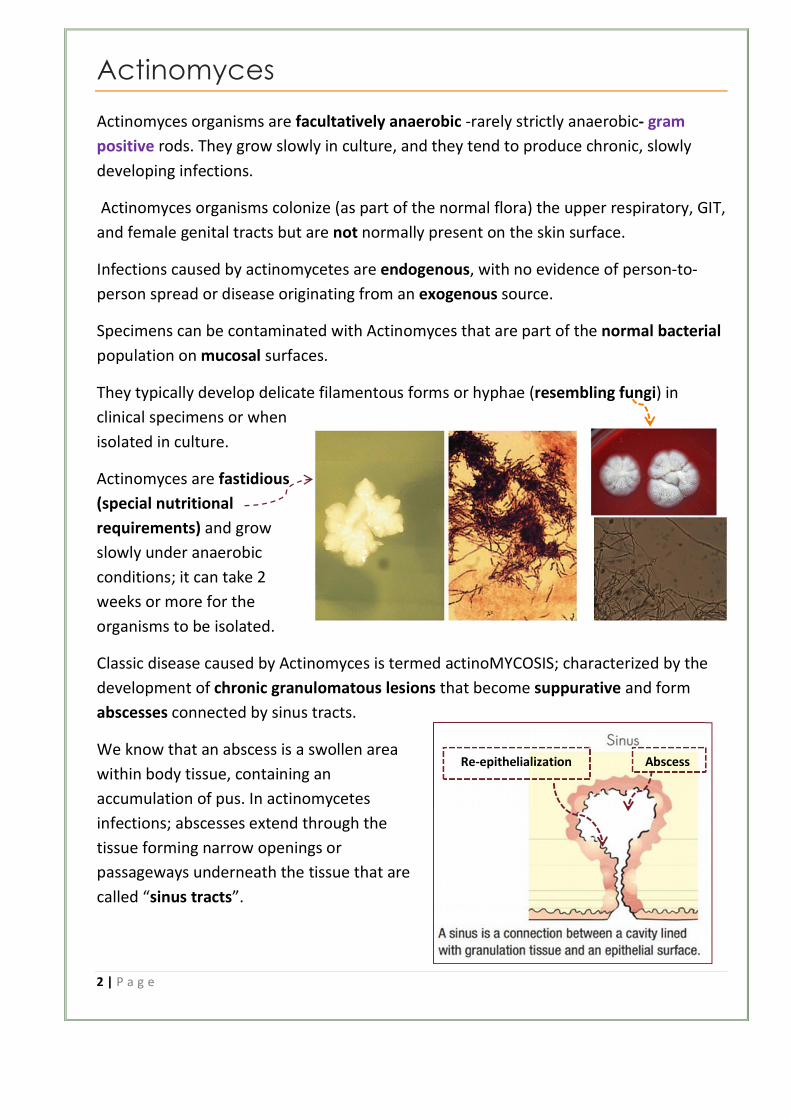
Classic disease caused by Actinomyces is termed actinoMYCOSIS; characterized by the
development of chronic granulomatous lesions that become suppurative and form
abscesses connected by sinus tracts.
We know that an abscess is a swollen area
within body tissue, containing an
accumulation of pus. In actinomycetes
infections; abscesses extend through the
tissue forming narrow openings or
passageways underneath the tissue that are
called “sinus tracts”.
Re-epithelialization Abscess

3 | P a g e
However, if theses passageways (channels) connect two anatomic spaces or cavities;
then they are called “fistulas”.
*To clear up:
A fistula is an abnormal pathway between two
anatomic spaces or a pathway that leads from an
internal cavity or organ to the surface of the body.
A sinus tract is an abnormal channel that originates
or ends in one opening.
Therefore, the finding of tissue swelling with fibrosis and scarring, as well as draining
sinus tracts along the angle of the jaw and neck, should alert the physician to the
possibility of actinoMYCOSIS.
The major sites of actinomycoses are
cervicofacial, abdominopelvic, and thoracic.
Most actinomycetes infections are cervicofacial
(following invasive dental procedure or oral
trauma).
Abdominal and pelvic infections are associated
with abdominal surgery, tuboovarian abscess,
ruptured appendicitis, and intrauterine
contraceptive devices (IUCD).
Treatment for actinomycosis involves the
combination of drainage of a localized abscess or
surgical debridement of the involved tissues
prolonged, and (removal of the necrotic tissue)
administration of antibiotics.
Nocardia (added here for similarity to
Actinomyces)
Nocardiae are strict aerobic rods that form branched
filaments in tissues and culture.
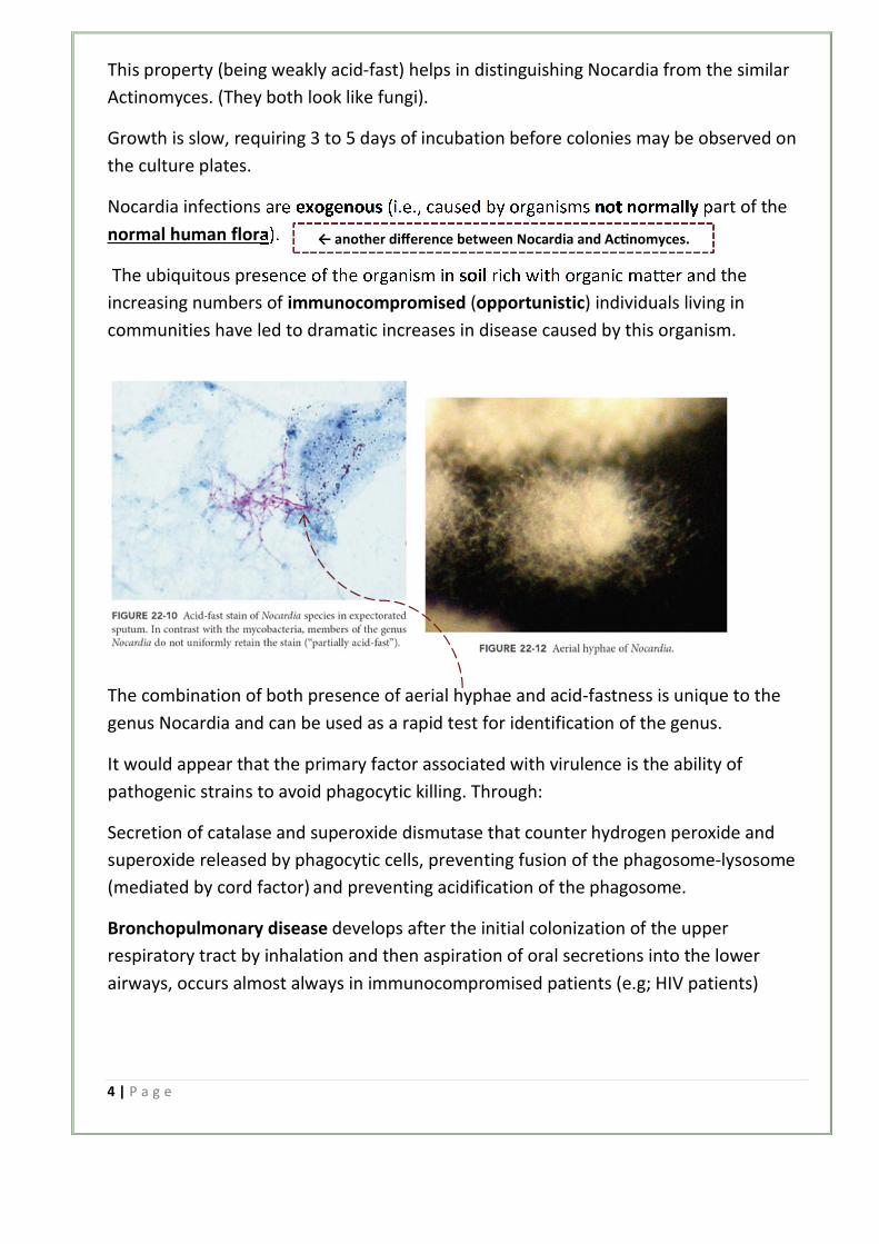
Nocardia is described as “weakly acid-fast”; that is, a weak decolorizing solution of
hydrochloric acid must be used to demonstrate the acid-fast property of nocardia.
Re-epithelializationTrue
epithelium
Looks like a molar tooth
(طاحونة)
4 | P a g e
This property (being weakly acid-fast) helps in distinguishing Nocardia from the similar
Actinomyces. (They both look like fungi).
Growth is slow, requiring 3 to 5 days of incubation before colonies may be observed on
the culture plates.
Nocardia infections are exogenous (i.e., caused by organisms not normally part of the
).normal human flora
The ubiquitous presence of the organism in soil rich with organic matter and the
increasing numbers of immunocompromised (opportunistic) individuals living in
communities have led to dramatic increases in disease caused by this organism.
The combination of both presence of aerial hyphae and acid-fastness is unique to the
genus Nocardia and can be used as a rapid test for identification of the genus.
It would appear that the primary factor associated with virulence is the ability of
pathogenic strains to avoid phagocytic killing. Through:
Secretion of catalase and superoxide dismutase that counter hydrogen peroxide and
superoxide released by phagocytic cells, preventing fusion of the phagosome-lysosome
(mediated by cord factor) and preventing acidification of the phagosome.
Bronchopulmonary disease develops after the initial colonization of the upper
respiratory tract by inhalation and then aspiration of oral secretions into the lower
airways, occurs almost always in immunocompromised patients (e.g; HIV patients)
← another difference between Nocardia and Ac�nomyces.

5 | P a g e
Primary cutaneous nocardiosis develops after traumatic introduction of organisms into
subcutaneous tissues, can present in the form of Mycetoma is characterized by a triad
of painless subcutaneous mass, multiple sinuses and discharge containing grains.
As many as one third of all patients with Nocardia infections have dissemination to the
brain, most commonly involving the formation of single or multiple brain abscesses.
Mycetoma is a chronic suppurative disease of the skin and subcutaneous tissue,
characterized by a symptomatic triad: tumors, fistulas and grains (seeds-like). It can be
caused by fungi (eumycetoma) and bacteria (actinomycetoma), with similar clinical
features.
Given its slow progression, painless nature, massive lack of health education and
scarcity of medical and health facilities in endemic areas, many patients present late
with advanced infection where amputation may be the only available treatment.
Lactobacillus
Lactobacillus species are facultatively anaerobic or
strictly anaerobic rods that ferment to yield lactic
acid.
They are found as part of the normal flora of the
mouth, stomach, intestines, and genitourinary tract.
In around 70% of women, a Lactobacillus species is
dominant in the female genital tract.
Commonly found in probiotics; some brands of
yogurt contain Lactobacillus as beneficial probiotics. They VERY rarely cause
infections.

6 | P a g e
Some Lactobacillus species are used as starter cultures in industry for controlled
fermentation in the production of yogurt, cheese, sauerkraut, pickles, beer, and cider
(to yield lactic acid).
Invasion into blood (sepsis) occurs -rarely- in one of the following three settings:
1. Transient bacteremia from a genitourinary source (e.g., after childbirth or a
gynecologic procedure).
2. Endocarditis.
3. Opportunistic septicemia in an immunocompromised patient.
Propionibacterium
Propionibacteria are small gram-positive rods often arranged in short
chains or clumps, commonly found on the skin (in contrast with the
Actinomyces), conjunctiva, external ear, oropharynx and female
genital tract.
The most commonly isolated species is “Propionibacterium acnes”.
P. acnes species is responsible for two types of infections:
1. Acne vulgaris in teenagers and young adults.
2. Opportunistic infections in patients with prosthetic devices or intravascular lines.
P. acnes species does not cause/originate acne vulgaris; it only triggers the disease
when it meets favorable dermato-physiological terrain; P. acnes colonization of the skin
is therefore necessary but not sufficient for the establishment of the pathology.

7 | P a g e
← Too much
sebum become
trapped inside
sebaceous glands,
causing them to
swell and form
black heads under
the skin which
clog the pores,
creating a
desirable environment for P.acnes to thrive, which can lead to inflammation by
releasing peptides and inflammatory mediators forming pimples or acnes.
Other Non-Spore Forming Anaerobic Gram Positive Rods
Mobiluncus
Members of the genus Mobiluncus are
when ( variable , gramobligate anaerobic
, negativegram or conclusive)staining is not
curved rods with tapered ends.
Despite their appearance in Gram stained
specimens (some appear gram negative and
others are not clear “gram-variable”); they
are classified as gram positive rods because
they have a gram positive cell wall, lack
endotoxin (which are found in gram
negative bacteria) , and are susceptible to
vancomycin, clindamycin, erythromycin, and
ampicillin but resistant to colistin (like some
other gram positive bacteria).
M. curtisii is rarely found in the vaginas of
healthy women but is abundant in women with bacterial vaginosis (shift in the vaginal
microbiota).

8 | P a g e
Bifidobacterium and Eubacterium
They are commonly found in the oropharynx, large intestine, and vagina. They usually
represent clinically insignificant contaminants (as they are parts of the normal flora).
Non-Spore Forming Aerobic Gram Positive Rods
They are a heterogeneous group of bacteria; some are well-recognized human
pathogens (Listeria monocytogenes, Corynebacterium diphtheria), others are primarily
animal pathogens that can cause human disease (Erysipelothrix rhusiopathiae), and
some are opportunistic pathogens that typically infect hospitalized or
immunocompromised patients (Corynebacterium jeikeium).
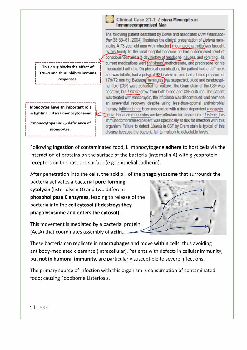
↑The doctor said that Listeria and Corynebacterium diphtheria are considered
opportunistic pathogens.
Listeria monocytogenes
L. monocytogenes is a short (0.4 to 0.5 × 0.5 to 2 μm), non-branching, gram positive,
short rods appear singly, . The -aerobicalthough it’s mostly - rod anaerobic facultatively
in pairs, or in short chains and can be mistaken for
Streptococcus pneumonia →.
The organisms are motile at room temperature but less so
at 37°C, and they exhibit a characteristic end-over-end
tumbling motion when a drop of broth is examined
microscopically.
L.monocytogenes exhibits weak β-hemolysis when grown on sheep blood agar plates.
These differential characteristics (Gram stain morphology, motility, and β-hemolysis)
are useful for the preliminary identification of Listeria.
Although the bacteria are widely distributed in nature, human disease is uncommon
and is restricted primarily to several well-defined populations: neonates, the elderly,
(Listeria is an and patients with defective cellular immunity pregnant women,
.pathogen) facultative intracellular opportunistic
9 | P a g e
Following ingestion of contaminated food, L. monocytogene adhere to host cells via the
interaction of proteins on the surface of the bacteria (internalin A) with glycoprotein
receptors on the host cell surface (e.g. epithelial cadherin).
After penetration into the cells, the acid pH of the phagolysosome that surrounds the
bacteria activates a bacterial pore-forming
cytolysin (listeriolysin O) and two different
phospholipase C enzymes, leading to release of the
bacteria into the cell cytosol (it destroys they
phagolysosome and enters the cytosol).
This movement is mediated by a bacterial protein,
(ActA) that coordinates assembly of actin.
These bacteria can replicate in macrophages and move within cells, thus avoiding
antibody-mediated clearance (intracellular). Patients with defects in cellular immunity,
but not in humoral immunity, are particularly susceptible to severe infections.
The primary source of infection with this organism is consumption of contaminated
food; causing Foodborne Listeriosis.
This drug blocks the effect of
TNF-α and thus inhibits immune
responses.
Monocytes have an important role
in fighting Listeria monocytogenes.
*monocytopenia: ↓ deficiency of
monocytes.

10 | P a g e
*People who are at higher risk for Listeria infection (elderly and immunocompromised
people) should avoid eating cold cuts (e.g. salami, turkey) to prevent FBL.
Human-to-human transmission can occur primarily from mother to child in utero or at
birth (neonatal → weak immunity).
Neonatal Disease can be an early-onset disease; acquired transplacentally (through the
placenta) in utero, or late-onset disease; acquired at or soon after birth.
Early-onset disease can result in abortion, stillbirth, or premature birth. Late-onset
disease occurs 2 to 3 weeks after birth in the form of meningitis or
meningoencephalitis with septicemia.
Most infections in pregnant women occur during the third trimester when cellular
immunity is most impaired.
Disease in healthy adults is self-limited and asymptomatic or in the form of a mild
influenza-like illness.
diphtheria Corynebacterium
Corynebacteria are aerobic or facultatively anaerobic, non-motile, and catalase positive.
They are ubiquitous in plants and animals, and they normally colonize the skin, upper
respiratory tract, gastrointestinal tract,
and urogenital tract in humans.
The most famous of these is C.
diphtheria -the etiologic agent of
diphtheria-.
C. diphtheriae is an irregularly staining,
pleomorphic rod -sometimes it looks
like rods and other times like cocci- (0.3
to 0.8 × 1.0 to 8.0 μm).
Humans are the only known reservoir
for this organism.
Respiratory droplets or skin contact transmits it from person to person.

11 | P a g e
Diphtheria toxin is the major virulence factor of C. diphtheria (it is an example of the
classic A-B exotoxin).
Three functional regions exist on the toxin molecule:
1. A catalytic region on the A
subunit.
2. A receptor-binding region.
3. A translocation region on the B
subunit.
The receptor for the toxin is heparin-
binding epidermal growth factor
precursor (HB-EGF) present on many
epithelial membranes and is
endocytosed by the cell.
After the toxin get into the cell, B
subunit (translocation region) become inserted into the endosomal membrane,
facilitating the movement of the A subunit (catalytic region) into the cytosol. The A
subunit then ADP-ribosylates (inactivates) elongation factor-2 (EF-2).
EF-2 is required for protein synthesis; when it is inactivated by the toxin, the host
cannot make protein and thus dies.
The clinical presentation of diphtheria is determined by:
1. Site of infection.
2. Immune status of the individual (opportunistic).
3. Virulence of the organism.
* Exposure to C. diphtheria may result in asymptomatic colonization in fully immune
people.
*Diphtheria toxin is produced at the site of the infection and then disseminates through
the blood to produce the systemic signs of diphtheria.

12 | P a g e
Cutaneous Diphtheria
A papule develops first and then evolves into a chronic, non-healing ulcer.
Respiratory Diphtheria
The symptoms of diphtheria
involving the respiratory tract
develop after a 2- to 4-day
incubation period.
The onset is sudden, with
malaise, sore throat, exudative
pharyngitis, and a low-grade
fever.
The exudate evolves into a
thick pseudomembrane
composed of bacteria,
lymphocytes, plasma cells,
fibrin, and dead cells that can
cover the tonsils, uvula, and
palate and can extend up into the nasopharynx or down into the larynx (difficult to
breathe).
Evidence of myocarditis can be detected in the majority of patients (systemic
complications involving the heart).
Diphtheria has become uncommon in the United
States because of an active immunization program,
as shown by the fact that more than 200,000 cases
were reported in 1921 but no cases have been
reported since 2003.
Read this clinical case and notice the symptoms, age
of the patient, and being unvaccinated.→

13 | P a g e
Anaerobic Gram-Negative Rods
These anaerobes are the predominant bacteria on most mucosal surfaces,
outnumbering aerobic bacteria 10 to 1000 fold.
Despite the abundance and diversity of these bacteria, most infections are caused by
relatively few species.
Bacteroides, Fusobacterium, Parabacteroides, Porphyromonas, and Prevotella.
Characteristically, Bacteroides growth is
stimulated by bile.
Although Bacteroides species grow rapidly
in culture, the other anaerobic gram
negative rods are fastidious; and cultures
may have to be incubated for 3 days or
longer before the bacteria can be detected.
Bacteroides species are pleomorphic in size
and shape and resemble a mixed population
of organisms in a casually examined Gram
stain.
Bacteroides have a typical gram negative cell
wall structure, which can be surrounded by a
polysaccharide capsule.
In contrast to the LPS molecules in the aerobic gram negative rods, the Bacteroides LPS
has little or no endotoxin activity; this is because the lipid [A] in Bacteroides lacks
certain structural components that are present in the LPS molecules in the anaerobic
gram negative rods “structural differences”.

14 | P a g e
To cause disease,
Bacteroides fragilis in the
resident flora are able to
spread by trauma or disease
from the normally colonized
mucosal surfaces to sterile
tissues or fluids.
Respiratory Tract Infections
Nearly half of the chronic infections of the sinuses (a cavity within the bones of the
skull) and ears, and virtually all periodontal (affecting the structures surrounding and
supporting the teeth) infections, involve mixtures of gram negative anaerobes, with
Prevotella, Porphyromonas, Fusobacterium, and non-fragilis Bacteroides the most
commonly isolated.
Bacteremia
Anaerobes were at one time responsible for more than 20% of all clinically significant
cases of bacteremia; however, these organisms now cause 3% to 10% of such infections.
_______________________________________________________________________
Intra-abdominal Infections
Anaerobes are recovered in virtually all of these infections, with B. fragilis the most
common organism.
________________________________________________________________
Skin and Soft-Tissue Infections
B. fragilis is the organism most commonly associated with significant disease.
Gastroenteritis
diarrhea by produce limited watery-selfStrains of enterotoxigenic B. fragilis that cause
a heat-labile zinc metalloproteas toxin (B. fragilis toxin).
Infections are usually
polymicrobial.

15 | P a g e
This toxin causes morphologic changes of the intestinal epithelium via F-actin
rearrangement, with the resultant stimulation of chloride secretion and fluid loss.
Anaerobic Gram Positive Cocci
The anaerobic gram positive cocci
normally colonize the oral cavity,
gastrointestinal (GI) tract,
genitourinary tract, and skin. They
produce infections when they spread
from these sites to normally sterile
sites.
Although anaerobic cocci can be
isolated from infections at all body
sites, a predisposition for certain sites
has been observed.
Peptostreptococcus species have been
recovered more often from
subcutaneous and soft tissue abscesses
and diabetes-related foot ulcers than
from intra-abdominal infections.
*Peptostreptococcus infections occur
more often in chronic infections.
*Many infections caused by
peptostreptococcus bacteria are
synergistic.
The End.