ha thanh dong1,2 emerging, re-emerging and new diseases of
TRANSCRIPT

1
Session 2
Ha Thanh Dong1,2
Emerging, re-emerging and new
diseases of tilapia
1King Mongkut’s University of Technology Thonburi, 2Fish Health
Platform, Centex Shrimp (Mahidol University/BIOTEC), Thailand.

Objective
• To update on emerging, re-emerging and
new diseases of tilapia
❖Emerging viral infections
❖Emerging bacterial infections
❖Emerging parasitic infection
❖Emerging unknown pathogen
2

Emerging Viral Infections
3

Viral Infections in Tilapia
4
Agent DNA/ RNA Geographical
Distribution
Ref.
Lymphocystis disease virus
(LCDV)
DNA North Tanzania Paperna, 1973
Infectious pancreatic
necrosis virus (IPNV)
RNA Taiwan Hedrick et al. 1983
Bohle virus DNA Australia Ariel and Owens, 1997
Iridovirus-like Canada McGrogan et al.1998
Viral nervous necrosis (VNN) RNA France,
Indonesia and
Thailand
Bigarre´ et al. 2009;
Prihartini et al. 2015; Keawcharoen et al. 2015
Infectious spleen and kidney necrosis virus (ISKNV)
DNA US Midwest,
Thailand
Subramaniam et al. 2015;
Suebsing et al. 2016
Tilapia larvae encephalitis
virus (TLEV)
DNA Israel Shlapobersky et al. 2010
Tilapia lake virus (TiLV) RNA Asia, Africa, and
South America
e.g. Eyngor et al. 2014;
Jansen et al. 2018

Emerging, re-emerging, new viral infections of tilapia
5
Lymphocystis disease
virus (LCDV)
Lymphocystivirus
Infectious pancreatic
necrosis virus (IPNV)
Aquabirnavirus
Bohle virus Ranavirus
Iridovirus-like Iridoviridae
Viral nervous necrosis
(VNN)
Betanodavirus
Infectious spleen and kidney necrosis virus
(ISKNV)
Megalocytivirus
Tilapia larvae encephalitis
virus (TLEV)
Herpesvirus
Tilapia lake virus (TiLV) Tilapinevirus
Case
reports
with little concern
New/ newly emerging
Re-emerging
Emerging
Global concern

IPNV re-emerged in tilapia
• Subclinical infection cases
• Its impact remains unknown
• Investigation should be initiated in tilapia farming countries
6
1983: Subclinical infection of
IPNV in tilapia in Taiwan was
reported
1987: Experimental challenge
indicated that IPNV is pathogenic
to tilapia (killed 25% fish)
……………
……………
2018: IPNV re-emerged in tilapia

Viral Nervous Necrosis (VNN) disease
❖ Causative agent: Betanodavirus
❖ Clinical signs: signs of neurological disorders: loss of balance,
erratic swimming
❖ Host: >30 species, mainly in marine fish
❖ Geographical distribution: worldwide
❖ Cases in tilapia (France, Thailand & Indonesia)
e.g. a case in tilapia hatchery
• 10 days-old larvae of tilapia
• Mortality 90-100%
• Histopathological manifestation of VNN disease
• 93.07–93.88% similarity to red-spotted grouper nervous
necrosis virus (RGNNV)
7
Keawcharoen et al. JFD 2015, 38, 49-54

Viral Nervous Necrosis (VNN) disease
8
❖ Histopathological feature: Vacuolation was observed in brain, eye and spinal cord of diseased fish
❖ Detection methods: PCR methods (OIE disease card)
Keawcharoen et al. JFD 2015, 38, 49-54

Infectious spleen and kidney necrosis disease (ISKND)
Subramaniam et al. (2016)
9
❖Synonym: Iridoviral disease
(common name), red sea bream
iridoviral disease (OIE)
❖Causative agent: Megalocytivirus
ISKNV
❖Clinical signs: darkening, pale gills
❖Host: wide range of both marine
and freshwater fish, including tilapia

Infectious spleen and kidney necrosis disease (ISKND)
10
A case in USA (Subramaniam et al. 2016)
• Tilapia fry/fingerlings
• Mortality 50-75%
In Thailand• Multiple infections of
ISKNV/Iridovirus was reported in
cage culture & a semi-nested PCR
was developed (Dong et al. 2016)
• Recent reports: vertical transmission
& LAMP detection method (Suebsing et
al. 2016)Presence of basophilic
hypertrophied cells (Subramaniam et al. 2016)

Tilapia larvae encephalitis virus (TLEV) disease
❖ Causative agent: TLEV/Herpes-like virus
❖ Clinical signs: spiral swimming
❖ Host: blue tilapia (O. aureus), red tilapia
(Oreochromis sp.), Nile tilapia (O. niloticus)
❖ Mortality: reach up to 98%
❖ Susceptible stages: 32-34 days post
fertilization
❖ Geographical distribution: Israel
❖ Histopathological feature: Not available
❖ PCR detection: available
TLEV-1(5′ TCGTGGGCCTTATCCCGCGT 3’)
TLEV-2 (5′ GAGACCAGAAAGTGCTTCTC 3′)
11Shlapobersky et al. Virology (2010) 399: 239-247
Lack of investigation in other countries

Tilapia lake virus disease (TiLVD)
12
Will be presented by other speakers

Emerging Bacterial Infections
13

Bacterial Diseases in Tilapia
❖ Streptococcosis – Emergence of S. agalactiae serotype IX
❖ Columnaris – Complexity of F. columnare
❖ Francisellosis – Emerging/re-emerging in some countries
❖ Hemorrhagic septicemiao A. hydrophila
o Non-A. hydrophila (A. veronii & A. jandaei) (Dong et al. JFD 2017)
❖ Edwardsiellosis caused by E. ictaluri
❖ Aerococcus viridans infection (Ke et al. Aquaculture 2012)
❖ Hahellosis/red egg disease (Senapin et al. Aquaculture 2016)
❖ Unknown diseases
14
New to tilapia
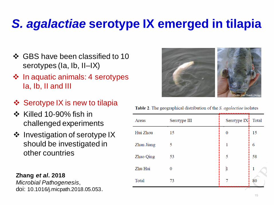
S. agalactiae serotype IX emerged in tilapia
15
Zhang et al. 2018
Microbial Pathogenesis, doi: 10.1016/j.micpath.2018.05.053.
❖ GBS have been classified to 10
serotypes (Ia, Ib, II–IX)
❖ In aquatic animals: 4 serotypes
Ia, Ib, II and III
❖ Serotype IX is new to tilapia
❖ Killed 10-90% fish in
challenged experiments
❖ Investigation of serotype IX
should be investigated in
other countries

16
Complexity of F. columnare in tilapia
Dong et al. J Fish Dis (2015) 38:901-913
Kayansamruaj et al. Infection, Genetics and Evolution 54 (2017) 7–17
❖ F. columnare is causative agent of
columnaris disease
❖ F. columnare in tilapia is a complex of
several unclassified taxa
dDDH supports taxonomic
reclassification of Fc originated from tilapia

Same same…but different…
17
head kidney
spleen
head kidney
spleen
What disease you think about?

Francisellosis of tilapia
Causative agent:
• Francisella noatunensis subsp. orientalis
• Previously known as Rickettsia-like organism, RLO
• Fastidious intracellular bacterium
Host range:
• Susceptible tilapia, ornamental cichlids
• Infection but does not kill the hosts: striped catfish, common carp
Cumulative mortality: 40-50%
Clinical signs: visceral white spots (eg spleen & head kidney)
Season: Cool weather (25-28 oC)
18

Francisellosis of tilapia
19
F. noatunensis subsp. orientalisF. noatunensis subsp. noatunensis
Adapted from Nguyen et al. 2015 Aquac Res. doi:10.1111/are.12802
Geographical distribution
China 2015
Mexico 2016

Francisellosis of tilapia
Presumptive Diagnosis
20
Wet mount examinationClinical sign
Photographs were taken in conjunction with the outbreaks described in Nguyen et al. 2015. Aquac Res & Dong et al. 2016. Dis Aquat Org.
diseased fish normal fish

Francisellosis of tilapia
Diagnosis
21
Rapid staining of smeared-head kidney with
Giemsa revealed presence of both intra- and
extra-cellular bacteria
Micrographs of
H&E stained
sections of the
spleen showed
typical granulomas
Photographs were taken in conjunction with the outbreaks described in Nguyen et al. 2015. Aquac Res & Dong et al. 2016. Dis Aquat Org.

Francisellosis of tilapia
• Genus specific PCR (Forsman et al. 1994)
• Real-time PCR (Duodu et al. 2012);
• ISH, genus-specific (Hsieh et al. 2007)
• Immunohistochemistry (Soto et al. 2012)
• Duplex PCR and ISH (Dong et al. 2016)
• Colorimetric LAMP (Pradeep et al. 2016)
• Recombinase polymerase amplification (RPA) (Shahin et al. 2018)
22
Molecular Diagnosis
Duplex PCR
Genus specific
Fno species-specific

Which one infected with F. noatunensis subsp. orientalis ?
23Photograph was taken in conjunction with the outbreaks described in Nguyen et al. 2015. Aquac Res

Edwardsiellosis of tilapia
Causative agent:
• Edwardsiella ictaluri
• Common in catfish but not common in non-catfish
• Does not kill tilapia in striped catfish ponds (personal
observation)
• 2012: first report of E. ictaluri in Nile tilapia in Western
Hemisphere (Soto et al. 2012)
• No reported in other countries
24

Edwardsiellosis of tilapia
Recent cases in Southeast Asia
• Red tilapia juveniles
• Killed 40-50% fish in the first month after stocking
• Presence of white spots in multiple internal organs
25
head kidney
spleen
• Presumptive diagnosis
based on clinical sign:
Francisellosis
• PCR negative for Fno
MS submitted

Edwardsiellosis of tilapia
26
Presumptive diagnosis
• Tissue smear, Gram
staining (take 5 min)
• Numerous Gram negative,
rod-shaped bacteria
• Suspected bacterial
infection
Gram staining of tissue smear
Intracellular bacteria

Edwardsiellosis of tilapia
• Bacterial isolation: pure pinpoint colonies on TSA
• Gram negative, rod-shaped bacteria
27
Edwardsiella ictaluri PH-0744 Ayu/Japan (AB453281)
Edwardsiella ictaluri 93-146 Channel catfish/USA (CP001600)
Edwardsiella ictaluri ATCC 33202 Channel catfish/USA (NR024769)
Edwardsiella ictaluri 2234 Red tilapia/Vietnam
Edwardsiella ictaluri 2254 Red tilapia/Vietnam
Edwardsiella ictaluri 2248 Red tilapia/Vietnam
Edwardsiella ictaluri UK1 Nile tilapia/Western Hemisphere (KM676418)
Edwardsiella ictaluri 2252 Red tilapia/Vietnam
Edwardsiella ictaluri T1-1 Striped catfish/Thailand (KR080248)
Edwardsiella anguillarum ET080813 (NR136429)
Edwardsiella hoshinae JCM1679 (NR024768)
Edwardsiella tarda ATCC 15947 (NR024770)
Serratia marcescens (NR036886)
67
58
64
71
0.005
Edwardsiella ictaluri
MS submittedPhylogenetic tree based on 16S rRNA

Edwardsiellosis of tilapia
Challenged experiments fulfilled Koch’s postulates– Fish reproduce the same clinical signs
– 95-100% mortality in 3-9 days (dose-dependent)
28MS submitted
Histopathological features of edwardsiellosis in
the experimental fish
Experimental fish
Spleen

Edwardsiellosis of tilapia
❖ E. ictaluri is an emerging pathogen of tilapia
aquaculture in Southeast Asia
❖ E. ictaluri infections in tilapia may have been
overlooked due to similar clinical signs between
Francisellosis & Edwardsiellosis
❖ Should be put on disease watchlist
29

A. veronii & A. jandaei infection
• are newly reported pathogens of tilapia
• may have been misidentified as A. hydrophila or previously overlooked
• both cause “hemorrhagic septicemia”
• Coinfections with other pathogens are very common
30
Dong et al. 2015 Aquaculture 448:427-435
Peepim et al. 2016 Aquaculture 464:399-409 Dong et al. 2017 J Fish Dis 40:1395–1403
Blood congestion Intestinal necrosis

Aerococcus viridans infection
• This work firstly reports the infection and histopathological changes of A.
viridans in tilapia
• Associated with 30-40% loss in Guangdong Province, China, 2010
• The major symptoms: serious congestion of the gill and the abdomen,
swelling gallbladder and a severe diffusion in liver. Some fish show
exophthalmia and spiral swimming.
• Experimental infection caused 45-85% mortality, fulfilled Koch’s postulates31

32
Hahellosis/Red egg disease
❖Occurred in a tilapia hatchery in Thailand since 2010
❖Mortality 10-50%
❖Occur during cold season (<24 0C)
❖ Causative agent: unknown
Senapin et al. Aquaculture (2016) 454:1-7
Normal eggs Red eggs Bacteria in the egg
C

33
Hahella chejuensis is a
marine bacteria
…occurred in tilapia hatcheries?
Bacterial isolation using TSA
Red pigmented bacteria was identified using 16S rRNA
Senapin et al. Aquaculture (2016) 454:1-7
Hahellosis/Red egg disease

34Senapin et al. Aquaculture (2016) 454:1-7
Specific PCR detection methods
were developed targeting 16S rRNA
H. chejuensis caused red egg
disease & reduced hatching rate in experimental challenge
Hahellosis/Red egg disease

35
Hahellosis/Red egg disease
Red egg
Ovary
Testis
Hahella-specific probe Unrelated probe
➢ H. chejuensis was found in red
eggs and brooders (ovary & testis)
➢ Possible of vertical transmission
Senapin et al. Aquaculture (2016) 454:1-7

36
➢ Reduction of loss: ~ $ 600,000 /year
➢ Calculation based on 30% mortality (range from 10-50%)

Emerging parasitic infection
37
• The first outbreak of Trypanosoma in Nile tilapia (~460 g) in
South America
• Unspecific signs such as anorexia, skin darkening and gill paleness

Trypanosomiasis
38de Jesus et al. 2018 Aquaculture 491: 169-176
❖ Trypanosoma sp. (combined
morphology & molecular analysis)
❖ 18S rDNA showed 95-98% identity to Trypanosoma sp.

Emerging unknown pathogen
50% fish (n=10) in a TiLV-positive cage showed a novel histopathological change (microsporidian-like?)
39
spore-like
normal muscle
infected areas

Comments
❖Emerging diseases are never ending threats in aquaculture industry
❖Preparedness for rapid response to emerging
diseases should be encouraged
❖Rapid pathogen discovery and early diagnosis will
limit its spread and reduce negative impact
❖SPF and autogenous inactivated vaccine programs
should be promoted for long-term development
40

41
Acknowledgments
41
Colleagues/collaborators