h circulatory system m
TRANSCRIPT
Summer 2005 H.1
Unit H: Circulatory System
Program Area: Health Occupations Education Course Title: Medical Sciences I Number: 7221 Unit Title: Circulatory System Suggested Time for Instruction: 6 class periods (90 minute classes) 12 class periods (55 minute classes) Course Percent: 7% Unit Evaluation: 100% Cognitive ------------------------------------------------------------------------------- Competency: MD08. Analyze the anatomy and physiology of the circulatory system. Specific Objectives: MD08.01 Describe the structure of the heart.
MD08.02 Analyze the function of the heart. MD08.03 Analyze circulation and the blood vessels.
MD08.04 Analyze characteristics and treatment of common cardiac and
circulatory disorders.

Summer 2005 H.2
Unit H Master Outline
H Circulatory System MD08.01 Explain the structure of the heart.
A. Size, shape and location 1. Size of closed fist 2. In thoracic cavity 3. Apex 4. Four chambers
B. Layers 1. Pericardium 2. Myocardium 3. Endocardium 4. Septum
C. Structures to and from heart 1. Superior and inferior vena cava 2. Pulmonary artery and vein 3. Aorta
D. Chambers and valves 1. Atria (atrium) 2. Ventricles (ventricle) 3. Tricuspid valve 4. Mitral (bicuspid) valve 5. Pulmonary semilunar valve 6. Aortic semilunar valve
MD08.02 Analyze the function of the heart
A. Four main functions of circulatory system a. Pump b. Blood transport system around body c. Carries oxygen and nutrients to cells, carries away waster
products d. Lymph system – returns excess tissue fluid to general circulation
B. Heart a. Ave. 72 beats per minute, 100,000 beats per day b. Superior and inferior vena cava bring deoxygenated blood to
right atrium c. Pulmonary artery takes blood from right ventricle to lungs d. Pulmonary veins bring oxygenated blood from lungs to left
atrium e. Aorta takes blood from left ventricle to rest of body f. Four heart valves permit flow of blood in one direction
C. Pump a. Heart is a double pump b. Right heart = right atrium tricuspid valve right ventricle
pulmonary semilunar valve pulmonary artery lungs (for oxygen)
c. Left heart = Lungs pulmonary veins left atrium mitral valve left ventricle aortic semilunar valve aorta general circulation
Summer 2005 H.3
D. Heart sounds (lubb dupp) E. Electrical activity
1. SA (sinoatrial) node = pacemaker, sends out electrical impulses, spreads impulse over atria and makes them contract 2. AV (atrioventricular) node = carries impulse to bundle of His 3. Bundle of His = conducting fibers in septum, divides into right
and left branches in ventricles to Purkinje fibers 4. Purkinje fibers = cause ventricles to contract
MD08.03 Analyze circulation and the blood vessels
A. Cardiopulmonary circulation – carries blood from heart to lungs 1. Oxygenated and deoxygenated blood 2. Oxygen/carbon dioxide exchange
B. General circulation 1. Coronary arteries 2. Aorta 3. Systemic circulation
C. Blood vessels 1. Arteries
a. Carry oxygenated blood away from the heart to the capillaries
b. Elastic, muscular and thick-walled c. Transport blood under very high pressure
2. Arterioles 3. Veins
a. Carry deoxygenated blood away from capillaries to heart b. Less elastic and muscular than arteries c. Thin walled, collapse easily when not filled with blood d. Superior and inferior vena cava carry blood to heart
4. Venules 5. Capillaries
a. Smallest blood vessels b. Only seen with microscope c. Connect arterioles and venules
e. Walls are one-cell thick, allow for selective permeability 6. Valves – permit flow of blood only in direction of heart 7. Capillaries 8. Jugular vein – located in neck 9. Carotid artery – carries blood to brain
D. Blood pressure 1. Systolic – ave = 120 (Systole is contraction phase) 2. Diastolic – ave = 80 (Diastole is relaxation phase)
E. Pulse – alternating expansion and contraction of an artery as blood flows through it 1. Brachial 2. Carotid 3. Femoral 4. Pedal 5. Popliteal 6. Radial

Summer 2005 H.4
MD08.04 Discuss characteristics and treatment of common cardiac and circulatory disorders. A. Heart diseases
1. Symptoms a. Arrythmia (dysrrhythmia) – any change from normal heart
rate or rhythm b. Bradycardia – slow heart rate (<60) c. Tachycardia – rapid heart rate (>100)
2. Coronary artery disease a. Angina pectoris – chest pain, lack of O2 to heart muscle,
treat with nitroglycerine b. Edema – fluid in tissues, often caused by poor circulation
3. Myocardial infarction (MI, heart attack) a. Lack of blood supply to myocardium b. Symps – severe chest pain radiating to left shoulder, arm,
neck and jaw, nausea, diaphoresis, dyspnea c. Rx – bedrest, oxygen, medication d. Morphine for pain e. Anticoagulant therapy to prevent further clots from forming f. Surgery may be necessary
4. Heart disease a. Congestive heart failure – ventricles unable to contract
effectively and blood pools in the heart, edema in lower extremities, blood backs up in lungs
b. Endocarditis – inflammation of membrane that lines heart and covers valves
c. Myocarditis – inflammation of heart muscle d. Pericarditis – inflammation of outer membrane of heart,
symptoms are chest pain, cough, dyspnea, tachycardia, fever
d. Mitral valve prolapse – valve closes imperfectly, symptoms in response to stress include fatigue, palpitations (racing heart) headache, chest pain, anxiety
e. Murmur – defect in heart valve, causing gurgling or hissing sound
B. Vascular diseases 1. Aneurysm – ballooning of an artery, thinning and weakening 2. Arteriosclerosis – arterial walls thicken and lose elasticity 3. Atherosclerosis – fatty deposits form on walls of arteries and
block circulation 4. Hypertension
a. High blood pressure b. Silent killer – usually no symptoms c. Leads to strokes, heart attacks, kidney failure d. Higher in African-Americans and post-menopausal women e. Risk factors – smoking, overweight, stress, high fat diets,
family history f. Treatment – relaxation, low fat diet, exercise, weight loss,
medication 5. Hypotension – low blood pressure, systolic <100 6. Embolism – traveling blood clot
Summer 2005 H.5
7. Varicose veins
a. Swollen, distended veins b. Heredity or due to poor posture, prolonged periods of
standing, physical exertion, age and pregnancy c. Hemorrhoid – varicose rectal vein
8. Phlebitis – inflammation of lining of vein, accompanied by clotting of blood, symptoms are edema, pain and redness
C. Diagnosis and treatment 1. Electrocardiogram – electrical tracing of the heart 2. Coronary bypass – healthy vein from leg removed and attached
before and after the coronary obstruction, creating an alternate route for blood supply to the myocardium
3. AED – automated external defibrillator 4. Defibrillation – electrical shock to bring the heart back to a
normal rhythm 5. CPR – cardiopulmonary resuscitation, used in presence of
cardiac arrest 6. Artificial pacemaker – when heart has conduction (electrical
impulse) defect, demand pacemaker fires when heart rate drops below minimum, causes heart to contract
7. Angiogram – x-ray of blood vessel using dye 8. Angioplasty – balloon used to open clogged vessels 9. Cardiac catheterization – catheter fed into heart, dye injected, x-
rays taken as dye moves through coronary arteries 10. Heart transplant
a. To replace irreparably damaged heart b. Problem – histocompatibility c. Rx – immunosuppressants d. Artificial hearts –experimental
11. Stent – tiny, expandable stainless steel tube that holds coronary artery open to improve blood flow
Summer 2005 H.6
Unit H: Circulatory System Competency MD08: Analyze the anatomy and physiology of the circulatory
system. Materials/Resources Scott, Ann Senisi and Elizabeth Fong. Body Structures & Functions. Delmar Publishers, Latest Edition. www.DelmarAlliedhealth.com National HOSA Handbook: Section B. Published by HOSA, Flower Mound, Texas. Current Edition. www.hosa.org Teaching/Learning Indicators: The following letters are used to indicate specific skills/areas required in the instructional activity. R Reading SS Social Studies
W Writing S Science M Math A The Arts H Health professional/parent/community involvement

Summer 2005 H.7
Objective MD08.01 Describe the structure of the heart.
Teaching/Learning Activities
• Cognitive S Have students label the diagram of the heart located in the appendix (Appendix MD08.01 B). After learning about the circulation, students can then color the different structures of the heart red or blue depending on the oxygenated or deoxygenated blood.
• Teamwork S, A
Have students work in teams to produce a 3-D model of the heart following the instructions in “Make a Heart 101” (Appendix MD08.01C). Each team member must show proof of participation. The teams will present the models to the class. Individual teams will decide on the method to produce the models. Before beginning the assignment, the teacher should obtain the following materials to be used with the activity: Latex gloves Paper cups (4 per group) Note cards Masking tape Markers
• Critical Thinking S, A
Have students draw a heart on a plain sheet of paper. Do not give any instructions other than to draw a heart. After the hearts are drawn, have students list all the words inside the heart that they can think of having to do with the circulatory system. The students must be able to explain the words to the class. It is good to require at least 5 words, but the number can vary.
• Special Needs Each student will reach the highest level of mastery in the least restrictive environment as recommended in the student’s IEP.

Summer 2005 H.8
Objective MD08.02 Analyze the function of the heart. Teaching/Learning Activities
• Critical Thinking S, M Have students complete the worksheet “Heart & Blood Math,” (Appendix MD08.2A). To be successful in this activity, the students and teacher will need to discuss why we have a pulse, and the teacher should teach students how to take a radial pulse.
• Cognitive S As a follow up to the previous activity, discuss the transparency “Blood and the Heart Fun Facts” (Appendix MD08.2B).
• Employability Skills S, H, W Invite a cardiovascular technologist to demonstrate and talk about the electrical activity of the heart, or visit the cardiovascular department at the local hospital and observe the cardiovascular technologist. After the presentation have students to write a summary of their conclusions about the function of the heart, based on their experiences with this activity. Note: Ask the speaker if he/she has an echocardiogram to share with the class. This shows the pumping mechanism of the heart and the structures.
• Basic Skills S, W Have students write a paper explaining the electrical activity of the heart. The paper should be imaginative while factual. For example, SA node could be Shawn Adams who gets a real charge out of life. The AV node could be Agnes Vernox who always gets a charge out of other people. Assign points for creativity and realism.
• Teamwork S, W Have students divide into groups of 3-5 depending on the class size. Complete the “New
Beginnings,” activity. (Appendix MD08.02C)
• Special Needs Each student will reach the highest level of mastery in the least restrictive environment as recommended in the student’s IEP.

Summer 2005 H.9
Objective MD08.03 Analyze the circulation and the blood vessels. Teaching/Learning Activities
• Cognitive S Have students participate in a class discussion about blood vessels and circulation. (Transparency Appendix MD08.03A)
• Teamwork S
Have students draw note cards with the pulse sites and their locations listed. Put one site on half the note cards and the location on the other half. Example:
Brachial Bend of the arm
Pin note cards on each student’s back and have him/her search for his/her mate. The students may ask only 3 questions of each person they meet. When they find their partner, the students should stay together until the activity is over.
• Critical Thinking S Show students how to feel pulses at various pulse sites and explain to the students what pulses are. Have students listen to the heartbeat with a stethoscope and explain the physiology behind the lubb dupp sound.
• Technology S, A Have students create a poster of the physiology of the blood vessels using the computer. Students may want to try the web site: www.innerbody.com/indexbody.htm/ as an anatomy and physiology resource.
• Teamwork S, A Have students become the different structures of the heart by making a sign to wear with the name of their structure on it. Assign one student per structure and any remaining students may be the blood. Assign each drop of blood a different direction to come from: tip of nose, bottom of ear lobe, etc. The first time a drop of blood goes through the heart, each structure holds up his/her sign to guide the way. The second time the blood goes through, no signs are shown, but the blood may ask the structures who they are. The third time the blood goes through, no questions are allowed. If the blood gets stuck, this could be a good learning example of a clot, etc.
• Teamwork S
Have students participate in the circulation race. (Appendix MD08.03B)
• Special Needs Each student will reach the highest level of mastery in the least restrictive environment as recommended in the student’s IEP.

Summer 2005 H.10
Objective MD08.04 Analyze characteristics and treatment of common cardiac and circulatory disorders.
Teaching/Learning Activities
• Employability S, H Invite a cardiologist to class to explain current methods used for treating heart attack patients, such as stints, cardiac bypass, angioplasty, transplants, etc.
• HOSA S, W
Using the Researched Persuasive Speaking guidelines, have students research a disorder assigned by the teacher, and then write a paper on the assigned topic. A main focus of the paper and speech will be to persuade people to change behavior related to the prevention or treatment of the disease assigned. Students will present their persuasive speech in class while classmates take notes. When two or more students are assigned the same topic, those speeches should be given, one after the other, to allow students to focus on one disorder at a time.
• HOSA S Using the guidelines for “Biomedical Debate” debate the topic “Heart Transplants – No Restrictions.”
• Cognitive S
This exercise will be useful as a review for the entire unit on the circulatory system. Have students play the game, “Speaking Of.” The game begins by the student making a statement concerning the heart. The statement must be true and must begin with “Speaking of.” For example: Speaking of the heart, it pumps 80 mL of blood with each heartbeat. A student may jump in when he/she can make a statement using any word in the previous sentence. For example: Speaking of each heartbeat, if you count it for 1 minute that will give you your pulse rate. The next student may pick up on pulse, heart, beat, count, rate, etc. Use for 5-10 minutes or until you are satisfied students understand.
NOTE: Make sure students understand how to play. Each student may be responsible for making at least one statement. If on teams, one team member may answer for the whole team if there is joint input.
• Teamwork S
This activity is best if it is done after the Researched Persuasive Speeches have been given. Have one team be a “host” panel. Each person on the team is allowed to ask the “guest” team two questions. At the end of the questions, the host panel has 30 seconds to confer and name the diseases. Students must answer questions truthfully, but should not volunteer any information that is not asked. If the host panel guesses correctly, the guests go home with nothing. If the host panel does not guess correctly, each host panel member gives each guest panel member a piece of miniature candy.
Summer 2005 H.11
Objective MD08.04 Analyze characteristics and treatment of common cardiac and circulatory disorders.
Teaching/Learning Activities (Continued)
• Teamwork S
Have students divide into four or five teams. Give them 15 minutes to write 5 questions about the diseases they studied. Each team gets to challenge any other team in the room with their questions. If the other team answers the question correctly and guesses the disease, they get the point. If not, the challenging team gets the point. Teammates who are challenged have 20 seconds to confer and then they must answer. The turn passes to the challenged team.
• Special Needs
Each student will reach the highest level of mastery in the least restrictive environment as recommended in the student’s IEP.
Summer 2005 H.12
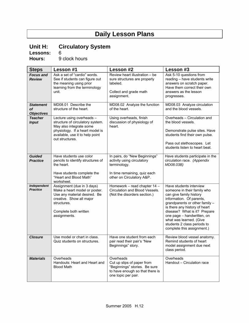
Daily Lesson Plans Unit H: Circulatory System Lessons: 6 Hours: 9 clock hours Steps Lesson #1 Lesson #2 Lesson #3 Focus and Review
Ask a set of “cardio” words. See if students can figure out the meaning using prior learning from the terminology unit.
Review heart illustration – be sure structures are properly labeled. Collect and grade math assignment.
Ask 5-10 questions from reading – have students write answers on scratch paper. Have them correct their own answers as the lesson progresses.
Statement of Objectives
MD08.01 Describe the structure of the heart.
MD08.02 Analyze the function of the heart.
MD08.03 Analyze circulation and the blood vessels.
Teacher Input
Lecture using overheads – structure of circulatory system. May also integrate some physiology. If a heart model is available, use it to help point out structures.
Using overheads, finish discussion of physiology of heart.
Overheads – Circulation and the blood vessels. Demonstrate pulse sites. Have students find their own pulse. Pass out stethoscopes. Let students listen to heart beat.
Guided Practice
Have students use color pencils to identify structures of the heart. Have students complete the “Heart and Blood Math” worksheet.
In pairs, do “New Beginnings” activity using circulatory terminology. In time remaining, quiz each other on Circulatory A&P.
Have students participate in the circulation race. (Appendix MD08.03B)
Independent Practice
Assignment (due in 3 days) Make a heart model or poster. Use any material desired. Be creative. Show all major structures. Complete both written assignments.
Homework – read chapter 14 – Circulation and Blood Vessels. (Not the disorders section.)
Have students interview someone in their family who can give family history information. Of parents, grandparents or other family – is there any history of heart disease? What is it? Prepare one page – handwritten, on what was learned. (Give students 2 class periods to complete this assignment.)
Closure Use model or chart in class. Quiz students on structures.
Have one student from each pair read their pair’s “New Beginnings” story.
Review blood vessel anatomy. Remind students of heart model assignment due next class period.
Materials Overheads Handouts: Heart and Heart and Blood Math
Overheads Cut up slips of paper from “Beginnings” stories. Be sure to have enough so that there is one topic per pair.
Overheads Handout – Circulation race
Summer 2005 H.13
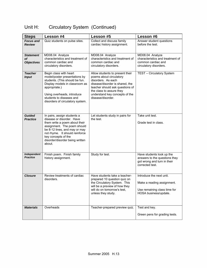
Unit H: Circulatory System (Continued) Steps Lesson #4 Lesson #5 Lesson #6 Focus and Review
Quiz students on pulse sites. Collect and discuss family cardiac history assignment.
Answer student questions before the test.
Statement of Objectives
MD08.04 Analyze characteristics and treatment of common cardiac and circulatory disorders.
MD08.04 Analyze characteristics and treatment of common cardiac and circulatory disorders.
MD08.04 Analyze characteristics and treatment of common cardiac and circulatory disorders.
Teacher Input
Begin class with heart model/poster presentations by students. (This should be fun. Display models in classroom as appropriate.) Using overheads, introduce students to diseases and disorders of circulatory system.
Allow students to present their poems about circulatory disorders. As each disease/disorder is shared, the teacher should ask questions of the class to assure they understand key concepts of the disease/disorder.
TEST – Circulatory System
Guided Practice
In pairs, assign students a disease or disorder. Have them write a poem about their assignment. The poem should be 8-12 lines, and may or may not rhyme. It should reinforce key concepts of the disorder/disorder being written about.
Let students study in pairs for the test.
Take unit test. Grade test in class.
Independent Practice
Finish poem. Finish family history assignment.
Study for test.
Have students look up the answers to the questions they got wrong and turn in their corrected test.
Closure Review treatments of cardiac disorders.
Have students take a teacher-prepared 10 question quiz on the Circulatory System. This will be a preview of how they will do on tomorrow’s test, unless they study.
Introduce the next unit. Make a reading assignment. Use remaining class time for HOSA business/update.
Materials Overheads
Teacher-prepared preview quiz.
Test and key. Green pens for grading tests.
Summer 2005 H.14
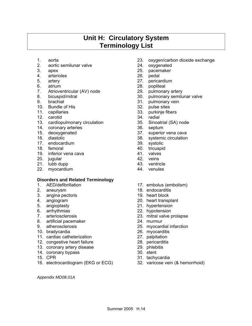
Unit H: Circulatory System Terminology List
1. aorta 2. aortic semilunar valve 3. apex 4. arterioles 5. artery 6. atrium 7. Atrioventricular (AV) node 8. bicuspid/mitral 9. brachial 10. Bundle of His 11. capillaries 12. carotid 13. cardiopulmonary circulation 14. coronary arteries 15. deoxygenated 16. diastolic 17. endocardium 18. femoral 19. inferior vena cava 20. jugular 21. lubb dupp 22. myocardium
23. oxygen/carbon dioxide exchange 24. oxygenated 25. pacemaker 26. pedal 27. pericardium 28. popliteal 29. pulmonary artery 30. pulmonary semilunar valve 31. pulmonary vein 32. pulse sites 33. purkinje fibers 34. radial 35. Sinoatrial (SA) node 36. septum 37. superior vena cava 38. systemic circulation 39. systolic 40. tricuspid 41. valves 42. veins 43. ventricle 44. venules
Disorders and Related Terminology 1. AED/defibrillation 2. aneurysm 3. angina pectoris 4. angiogram 5. angioplasty 6. arrhythmias 7. arteriosclerosis 8. artificial pacemaker 9. atherosclerosis 10. bradycardia 11. cardiac catheterization 12. congestive heart failure 13. coronary artery disease 14. coronary bypass 15. CPR 16. electrocardiogram (EKG or ECG)
17. embolus (embolism) 18. endocarditis 19. heart block 20. heart transplant 21. hypertension 22. hypotension 23. mitral valve prolapse 24. murmur 25. myocardial infarction 26. myocarditis 27. palpitation 28. pericarditis 29. phlebitis 30. stent 31. tachycardia 32. varicose vein (& hemorrhoid)
Appendix MD08.01A

Summer 2005 H.15
The Heart
Label the following structures of the heart: 1. right atrium 2. left atrium 3. right ventricle 4. left ventricle 5. septum 6. mitral valve 7. tricuspid valve 8. superior vena cava 9. inferior vena cava 10. aorta 11. myocardium 12. endocardium 13. pericardium Appendix MD08.01B
Summer 2005 H.16
Make a Heart 101
Your assignment is to work in you assigned groups (do not change them) and using the materials in your heart packet, construct a heart. You may use the finger tips of the gloves for your valves. You may use up to four cups, but you do not have to use all
of these if you are doing something else creatively. Your heart must have all the chambers, valves, arteries, veins, etc. The note cards can be cut into small pieces and used as labels. Your heart must be labeled. The tape needs to be returned to the teacher at the end of class to be used with other classes. The most accurate and creative heart will win a prize. To complete this assignment you must write a group essay in which you use your particular model to teach a person about the heart. Make sure you make reference to the different structures and the materials you have used to make those structures. This essay will be graded for content, grammar, and spelling. Please use paragraphs. Use other people in your group to proof-read your essay. You have only this class period to complete this assignment, so do not waste time. For maximum success, put your heart into this assignment! Appendix MD08.01C
Summer 2005 H.17
Heart and Blood Math
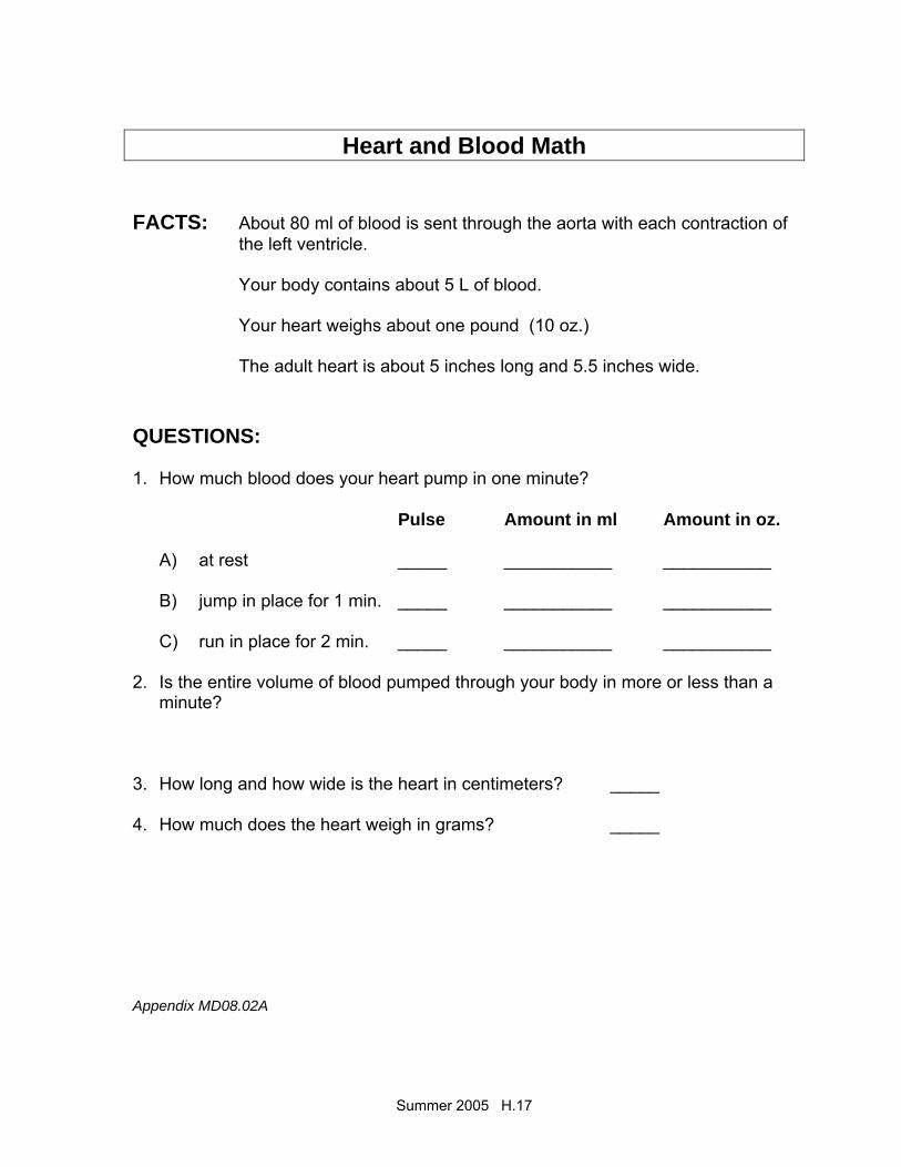
FACTS: About 80 ml of blood is sent through the aorta with each contraction of the left ventricle.
Your body contains about 5 L of blood. Your heart weighs about one pound (10 oz.) The adult heart is about 5 inches long and 5.5 inches wide. QUESTIONS: 1. How much blood does your heart pump in one minute?
Pulse Amount in ml Amount in oz. A) at rest _____ ___________ ___________ B) jump in place for 1 min. _____ ___________ ___________ C) run in place for 2 min. _____ ___________ ___________
2. Is the entire volume of blood pumped through your body in more or less than a minute?
3. How long and how wide is the heart in centimeters? _____ 4. How much does the heart weigh in grams? _____ Appendix MD08.02A

Summer 2005 H.18
BLOOD AND THE HEART FUN FACTS
♦ An average adult human contains
about 5 liters (5.3qt) of blood. ♦ The blood makes up about one-
thirteenth of the body’s weight. ♦ The adult heart weighs about 280 grams (10 oz.) ♦ At rest, the heart pumps out about 80 millimeters
(2.6 oz) of blood with each beat. ♦ The heart beats, on average, 70 times each
minute at rest. ♦ This means all the blood is circulated (goes
round the body once) in about one minute. ♦ During strenuous exercise the heart can pump
six to eight times the amount of blood that it pumps at rest.
Appendix MD08.02B
Summer 2005 H.19
New Beginnings
In a container place the slips of paper containing the “New Beginnings.” Have each team to draw one “new beginning” out of the container. The team must take their beginning and complete their story using ten of the anatomy terms found on the terminology list in the appendix. Each team member must participate. Allow 20 minutes for the exercise. Terms must be used appropriately. Examples of “New Beginnings” John and Jillian were studying together when all of a sudden. . . . . . Peter thought Chris was meeting him at the bus station. When he walked through the door soaking wet. . . . . . As the train careened around a steep turn. . . . . . It was a bright and sunny day.. . . . . . . . . . Newsflash! This is amazing!. . . . . . . Help!. . . . . . Appendix MD08.02C
Summer 2005 H.20
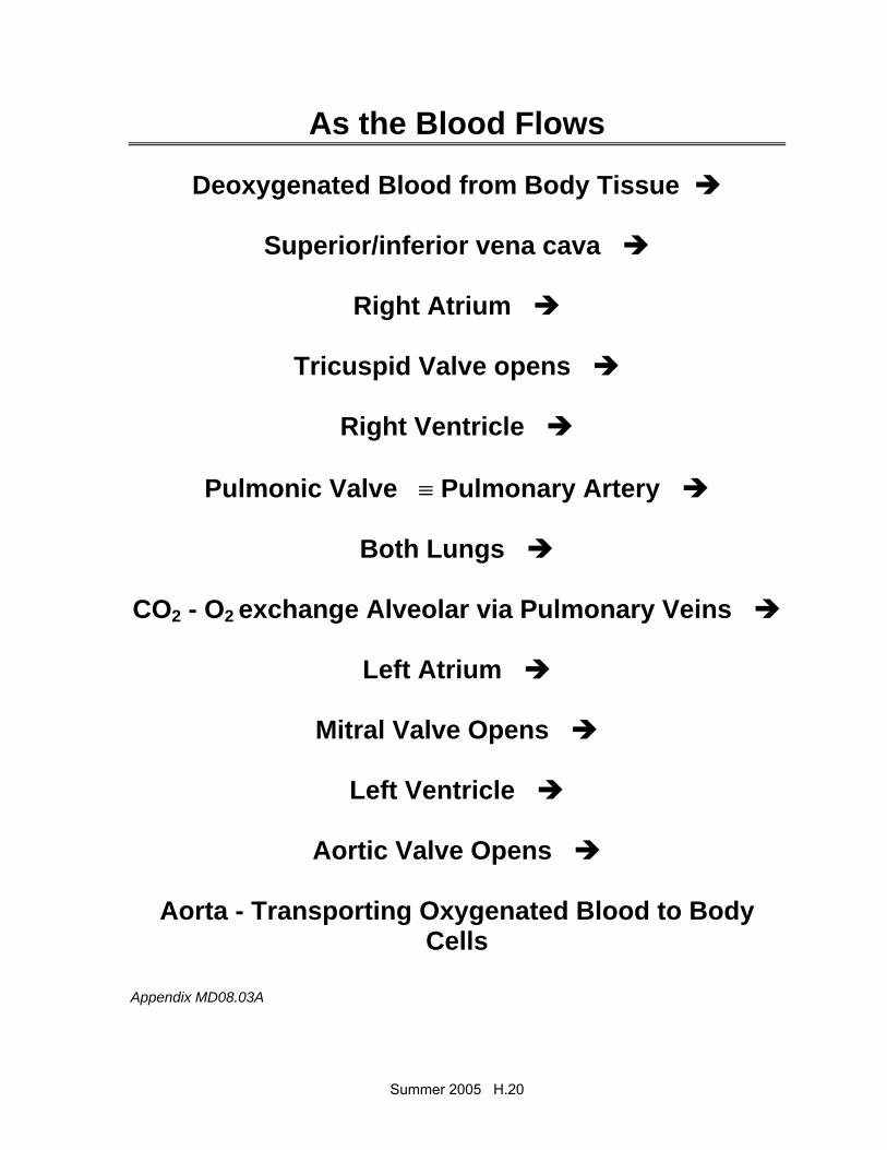
As the Blood Flows
Deoxygenated Blood from Body Tissue
Superior/inferior vena cava
Right Atrium
Tricuspid Valve opens
Right Ventricle
Pulmonic Valve ≡ Pulmonary Artery
Both Lungs
CO2 - O2 exchange Alveolar via Pulmonary Veins
Left Atrium
Mitral Valve Opens
Left Ventricle
Aortic Valve Opens
Aorta - Transporting Oxygenated Blood to Body Cells
Appendix MD08.03A
Summer 2005 H.21
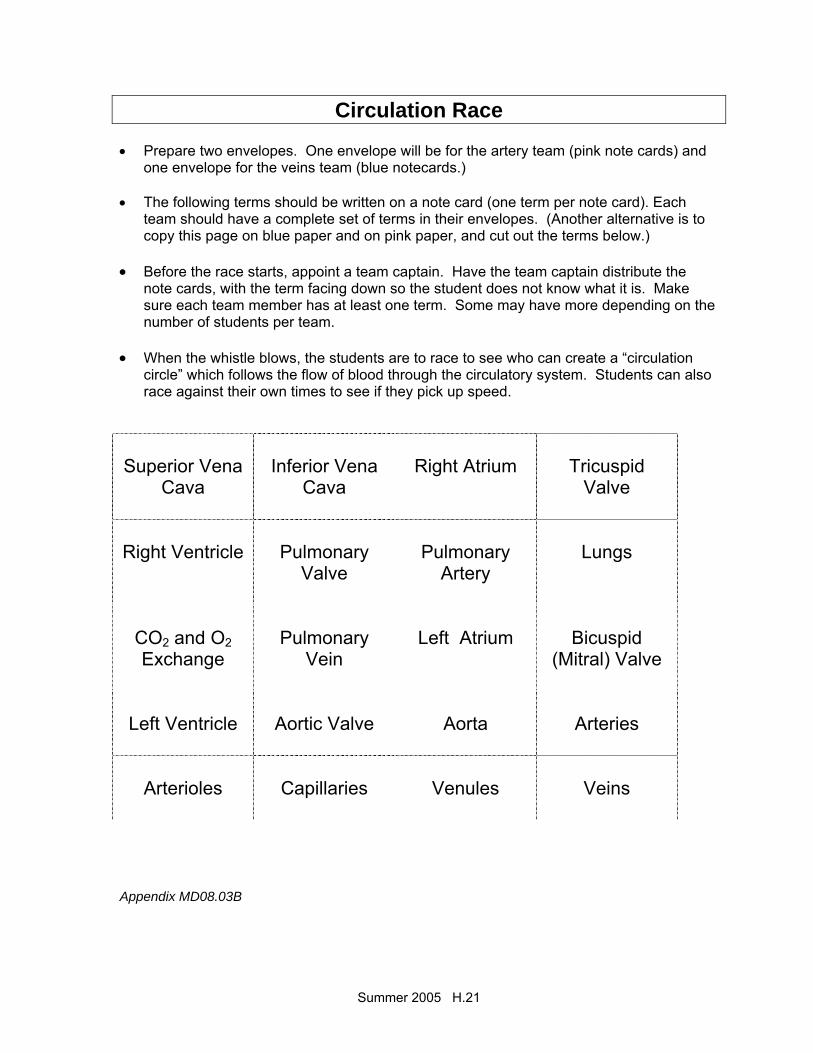
Circulation Race • Prepare two envelopes. One envelope will be for the artery team (pink note cards) and
one envelope for the veins team (blue notecards.) • The following terms should be written on a note card (one term per note card). Each
team should have a complete set of terms in their envelopes. (Another alternative is to copy this page on blue paper and on pink paper, and cut out the terms below.)
• Before the race starts, appoint a team captain. Have the team captain distribute the
note cards, with the term facing down so the student does not know what it is. Make sure each team member has at least one term. Some may have more depending on the number of students per team.
• When the whistle blows, the students are to race to see who can create a “circulation
circle” which follows the flow of blood through the circulatory system. Students can also race against their own times to see if they pick up speed.
Superior Vena
Cava
Inferior Vena
Cava
Right Atrium
Tricuspid
Valve
Right Ventricle
Pulmonary
Valve
Pulmonary
Artery
Lungs
CO2 and O2 Exchange
Pulmonary
Vein
Left Atrium
Bicuspid
(Mitral) Valve
Left Ventricle
Aortic Valve
Aorta
Arteries
Arterioles
Capillaries
Venules
Veins
Appendix MD08.03B

Summer 2005 H.22
Unit H: Circulatory System
OVERHEAD TRANSPARENCY
MASTERS
Summer 2005 H.23
Functions 1. Pump 2. Blood transport system around body 3. Carries O2 and nutrients to cells, carries away
waste products 4. Lymph system – returns excess tissue fluid to
general circulation Structure – Circulatory system involves: • Heart • Arteries • Veins • Capillaries • Blood and lymph are part of circulatory system Major Blood Circuits • General (Systemic) circulation • Cardiopulmonary circulation

Summer 2005 H.24

Summer 2005 H.25
The Heart
• Muscular organ • Size of a closed fist • Weighs 12-13 oz • Location – thoracic
cavity • APEX – conical tip, lies
on diaphragm, points left
• Stethoscope – instrument used to hear the heartbeat
Structure
Hollow, muscular, double pump that circulates blood
At rest = 2 oz blood with each beat, 5 qts./min., 75 gallons per hour
Ave = 72 beats per minute 100,000 beats per day PERICARDIUM – double layer of fibrous tissue
that surrounds the heart MYOCARDIUM – cardiac muscle tissue ENDOCARDIUM – smooth inner lining of heart

Summer 2005 H.26
SEPTUM – partition (wall) that separates right half from left half
Superior vena cava and
inferior vena cava – bring deoxygenated blood to right atrium
Pulmonary artery – takes blood away from right ventricle to the lungs for O2
Pulmonary veins – bring oxygenated blood from lungs to left atrium
Aorta – takes blood away from left ventricle to rest of the body
Chambers and Valves • SEPTUM divides into R and L halves • Upper chambers – RIGHT ATRIUM and LEFT
ATRIUM • Lower chambers – RIGHT VENTRICLE and
LEFT VENTRICLE • Four heart valves permit flow of blood in one
direction
Summer 2005 H.27
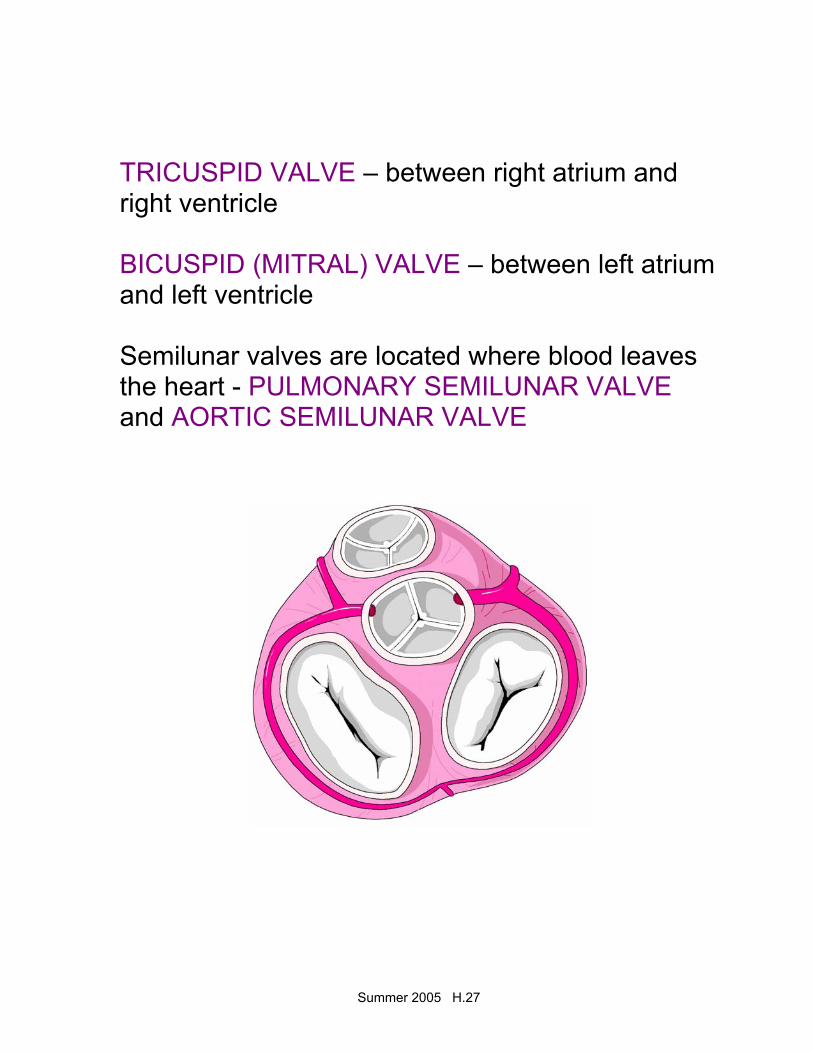
TRICUSPID VALVE – between right atrium and right ventricle BICUSPID (MITRAL) VALVE – between left atrium and left ventricle Semilunar valves are located where blood leaves the heart - PULMONARY SEMILUNAR VALVE and AORTIC SEMILUNAR VALVE
Summer 2005 H.28
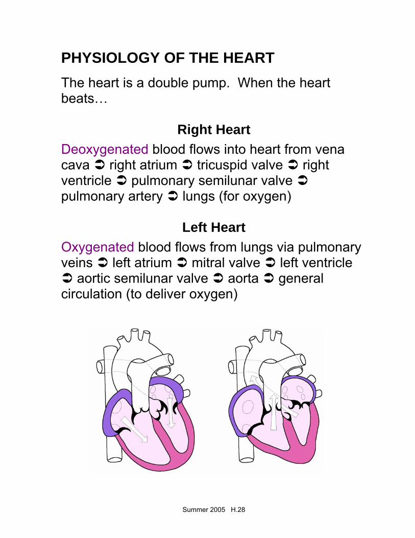
PHYSIOLOGY OF THE HEART
The heart is a double pump. When the heart beats…
Right Heart Deoxygenated blood flows into heart from vena cava right atrium tricuspid valve right ventricle pulmonary semilunar valve pulmonary artery lungs (for oxygen)
Left Heart Oxygenated blood flows from lungs via pulmonary veins left atrium mitral valve left ventricle
aortic semilunar valve aorta general circulation (to deliver oxygen)
Summer 2005 H.29
Blood Supply to the Heart – from CORONARY ARTERIES Heart Sounds = lubb dupp Control of Heart Contractions SA (sinoatrial) NODE = PACEMAKER • Located in right atrium • SA node sends out electrical impulse • Impulse spreads over atria, making them
contract • Travels to AV Node AV (atrioventricular) NODE • Conducting cell group between atria and
ventricle • Carries impulse to bundle of His BUNDLE OF HIS • Conducting fibers in septum • Divides into R and L branches to network of
branches in ventricles (Purkinje fibers) PURKINJE FIBERS • Impulse shoots along Purkinje fibers causing
ventricles to contract
Summer 2005 H.30
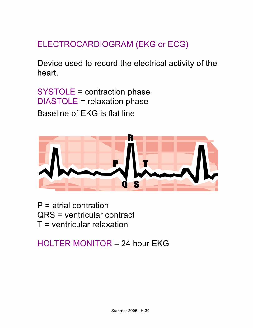
ELECTROCARDIOGRAM (EKG or ECG) Device used to record the electrical activity of the heart. SYSTOLE = contraction phase DIASTOLE = relaxation phase Baseline of EKG is flat line
P = atrial contration QRS = ventricular contract T = ventricular relaxation HOLTER MONITOR – 24 hour EKG
Summer 2005 H.31
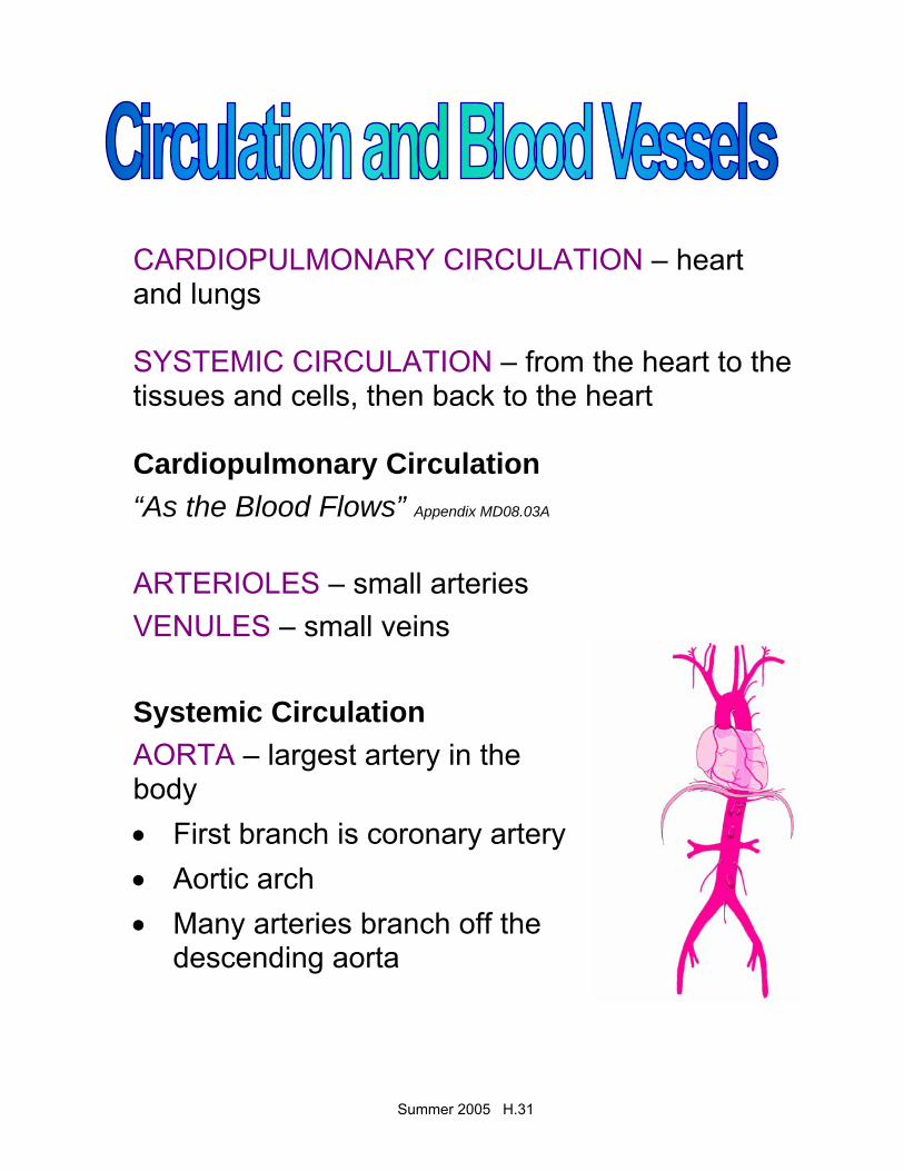
CARDIOPULMONARY CIRCULATION – heart and lungs SYSTEMIC CIRCULATION – from the heart to the tissues and cells, then back to the heart Cardiopulmonary Circulation “As the Blood Flows” Appendix MD08.03A
ARTERIOLES – small arteries VENULES – small veins Systemic Circulation AORTA – largest artery in the body • First branch is coronary artery • Aortic arch • Many arteries branch off the
descending aorta
Summer 2005 H.32
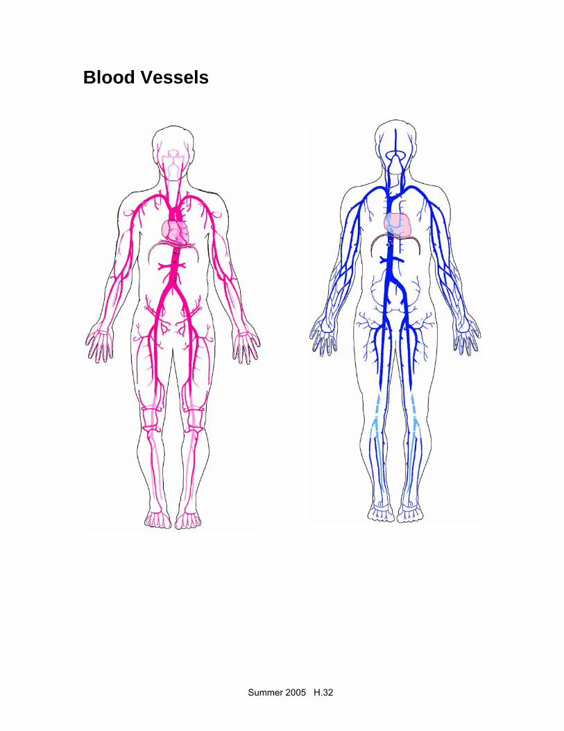
Blood Vessels

Summer 2005 H.33
ARTERIES • Carry oxygenated blood away from the heart to
the capillaries • Elastic, muscular and thick-walled • Transport blood under very high pressure CAPILLARIES • Smallest blood vessels, can only be seen with
a microscope • Connect arterioles with venules • Walls are one-cell thick and extremely thin –
allow for selective permeability of nutrients, oxygen, CO2 and metabolic wastes
VEINS • Carry deoxygenated blood away from
capillaries to the heart • Veins contain a muscular layer, but less elastic
and muscular than arteries • Thin walled veins collapse easily when not filled
with blood • VALVES – permit flow of blood only in direction
of the heart • JUGULAR vein – located in the neck
Summer 2005 H.34
Blood Pressure Surge of blood when heart pumps creates pressure against the walls of the arteries SYSTOLIC PRESSURE – measured during the contraction phase DIASTOLIC PRESSURE – measured when the ventricles are relaxed Average systolic = 120 Average diastolic = 80 PULSE – alternating expansion and contraction of an artery as blood flows through it. Pulse sites: • BRACHIAL • CAROTID • RADIAL • POPLITEAL • PEDAL
Summer 2005 H.35
DDiisseeaasseess ooff tthhee HHeeaarrtt
ARRHYTHMIA (or dysrrhythmia) – any change from normal heart rate or rhythm BRADYCARDIA – slow heart rate (<60 bpm) TACHYCARDIA – rapid heart rate (>100 bpm) MURMURS – indicates defect in heart valve – valves fail to close properly, causing gurgling or hissing sound. MITRAL VALVE PROLAPSE – mitral valve closes imperfectly – symptoms occur in response to stress, including fatigue, PALPITATIONS (heart feels like it is racing) headache, chest pain, and anxiety. Infectious Diseases of the Heart Cause = virus or bacteria Treatment = antibiotics PERICARDITIS – inflammation of outer membrane covering the heart – symptoms are chest pain, cough, dyspnea (difficulty breathing), tachycardia, and fever.
Summer 2005 H.36
MYOCARDITIS – inflammation of heart muscle – symptoms the same as pericarditis ENDOCARDITIS – inflammation of the membrane that lines the heart and covers the valves, causes rough spots in the endocardium which may lead to the development of a thrombus Coronary Artery Disease ANGINA PECTORIS – chest pain, caused by lack of oxygen to heart muscle, treat with nitroglycerin to dilate coronary arteries MYOCARDIAL INFARCTION • MI or heart attack • Lack of blood supply to myocardium causes
damage • Due to blockage of coronary artery or blood clot
atherosclerosis – plaque build-up on arterial walls, or arteriosclerosis – loss of elasticity and thickening of wall.
• Amount of damage depends on size of area deprived of oxygen
Summer 2005 H.37
• Symptoms – severe chest pain radiating to left shoulder, arm, neck and jaw. Also nausea, diaphoresis, dyspnea.
• Immediate medical care is critical • Rx – bedrest, oxygen, medication • Morphine for pain, tPA to dissolve clot • Anticoagulant therapy to prevent further clots
from forming • Angioplasty and by-pass surgery may be
necessary CONGESTIVE HEART FAILURE • Ventricles unable to contract effectively and
blood pools in the heart • Edema in lower extremities • Blood backs up into lungs • Rx – drugs to strengthen heart beat (digoxin)
and diuretics to reduce fluid Heart Surgery ANGIOPLASTY – procedure to help open clogged vessels – may also be called “balloon surgery.”
Summer 2005 H.38
CORONARY BY-PASS – usually, a healthy vein from the leg removed and attached before and after the coronary obstruction, creating an alternate route for blood supply to the myocardium. HEART TRANSPLANT • Why? Irreparably damaged myocardium,
valves or blood vessels, or baby/child with congenital heart defect
• Problem? Histocompatibility • Rx? Immunosuppressants • Artificial hearts? First used in 1982. What is
the current status? PACEMAKERS Demand pacemaker – fires only when heart rate drops below programmed minimum STENT Tiny, expandable stainless steel tube that holds coronary artery open following angioplasty CPR – cardiopulmonary resuscitation, used in the presence of cardiac arrest
Summer 2005 H.39
DEFIBRILLATION – electrical shock to bring the heart back to a normal rhythm. AED – automated external defibrillator HEART BLOCK – disturbance in electrical conductivity of the heart beat
Disorders of the Blood Vessels
ANEURYSM – ballooning of an artery, thinning and weakening ARTERIOSCLEROSIS – arterial walls thicken, lose elasticity ATHEROSCLEROSIS – fatty deposits form on walls of arteries PHLEBITIS – inflammation of lining of vein, accompanied by clotting of blood – symptoms are edema, pain and redness EMBOLISM – traveling blood clot VARICOSE VEINS – swollen, distended veins – heredity or due to posture, prolonged periods of standing, physical exertion, age and pregnancy
Summer 2005 H.40
HEMORRHOIDS - varicose rectal veins HYPOTENSION – low blood pressure, systolic <100 HYPERTENSION • High blood pressure • “silent killer” – usually no symptoms • Condition leads to strokes, heart attacks, and
kidney failure • 140/90 or higher • Higher in African-Americans and post-
menopausal women • Risk factors = smoking, overweight, stress, high
fat diets, family history • Treatment = relaxation, low fat diet, exercise,
weight loss, medication Diagnostic Tests
CARDIAC CATHETERIZATION – catheter fed into heart, dye injected, x-rays taken as dye moves through coronary arteries ANGIOGRAM – x-ray of a blood vessel using dye ELECTROCARDIOGRAM – electrical tracing of the heart