guidewire breakage during neurointerventional … of guidewires during interventional procedures is...
TRANSCRIPT

638 Korean J Radiol 12(5), Sep/Oct 2011 kjronline.org
INTRODUCTION
Breakage of guidewires during interventional procedures is a rare event (1-3). A few cases of its occurrence have been reported, but not in the neurointerventional field. This paper reports on two cases of guidewire fracture during neurointerventional procedures. We describe causative factors and suggest several technical tips for avoidance of this problem.
CASE REPORTS
Case 1A 47-year-old woman with an un-ruptured ophthalmic
artery aneurysm in the left internal carotid artery (ICA)
Guidewire Breakage during Neurointerventional Procedures: a Report of Two CasesMyeong Sub Lee, MD1, Kum Whang, MD2, Hun Ju Kim, MD2, O-Ki Kwon, MD3; Brain Research GroupDepartments of 1Radiology and 2Neurosurgery, Wonju College of Medicine, Yonsei University, Gangwon-do 220-701, Korea; 3Department of Neurosurgery, Seoul National University Bundang Hospital, Gyeonggi-do 463-707, Korea; Brain Research Group
We report on two cases of microguidewire breakage that occurred during endovascular treatment of intracranial aneurysms. The microguidewire can be broken when a part of the wire is stuck due to vascular tortuosity, and, subsequently, application of excessive rotational movement. The mechanical and physical properties of a microguidewire are also important factors in microguidewire breakage. We also suggest technical tips for avoidance of this problem.Index terms: Aneurysm; Microguidewires; Mechanical properties
Received January 4, 2011; accepted after revision March 25, 2011.Corresponding author: O-Ki Kwon, MD, Department of Neurosurgery, Seoul National University Bundang Hospital, 300 Gumi-dong, Bundang-gu, Seongnam-si, Gyeonggi-do 463-707, Korea. • Tel: (8231) 787-7163 • Fax: (8231) 787-4059• E-mail: [email protected] is an Open Access article distributed under the terms of the Creative Commons Attribution Non-Commercial License (http://creativecommons.org/licenses/by-nc/3.0) which permits unrestricted non-commercial use, distribution, and reproduction in any medium, provided the original work is properly cited.
Case Reporthttp://dx.doi.org/10.3348/kjr.2011.12.5.638pISSN 1229-6929 · eISSN 2005-8330Korean J Radiol 2011;12(5):638-640
was treated with endovascular coiling under general anesthesia. The aneurysm measured 5 mm in size and was superomedially directed. Conjoined with a guiding catheter (Envoy 6 Fr, Codman, MA), a microcatheter (Prowler 14, Codman, MA; 0.4 mm inner diameter) with a microguidewire (Transend 014, Boston Scientific, MA; 0.37 mm outer diameter) was used in selection of the aneurismal sac. Initially, several attempts were made to select the aneurysm using the steam-shaped microcatheter and microguidewire. However, due to the tortuosity of the ICA, entry into the aneurysm sac was very difficult. During this process, the wire was rotated many times in a clockwise and counterclockwise direction. At one point, we noticed that the tip no longer moved with the rotation of the guidewire. The distal part of the wire was fractured (Fig. 1A). Using a microsnare (Microvena, Boston Scientific, MA), it was successfully removed without complication (Fig. 1B). The aneurysm was then selected using a new guidewire and successfully coiled without further problems.
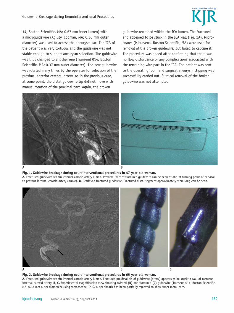
Case 2A 65-year-old female patient with a ruptured anterior
communicating artery aneurysm was treated with endovascular coiling. Conjoined with a guiding catheter (Envoy 6 Fr, Codman, MA), a microcatheter (Excelsior
Korean J Radiol 12(5), Sep/Oct 2011kjronline.org 639
Guidewire Breakage during Neurointerventional Procedures
14, Boston Scientific, MA; 0.67 mm inner lumen) with a microguidewire (Agility, Codman, MA; 0.36 mm outer diameter) was used to access the aneurysm sac. The ICA of the patient was very tortuous and the guidewire was not stable enough to support aneurysm selection. The guidewire was thus changed to another one (Transend 014, Boston Scientific, MA; 0.37 mm outer diameter). The new guidewire was rotated many times by the operator for selection of the proximal anterior cerebral artery. As in the previous case, at some point, the distal guidewire tip did not move with manual rotation of the proximal part. Again, the broken
guidewire remained within the ICA lumen. The fractured end appeared to be stuck in the ICA wall (Fig. 2A). Micro-snares (Microvena, Boston Scientific, MA) were used for removal of the broken guidewire, but failed to capture it. The procedure was ended after confirming that there was no flow disturbance or any complications associated with the remaining wire part in the ICA. The patient was sent to the operating room and surgical aneurysm clipping was successfully carried out. Surgical removal of the broken guidewire was not attempted.
A BFig. 1. Guidewire breakage during neurointerventional procedures in 47-year-old woman. A. Fractured guidewire within internal carotid artery lumen. Proximal part of fractured guidewire can be seen at abrupt turning point of cervical to petrous internal carotid artery (arrow). B. Retrieved fractured guidewire. Fractured distal segment approximately 9 cm long can be seen.
A B CFig. 2. Guidewire breakage during neurointerventional procedures in 65-year-old woman. A. Fractured guidewire within internal carotid artery lumen. Fractured proximal tip of guidewire (arrow) appears to be stuck in wall of tortuous internal carotid artery. B, C. Experimental magnification view showing twisted (B) and fractured (C) guidewire (Transend 014, Boston Scientific, MA; 0.37 mm outer diameter) using stereoscope. In C, outer sheath has been partially removed to show inner metal core.

Korean J Radiol 12(5), Sep/Oct 2011 kjronline.org640
Lee et al.
DISCUSSION
Microguidewires for neurointervention are commonly used for complex and intricate intracranial vascular pathology; therefore, they must meet a high standard of quality. These guidewires are frequently subject to multiple strong axial and lateral stresses that can cause bending and kinks. Repeated overextension or excessive rotational movement can also cause failure of guidewire components. The mechanical and physical properties of an optimal microguidewire are important facets of their quality. Using torsion-testing equipment, Ceschinski et al. (4) measured the torsional rigidity of various guidewires on the market. They found significant variation in torsional rigidity, depending on materials and design characteristics. The fractured guidewire in our series was the Transend EX 014 (Boston Scientific, MA). The basic structure of this microguidewire consists of a scitanium alloy core metal, which is covered with polyurethane. As an experiment, the authors rotated the Transcend microguidewire many times in one direction within a microcatheter, which is placed on a table with simulation of the tortuosity of the carotid artery and cerebral arteries (Fig. 2B, C). We found that the outer polyurethane sheath at the middle or distal part of the wire became twisted segmentally with manual rotation of the proximal wire part (Fig. 2B). After 20-30 rotations in one direction, the inner metal core was often fractured too. This twisting and fracturing occurred only when the microcatheter was placed as if in simulation of vascular tortuosity. In this situation, a part or parts of the guidewire became relatively fixed to part of the microcatheter lumen. As a result, rotational force can be transmitted unevenly through the microwire. Consequently, the fixed part does not rotate while the proximal part does. This discrepancy in rotational motion causes the two components (the core metal and the outer polyurethane cover) to move separately. The outer part is twisted more than the inner metal core. Soon, the inner metal core begins to become twisted;
if excessive, it can be fractured (Fig. 2C). As the same guidewire has been fractured in several neurointerventional cases, we suspect that the Transend guidewire is more brittle than guidewires produced by other companies.
Technical errors committed by the operator are another important cause of guidewire fracture. In the cases reported here, excessive torsional rotatory manipulation or handling, especially in one direction, was the apparent cause of the wire fracture. To avoid this complication, operators need to make note of or recognize how they manipulate microwires during the procedure. If vessels are particularly tortuous, they should be more careful. Excessive rotation of the microguidewire may cause its fracture.
CONCLUSION
We report on microguidewire fracture during neurointerventional procedures in two patients with severe vascular tortuosity. Operators should understand the mechanical and physical characteristics of commonly used microwires, and should try to avoid excessive manipulation that could exceed the capacity of the device.
REFERENCES
1. Stellin G, Ramondo A, Bortolotti U. Guidewire fracture: an unusual complication of percutaneous transluminal coronary angioplasty. Int J Cardiol 1987;17:339-342
2. Sethi GK, Ferguson TB Jr, Miller G, Scott SM. Entrapment of broken guidewire in the left main coronary artery during percutaneous transluminal coronary angioplasty. Ann Thorac Surg 1989;47:455-457
3. Kilic H, Akdemir R, Bicer A. Rupture of guide wire during percutaneous transluminal coronary angioplasty, a case report. Int J Cardiol 2008;128:e113-114
4. Ceschinski H, Henkes H, Weinert HC, Weber W, Kuhne D, Monstadt H. Torquability of microcatheter guidewires: the resulting torsional moment. Biomed Mater Eng 2000;10:31-42