guidelines on the diagnosis and management of stable angina angina guidelines issue 1... · angina...
TRANSCRIPT

Guidelines on theDiagnosis and
Management ofStable Angina
INTR
OD
UC
TIO
N
Guidelines on theDiagnosis and
Management ofStable Angina
INTR
OD
UC
TIO
N

By kind permission, these guidelines are adapted from the Cheshire andMerseyside Cardiac Network Guidelines and represent the consensus viewsof the North Wales Cardiac Network (NWCN).
They were developed following consideration of the available evidenceand aim to ensure equity and best practice.
Health professionals are asked to take them into account when exercisingtheir clinical judgement and are encouraged to discuss with colleaguesthose cases where the assessment of likely benefit from a particularintervention is equivocal. The guidelines do not override the responsibilityof health professionals to make appropriate decisions in the circumstancesof the individual patient in consultation with the patient and/or guardianor carer.

CONTENTS1
CONTENTS...........................................................................................................................................1OVERVIEW OF GUIDELINES ..........................................................................................................2INTRODUCTION........................................................................................................................................4
1 What is Angina?2 Why Is Angina Important?3 The Need for Guidelines4 Principles Underpinning Guidelines
STAGE 1 CASE MANAGEMENT AND REFERRAL PATHWAYS ..............81 Patients with Known Stable Angina2 Patients with Worsening Known Angina3 New Patients with Recent Onset Chest Pain - Suspected Angina4 Communications
STAGE 2 DIAGNOSIS ..............................................................................................................131 Use of Algorithm 22 Clinical Assessment3 Pre-test Probability of CAD4 Non-invasive Diagnostic Testing to Produce Post-test Probability of CAD
STAGE 3 RISK STRATIFICATION ....................................................................................23STAGE 4 CORONARY ANGIOGRAPHY ...................................................................26STAGE 5 LIFESTYLE AND RISK FACTOR MODIFICATION......................29
1 Cardiac Rehabilitation2 Refractory Angina
STAGE 6 DRUG TREATMENT...........................................................................................401 Drug Treatment of Acute Episode2 Prophylactic Drug Treatment of Symptoms3 Drug Treatment to Improve Prognosis in Stable Angina
STAGE 7 REVASCULARISATION....................................................................................491 Selection of Patients for Revascularisation Therapy2 Selection of Method of Revascularisation3 Contraindications to Myocardial Revascularisation4 Specific Patient and Lesion Subsets
STAGE 8 ASSESSMENT AND MANAGEMENT OF CVD RISKPRIOR TO NON-CARDIAC SURGERY ................................................55
1 Use of Algorithm 32 Mechanisms of Perioperative Acute Coronary Syndrome (ACS)3 Accuracy of Non-invasive Testing in Pre-operative Assessment4 Classical Approach5 Preferred (Modern) Approach
APPENDIX TO STAGE 8 ASSESSMENT ANDMANAGEMENT OF CVD RISK OF POTENTIALCANDIDATES FOR RENAL TRANSPLANTATION ......................62
1 Objectives2 Special Considerations (Why Renal Transplant and Pancreatic–renal
Transplant Differ)3 CMCN Pathway4 Re-assessment of Patients Accepted on to Transplant Waiting List

OVERVIEW OF GUIDELINES2
OVERVIEW OF GUIDELINES
Wherever possible the guidelines are in the format of clinical algorithms supportedby tables with a minimum of text. The component parts of the Stable AnginaPathway are discussed in eight management stages as follows and the overallpathway is outlined in Algorithm 1.
Table 1
OVERVIEW OF GUIDELINESINTRODUCTION
STAGE 1 Case management and referral pathwaysModels of care within primary care and between primary andsecondary care.
STAGE 2 Diagnostic pathwayInitial clinical assessment giving a pre-test probability of angina;then appropriate non-invasive investigations; followed by are-assessment of the likelihood of angina modified by theinvestigation results giving a post-test probability.
STAGE 3 Risk StratificationBased on clinical and non-invasive investigational data.
STAGE 4 Coronary angiographyIndications for, and interpretation of, results of invasiveinvestigation by coronary angiography (and left ventricularangiography) to confirm diagnosis, refine prognosis and identifyneed for interventional therapy.
STAGE 5 Lifestyle and risk factor modificationAs a complement to medical and/or interventional treatmentincluding the roles of cardiac rehabilitation and refractory anginamanagement.
STAGE 6 Drug treatmentAcute and prophylactic drug treatment, either as a primarytreatment modality or as an adjunct to cardiac revascularisation.
STAGE 7 RevascularisationInterventional treatment including the indications and standardsfor either percutaneous coronary intervention (PCI) or coronaryartery bypass graft surgery (CABG).
STAGE 8 Assessment and management of patients for non-cardiac surgeryFocussing on patients undergoing elective surgery.In addition, the specific problems relating to renal transplantationare addressed in an appendix.

ALGORITHM 1STABLE ANGINA PATHWAY
Lifestyle/risk factormodificationCardiac rehabRefractory angina
Coronary angionot indicated
Coronary angio indicatedfor diagnosis/prognosis
Revasc.PCI
CABG
Non-cardiac pre-opassessment/
management
Worseningangina
Symptomseverity
assessment
Riskstratification
Knownangina
Diagnosis- pre-test probability- testing- post-test probability
New/suspectedangina
OV
ERVIEW
OF G
UID
ELINES
3Drug treatment

INTRODUCTION4
INTRODUCTION
1. What Is Angina?
Angina pectoris (angina) is a clinical syndrome characterised by discomfort in thechest, jaw, shoulder, back or arms, brought on by exercise or emotion and relievedby rest or nitroglycerin. Conventionally, the term angina is reserved for cases inwhich the discomfort is due to myocardial ischaemia resulting from atheromatouscoronary artery disease (CAD).
Less commonly anginal-type chest pain sounding similar, or even identical, to trueangina can arise in the absence of CAD due to:
- dynamic coronary artery problems (e.g. coronary spasm, cardiac syndrome X,endothelial dysfunction),
- non-coronary cardiac problems (e.g. aortic stenosis, cardiomyopathy, vasculitisetc),
- non-cardiac causes mimicking angina (e.g. oesophageal, musculo-skeletal orpsychogenic problems).
Myocardial ischaemia, which underlies true angina, results from an imbalancebetween the supply of and demand for myocardial oxygen.
Myocardial oxygen supply is essentially the coronary flow and is itself dependent onthe luminal cross-sectional area of the coronary artery and coronary arterial tone,both adversely affected by atherosclerotic plaque.
Myocardial oxygen demand is determined by heart rate, myocardial contractility(force of contraction) and wall stress, all of which increase with exercise andemotion.
Imbalance, caused by demand exceeding supply, initiates a sequential ischaemiccascade of metabolic abnormalities, perfusion mismatch, contractile dysfunction,ECG changes and then angina. The pain of angina is mediated by the release ofadenosine, from ischaemic myocardium, that stimulates A1 receptors on cardiacnerve endings.
The stable angina threshold frequently varies from day to day or even during thesame day. This symptom variability, including the occurrence of rest pain, resultsfrom dynamic factors, especially the degree of vasoconstriction at the site ofunderlying fixed atheromatous plaques (dynamic stenosis) or at the distal coronaryvessels, and from factors such as ambient temperature, mental stress andneurohumoral influences.

INTRODUCTION5
The Canadian Cardiovascular Society (CCS) has produced a classification systemwhich has been widely adopted:
CCS Ordinary physical activity such as walking, climbing stairs does not causeCLASS I angina. Angina occurs with rapid or prolonged exertion at work or
recreation.
CCS Slight limitation of ordinary activity. Angina occurs on walking or climbingCLASS II stairs rapidly, walking uphill, walking or stair climbing after meals, or in
cold or wind or under emotional stress or only during the few hoursafter waking. Angina occurs after walking more than two blocks on thelevel or climbing more than one flight of ordinary stairs at a normal paceand in normal conditions.
CCS Marked limitations of ordinary physical activity. Angina occurs onCLASS III walking one to two blocks on the level and climbing one flight of stairs
in normal conditions and at a normal pace.
CCS Inability to carry on any physical activity without discomfort – anginalCLASS IV symptoms may be present at rest.
2. Why Is Angina Important?
The main importance of angina is that it is a symptom suggesting that the individualmay have underlying CAD. Ischaemic heart disease (IHD) resulting from CAD iscommon and remains the major cause of death and morbidity in the Western world.The Health Survey for England (2003) reported a standardised prevalence of IHD ininformants aged 35 and over in the North West of 9.4% in men and 6.6% in women.
It is important to understand that CAD produces adverse effects either: - predictably – via a gradual increase in arterial obstruction (enlarging plaque),worsening the severity of stable angina, and/or - unpredictably – by sudden and unheralded complications, usually due to plaqueerosion or rupture, causing a heart attack (myocardial infarction [MI]), unstableangina or sudden death.
Annual mortality rates in stable angina vary from 0.9 to 1.4%, with an annualincidence of non-fatal MI of 0.55 to 2.6%. However, critically within the stableangina population there can be up to tenfold variation in an individual’s prognosis.A prognostic assessment, termed risk stratification is therefore an essential part ofthe management of patients with stable angina.
3. The Need for Guidelines
Despite a decline in the rate of major coronary events in recent years, data from theBritish Regional Heart Study based on GP records has shown a 2.6% annual increasein new diagnoses of angina in the 20 years of follow up to 2000 in males aged 40–59at entry.

INTRODUCTION6
The National Service Framework (NSF) for CHD, Government targets and financialconstraints within the NHS mandate the more rapid identification of patients, theapplication of evidence-based choices in ensuring best practice and the cost-effectiveuse of scarce resources.
The management of angina is now truly multi-disciplinary. Guidance is thusrequired to promote seamless, consistent and equitable management acrossorganisational boundaries. The target audience for these guidelines includes all therelevant healthcare professionals but in addition, it is intended to encourageinvolvement by patients in decisions about their own care.
These guidelines are not meant to be a comprehensive review of all angina-relatedliterature, for which the reader is referred to the following publications that haveinformed this local guidance:
• American College of Cardiology and American Heart Association (ACC/AHA)2002 Guideline Update for the Management of Patients with Chronic StableAngina.1
• European Society of Cardiology (ESC) Guidelines on the Management of StableAngina Pectoris (2006).2
• Scottish Intercollegiate Guidelines Network Management of Stable Angina(2007).3
4. Principles Underpinning Guidelines
This is inevitably a consensus document combining the views of a number of multi-disciplinary task groups set up by the Cheshire and Merseyside Cardiac Network(CMCN). Whenever possible guidance is evidence-based and designed to bedeliverable within the NHS locally. It is recognised, however, that some parts willcurrently be aspirational since not all health economies will be able to deliver everyaspect within current resource constraints. Therefore, where appropriate,acceptable alternatives to best practice have been identified.
These guidelines have adopted the following principles:
• The guidance addresses not only clinical practice but also relevant models ofcare, standards of service provision and audit.
• The diagnosis and management of angina usually starts and ends in theprimary/community care setting with secondary and tertiary services providingkey interventions within the framework of the patient’s long-term care.
• The diagnosis of angina is rarely definitive and the concept of probability orlikelihood of disease is used.
• The management of angina requires, in addition to symptomatic relief, theamelioration of adverse events or complications and thus prognostic riskstratification is a central feature.

INTRODUCTION7
• In practice, diagnostic and prognostic assessments are conducted in tandemrather than sequentially as the same clinical and investigational tools are usedfor both. However, for clarity of understanding and presentation, these linkedassessments are described separately.
• Modern medical management now includes a number of effective and locallywell-developed treatment modalities in addition to drug therapy, includingcardiac rehabilitation and refractory angina management. This guidanceattempts to define their place in patient management.
• Invasive and interventional approaches carry risks as well as benefits and are notinfinitely available. Guidance is given on appropriate indications, treatmentchoices and patient prioritisation to allow best possible use of local resourcescompatible with good standards of care.
• Successful implementation of these guidelines will require investment in theongoing education of a large constituency of relevant healthcare professionals.
Where appropriate the customary ACC/AHA classifications of recommendation havebeen adopted:
CLASS I Conditions for which there is evidence and/or general agreement that agiven procedure/treatment is useful and effective
CLASS II Conditions for which there is conflicting evidence and/or divergence ofopinion about the usefulness/efficacy of a procedure/treatmentIIa Weight of evidence/opinion is in favour of usefulness/efficacyIIb Usefulness/efficacy is less well established by evidence/opinion
CLASS III Conditions for which there is evidence and/or general agreement that agiven procedure/treatment is not useful/effective and in some cases maybe harmful
1ACC/AHA 2002 Guideline Update for the Management of Patients With Chronic Stable Angina. www.acc.org2Guidelines on the management of stable angina pectoris. The Task Force on the Management ofStable Angina Pectoris of the European Society of Cardiology. European Heart Journal 2006.doi:1093/ eurheartj/ehl10023www.sign.ac.uk

STAGE 1CASE MANAGEMENT AND REFERRAL PATHWAYS
STA
GE
1
STAGE 1CASE MANAGEMENT AND REFERRAL PATHWAYS
STA
GE
1

STAGE 1: CASE MANAGEMENT AND REFERRAL PATHWAYS8
Summary
Table 2
STAGE 1CASE MANAGEMENT and REFERRAL PATHWAYS
RECOMMENDED CASE MANAGEMENT
Reason for referral: Refer to: Recommended timeframe from referral topatient being seen:
Stable known angina:- further cardiological
advice about ongoingmanagement
General cardiologyoutpatient clinic
4-6 weeks
Worsening known angina:• Stable angina:
- early assessment and treatment
Rapid access clinic orgeneral cardiologyoutpatient clinic
14 days
• Possible or probableunstable angina:
- urgent treatment
A&E , Acute MedicalAssessment Unit (AMU) orHeart Emergency Centre(HEC)
immediate
New onset chest painsuspected angina:- confirmation of diagnosis - risk stratification- management plan
Rapid access clinic orreserved slots in generalcardiology outpatient clinic
14 days

STAGE 1: CASE MANAGEMENT AND REFERRAL PATHWAYS9
STAGE 1CASE MANAGEMENT and REFERRAL PATHWAYS
1. Patients with Known Stable Angina
This section refers to patients in whom a diagnosis of angina has previously beenmade and confirmed by specialist assessment either within general practice or moreusually within secondary/tertiary care. The principles of management should be:
• GPs should ensure that unless contraindicated by infirmity or co-morbidity, allpatients with a diagnosis of angina should have undergone diagnostic testingand risk stratification (see Stages 2 and 3). This should be undertaken withingeneral practice or by referral to a general cardiology clinic in secondary care.
• Long-term management should be delivered in the community on a structuredbasis, ideally within a multi-disciplinary management programme in primarycare. The focus should be on symptomatic anti-anginal treatment; drugtreatment as secondary prevention of future events (see Stage 6); and riskfactor/lifestyle modification (see Stage 5).
• Specialist advice on specific management issues e.g. in relation to medication,perioperative risk, air travel etc should be obtained via a referral to a generaloutpatient cardiology clinic, using the local general cardiology referral form.The patient should be seen within 4-6 weeks. In some areas, there may bearrangements in place for such advice to be obtained by telephone.
• It is important to identify changes in clinical status indicative of worsening stableangina requiring assessment or re-assessment for revascularisation or ofunstable angina mandating admission to hospital. See Section 2 below.
This process will be aided by consideration of the following questions duringeach primary care follow up:
- Has the patient decreased his or her level of physical activity since the lastvisit?
- Have the patient’s anginal symptoms increased in frequency and becomemore severe since the last visit?
- How well is the patient tolerating therapy?- How successful has the patient been in modifying risk factors and improving
knowledge about ischaemic heart disease?- Has the patient developed any new co-morbid illness or has the severity or
treatment of any co-morbid illness worsened the patient’s angina?
STAGE 1: CASE MANAGEMENT AND REFERRAL PATHWAYS10
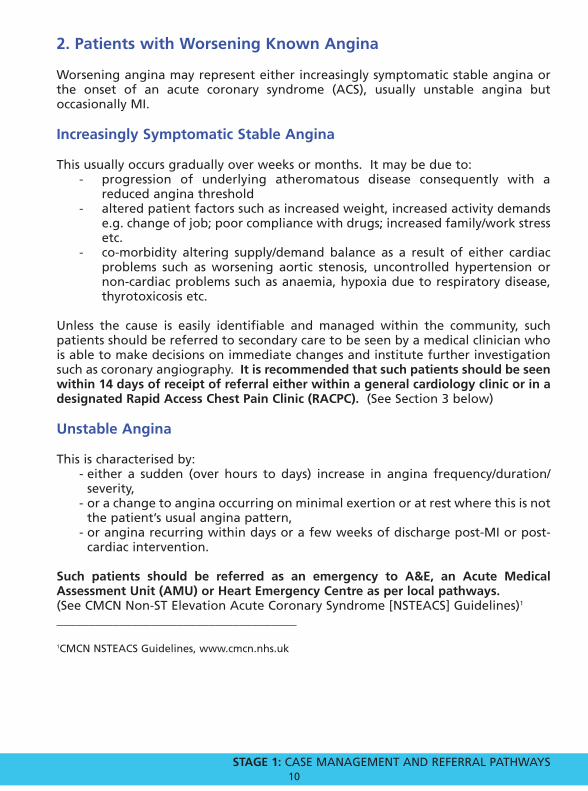
2. Patients with Worsening Known Angina
Worsening angina may represent either increasingly symptomatic stable angina orthe onset of an acute coronary syndrome (ACS), usually unstable angina butoccasionally MI.
Increasingly Symptomatic Stable Angina
This usually occurs gradually over weeks or months. It may be due to:- progression of underlying atheromatous disease consequently with a
reduced angina threshold- altered patient factors such as increased weight, increased activity demands
e.g. change of job; poor compliance with drugs; increased family/work stressetc.
- co-morbidity altering supply/demand balance as a result of either cardiacproblems such as worsening aortic stenosis, uncontrolled hypertension ornon-cardiac problems such as anaemia, hypoxia due to respiratory disease,thyrotoxicosis etc.
Unless the cause is easily identifiable and managed within the community, suchpatients should be referred to secondary care to be seen by a medical clinician whois able to make decisions on immediate changes and institute further investigationsuch as coronary angiography. It is recommended that such patients should be seenwithin 14 days of receipt of referral either within a general cardiology clinic or in adesignated Rapid Access Chest Pain Clinic (RACPC). (See Section 3 below)
Unstable Angina
This is characterised by:- either a sudden (over hours to days) increase in angina frequency/duration/
severity, - or a change to angina occurring on minimal exertion or at rest where this is not
the patient’s usual angina pattern,- or angina recurring within days or a few weeks of discharge post-MI or post-
cardiac intervention.
Such patients should be referred as an emergency to A&E, an Acute MedicalAssessment Unit (AMU) or Heart Emergency Centre as per local pathways.(See CMCN Non-ST Elevation Acute Coronary Syndrome [NSTEACS] Guidelines)1
______________________________________
1CMCN NSTEACS Guidelines, www.cmcn.nhs.uk

STAGE 1: CASE MANAGEMENT AND REFERRAL PATHWAYS11
3. New Patients with Recent Onset Chest Pain – Suspected Angina
Patients suspected to have new onset angina should be referred to a dedicatedoutpatient clinic (RACPC) or reserved rapid access slots within general cardiologyclinics and seen within 14 days of receipt of referral in accordance with mandatorynational requirements.
The rapid access provision should meet the following standards:
• RACPCs should work under protocols set up by a cardiologist. As a minimumstaff must be able to call on a consultant cardiologist although he/she does nothave to be present at all times.
• Initial assessment of the patient should be performed by practitioners skilledand experienced in assessing patients with chest pain and in the interpretationof an exercise ECG.
• The service should have the following diagnostic facilities with results reportedduring the patient’s attendance:
- Essential12 lead resting ECGExercise ECG
- DesirableEchocardiogramChest X-Ray – film to be available to cardiologist and reported by radiologistlater
• Patients who cannot exercise should be seen within 14 days of referral and ifindicated, referred for alternative investigations e.g. perfusion imaging, stressecho, or coronary angiography. (See Stages 2,3 and 4.)
• Patients who are given a confirmed diagnosis of stable angina requireimmediate access to an appropriately trained practitioner to commenceeducation and arrange cardiac rehabilitation follow-up. (See Stage 5.)
• The quality of local GPs’ referral practice to rapid access services should beregularly reviewed by PCTs to ensure it continues to be appropriate.

STAGE 1: CASE MANAGEMENT AND REFERRAL PATHWAYS12
4. Communications
Referral to the rapid access services should be made by the patient’s GP using thedesignated rapid access form delivered electronically or by fax. See sample at end of chapter.
A response to the patient’s GP via fax/e-mail/patient delivery should be madewithin 24 hours of the patient being seen. The content of the response shouldinclude:
• Diagnosis (where this has been made).• Results of investigations.• Follow-up appointments/investigations which have been arranged.• Information as to what treatment changes have been made by the clinic
(e.g. medication changes).• Treatment changes which the GP is asked to make.• Information/advice which has been given to the patient.


STAGE 2DIAGNOSIS
STA
GE
2
STAGE 2DIAGNOSIS
STA
GE
2

STAG
E 2:DIA
GN
OSIS
13
Summary
ALGORITHM 2DIAGNOSIS
STAGE 2DIAGNOSIS
CLINICAL ASSESSMENTChest pain features Table 3 History and risk factors Table 4
Physical examination Table 5 12 lead ECG and bloods
<20 %
Known CAD
Unable to exercise/Ex. ECG contra-indicated Table 7
Exercise ECG/stress imaging
for riskstratification only
if appropriate
Manage riskExclude other causes - echoFurther cardiac Ix - only ifexclusion of CAD essential
Coronary angiographyunless contra-indicatedor unlikely to affectmanagement
RISK STRATIFICATION Tables 12,13,14
Review medical treatment,risk factor modification
Exercise ECG usingBruce protocol
AbnormalNormal
No known CAD-suspected angina
CADunlikely
Contraindicated Table 9 or inconclusive.Consider angiography for diagnosis
Consider angiography ifrevascularisation to beconsidered for symptoms
>80%
Inconclusive
Consider angiography ifrevascularisation to beconsidered for symptoms
PRE-TEST PROBABILITY OF CAD Table 6
POST-TEST PROBABILITYOF CAD Table 8
>20 - <30% 30 - <80%
Stress imaging>50%CAD
probable
<20%CAD
unlikely
>20 - <50%CAD possible
HighIntermediateLow
Select techniqueTable 11

STAGE 2: DIAGNOSIS14
1. Use of Algorithm 2
Algorithm 2 demonstrates the recommended investigational pathway for diagnosisand risk stratification of patients. In practice, these assessments are conducted intandem as the same clinical and investigational tools are used for both. However,for clarity of understanding and presentation, they are described sequentially asstages 2 and 3 and coronary angiography which is used for both diagnostic andprognostic purposes is described separately as stage 4.
The algorithm demonstrates the pathways for patients presenting with suspectedstable angina without previously known CAD and for patients with known CADwho present with recurrent or worsened stable angina. In this latter group, thediagnosis is rarely in doubt and further diagnostic tests add little since they are ahigh prevalence population with high pre-test probability. Therefore, the focus ofnon-invasive testing should be risk stratification and prognosis.
Use of Algorithm 2 will lead to a logical, evidence-based and cost-effectiveapproach. It should be used as the basis for decision-making as to theappropriateness of invasive coronary angiography with a view to interventionaltreatment by percutaneous coronary intervention (PCI) or coronary arterial bypassgraft (CABG).
The algorithm also recognises and endorses as good practice, the direct applicationof coronary angiography without preliminary non-invasive testing in some patientgroups. Thus in many patients with known CAD, or those with a very high (≥ 80%)pre-test probability of CAD who, despite appropriate medical treatment, havesevere limiting or worsening typical angina, non-invasive testing is oftenunnecessary and direct listing for coronary angiography is appropriate.
2. Clinical Assessment
History
This should include consideration of the following aspects:
• Chest pain - the initial suspicion or presumptive diagnosis of angina is usuallybased on the patient’s description of the pain.
Table 3
STAGE 2DIAGNOSIS
CLASSIFICATION OF CHEST PAINTypical angina Substernal chest discomfort with characteristic quality and duration
Provoked by exertion or emotional stress Relieved by rest or nitroglycerin (GTN)
Atypical angina Meets two of the above characteristics
Non-cardiac chest pain
Meets one or none of the above characteristics

STAGE 2: DIAGNOSIS15
• Patient setting - the likelihood of a chest pain being angina whatever itsfeatures is highly dependent on the patient setting in which it occurs. Evidenceof previous/known CVD or the co-existence of vascular risk factors increases thelikelihood of angina.
Table 4
Examination
This is usually diagnostically less helpful than the history but signs may existsupporting an ischaemic origin, suggesting an alternative cardiac cause, or pointingpositively to a non-cardiac cause.
Table 5
Baseline Tests
• Blood tests – all patients should have haemoglobin; blood glucose (preferablyfasting); lipid profile including total cholesterol, HDL cholesterol andtriglycerides; urea and electrolytes. Where appropriate other tests may benecessary including thyroid function, liver function, troponin etc.
PATIENT SETTINGEvidence of CVD Risk factors
Known IHDPrevious CVA, TIAKnown PVD
Age- M >40 yrs; F >50 yrsGender - M > FFamily IHD history -especially premature M<50 yrs ; F<60 yrs SmokingDyslipidaemia HypertensionDiabetes Mellitus
PHYSICAL EXAMINATION
Ischaemia Non- ischaemic cardiac Non-cardiac
Usually normal.Arrhythmia
-AF, SVT, VT, bradycardia.
LV dysfunction-S3,pulmonary oedema.
Pericardial rub.Valvular disease
-especially AS.Cardiomyopathy
-LVH, CCF.Aortic dissection
-AR, differential armpulses or BP.
Musculo-skeletal-chest wall tenderness, positive physical manoeuvres.
Respiratory-pleural rub, pneumothorax, consolidation.
Other-pyrexia, rash, epigastric Tenderness

STAGE 2: DIAGNOSIS16
• ECG – a 12 lead resting ECG should be done in all patients. It will be normal in≥50% of patients. This does not exclude CAD but does strongly imply normalresting left ventricular (LV) function and hence a favourable prognosis. Evidenceof prior Q wave MI, LV hypertrophy (LVH), or ST/T wave changes, consistent withmyocardial ischaemia, favour a diagnosis of angina. An ECG done during painadds greatly to its otherwise poor diagnostic ability.
• Chest X-ray – this does not add specific diagnostic or prognostic information andis therefore not a routine test. It should be done in patients with suspectedheart failure, valvular disease or pulmonary disease, (including smokers whohave not had a chest X-ray in the last year).
• Echocardiogram – this is not a routine test for angina assessment. It should onlybe requested for (a) patients with a systolic murmur suggestive of aortic stenosis,mitral regurgitation or hypertrophic cardiomyopathy or (b) to assess LV functionin patients with signs, symptoms or ECG suggestive of heart failure or LVdysfunction.
3. Pre-Test Probability of CAD
Concept of Disease Probability or Likelihood
The presence of CAD and hence a diagnosis of angina cannot be confirmed or refutedwith 100% certainty by non-invasive means i.e. clinical assessment or non-invasivetesting. A Bayesian approach to diagnosis that deals with probabilities shouldtherefore be adopted. This approach uses the clinician’s pre-test estimate of diseaselikelihood and then modifies it on the basis of the results of diagnostic tests togenerate individualised post-test disease probabilities for a given patient.
The pre-test probability depends on the prevalence of the disease in the populationstudied as well as the individual’s clinical features especially age, gender, risk factorprofile and chest pain type.
In populations with a low prevalence of CAD e.g. young, female, with no risk factorsand atypical pain, a positive test result will have a much higher chance of being afalse positive than the same result in a high prevalence population e.g. 60 years old,male, with diabetes and with typical chest pain.
See Table 6.


STAG
E 2:DIA
GN
OSIS
17
Table 6
PRE-TEST PERCENTAGE PROBABILITY OF CAD ADJUSTED FORTHE RISK FACTORS: SMOKING, DIABETES AND HYPERLIPIDAEMIA1
MEN
WOMEN
No of risk factors No of risk factors No of risk factors
No of risk factors No of risk factors No of risk factors
Non-cardiac chestpain
Atypical chest pain Typical angina
Non-cardiac chestpain
Atypical chest pain Typical angina
0 1 32 0 1 32 0 1 32
1 7 1813 2 14 3926 10 32 7855
2 8 2115 5 17 4330 20 39 7959
4 11 2518 10 22 4734 38 52 8267
9 15 2922 20 30 5140 56 65 8474
0 1 32 0 1 32 0 1 32
3 14 3525 8 25 5942 30 49 8868
9 21 4734 21 37 7053 51 64 9278
23 35 5947 45 56 7967 80 85 9590
49 55 6962 71 76 8681 93 94 9795
Chest paintype
Chest paintype
Age
30-39
40-49
50-59
60+
Age
30-39
40-49
50-59
60+
Definitions ECG%Probability
Smoking: > half a pack per day in lastfive years or 25 pack yearsHyperlipidaemia: cholesterol >6.5mmol/l
The above values are for patientswith a normal resting ECG.If ST-T wave changes or Q wavesare present, the likelihood ofCAD will be higher
> 80
> 30 < 80
> 20 < 30
< 20

STAGE 2: DIAGNOSIS18
4. Non-Invasive Diagnostic Testing to Produce Post-testProbability of CAD
These tests are used to detect signs of myocardial ischaemia during stress and henceto improve the clinically based pre-test estimate of probability of CAD. Relevantinvestigations are exercise/stress tolerance testing with ECG recording (exerciseECG), usually using a motorised treadmill or occasionally a bicycle; myocardialperfusion scintigraphy (MPS); stress echo using dobutamine (DSE)
Exercise ECG
For reasons of availability and cost, an exercise ECG using the Bruce protocol is theinitial test of choice. It should be performed for diagnostic purposes in all patientsin whom the pre-test probability lies in the range ≥ 30- <80% and in whom there areno contra-indications.
Table 7
Outside the range ≥ 30% to <80%, the test has little diagnostic accuracy producingexcessive false positives in very low prevalence populations and failing to add to thepre-test probability in very high prevalence populations. Within this range, itsaccuracy is reasonable having an overall sensitivity of 68% and a specificity of 77%for the detection of significant CAD using a diagnostic threshold of 1mm horizontalor down-sloping ST depression.
CONTRA-INDICATIONS TO DIAGNOSTIC EXERCISE ECG TESTINGAbsolute contra-indications- Uncontrolled hypertension:>200 mmHg systolic and/or >110 mmHg diastolic- LBBB on ECG- Pre-excitation pattern i.e. delta waves- Paced rhythm- Uncontrolled arrhythmia- Suspected unstable angina - More than 2 mm resting ST depression, particularly if the patient is on digoxin- Acute myocarditis or pericarditis- Uncontrolled, symptomatic heart failure- Symptomatic severe aortic stenosis- Inability to perform exercise ECG due to co-morbidity or disability
Relative contra-indications – caution required- Suspected significant outflow tract obstruction due to moderate aortic stenosis
or hypertrophic obstructive cardiomyopathy- Other significant valvular disorder e.g. mitral stenosis or aortic regurgitation- Recent ACS: MI/high risk unstable angina ≤ 3 weeks; known left main stem stenosis- High degree atrioventricular block


STAG
E 2:DIA
GN
OSIS
19
The degree of ST-depression is the principal diagnostic, as opposed to prognostic, outcome from an exerciseECG. This should be factored into known pre-test clinical variables to obtain a refined and individualisedpost-exercise ECG probability of CAD as shown in Table 8 below.
Table 8
POST-EXERCISE ECG PROBABILITY OF CAD 2
MEN
WOMEN
ST - depression mm
Non-cardiac chest painChestpain type
Age
30-39
40-49
50-59
60+
Atypical chest pain Typical angina
ST - depression mm ST - depression mm
ST - depression mm
Non-cardiac chest painChestpain type
Age
30-39
40-49
50-59
60+
Atypical chest pain Typical angina
ST - depression mm ST - depression mm
<0.4 0.5 -0.9
1.5-1.91-1.4 >2.52.0-
2.4
1 5 1910 6839
4 13 4126 8765
6 20 5337 9175
8 26 6245 9481
<0.4 0.5 -0.9
1.5-1.91-1.4 >2.52.0-
2.4
6 21 5538 9276
16 44 7864 9791
25 57 8675 9894
32 65 8981 9996
<0.4 0.5 -0.9
1.5-1.91-1.4 >2.52.0-
2.4
25 68 9183 9996
61 86 9794 >9999
73 91 9896 >9999
79 94 9997 >9999
<0.4 0.5 -0.9
1.5-1.91-1.4 >2.52.0-
2.4
<1 1 32 248
1 3 116 5324
2 8 2816 7850
5 17 4933 9072
<0.4 0.5 -0.9
1.5-1.91-1.4 >2.52.0-
2.4
1 4 159 6333
3 12 3925 8663
10 31 6750 9584
21 52 8372 9893
<0.4 0.5 -0.9
1.5-1.91-1.4 >2.52.0-
2.4
7 24 5942 9379
22 53 8472 9893
47 78 9489 9998
69 90 9895 9999
%Probability > 50 > 20 <50 < 20

STAGE 2: DIAGNOSIS20
Stress Imaging
Stress imaging should be considered if there are no absolute contra-indications (seeTable 9), in the following situations:• When pre-test probability of CAD is in the range ≥ 20 <30%. (See Table 6)• When exercise ECG is contra-indicated. (See Table 7)• When post-exercise ECG probability is in the range ≥ 20 <50 %. (See Table 8)
Table 9
The two techniques currently appropriate in the CMCN area are:• Myocardial perfusion scintigraphy (MPS) involving single photon emission
computed tomography (SPECT); technetium (sesta) methoxy-isobutyl-isonitrile(MIBI) as the radiotracer; and adenosine, dipyridamole, dobutamine or exerciseas the stress agent.
• Stress echocardiography with dobutamine as the stress agent.
There is little difference between the two techniques in terms of accuracy. (Table 10)
Table 10
In the majority of cases, the choice of technique should be based on local availability,including waiting times, and expertise. In some clinical scenarios, however, one orother test is to be preferred. (See Table 11)
ABSOLUTE CONTRA-INDICATIONS TO STRESS IMAGING
- Uncontrolled hypertension: >200 mmHg systolic and/or >110 mmHg diastolic- Suspected unstable angina or acute MI- Acute myocarditis or pericarditis- Uncontrolled, symptomatic heart failure- Symptomatic severe aortic stenosis
PHYSICAL EXAMINATION
Test Sensitivity % Specificity %
Exercise MPS 85-90 70-75
Vasodilator stress MPS 83-94 64-90
Dobutamine stress echo 40-100 62-100

STAGE 2: DIAGNOSIS21
Table 11
If there is a lengthy delay in the local waiting time for stress imaging the clinicianmay consider in individual cases that it is in the patient’s interest to proceed directlyto coronary angiography.
STRESS IMAGING: COMPARATIVE INDICATIONS
Indication MPS with SPECT Stress Echocardiography
Suitability Comment Suitability Comment
Diagnosis of ischaemia Yes More sensitive yes More specific
Assessment ofmyocardial viability
Yes More sensitive yes More specific
Patients with pacedrhythm/LBBB
Yes no
Poor echo subject Yesyes withlimitations
Contrast mayovercomelimitation
Patients withasthma/COPD
yes withlimitations
Use dobutamine inpharmacologicstress imaging sinceadenosine anddipyridamole arecontra-indicated
yes withlimitations
Reduced imagequality likely
More comprehensivecardiac examinationrequired
No yes
Assessment of valvularlesions (low-grade AS withLV dysfunction, MS/MR)
No yes

STAGE 2: DIAGNOSIS22
Other Imaging Techniques
Cardiac magnetic resonance is rapidly emerging as a technique capable of providinghighly accurate diagnostic information regarding myocardial ischaemia andmyocardial viability as well as cardiac anatomy and function. It is currently limitedby availability and cost and should be reserved for special cases after discussion withthe appropriate provider.
The spatial and temporal resolutions of computed tomography (CT) have improvedenormously. Multi-detector or multi-slice CT (MDCT) shows great promise for non-invasive coronary imaging – 90-94% sensitivity, 95-97% specificity, 93-99% negativepredictive accuracy for 64 detector scanners. At present, its place in theinvestigational hierarchy for angina is unknown. A cardiac MDCT service is currentlyunavailable locally.
1 Adapted from ACC/AHA Guidelines. Ref Duke Database: Pryor et al. Ann of Intern Med 1993; 118: 81-902 Adapted from ESC Guidelines. Ref Diamond GA et al Application of information theory to clinicaldiagnostic testing. The electrocardiographic stress test. Circulation 1981; 63:915-21


STAGE 3RISK STRATIFICATION
STA
GE
3
STAGE 3RISK STRATIFICATION
STA
GE
3
STAG
E 3:RISK
STRA
TIFICA
TION
23
Summary
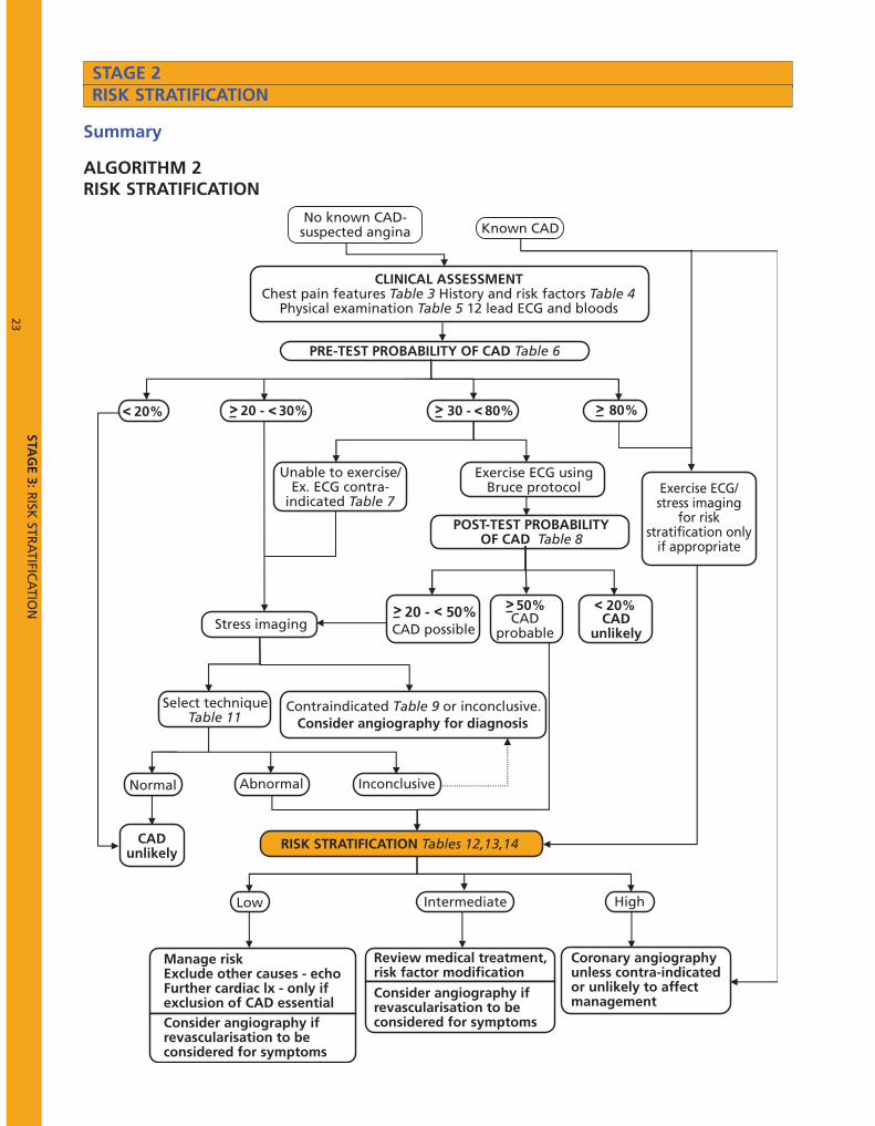
ALGORITHM 2RISK STRATIFICATION
STAGE 2RISK STRATIFICATION
<
Abnormal Normal
Contraindicated Table 9 or inconclusive.Consider angiography for diagnosis
Inconclusive
< <
<20 - < 50%
HighIntermediateLow
No known CAD-suspected angina Known CAD
CLINICAL ASSESSMENTChest pain features Table 3 History and risk factors Table 4
Physical examination Table 5 12 lead ECG and bloods
PRE-TEST PROBABILITY OF CAD Table 6
20% 20 - 30% 30 - 80% 80%
Unable to exercise/Ex. ECG contra-
indicated Table 7
Exercise ECG usingBruce protocol Exercise ECG/
stress imagingfor risk
stratification onlyif appropriate
POST-TEST PROBABILITYOF CAD Table 8
Stress imaging CAD possibleCAD
probable
50%CAD
unlikely
20%
Select techniqueTable 11
CADunlikely
RISK STRATIFICATION Tables 12,13,14
Manage riskExclude other causes - echoFurther cardiac lx - only ifexclusion of CAD essentialConsider angiography ifrevascularisation to beconsidered for symptoms
Review medical treatment,risk factor modification
Consider angiography ifrevascularisation to beconsidered for symptoms
Coronary angiographyunless contra-indicatedor unlikely to affectmanagement

STAGE 3: RISK STRATIFICATION24
The long-term prognosis of stable angina varies widely from individual to individual,perhaps up to tenfold. The assessment of risk helps to determine the optimumtreatment regime for an individual and in these guidelines risk is defined in termsof annual CV mortality as: - high >3%- intermediate 1-3%- low <1%A reasonable estimate of risk can be obtained simply from clinical evaluation(history and ECG) or from invasive coronary angiography. However, it isrecommended that initially a quantitative estimate be obtained wherever possiblefrom the results of the same non-invasive tests used for diagnostic purposes.
Table 12 outlines the use of the Duke Treadmill Score for risk stratification byexercise ECG whilst Tables 13 and 14 give prognostic stratification criteria for MPSand stress echocardiography respectively.
Table 12
STAGE 3RISK STRATIFICATION
RISK STRATIFICATION: EXERCISE ECGDuke Treadmill Score calculationTotal score = time in minutes on Bruce protocol – 5 x maximum ST-deviation in any lead in mm – 4 x angina index (0 = no angina, 1 = angina not limiting, 2 = limiting angina)
Duke Treadmill Score Risk Group• ≤ –11 High
• -10 to +4 Intermediate
• ≥ +5 Low

STAGE 3: RISK STRATIFICATION25
Table 13
Table 14
RISK STRATIFICATION: MYOCARDIAL PERFUSION SCINTIGRAPHY
Finding Risk group• Stress-induced large perfusion defect (particularly if
anterior)• Stress-induced multiple perfusion defects of moderate size• Large fixed perfusion defect with LV dilation or increased
lung uptake (thallium-201)• Stress induced moderate perfusion defect with LV dilation
or increased lung uptake (thallium-201)
High
• Mild/moderate resting LV dysfunction (LVEF = 35% - 49%)• Stress induced moderate perfusion defect without LV
dilation or increased lung intake (thallium-201)
Intermediate
• Normal or small myocardial perfusion defect at rest orwith stress**
Low
** Patient with these findings will probably not be at low risk in the presence of either a highrisk treadmill score or severe resting left ventricular dysfunction (LVEF<35%))
RISK STRATIFICATION: STRESS ECHOCARDIOGRAPHY
Finding Risk group• Extensive ischaemia with wall motion abnormality
involving >2 segments developing at low dose ofdobutamine
• (< 10mg/kg/min) or at a low heart rate (<120 beats/min)
High
• Mild/moderate resting LV dysfunction (LVEF 35% - 49%)
• Limited ischaemia with a wall motion abnormality involving< 2 segments only at higher doses of dobutamine
Intermediate
• Normal wall motion or no change of limited resting wallmotion abnormalities during stress**
Low
** Patient with these findings will probably not be at low risk in the presence of either a highrisk treadmill score or severe resting left ventricular dysfunction (LVEF<35%))

STA
GE
4STAGE 4CORONARY ANGIOGRAPHY ST
AG
E 4STAGE 4
CORONARY ANGIOGRAPHY

STAG
E 4:CO
RO
NA
RY A
NG
IOG
RA
PHY
26
Summary
ALGORITHM 2CORONARY ANGIOGRAPHY
STAGE 4CORONARY ANGIOGRAPHY
<
Abnormal Normal
Contraindicated Table 9 or inconclusive.Consider angiography for diagnosis
Inconclusive
< <
<20 - < 50%
HighIntermediateLow
No known CAD-suspected angina Known CAD
CLINICAL ASSESSMENTChest pain features Table 3 History and risk factors Table 4
Physical examination Table 5 12 lead ECG and bloods
PRE-TEST PROBABILITY OF CAD Table 6
20% 20 - 30% 30 - 80% 80%
Unable to exercise/Ex. ECG contra-
indicated Table 7
Exercise ECG usingBruce protocol Exercise ECG/
stress imagingfor risk
stratification onlyif appropriate
POST-TEST PROBABILITYOF CAD Table 8
Stress imaging CAD possibleCAD
probable
50%CAD
unlikely
20%
Select techniqueTable 11
CADunlikely
RISK STRATIFICATION Tables 12,13,14
Manage riskExclude other causes - echoFurther cardiac lx - only ifexclusion of CAD essentialConsider angiography ifrevascularisation to beconsidered for symptoms
Review medical treatment,risk factor modification
Consider angiography ifrevascularisation to beconsidered for symptoms
Coronary angiographyunless contra-indicatedor unlikely to affectmanagement

STAGE 4: CORONARY ANGIOGRAPHY27
Coronary angiography holds a fundamental position in the investigation of patientswith stable angina. It provides reliable anatomical information to identify thepresence or absence of coronary lumen stenosis; defines therapeutic optionsincluding the suitability of medical treatment or myocardial revascularisation anddetermines prognosis by defining the extent and severity of coronary artery stenosis.This allows classification into one-two-three vessel disease or left main stem CAD.
However, it is important to recognise its limitations which include the following:- It does not diagnose coronary atheroma since vessel wall disease may be present
when the lumen is normal.- It does not give information on myocardial ischaemia since it does not assess the
functional importance of any anatomical stenosis.- It is insensitive in detection of a thrombus.- It is ineffective in determining which plaques have characteristics likely to lead
to acute coronary events. Plaques resulting in unstable angina and MIcommonly produce less than 50% stenosis before the acute event and willtherefore be angiographically "silent".
Complications and Consent
In the vast majority of cases, coronary angiography for stable angina should be a daycase procedure. It can be carried out with adequate quality and safety in either atertiary or DGH setting. However, it is an invasive investigation and as such hasinherent risks and complications. The composite rate of death, MI or strokeassociated with routine diagnostic catheterisation in patients is of the order of 0.1%to 0.2%. The composite rate of major complications is about 1%. Where possiblethe complication rate of the relevant hospital should be known and quoted. Inmaking the decision to proceed to invasive coronary angiography, it is important totake into account the patient’s willingness to accept the risks of the procedure andhis/her willingness to proceed to any therapeutic intervention that might arise.Obviously, it is important to consider the patient’s suitability on the basis ofcomorbidity and frailty.
STAGE 4CORONARY ANGIOGRAPHY

STAGE 4: CORONARY ANGIOGRAPHY28
Recommendations for Angiography
Algorithm 2 indicates the group of patients identified as high risk on clinicalassessment or after non-invasive investigation who should undergo coronaryangiography on prognostic grounds unless contra-indicated or unlikely to affectmanagement. (ACC/AHA Class I recommendation)
The algorithm also identifies patients at low or intermediate risk for whomrevascularisation might be considered for symptom control. (ACC/AHA Class Irecommendation for patients with CCS Class III and IV angina despite medicaltherapy.)
Table 15 lists the above indications and identifies additional patient groups whomay benefit from angiography:
Table 15
Audit
Each institution should make adequate provision for quality assurance and audit ofits catheter laboratory procedures. Quality assurance requires that angiography becarried out by practitioners competent in the procedure or by trainees underadequate supervision and that operators carry out sufficient cases per year tomaintain competence. In addition, CMCN has established a data set for coronaryangiography which will aid quality assurance, allow bench marking and underpinaudit.
RECOMMENDATIONS FOR CORONARY ANGIOGRAPHYPatient groups ACC/AHA
ClassIdentified as high risk (annual CV mortality >3%) IPatients with CCS Class III and IV for whom revascularisation might beconsidered for symptom control
I
Patients with angina who have survived sudden cardiac death or seriousventricular arrhythmia
I
Patients with angina and symptoms and signs of congestive cardiac failure IPatients with an uncertain diagnosis after non-invasive testing in whomthe benefit of a more certain diagnosis outweighs the risk of coronaryangiography
IIa
Patients who cannot undergo non-invasive testing due to disability,illness or morbid obesity
IIa
Patients with an occupational requirement for a definitive diagnosis IIaPatients with inadequate prognostic information IIa

STAGE 5LIFESTYLE AND RISK FACTOR MODIFICATION
STA
GE
5
STAGE 5LIFESTYLE AND RISK FACTOR MODIFICATION
STA
GE
5

STAGE 5: LIFESTYLE AND RISK FACTOR MODIFICATION29
Summary
Comprehensive angina management requires, in addition to drug therapy (seeStage 6) and revascularisation (see Stage 7), close attention to:
• Lifestyle issues including advice on smoking, diet, weight reduction, alcoholconsumption, driving, sexual intercourse and physical activity/exercise.
• Modifiable risk factors for atherosclerosis including hypertension, diabetes anddyslipidaemia.
• Psychosocial factors including depression, anxiety, misconceptions, negativebehaviour patterns and poor coping mechanisms.
• Education of patient’s family and carers.
Whilst all relevant healthcare professionals should deal with aspects of these issuesduring the patient’s multiple contacts in primary care and hospital, they are mosteffectively and comprehensively addressed by cardiac rehabilitation practitioners. Provision should be made locally for all patients with newly diagnosed stableangina to have at least an initial consultation with a cardiac rehabilitationpractitioner. Local resource constraints may mean that it is not possible to offer allpatients follow-up in a comprehensive programme tailored to their needs.However, the initial consultation will as a minimum, enable the practitioner to assessthe patient, provide him/her with a personal plan and identify any major areas ofconcern which will require specific professional intervention.
Continuing physical or psychosocial issues not resolved by treatment (drugs orrevascularisation), or by standard rehabilitation should lead to referral to arefractory angina service.
STAGE 5LIFESTYLE AND RISK FACTOR MODIFICATION

STAGE 5: LIFESTYLE AND RISK FACTOR MODIFICATION30
1. Cardiac Rehabilitation
Comprehensive cardiac rehabilitation consists of exercise training together witheducation and psychological support. The purpose of these interventions is tofacilitate a return to normal living and to encourage patients to make lifestylechanges in order to prevent further events. It has much in common with, and linksto, the provision of long-term secondary prevention co-ordinated in general practicebut takes the opportunity, following a significant cardiac event to provide a patientand his/her family with more intensive support. The support is given over arelatively short period, through a programme delivered in a group or individuallythat should be tailored to meet the patient’s particular needs.
Several meta-analyses of randomised controlled trials (RCTs) in exercise basedcardiac rehabilitation programmes since the 1980’s have demonstrated that theseprogrammes reduce all cause and cardiac mortality in patients with coronary arterydisease.1 Although most of the trials have been in patients post-MI, the presence ofthe same underlying pathology in patients with stable angina means that the likelybenefit to this group should not be ignored.
A more recent meta-analysis of 63 RCTs demonstrated that benefit was notrestricted to exercise based programmes.2 It covered three types of programmedelivered individually or in a group: (a) combined education, risk factor counsellingand supervised exercise; (b) combined education and risk factor counselling with nosupervised exercise; and (c) supervised exercise alone. All types of programme werefound to improve processes of care, coronary risk factor profiles, functional statusand quality of life. In all types, a reduction in all cause mortality was absent at 12months but was demonstrable at two years and sustained at five years.
Effective cardiac rehabilitation recognises the need to design services that takeaccount of the diverse needs of the population in terms of age, gender, impairment,literacy, ethnicity, religious practice, cultural diversity, income, employment,dependents, and carers. Failure to design services appropriately has been shown tolead to inequality of patient access and poor compliance.
The design of trials has been such that the optimal mix of interventions, includingfrequency and duration and the incremental benefit of the various components,remains unclear. The detail described below reflects the local consensus viewinformed by current international guidelines.
All cardiac rehabilitation services should use the national data set3 to ensureconsistent record keeping of the patient’s ongoing management, effectivemonitoring of patient outcomes to inform future service development andcomprehensive audit.
STAGE 5LIFESTYLE AND RISK FACTOR MODIFICATION

STAGE 5: LIFESTYLE AND RISK FACTOR MODIFICATION31
Priorities for Referral
In addition to the NSF requirements of post-MI and post-revascularisation, it isrecommended that the following cohorts of patients with stable CAD be offeredcardiac rehabilitation in the priority order indicated:
1. All patients newly diagnosed with stable angina.
2. Patients previously diagnosed with stable angina who are experiencing severeadaptation problems such as issues with activity levels, weight control, adherence tomedication etc.
3. Patients waiting for coronary revascularisation who will benefit from input toprepare for the procedure (‘pre-hab’) and afterwards to aid recovery.
Pathway at Point of Diagnosis
The practitioner initially informing the patient of his/her diagnosis of angina shouldensure that the steps indicated in Table 16 have been taken.
Table 16
Initial Consultation
The initial consultation should include:
• Assessment of psychological well beingA psychological tool for assessment should be used to identify problems.4 It isimportant to recognise that psychological factors can often act as a barrier to therehabilitation process. Concerns related to living with angina can influence mood,degree of disability and quality of life. Beliefs and misconceptions about heartdisease have been shown to influence outcome, as has presence of depression andanxiety. If serious or pre-existing psychological problems are identified, see relevantsub-section below.
PATHWAY AT POINT OF DIAGNOSIS• A clear easily understood explanation about what will happen next should be given.
• A leaflet/booklet explaining about angina, such as a British Heart Foundation advicebooklet or locally developed version should be provided. (With appropriatealternatives for non-English speakers and patients with vision or reading difficulties.)
• A referral should be made to the local Cardiac Rehabilitation Service and contactdetails of the service should be provided to the patient.
• Patients should be encouraged to continue normal daily activities, unless there arecompelling reasons to the contrary. To advise against activity can be unhelpful to thepatient’s future rehabilitation prospects and can cause undue distress.

STAGE 5: LIFESTYLE AND RISK FACTOR MODIFICATION32
• Risk factor profile assessment and individual advice on managementLifestyle intervention to discontinue smoking, make healthier food choices, increaseaerobic physical activity, achieve optimal weight, and weight distribution is centralto CVD prevention. These lifestyle issues and the control of blood pressure,cholesterol and glucose can be addressed more intensively in a comprehensiverehabilitation programme. However, the consultation at least provides anopportunity to set goals with patients in relation to their modifiable risk factors.
• Fitness assessment and adviceThis will be informed by the cardiologist’s/GP’s assessment, exercise ECG results andthe patient’s pre-morbid and current fitness/activity levels. Recognised fitnessassessment tools may be used, for example the Shuttle Walk test. The assessment isprimarily intended to plan the supervised exercise element of a comprehensiveprogramme that will be carried out in a group or individually at home. However, itwill also provide useful information about safe levels of exercise during dailyactivities.
• Patient educationThis will include advice about medication, angina, symptoms, how to recognise chestpain, how to treat it and when to call for help. It is important to dispel mythsaround angina and promote the importance of adherence to medication.
Advice should also be given about social, cultural, family and carer needs, and issuessuch as returning to work, travel, driving, insurance cover and sexual relationships.
Choice of Programme
Following this consultation and in discussion with the patient, an agreed decisionwill be made on the appropriate individualised cardiac rehabilitation pathway to befollowed. Outcomes from the consultation may also reveal a need for referral ontospecialist services such as smoking cessation, diabetic and psychology.
Comprehensive Rehabilitation Programme
The optimal provision for patients with newly diagnosed stable angina is anindividualised programme based on the needs identified at the initial consultationand which includes the following:
• Lifestyle advice e.g. healthy eating, smoking cessation, weight reduction• Goal setting and targeting• Medication advice to ensure adherence• Stress management• Relaxation• Education about the heart, its function and the disease process.• Individually tailored exercise programme• Monitoring and evaluation of outcomes
The principles of adult learning should be adopted in order to improve patients’understanding.
STAGE 5: LIFESTYLE AND RISK FACTOR MODIFICATION33
Exercise programmes should include a mixture of warm-up, pulse-raising activities,strength work and cool-down exercises (as outlined in British Association CardiacRehabilitation [BACR]/American College of Sports Medicine [ACSM] guidelines).They should be delivered by appropriately trained cardiac rehabilitationpractitioners or by suitably qualified exercise professionals (BACR Level IV), working,for example, in leisure centres.
There is no current evidence on the most effective length of a supervised exerciseprogramme. The NSF for CHD suggests that cardiac rehabilitation services shouldaim to provide around two supervised exercise sessions per week for at least sixweeks. It is therefore reasonable to expect that angina programmes work towardsthis level.
Home-Based Programme
Where a centre-based comprehensive programme is unavailable or unsuitable, orwhen a patient decides against this approach due to personal preference, a home-based programme such as The Angina Plan5 or equivalent locally-devisedprogramme should be provided. For some patients, the greater flexibility andfamiliarity of a home-based programme will improve their compliance.
The patient should be assessed by a cardiac rehabilitation practitioner and amanagement plan agreed. The plan requires professional review and monitoringthrough regular contact, usually by telephone. Patients will have a copy of the planthat will be evaluated upon completion. Where patient goals have not been met, areassessment should take place.
The Angina Plan can be delivered by a variety of health professionals who haveundergone the required training and assessment.
Serious Pre-existing Psychological Problems
Patients identified as having serious or pre-existing psychological problems atassessment should be referred in consultation with their GP to local specialisedservices. If there is a significant delay between referral and consultation, regularcontact should be maintained with the patient by the Cardiac Rehabilitation Servicewherever possible.
Continuing Angina
Cardiac rehabilitation practitioners should consider referring those patients whocontinue to have adaptation/coping difficulties, despite intervention from the localservice, to the National Refractory Angina Centre (NRAC) for further managementincluding motivational psychotherapy and multidisciplinary cognitive behaviouraltherapy (CBT).
For further details about refractory angina and sample NRAC referral form, seeSection 2.

STAGE 5: LIFESTYLE AND RISK FACTOR MODIFICATION34
Communication with Primary Care
Clear communication with primary care should take place for all patients at thefollowing key points:
• Following initial diagnosis.• After initial consultation with cardiac rehabilitation practitioner, outlining the
next steps which will have taken account of the patient’s preferences and theservices available locally
• Following the patient’s completion of the rehabilitation programme.
Long Term Management
The patient’s GP practice should ensure that the patient is placed on the heartdisease register and offered regular follow-ups. These follow-ups will include checkson blood pressure and cholesterol, weight management and medication review. Itis an opportunity to assess angina status and reinforce positive lifestyle choices.
In order to help patients maintain the beneficial changes achieved by cardiacrehabilitation, referral to an exercise programme run in the patient’s localcommunity should be made. Usually described as Phase IV cardiac rehabilitation,these programmes are often provided by local authority and voluntary organisationssuch as heart support groups. Personnel delivering these programmes should bequalified to BACR IV trainer level and should be in regular contact with the localCardiac Rehabilitation Service. This will help ensure continuity of care.
Staffing
Cardiac rehabilitation practitioners will have a variety of professional backgroundsbut should be trained and experienced in line with the competencies identified inthe NHS Knowledge and Skills Framework.6 In particular, they should be assessed aspossessing the following:
Specialist knowledge• Cardiopulmonary anatomy and physiology. • Cardiovascular disease process, major diagnoses, the implications of modifiable
risk factors, frequently used drugs and their complications.• Research-based evidence of the impact of environmental, social, lifestyle and
behavioural factors on the incidence of CVD and of the impact of CVD onindividuals and their families.
• Principles and practice of adult learning.
Specialist skills• Cardiac risk stratification • Assessment of psychological, social and emotional needs • Assessment of cardiopulmonary capacity • Monitoring cardiovascular and pulmonary responses to exercise• Methods of monitoring to ensure patient safety

STAGE 5: LIFESTYLE AND RISK FACTOR MODIFICATION35
2. Refractory Angina
Definition and Epidemiology of Refractory Angina
Chronic refractory angina pectoris (CRA) is a clinical diagnosis. It is based on thepresence of symptoms of stable angina due to myocardial ischaemia resulting fromadvanced CAD which persists despite optimal anti-anginal therapy. Such patientstherefore present with continuing angina which significantly impairs the quality oftheir lives and which is not responding to optimal anti-anginal drug therapy and isnot amenable to any form of coronary intervention such as PCI or CABG.
It is a distressing chronic pain condition that causes severe reduction in the qualityof life of both patients and their families. Typically such patients complain of amyriad of problems causing repeat and often protracted hospital admissions. Theseproblems can be exacerbated by lack of support and understanding of theircondition that in turn adds to their continuing general deterioration.
Patients attending a refractory angina clinic are a heterogeneous cohort made up ofsub-groups whose angina is refractory to conventional medical therapy for one ormore of the following reasons:
• PCI and CABG are not technically feasible e.g. because of severe distal disease. • PCI and/or CABG have already been carried out on one or more occasion and
further intervention is deemed to carry unjustifiable risks.• Comorbidity precludes coronary intervention. • Patient choice not to undergo further coronary intervention.
The current prevalence of CRA is said to be one in ten thousand and its incidenceone in twenty thousand with both rates increasing year on year. The average age issixty three years and 70% are male. Generally, there is a long history of CAD (>8years) which is usually three vessel disease (>68%) and often in association withsome LV impairment.
It has been estimated that there will be approximately 1800 patients with recurrentmoderate to severe angina post-CABG in the North West region within the next tenyears. Two thirds of this group (1200 patients) will not be amenable to furtherrevascularisation and hence will become CRA patients. In addition, within the sameperiod an approximately equal number of patients will have CRA without priorCABG due to unrevascularisable disease.

STAGE 5: LIFESTYLE AND RISK FACTOR MODIFICATION36
Refractory Angina Management at National Refractory AnginaCentre (NRAC)
Treatment aims and the treatment contract
The primary aim of therapy is to maximise the patient’s quality of life byameliorating the effects of the condition without jeopardising quantity of life. Thepatient and their carers need time and help to define how angina impairs theirquality of life and what level of recovery would be acceptable. These are difficultconcepts in the present care system which is a largely pathology or disease-basedmedical treatment paradigm.
The ideal doctor/patient relationship exists when the patient, their carers and doctorcan openly 'negotiate' a treatment contract with clearly stated aims and objectives.In this way the choice of therapy becomes simplified for the doctor and it enablesthe patient to make more rational decisions about which therapy is mostappropriate to his/her particular needs.
Core Programme
• Patient Education
Education to promote both the patient’s and carer’s understanding of theircondition and the available treatments is a core component of the NRAC care modelin line with internationally accepted best practice guideline recommendations.
The programme consists of an initial three-hour multi-disciplinary assessment anddiagnostic day case clinic. On entry into the programme, patients are invited todefine their objectives. All patients are given the Angina Plan, the NRAC anginamanual and other relevant material including information on each of the treatmentmodalities offered.

STAGE 5: LIFESTYLE AND RISK FACTOR MODIFICATION37
• Therapeutic options for pain management
A broad menu of available therapeutic options has been developed for painmanagement. There is a hierarchy with patients starting with the simplest andmoving upwards in order to try to achieve adequate pain control.
- TENS (transcutaneous electrical nerve stimulation) – This is the applicationand stimulation of electrically conducted pads to the skin resulting inactivation of the large diameter nerve fibres which in turn inhibit theonward transmission of painful stimuli to the brain.
- Temporary sympathectomy – This is carried out using local anaestheticinfiltrated around the left stellate ganglion at level C5/6 of the cervicalvertebra and in the para-vertebral region of T3/4. Usually stellate blockadeis tried first with three attempts at four to six week intervals. This is theaverage period of remission gained by a successful blockade. If this failsthen a series of three para-vertebral blocks again at four to six weekintervals is used.
- Opioids – Although there is limited evidence for the effectiveness of opioidsin CRA, in clinical practice oral and transdermal opioids can be effective.Furthermore, epidural or intrathecal opioids are sometimes beneficial.
- Enhanced external counter pulsation therapy (EECP) – This modality is usedfor patients suffering from debilitating effects of CRA or heart failure. Itconsists of diastolic gated sequential leg compression which is a non-invasive and low-risk outpatient procedure enhancing coronary blood flowby diastolic augmentation. EECP is safe and there is some evidence base forits efficacy.
- Spinal Cord Stimulation – This is a form of neuro-modulation in whichsuppression of pain is induced by application of an electronic device tomodify nervous system function. The underlying principles are based on thegate theory of pain control i.e. that non–destructive stimuli can interferewith the transmission of pain within the central nervous system and therebyprevent the pain messages from reaching the brain.
- Intrathecal pumps – These are used for the suppression of pain by theinfusion of prescribed medication such as hydromorphone directly into theintrathecal space. However, dose related side effects are common and manypatients find these intolerable.

STAGE 5: LIFESTYLE AND RISK FACTOR MODIFICATION38
Psychological Support Modalities
- Group cognitive behavioural therapy (CBT) – This format of CBT is felt tooptimise patient empowerment and is designed to address a number ofissues including demystifying angina; graduated exercise using goal settingand pacing; relaxation and stress management; optimization of medication;lifestyle modification; understanding therapeutic alternatives and dealingwith setbacks.
- Motivational psychotherapy – Patients in whom an appropriatepsychological assessment tool e.g. the Hospital Anxiety and Depression(HAD) score indicates the need, should undergo professional psychologicalassessment in order to decide whether formal psychotherapy might bebeneficial.
- Counselling – This enables a realistic and achievable "treatment contract" tobe decided upon so that the patient, their family and the clinical treatmentteam have an agreed objective.
Patient Referral
Currently, access is limited to referral from consultants within secondary and tertiarycardiological care. It is hoped that access will be expanded in the future to includereferral from GPs. Referrals should preferably be made on the designated NRACreferral formDirect referral by letter is also acceptable providing that adequate details are givenincluding cardiac history, clinical features, relevant investigation results, risk factorsand current treatment.
1Taylor R.S. et al Am J Med. 2004;116: 682-6922Clark A.M et al Meta-analysis: Secondary prevention programmes for patients with coronary arterydiseases Ann. Internal Med 2005 ;143:659-6723National Audit for Cardiac Rehabilitation. NHS Information Centre for Health and Social CareCentral Cardiac Audit Database.4There are a number of suitable tools but a score using the Hospital Anxiety and Depression Scale(HADs) is currently part of the NACR dataset for cardiac rehabilitation and will therefore provideuseful benchmarking information.5The Angina Plan. Lewin R et al British Journal of General Practice 2002;52:194-2016Coronary Heart Disease National Workforce Competence Framework Guide Version 2 2005. Skillsfor Health Council.

STAGE 5: LIFESTYLE AND RISK FACTOR MODIFICATION39
Datedd/mm/yy
Referrername email
GP; Specialist nurse; matronhospital doctor
PATIENT GGPPNameAddressPost codeDOBTel FaxMobileEmailNHS NoNRAC NoCLINICAL FEATURES
PLEASE GIVE Y/N OR VALUEINVESTIGATIONRESULTS
TREATMENTS [NAME & DOSE ]
Angina G33 Haemoglobin 423 Aspirin 8B63-plavixPrior MI G30 Creatinine 44J3 Betablocker 8B69
Hyperlipid C324 Cholesterol 44P Statin 8B28
FH CHD TC:HDL 44PF FibrateHypertension G20 K 44I4 ACE inhibitor 8B6B
BP 246 / Glucose 44G Ca Ch BlockerDiabetes C10 HbA1C K Ch opener Nicorandil
FH Premature CHD 12CA
TSH 442 Nitrate sprayNitrate tab
Ave per day
CABG 792PTCA 7928
OTHER FACTORS
AF G5730 ECG normal 3216 Obesity C380 BMI 22K
PVD G73 ECG abn 3217 Weight 22A Kg
Lb
COPD H3
Smoker 137R CXR norm 5352 Waist 22NO cm
inch
Asthma H33
Daily cigarettes 137 CXR abn 5353 Waist-Hip ratio PFR 3395
Alcohol u/wk 136 TIA G65 CVA G66 Heart Failure G58 Murmur 24D
Most recent angiogram DateMost recent Echo assessment Date Normal Mild moderate severe impairment
PLEASE GIVE YOUR ASSESSMENT IT WILL HELP US PREPARE FOR THE FIRST ASSESSMENT CLINICPatientʼs understanding of angina good adequate poorCarerʼs understanding of angina good adequate poorEffect of other chronic pain on QoL very significant some relevance irrelevantEffect of other chronic illness on Qol very significant some relevance irrelevantEffect of fear on QoL very significant some relevance irrelevantEffect of frustration on Qol very significant some relevance irrelevant
Thalium normalabnormal
NATIONAL REFRACTORY ANGINA CENTRE REFERRAL FORM


STAGE 6 DRUG TREATMENT
STA
GE
6
STAGE 6 DRUG TREATMENT
STA
GE
6

STAGE 6: DRUG TREATMENT40
Summary
In the acute episode, glyceryl trinitrate (GTN) is the drug of choice.
The prophylactic treatment of symptoms requires a strategic approach commencingwith monotherapy using a first-line agent in small dosage. If necessary, titrateupwards to maximally tolerated dose and if symptoms continue, add a second-lineagent.
The contribution which can be made by drug therapy to the reduction of risk factorsfor the progression of atherosclerosis and occurrence of acute cardiac adverse eventsincludes anti-platelet therapy, lipid lowering therapy and anti-anginal drugs.
STAGE 6DRUG TREATMENT

STAGE 6: DRUG TREATMENT41
Introduction
This chapter covers the use of drugs to prevent and treat angina and to improveprognosis. Key details about individual drugs will be given in the following sectionsbut full information on dosages, formulations, contraindications, use in pregnancyand during lactation and side effects should be sought from the British NationalFormulary (BNF) and summaries of product characteristics (SMPCs).
1. Drug Treatment of Acute Episode
Glyceryl trinitrate (GTN) is the drug of choice. It reduces pre-load and after-load andinduces coronary vasodilatation. It is effective quickly (usually < five minutes) andlasts 20-30 minutes. It can be repeated as necessary but recurrent chest pain over ashort period should raise the question of worsening angina or acute coronarysyndrome.
It can be taken in different formulations, tablets, spray and buccal tablets, whichshould be tailored to suit the patient and context. Attention should be paid to thelikelihood of headache on first use when tablets are particularly appropriate.
2. Prophylactic Drug Treatment of Symptoms
Four classes of drug are widely accepted as effective for symptomatic prophylaxis inreducing the frequency and severity of anginal chest pain and/or breathlessnesswhere this is an angina-equivalent due to ischaemia. A fifth class has recently beenlicensed and others with novel modes of action are in development.
There is no universally acknowledged strategy for the optimal cost effective use ofthese drugs and the following reflects local consensus. However, individual patientsreact very differently in terms of benefit and efficacy and so in practice choice isoften determined by patient response.
Recommended Strategy
• Commence with monotherapy using a first-line agent in small dosage.
• Titrate dose upwards until the usual dose or maximally-tolerated dose is reached.
• If symptoms continue, add a second-line agent to optimise symptom control,assessed subjectively by clinician and objectively by exercise ECG.
STAGE 6DRUG TREATMENT

STAGE 6: DRUG TREATMENT42
• There is little objective evidence of added benefit from a third or fourth drugalthough it is acceptable to try with the proviso that they be stopped if thepatient does not respond.
• Consider the potential adverse effects on blood pressure (BP), heart rate (HR)and left ventricular function (LVF).
• The failure of a maximally tolerated drug treatment to control symptomsadequately constitutes an indication for consideration of revascularisation evenin the absence of other indications.
• Anti-anginal agents, especially beta blockers, when no longer required, shouldbe tailed off rather than stopped abruptly unless they are causing significantside effects.
Table 17 overleaf summarises the agents which are described in detail in thesections that follow.


STAG
E 6:DR
UG
TREA
TMEN
T43
Table 17
PR
OP
HY
LA
CT
ICT
RE
AT
ME
NT
OF
SY
MP
TO
MS
Dru
gT
ype
Co
mm
on
Exam
ples
Usefu
linP
atient
Su
b-G
rou
ps
Main
Co
ntra-in
dicatio
ns
Cau
tion
Ad
visedM
ainS
ide
Effects
Bisoprolol
Metoprolol
Hypertension
Tachycardia
Atrialfibrillation
Anxiety/em
otionC
ontrolledheartfailure
Asthm
a2-3
oA
Vblock
Uncontrolled
heartfailureH
ypotensionC
oronaryspasm
Bradycardia
PV
DD
iabetesC
OP
D
Bradycardia
Broncho
-spasmW
orsenedclaudication
Cold
extremities
Fatigue
Impotence
Beta
blo
cker(B
B)
-Non-Intrinsic
sympathom
imetic
activity(N
on-IS
A)
-Cardio-se
lectivepreferred
Atenolol
As
aboveA
sabove
and-A
nyheartfailure
Mod/severe LV
dysfunction
As
aboveA
sabove
Diltiazem
Hypertension
Atrialfibrillation
PV
DA
sthma/C
OP
DD
iabetesT
achycardia
2-3o
AV
Block
Any
heartfailureH
ypotensionM
od/severe LVdysfunction
Bradycardia
Com
binedw
ithB
BB
radycardiaP
eripheraloedema
TSRIF ENIL
Rate-lim
iting
calcium
-chan
nel
blo
cker(C
CB
)
(IfBB
nottoleratedor
contra-indicated)
Verapam
ilA
sabove
As
aboveand-
shouldnotbe com
binedw
ithB
Bs
exceptunder specialistsupervision
Bradycardia
Constipation
No
n-rate-lim
iting
CC
BA
mlodipine
Felodipine
Hypertension
PV
DA
sthma/C
OP
DC
ombined
with
BB
Diabetes
Unstable
angina(m
onotherapy)S
evereaortic
stenosisH
ypotension
Peripheraloedem
aC
onstipationF
lushingP
alpitations
Po
tassium
chan
nel
activator
Nicorandil
Asthm
a/CO
PD
PV
DD
iabetes
Hypotension
Phosphodiesterase
type-5inhibitors
e.g.sildenafil
Mod/severe
LVdysfunction
Driving:patients
shouldbe
warned
nottodrive
or operate
machinery
untilit is
establishedthattheir
performance
isunim
paired
Headache
Flushing
Dizziness
Oraland
analulceration
Nitrates
Modified
releaseisosorbide
mononitrate
(ISM
N)
Asthm
a/CO
PD
PV
DD
iabetesH
eart failure
Hypotension
Hypovolaem
iaH
OC
M/A
SP
hosphodiesterasetype-5
inhibitorse.g.sildenafil
Migraine
Headaches
Flushing
Posturalhypotension
Tachycardia
ENILDNOCES
Iƒcu
rrent
inh
ibito
rP
rescribeunder
specialistsupervisiononly
IvabradineIn
normalsinus
rhythmw
henbeta
blockersare
contra-indicatedor
nottolerated
Bradycardia
Sino-atrialdisorder
Com
binedw
ithB
BB
radycardiaA
lteredcolour
vision

First Line Agents
Beta Blockers (BBs)
BBs are the recommended first choice for exercise or emotion induced angina.Whilst no more effective in reducing symptoms than other agents, extrapolationfrom successful post-MI trials suggests they may also reduce mortality in patientswith stable angina although this has not been proved in a placebo controlledtrial. See also section 3 ‘Drug Treatment to Improve Prognosis in Stable Angina:Anti-anginal Drugs’.
BBs work by blockade of the adrenoceptor, so reducing myocardial oxygendemand (especially on exercise) by slowing the heart rate, lowering the bloodpressure and reducing myocardial contractility. Perfusion of ischaemic areas may beimproved by prolonging diastole and by ‘reverse coronary steal’ due to increasedvascular resistance in non-ischaemic areas.
Cardio-selective BBs are recommended because they preferentially block adrenoceptors in the heart and blood vessels rather than elsewhere especially in thebronchi.
Some BBs such as oxprenolol, pindolol, acebutolol and celiprolol also have a partialagonist activity. This causes simultaneous activation termed intrinsicsympathomimetic activity (ISA). The importance of this is unproven but these drugscause less bradycardia and may therefore be less efficacious.
In contrast to standard BBs, some newer agents such as carvedilol and nebivolol,have an additional arteriolar vasodilating action. Since this reduces the likelihoodof cold peripheries and worsened claudication such drugs may be useful where theseare concerns.
All BBs are effective in angina but Table 17 will help guide the choice of agent foran individual patient.
Rate Limiting Calcium-channel Blockers (CCBs)
CCBs work by interfering with the inward displacement of calcium ions through theslow (L-type) channels of active membranes. The target tissues are therefore themyocardium (reduced contractility), cardiac conducting system (reduced heart rate)and vascular smooth muscle (peripheral vasodilation). However, CCBs are aheterogeneous group of compounds with important differences in pharmacologicalaction due largely to variable effects on the target tissues. CCBs are generallydivided into two subgroups with similar but not identical properties.
The rate-limiting CCB group consists of diltiazem and verapamil. These principallyreduce myocardial oxygen demand through their major effects on the conductingsystem (bradycardia, impaired AV conduction) and the myocardium (negativeinotropic response) reducing contractility. They also have a moderate effect onvascular smooth muscle causing some coronary and peripheral vasodilation.
STAGE 6: DRUG TREATMENT44
1
1
1

STAGE 6: DRUG TREATMENT45
They are recommended as first-line agents when BBs are contra-indicated or nottolerated, especially when used as monotherapy. They are also safe and effective assecond-line agents in combination with other anti-anginal classes but specialprecautions are necessary with BBs see Table 17.
Second-Line Agents
Non-Rate Limiting CCBs
The larger non-rate limiting group includes all other CCB agents, the vast majorityof which are analogues of the prototype dihydropyridine drug, nifedipine.
These agents have a major effect on the vascular smooth muscle, causing markedcoronary and especially peripheral vasodilation and lesser effects on themyocardium and conducting system.
These are therefore second-line agents and ideally combined with a BB because thelatter will block the reflex tachycardia often associated with vasodilation.
Potassium-Channel Activators
Nicorandil is the only available drug in this group. It has both arterial and venousdilating properties due to its potassium channel activation and an associated nitrateactivity. It is licensed for angina treatment and prophylaxis as monotherapy (second-line agent). It is also useful in combination with other drug classes. (Off licence.)
Nitrates
Nitrates are veno- and vasodilatory. Their principal benefit in angina derives fromreduced preload due to venodilation but also from coronary vasodilation. Threemain agents are available: GTN, isosorbide mononitrate (ISMN) and isosorbidedinitrate (ISDN), which is converted to its active metabolite ISMN.
GTN, either sub-lingually or preferably by the buccal route, is valuable for situationalprophylaxis prior to exertion. (For use in the relief of an acute episode, see above.)For long-term prophylaxis, ISMN is preferred to ISDN because it avoids the variabilityof conversion to ISMN. It is also preferred to GTN (buccal, patch etc) because of itsease of use and patient acceptability.
The problems with nitrates relate to headaches (which can be severe but tend toreduce with continued use) and reduced efficacy (nitrate tolerance) on continuoususage with long-lasting or transdermal preparations. Nitrate tolerance can beavoided by appropriate dosing techniques, which allow 8-12 hour nitrate low ornitrate-free periods. This can be achieved by limiting use of ISMN to a once-dailymodified release preparation or to two asymmetrically timed conventionalformulations or by removing the patch at night.
The choice between different preparations of ISMN will depend on price and patientpreference but in general, a single daily dose of a modified release preparation ispreferred.

STAGE 6: DRUG TREATMENT46
Iƒ Current Inhibitors
Ivabradine is the only currently available drug in this group. It inhibits the cardiacpacemaker Iƒ current in the sino-atrial node selectively and specifically resulting inreduced heart rate but not reduced BP. By reducing myocardial oxygen demand, itis as symptomatically effective as atenolol but has no BB related side effects. It islicensed as an alternative agent in patients who cannot tolerate a BB. It is restrictedto use in sinus rhythm and has no value in atrial fibrillation. It should be prescribedunder specialist supervision only.
3. Drug Treatment to Improve Prognosis in Stable Angina
An essential component of stable angina management is the optimal control of riskfactors for progression of atherosclerosis and for the occurrence of acute cardiacadverse events. This involves optimal control of hypertension, dyslipidaemia anddiabetes as well as smoking cessation using both non-pharmacological methods anddrug therapy. A detailed account of these aspects is outside the scope of theseguidelines but this section summarises recommendations on drug therapy forsecondary prophylaxis whilst Stage 5 addresses aspects of lifestyle modification andnon-pharmacological risk factor management.
Anti-Platelet Therapy
• Low dose aspirin is the drug of choice and is the cornerstone of pharmacologicalprevention of arterial thrombosis. It irreversibly inhibits cyclo-oxygenase-1(COX-1) in platelets and this prevents thromboxane production. Optimal anti-thrombotic dosage is 75mg since gastro-intestinal side effects, especiallybleeding, increase as the dose increases.
• Clopidogrel is a thienopyridine, which non-competitively blocks the plateletadenosine diphosphate (ADP) receptor, producing anti-thrombotic effectssimilar to aspirin. It is not recommended as first-line anti-platelet medicationexcept in patients with contra-indications to low dose aspirin. The combinationof clopidogrel with aspirin is not recommended in the management of stableangina unlike its recommended use in management post-NSTEACS or post-PCI.(See CMCN Guidelines on the Use of Clopidogrel in the Management of CAD.)
• Dipyridamole and warfarin are not recommended for anti-thromboticprophylaxis in stable angina. As part of treatment for other conditions andunder specialist supervision, they may be prescribed with aspirin. (Off licence.)
Lipid-Lowering Therapy
• Statins reduce the risk of fatal and non-fatal vascular events in secondaryprophylaxis, including stable angina. Reference should be made to the CMCNguidelines Statins in the Secondary Prevention of CVD.

STAGE 6: DRUG TREATMENT47
• Fibrates, nicotinic acid (NIACIN), ezetemibe and resins may be needed, alone orin combination with statins, to control severe dyslipidaemia especially when lowHDL and/or high triglycerides predominate or remain after cholesterolreduction. However, there is no strong evidence to support their routine useand they are not recommended as standard therapy in stable angina.
Anti-Anginal Drugs
• BBs without ISA in addition to reducing anginal symptoms have also beenshown to reduce mortality by 24% during long-term secondary prophylaxis post-MI. Some BBs (metoprolol, bisoprolol, carvedilol) also effectively reduce cardiacevents in patients with heart failure, commonly in the setting of CAD.
• Calcium antagonists are not recommended for use on prognostic grounds inuncomplicated angina though rate-limiting CCBs may be used post-MI, in theabsence of heart failure, as alternatives when BBs are not tolerated.
• Nitrates have no beneficial effects on prognosis in stable angina and aretherefore not recommended for secondary prophylaxis.
• Potassium channel openers. Nicorandil was shown to have some cardio-protective properties in the IONA trial, reducing major coronary events(predominantly the soft endpoint of recurrent hospital admission for chestpain), in stable angina as an add-on to conventional therapy. The size andimportance of this effect remains a subject of debate.
Other Drugs
• ACE Inhibitors are well established for the treatment of hypertension, heartfailure and diabetes with microalbuminuria. Three trials (HOPE, EUROPA andPEACE), have studied their effects in secondary prophylaxis in stable CAD or inpatients at very high risk of developing it (diabetes with risk factor). Costefficacy has been shown for the high- and medium-risk cohorts but not provenin the low- risk. Therefore, routine use of ACE inhibitors (ramipril or perindoprilin relevant trial doses) is only recommended for stable angina patients with co-existing hypertension, diabetes, heart failure, asymptomatic LV dysfunction orpost-MI.
• Angiotensin receptor blocking drugs (ARBs) are appropriate treatment forhypertension, heart failure or diabetic renal dysfunction in angina patients onlywhen ACE inhibitors are indicated but not tolerated. They are notrecommended as first-line agents especially in non-diabetics with preserved LVfunction.
• Hormone Replacement Therapy (HRT). Although epidemiological evidencesupported substantial cardiovascular benefits of HRT in post-menopausalwomen, subsequent prospective trials have shown no benefit or potential harm.Routine use of HRT is thus not recommended on cardiovascular grounds andcurrent users should discontinue if possible, or taper doses to the minimumrequired for non-cardiovascular purposes.

STAGE 6: DRUG TREATMENT48
• COX-2 Inhibitors and NSAIDS. COX-2 inhibitors should be avoided in stableangina because, unlike aspirin (a COX-1 inhibitor), they reduce the formation ofprostacyclin which has beneficial vaso-dilatory and platelet inhibiting effects, sopredisposing to elevated BP, accelerated atherogenesis, stroke and thrombosisor plaque rupture.
Standard NSAIDS are non-selective, reversible COX inhibitors. Their effects onplatelet function and thrombosis are thus unpredictable. Paracetamol should be thepreferred analgesic but if NSAIDs are needed they should be used in the lowest dosefor shortest time and wherever possible in combination with low-dose aspirin toassure effective platelet inhibition.


STAGE 7 REVASCULARISATION
STA
GE
7
STAGE 7 REVASCULARISATION
STA
GE
7
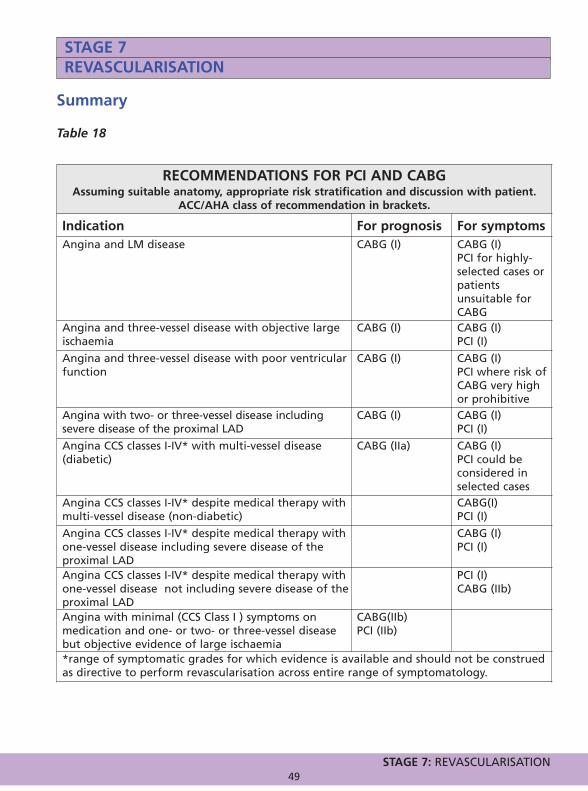
STAGE 7: REVASCULARISATION49
Summary
Table 18
STAGE 7REVASCULARISATION
RECOMMENDATIONS FOR PCI AND CABGAssuming suitable anatomy, appropriate risk stratification and discussion with patient.
ACC/AHA class of recommendation in brackets.
Indication For prognosis For symptomsAngina and LM disease CABG (I) CABG (I)
PCI for highly-selected cases orpatientsunsuitable forCABG
Angina and three-vessel disease with objective largeischaemia
CABG (I) CABG (I) PCI (I)
Angina and three-vessel disease with poor ventricularfunction
CABG (I) CABG (I)PCI where risk ofCABG very highor prohibitive
Angina with two- or three-vessel disease includingsevere disease of the proximal LAD
CABG (I) CABG (I)PCI (I)
Angina CCS classes I-IV* with multi-vessel disease(diabetic)
CABG (IIa) CABG (I)PCI could beconsidered inselected cases
Angina CCS classes I-IV* despite medical therapy withmulti-vessel disease (non-diabetic)
CABG(I) PCI (I)
Angina CCS classes I-IV* despite medical therapy withone-vessel disease including severe disease of theproximal LAD
CABG (I)PCI (I)
Angina CCS classes I-IV* despite medical therapy withone-vessel disease not including severe disease of theproximal LAD
PCI (I)CABG (IIb)
Angina with minimal (CCS Class I ) symptoms onmedication and one- or two- or three-vessel diseasebut objective evidence of large ischaemia
CABG(IIb)PCI (IIb)
*range of symptomatic grades for which evidence is available and should not be construedas directive to perform revascularisation across entire range of symptomatology.

STAGE 7: REVASCULARISATION50
Following coronary angiography and assessment of left ventricular function (eitherby left ventricular angiography or non-invasively), patients may be considered forcoronary revascularisation.
There are two well-established approaches to revascularisation for the treatment ofchronic stable angina caused by coronary atherosclerosis: surgical revascularisation(CABG) and percutaneous coronary intervention (PCI). As in the case ofpharmacological therapy, the potential objectives of revascularisation are two-fold:firstly to improve survival or survival free of infarction and secondly to diminish oreradicate symptoms. The individual risk of the patient as well as symptomatic statusmust be a major factor in the decision-making process.
Currently both methods of revascularisation are facing rapid development.Advances in CABG techniques include the greater use of arterial conduits, theintroduction of minimally-invasive techniques and the increase in use of off-pumpsurgery. PCI has seen a rapid development in the movement away from simpleballoon angioplasty towards the insertion of metal stents and most recently theincreased use of drug–eluting stents (DES), which are metal stents coated with anti-proliferative or anti-mytotic agents, designed to reduce the rate of in-stent re-stenosis. Current and future guidelines on the practice of PCI and CABG will have adirect bearing on the management of stable angina.
This chapter is based on not only the American and European Guidelines on stableangina referred to in the Introduction but also on the current leading references forPCI and CABG:
• Guidelines for percutaneous coronary interventions. The Task Force forPercutaneous Coronary Interventions of the European Society of Cardiology.1
• ACC/AHA 2004 guideline update for coronary artery bypass graft surgery.2
1. Selection of Patients for Revascularisation Therapy
In general, patients who have indications for coronary angiography and in whomcatheterisation reveals severe coronary artery stenosis are potential candidates formyocardial revascularisation. The principal indications for revascularisation are:
• To improve prognosis – this relates to effects on mortality or morbidity especiallyMI. This indication is relevant where coronary angiography has shown anatomyassociated with a high risk of adverse events which can be ameliorated by CABGand includes significant stenosis of the left mainstem (LM), significant proximalthree vessel disease, and significant two-vessel disease including high gradestenosis of the proximal left anterior descending artery (LAD).
STAGE 7REVASCULARISATION

STAGE 7: REVASCULARISATION51
• To control symptoms – when maximally tolerated medical treatment has failedto suppress symptoms to the point where the patient is happy with the qualityof his/her life. This relates to change in angina class, exercise duration, time toangina on treadmill testing, repeat hospitalisation for angina or otherparameters of functional capacity or quality of life.
Where such indications for revascularisation exist, the following should also beconsidered before deciding on a patient’s eligibility for intervention:
• The patient prefers an interventional rather than a medical approach havingbeen fully informed of the risks and benefits to be expected.
• There is a high likelihood of technical success.• The risks (mortality and morbidity) of the procedure are acceptable.
An adequate response to therapy must be judged in consultation with the patient.For some, CCS Class 1 symptoms (angina only on strenuous exertion but not duringordinary activity) are acceptable but others may wish for complete abolition of theirsymptoms. What is an acceptable risk of morbidity and mortality should beconsidered on an individual basis for each patient. Patients should not be advisedto have a procedure for which the procedural mortality exceeds their estimatedannual mortality, unless there is evidence of substantial prognostic benefit in thelonger term, or symptoms are having a serious impact on their quality of life despiteappropriate medical therapy.
2. Recommendations for Revascularisation
The current evidence-based indications for revascularisation fall into the twocategories identified above. Specific recommendations are detailed below.
To Improve Prognosis in Patients with Stable Angina
Class I • CABG for significant LM CAD or its equivalent (i.e. severe stenosis of
ostial/proximal segment of left descending and circumflex coronary arteries).• CABG for significant proximal stenosis of three major vessels, particularly in
those patients with abnormal LV function, or with early or extensive reversibleischaemia on functional testing.
• CABG for single or two vessel disease with high grade stenosis of proximal LADwith reversible ischaemia on non-invasive testing.
• CABG for significant disease with impaired LV function and viabilitydemonstrated by non-invasive testing.
Class IIa • CABG for single- or two-vessel CAD without significant proximal LAD stenosis in
patients who have survived sudden cardiac death or sustained ventriculartachycardia.
• CABG for significant three-vessel disease in diabetics with reversible ischaemiaon functional testing.
• PCI or CABG for patients with reversible ischaemia on functional testing andevidence of frequent episodes of ischaemia during daily activities.

STAGE 7: REVASCULARISATION52
To Improve Symptoms in Patients with Stable Angina
Class I • CABG for multi-vessel disease (MVD) technically suitable for surgical
revascularisation in patients with moderate to severe symptoms not controlled bymedical therapy, in whom risks of surgery do not outweigh potential benefits.
• PCI for single-vessel disease technically suitable for percutaneousrevascularisation in patients with moderate to severe symptoms not controlled bymedical therapy, in whom procedural risks do not outweigh potential benefits.
• PCI for MVD without high-risk coronary anatomy, technically suitable forpercutaneous revascularisation in patients with moderate to severe symptomsnot controlled by medical therapy and in whom procedural risks do notoutweigh potential benefits.
Class IIa • PCI for single-vessel disease technically suitable for percutaneous
revascularisation in patients with mild to moderate symptoms which arenonetheless unacceptable to the patient, in whom procedural risks do notoutweigh potential benefits.
• CABG for single-vessel disease technically suitable for surgical revascularisationin patients with moderate to severe symptoms not controlled by medicaltherapy, in whom operative risk does not outweigh potential benefit.
• CABG in MVD technically suitable for surgical revascularisation in patients withmild to moderate symptoms, which are nonetheless unacceptable to the patient,in whom operative risk does not outweigh potential benefit.
• PCI for MVD technically suitable for percutaneous revascularisation in patientswith mild to moderate symptoms, which are nonetheless unacceptable to thepatient, in whom procedural risks do not outweigh potential benefits.
Class IIb • CABG in single-vessel disease technically suitable for surgical revascularisation in
patients with mild to moderate symptoms, which are nonetheless unacceptable tothe patient, in whom operative risk is not greater than estimated annual mortality.
3. Selection Of Method Of Revascularisation
Selection of the method of revascularisation should be based on:
• Risk of periprocedural mortality and morbidity. • Likelihood of success, including factors such as technical suitability of lesions for
angioplasty or surgical bypass. • Risk of re-stenosis or graft occlusion. • Completeness of revascularisation. If considering PCI for MVD, is there a high
probability that PCI will provide complete revascularisation or at least in thesame range as CABG?
• Diabetic status. • Local hospital experience in cardiac surgery and interventional cardiology. • Patient preference.

STAGE 7: REVASCULARISATION53
4. Contraindications to Myocardial Revascularisation
Contraindications to myocardial revascularisation are described in Table 19 below.
Table 19
5. Specific Patient and Lesion Subsets
• Patients with severely depressed LV function and/or high surgical risk, patientswith LM disease, patients with diabetes and MVD, and patients with previousbypass surgery warrant particular consideration when selecting revascularisationoptions.
• Patients in whom surgical risk is prohibitively high may benefit fromrevascularisation by PCI, particularly when residual viability can be demonstratedin the myocardium perfused by the target vessel(s). This issue is currentlyaddressed in two large randomised studies, the STICH3, and the HEART UK4
trials.
• Although PCI in LM stem disease is feasible, and good results have been achievedin registries comparing DES and bare metal stents, surgery should remain thepreferred approach until the outcome of further trials are known.
• Subgroup analyses of randomised trials have shown reduced mortality withbypass surgery compared with PCI in diabetic patients with MVD. The BARI5 trialwas the largest of these trials and the only one in which a statistical differencein mortality of patients with diabetes was detected between the treatmentgroups. A limitation of these trials is that they were conducted before thewidespread use of DES stents or adjuvant periprocedural antiplatelet therapy.
Two major trials are underway to address this important issue: BARI 2D6, andFREEDOM7. However, for the present, due consideration should be given to theevidence available and PCI should be used with reservation in diabetics with MVDuntil the results of further trials are known.
CONTRAINDICATIONS TO MYOCARDIAL REVASCULARISATION• Patients with one- or two-vessel CAD without significant proximal LAD stenosis who
have mild or no symptoms and have not received an adequate trial of medical therapyor have no demonstrable ischaemia or only a limited area of ischaemia/viability on non-invasive testing
• Borderline (50–70%) coronary stenosis in location other than LM and no demonstrableischaemia on non-invasive testing
• Non-significant (<50%) coronary stenosis
• High risk of procedure-related morbidity or mortality (>10–15% mortality risk) unlessthe risk of the procedure is balanced by an expected significant improvement insurvival or the patient's quality-of-life without the procedure is extremely poor

STAGE 7: REVASCULARISATION54
• There are no randomised controlled trials comparing treatment options inpatients with previous bypass surgery. Re-do surgery may be undertaken onsymptomatic grounds where the anatomy is suitable. However, the operativerisk of re-do bypass surgery is as high as three-fold greater than initial surgery,and for those with a patent internal mammary artery (IMA) graft there is theadditional risk of damage to this graft during surgery. On the other hand PCIcan be performed following previous surgical revascularisation, either in thevein graft or arterial graft, or the native coronary tree beyond the graft whichis not revascularised, and may provide a useful alternative to re-do surgery forsymptomatic relief.
• In the case of a chronic total occlusion that cannot be crossed in patients withMVD, failure to treat the chronic total occlusion will result in incompleterevascularisation, which could be avoided if the patient were to be referred forbypass surgery.
1 Eur Heart J. 2005 Apr; 26 (8):804-47.2 J Am Coll Cardiol. 2004 Sep 1; 44(5):e213-310.3 The Surgical Treatment for Ischemic Heart Failure Trial (STICH).4 Heart failure revascularisation trial (HEART).5 Bypass Angioplasty Revascularisation Investigation (BARI) Investigators. Comparison of coronary
bypass surgery with angioplasty in patients with multi-vessel disease. N Engl J Med 1997;336:147 and N Engl J Med 1996;335:2).17-25.
6BARI 2 Diabetes.7Future Revascularisation Evaluation in Patients with Diabetes Mellitus (FREEDOM) Trial.
STAGE 8ASSESSMENT AND MANAGEMENT OF
CVD RISK PRIOR TO NON-CARDIAC SURGERY
STA
GE
8
STAGE 8ASSESSMENT AND MANAGEMENT OF
CVD RISK PRIOR TO NON-CARDIAC SURGERY
STA
GE
8
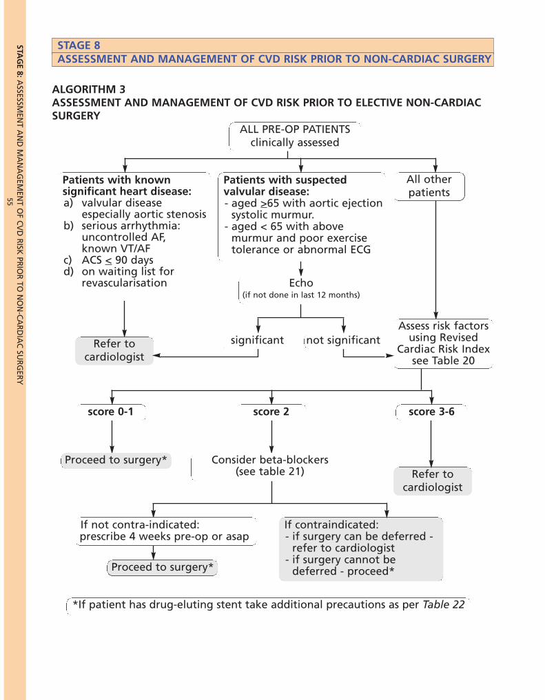
55STA
GE 8:A
SSESSMEN
T AN
D M
AN
AG
EMEN
T OF C
VD
RISK
PRIO
R TO
NO
N-C
AR
DIA
C SU
RG
ERY
STAGE 8ASSESSMENT AND MANAGEMENT OF CVD RISK PRIOR TO NON-CARDIAC SURGERY
Patients with suspectedvalvular disease:- aged ≤ 65 with aortic ejectionsystolic murmur.- aged < 65 with above murmurand poor exercise tolerance orabnormal ECG
score 0-1
Proceed to surgery*Refer to
cardiologist
Proceed to surgery*
If contraindicated:- if surgery can be deferred -
refer to cardiologist- if surgery cannot be
deferred - proceed*
*If patient has drug-eluting stent take additional precautions as per Table 22
ALGORITHM 3ASSESSMENT AND MANAGEMENT OF CVD RISK PRIOR TO ELECTIVE NON-CARDIACSURGERY
All otherpatients
Patients with knownsignificant heart disease:a) valvular disease
especially aortic stenosisb) serious arrhythmia:
uncontrolled AF,known VT/AF
c) ACS < 90 daysd) on waiting list for
revascularisation
Patients with suspectedvalvular disease:- aged >65 with aortic ejection
systolic murmur.- aged < 65 with above
murmur and poor exercisetolerance or abnormal ECG
ALL PRE-OP PATIENTSclinically assessed
Echo(if not done in last 12 months)
Assess risk factorsusing Revised
Cardiac Risk Indexsee Table 20
score 3-6score 2
Consider beta-blockers(see table 21)
If not contra-indicated:prescribe 4 weeks pre-op or asap
significantRefer tocardiologist
not significant
STAGE 8: ASSESSMENT AND MANAGEMENT OF CVD RISK PRIOR TO NON-CARDIAC SURGERY56
STAGE 8ASSESSMENT AND MANAGEMENT OF CVD RISK PRIOR TONON-CARDIAC SURGERY
1. Introduction
Emergency and elective surgery differ significantly in terms of logistics, time frameand clinical considerations such as haemodynamic stability, blood volume etc. TheCMCN Task Group concluded that the risk assessment for emergency surgery shouldbe undertaken with the anaesthetists on an individual patient basis. Therefore, thefollowing guidelines relate entirely to elective procedures and in this area, the useof Algorithm 3 will allow a logical evidence-based and cost-effective approach.
2. Mechanisms of Perioperative Acute Coronary Syndrome (ACS)
The pathophysiology of ACS perioperatively differs from that in stable CAD. In thesetting of pre-existing stable CAD, acute ischaemic syndromes occur because ofdestabilisation (erosion, rupture) of atheromatous plaques. The pathophysiology ofperioperative ischaemic events is less clear but current belief is that half result fromplaque destabilisation and half from prolonged imbalance of the supply anddemand of myocardial oxygen. This imbalance may result from:
- increased myocardial demand due to increased heart rate and bloodpressure often sympathetically mediated via pain, or due to large fluidvolume shiftsor
- reduced coronary supply due to anaemia, hypotension or increased coronaryvascular tone (coronary spasm).
Standard non-invasive testing may be of value in assessing pre-existing stable CADand the propensity to plaque rupture but would be of limited value in predictingprolonged supply-demand imbalance.
3. Accuracy of Non-Invasive Testing in Pre-operative Assessment
Non-invasive cardiac tests (exercise ECG, MPS, DSE) allow a reasonably accurateassessment of prognosis (death, serious cardiac events) during medium- and long-term follow-up i.e. in the setting of chronic CAD. They have proved much lessreliable in predicting such events over the ninety-six hour perioperative periodwhich is required for accurate pre-operative assessment.

STAGE 8: ASSESSMENT AND MANAGEMENT OF CVD RISK PRIOR TO NON-CARDIAC SURGERY57
This inaccuracy results from the following:- the mechanisms of pre-operative ischaemia/infarction differ from those in
stable CAD (see Section 2 above) - patients who are assessed for non-cardiac surgery are usually a low risk
population with only about 2% actually suffering a perioperative cardiacevent. In this type of low prevalence population, non-invasive tests havemodest sensitivity and very poor specificity (high false positive rates).
The problem is how to identify accurately a small number of high-risk patients in thispredominately low-risk population. The high false positive rate of non-invasivetesting has often led to unnecessary, further and increasingly invasive investigationsthat multiply costs, increase delays and expose patients to unnecessary risks.
4. Classical Approach
The current standard or classical approach is that advocated in the 2002 ACC/AHAGuidelines1 and involves pre-operative, non-invasive testing for a large proportionof patients. A brief summary is given below:
• Step 1 – identification of clinical predictors of perioperative risk, based onhistory, examination and ECG. Patients are grouped into those having major,intermediate or minor predictors.
• Step 2 – assessment of the patient’s functional capacity in METs based onactivities of daily living using the Duke Activity Status Index. Higherperioperative risk is associated with inability to meet four METs demand.
• Step 3 – classification of the proposed non-cardiac surgical procedure into high,intermediate or low risk operative groups.
• Step 4 – the results of steps 1-3 determine the most appropriate of threemanagement strategies:- High risk - delay or cancel non-cardiac surgery to allow for optimisation of
medical therapy, risk factor modification and coronary angiography with aview to revascularisation
- Intermediate risk – undertake non-invasive investigation(s) and dependingon results, coronary angiography.
- Low risk - proceed to non-cardiac surgery without further investigation ortreatment.
Unfortunately the majority of patients fall into the intermediate group requiringnon-invasive testing. The CMCN Task Group did not regard this as the optimalapproach for the following reasons:
- Heavy burden placed on non-invasive testing resources.- Poor predictive accuracy of such tests.- Delays and costs incurred.- Excessive and unnecessary need for coronary angiography resulting from the
inevitable false positive results.

STAGE 8: ASSESSMENT AND MANAGEMENT OF CVD RISK PRIOR TO NON-CARDIAC SURGERY58
5. Preferred (Modern) Approach
The CMCN Task Group recommends a more modern approach outlined in Algorithm3. This shifts the emphasis away from pre-operative risk stratification, which usesnon-invasive testing, to risk modification with drug therapy aimed at reducingperioperative ischaemia and hence the likelihood of adverse cardiac events.
The principles underlying the preference for this strategy are:
• No evidence exists that non-invasive testing leads to a therapeutic strategy thatreduces perioperative risk of MI or death.
• Non-invasive testing has a uniformly low positive predictive accuracy which doesnot improve on information available from clinical assessment alone.
• The most robust clinical assessment scheme currently available is the RevisedCardiac Risk Index (RCRI)2 as used by Grayburn and Hillis.3
• Beta blockers and to some extent statins, have recently been shown to reducethe risk of perioperative cardiac events whilst routine coronary revascularisationhas not.
The approach is easy to use by a range of healthcare professionals and minimises thepressure on non-invasive testing resources by focussing them on patients most likelyto accrue benefit from their application. However, it is recognised that the pathwayoutlined in Algorithm 3 will not cover every possible set of circumstances andclinicians may consider modification is justified in individual cases.
The pathway involves the application of the RCRI, a simple bedside clinical index, toall patients requiring pre-operative assessment who do not have known significantheart disease, including significant valvular disease, especially aortic stenosis, seriousarrhythmia (uncontrolled AF, VT/VF), recent ACS (< 90 days), or are on arevascularisation waiting list. The index and scoring system is described in Table 20.
Cardio-pulmonary exercise testing (CPET) consists of exercise testing withsimultaneous ECG and gas exchange measurements. It is increasingly used incardiology to assess objectively functional capacity, stress inducible myocardialischaemia, the cause of unexplained breathlessness and to risk stratify prior tocardiac transplantation. Recently, it has also been shown to predict patients at highrisk of perioperative cardiac complications.
Conclusive evidence of its accuracy and cost-effectiveness in prospective studies pre-operatively is still lacking and in comparison to a standard exercise ECG it is morecomplex, more time consuming and requires added skills. For these reasons, CPET isnot recommended as a routine pre-operative assessment but its value toanaesthetists in certain surgical cases is recognised and where such a service is inexistence its use may well add important information.

STAGE 8: ASSESSMENT AND MANAGEMENT OF CVD RISK PRIOR TO NON-CARDIAC SURGERY59
Table 20
Pre-operative management with beta blockers
It is recommended that patients with an RCRI score of two should be started on betablockers, ideally four weeks prior to surgery but if time does not allow then as soonas possible and definitely on admission. It is not known if, or when, they should bestopped but certainly, abrupt withdrawal should be avoided. Consensus advice isgiven in Table 21.
Table 21
REVISED CARDIAC RISK INDEX (RCRI)Score 1 for each risk factor present
RISK FACTORS• High risk surgical procedure – (intra-peritoneal, intra-thoracic, or suprainguinal
vascular reconstruction).
• History of IHD (history of MI, history of positive exercise test, current complaint ofchest pain considered to be secondary to myocardial ischaemia, symptomatic need fornitrate therapy, ECG with pathological Q waves). Patients with prior coronary revascularisation who are potentially protected should notbe included unless there has been a prior MI or evidence of ongoing myocardialischaemia such as a positive exercise test or angina.
• History of congestive heart failure.
• History of stroke or TIA.
• Pre-operative treatment with insulin.
• Pre-operative serum creatinine >177µmol/L.
TOTAL SCORE0-1 proceed to surgery*
2 proceed to surgery after pre-treatment with beta blockers*3-6 refer to cardiologist for assessment
*with additional precautions for patients with pre-existing drug-eluting stents in situ.
BETA BLOCKERSAgents - Bisoprolol, atenolol and metoprolol. Evidence favours the longer acting agents andatenolol has been shown to be superior to metoprolol.
Contraindications - asthma, uncontrolled heart failure; hypotension (SBP>100) orcardiogenic shock; bradycardia (<60 bpm); 2o – 3o AV heart block; sino-atrial disorder (sicksinus syndrome); severe peripheral vascular disease (but can be used with caution in stableand non-critical PVD); known coronary artery spasm or phaeochromocytoma.
Dosage – Bisoprolol 5-10mg daily; atenolol 25-100mg daily; metoprolol 50-100mg twicedaily. Start with the lower dose and increase after 3 days to get resting heart rate to ≤ 60bpm providing SBP ≥ 100 and no side effects.
Duration - Start 4 weeks pre-op or if time does not allow as soon as possible and definitelyon admission. Continue daily until discharge. No evidence for longer duration of treatmentexists except where there are other indications e.g. hypertension, angina.

STAGE 8: ASSESSMENT AND MANAGEMENT OF CVD RISK PRIOR TO NON-CARDIAC SURGERY60
Statins
The situation regarding statins is less clear. A significant number of perioperativeevents are caused by rupture of vulnerable plaques during the unusual stressesassociated with anaesthesia and surgery. A role for rapid plaque stabilisation withstatins is therefore logical and is now supported by growing evidence. However, theCMCN Task Group did not consider current evidence was strong enough torecommend routine initiation of pre-operative statins but they should not bewithdrawn in patients already taking them.
Other drugs
Although other drugs including aspirin, clopidogrel, alpha blockers and calcium-channel blockers have been considered for pre-operative use, the evidence does notsupport their recommendation.
CABG
The routine use of CABG pre-operatively for patients with stable, significant CAD doesnot reduce risk and is not recommended. However, if irrespective of the non-cardiacsurgery, cardiac symptoms are unstable or if CABG would be recommended onstandard symptomatic or prognostic grounds, then it is sensible for this to be carriedout prior to the non-cardiac procedure if time allows.
Studies have shown a significantly higher risk of death and/or cardiac complicationsin patients undergoing non-cardiac surgery early after CABG. The optimal safeperiod to delay non-cardiac surgery after CABG is unknown and will depend on therisk/benefit balance in individual patients. However, for most cases it is prudent toavoid elective non-cardiac surgery for at least one month and, where possible, forsix months.
PCI
PCI is not recommended for routine prophylactic use prior to non-cardiac surgeryand indications for it should be as in the non-operative setting.
After balloon angioplasty, a minimum of one week should be left to allow forhealing of the traumatised vessel prior to non-cardiac surgery.
Patients with pre-existing in-situ stents. These patients risk either stent thrombosisdue to the discontinuation of dual anti-platelet therapy or haemorrhage due to itscontinuation. With bare metal stents, ideally four to six weeks should elapse toallow for full re-endothelialisation.
The situation with regard to drug-eluting stents (DES) is less clear. DES delay re-endothelialisation and hence patients require a longer period on dual anti-platelettherapy before non-cardiac surgery is undertaken. Recent data has also raisedconcerns about late thrombosis with DES. No evidence based guidelines exist for themanagement of DES patients prior to non-cardiac surgery but the followingprinciples have consensus agreement.

STAGE 8: ASSESSMENT AND MANAGEMENT OF CVD RISK PRIOR TO NON-CARDIAC SURGERY61
Table 22
1ACC/AHA 2002 Guideline Update on Perioperative Cardiovascular Evaluation for Non-cardiacSurgery www.acc.org2Lee TH et al. Derivation and prospective validation of a simple index of cardiac risk of major non-cardiac surgery. Circulation 1999; 100:1043-93Grayburn PA.and Hillis LD. Cardiac Events in Patients Undergoing Non-cardiac Surgery: Shifting theParadigm from Non-invasive Risk Stratification to Therapy. Ann. Internal Med 2003; 138: 506-114L. Broad et al. Successful management of patients with a drug-eluting coronary stent presenting forelective, non-cardiac surgery Br J Anaesth 2007; 98: 19–22
PATIENTS WITH DRUG ELUTING STENTS (DES): PRE- and PERIOPERATIVE ADVICE
1. Whenever possible advice should be sought from the cardiologist who implanted theDES with regard to perioperative management.
2. Whenever possible, elective PCI should be delayed until after any foreseeable elective,non-cardiac surgery has been undertaken. Obviously, where PCI is for ACS or intractablesymptoms it should not be delayed.
3. Non-urgent elective surgery should be postponed for 12 months after DES insertionwhen clopidogrel can be stopped. Low dose aspirin should be continued unless thesurgeon or type of surgery precludes it, when it should be restarted as soon as possiblepost-operatively.
4. Urgent elective surgery which cannot be delayed until 12 months have elapsed e.g. forcancer, should be undertaken on dual anti-platelet therapy if possible. If the surgeonor type of surgery precludes this, then aspirin alone should be continued (other than inexceptional circumstances) and clopidogrel restarted as soon as possible post-operatively. If the surgery is likely to be delayed for > 5 days and clopidogrelcontinuation is not acceptable then the following regime4 has been suggested whilstcontinuing low dose aspirin throughout:-
• 5 days pre-op: Stop clopidogrel
• 3 days pre-op: Admit to hospitalCommence short acting GpIIb/IIIa inhibitor (tirofiban) infusion Commence unfractionated heparin infusion
• 6 hours pre-op: Stop tirofiban infusionStop heparin infusion
• 1st post-op day: Give oral loading dose of 300mg of clopidogrel
• 2nd post-op day: Start maintenance oral clopidogrel 75mg daily
5. Emergency or urgent non-elective surgery which needs to be undertaken immediatelyor at very short notice should go ahead when clinically indicated. In these circumstances,the question of pre-operative cessation of anti-platelet agents does not arise.

STAG
E 8:ASSESSM
ENT A
ND
MA
NA
GEM
ENT O
F CV
D R
ISK PR
IOR
TO N
ON
-CA
RD
IAC
SUR
GERY
62
APPENDIX TO STAGE 8ASSESSMENT AND MANAGEMENT OF CVD RISK OF POTENTIAL CANDIDATES FORRENAL TRANSPLANTATION
History, exam, bloods, resting ECG
Patients with known significant heart disease:a) valvular disease especially aortic stenosisb) serious arrhythmia: uncontrolled AF,
known VT/VFc) ACS < 90 daysd) on waiting list for revascularistion
All other patients
Newcastle riskassessment scoreTables 23 & 24
9-12<8>13Referral to cardiologist- clinical assessment- echo- stress imaging
Tx contraindicatedHigh riskLow risk
Cardiac catheter
No prognosticdisease
Prognosticdisease
Defer Tx-referto
cardiologist onclinical grounds
Advise Tx teamto add to
waiting list
Defer Tx,reconsider after
treatment ifappropriate
Advise Tx teamnot to add towaiting list
Summary
ALGORITHM 4CV RISK ASSESSMENT FOR POTENTIAL RENAL TRANSPLANT RECIPIENTS

STAGE 8: ASSESSMENT AND MANAGEMENT OF CVD RISK PRIOR TO NON-CARDIAC SURGERY63
APPENDIX TO STAGE 8ASSESSMENT AND MANAGEMENT OF CVD RISK OFPOTENTIAL CANDIDATES FOR RENAL TRANSPLANTATION
For a number of reasons outlined below, the CMCN Task Group considered thatassessment prior to renal transplantation (RTx) and pancreatic-renal transplantation(PRTx) constituted special situations not adequately addressed by national orinternational guidelines. The essential principles of the ‘modern’ approach (knownpathology, validated clinical risk tool, proven risk modification by drugs) do not holdin these situations and hence the decision to develop a version of the classical (riskstratification) approach modified to take account of the limited evidence available.The guidance, which is outlined in Algorithm 4 and detailed below, is thereforerecommended but not fully validated and constitutes the consensus view of the TaskGroup.
1. Objectives
The aims of the pre-operative assessment prior to RTx and PRTx are wider than forother non-cardiac surgery.
• To quantify perioperative risk to inform discussion with patients on consent.
• To prioritise the use of a severely limited supply of organs by enabling anassessment of intermediate outcomes to be made. The European TransplantGuidelines1 recommend that recipients have a pre-transplant life expectation ofmore than two years in order to justify organ usage.
• To reduce perioperative risk where possible and initiate specialist cardiovascularfollow up where appropriate.
2. Special Considerations (Why RTx and PRTx Differ)
There are a number of major differences from other non-cardiac surgery, whichinvalidate the strategy hitherto recommended, including the following:
• The surgery, whilst elective, is carried out at very short notice.
• Unlike the usual pre-operative population, which is low risk (low diseaseprevalence), RTx and PRTx patients are relatively high risk both for perioperativeevents and for current or future CAD. This and the need to ensure year lifeexpectancy beyond two years, means that the risk to be assessed is not just shortterm due to surgery but also intermediate and long term.

STAGE 8: ASSESSMENT AND MANAGEMENT OF CVD RISK PRIOR TO NON-CARDIAC SURGERY64
• Clinical features are modified with a much higher prevalence of silentmyocardial ischaemia due to poor exercise tolerance, anaemia and neuropathy.Exercise ECG with Bruce protocol is usually of little value especially in thepresence of LVH.
• The RCRI has not been validated in this group of patients. The NewcastleScoring System for Clinical Risk Assessment (Table 23) has been specificallyderived for RTx and validated to some extent. High scores (> 13) have beenshown prospectively to correlate with longer duration of delayed graft function,poorer glomerular filtration rate at six months, inferior patient and graftsurvival over five years of follow up, together with an increased relative rate (>4)of early graft loss within sixty days.
• In this higher prevalence population, non-invasive imaging (MPS, DSE) is moreaccurate for risk prediction but has a reduced diagnostic predictive accuracy (upto 20% false negatives).
• There is no evidence base for pre-operative risk modification by either drugtherapy or revascularisation.
• Coronary angiography, PCI and CABG are inherently more risky in these patientsand their therapeutic value lessened.
• Coronary angiography has some value, albeit limited, in predicting futurecardiac events. However, it retains its accuracy in identifying patterns ofsignificant CAD, which have been shown to benefit from revascularisation.
3. CMCN Pathway
The CMCN recommended pathway for pre-operative assessment of RTx and PRTxpatients is outlined in Algorithm 4. The principles of the pathway are:
• Patients who have significant heart disease including serious arrhythmia(uncontrolled AF, VT/VF), significant valvular disease especially aortic stenosis,recent ACS (< 90 days), or are on a revascularisation waiting list should be referreddirectly to a cardiologist. Specialist assessment will decide whethertransplantation should proceed, is completely contraindicated or whether invasiveinvestigation by cardiac catheterisation is necessary to make this decision.
• In all other patients, a relevant health care professional (not a cardiologist)should undertake clinical assessment and risk assessment using the Newcastlescoring tool. The Newcastle score should be used to direct further actions anddecision making as indicated in Algorithm 4 and Table 24.

STAGE 8: ASSESSMENT AND MANAGEMENT OF CVD RISK PRIOR TO NON-CARDIAC SURGERY65
Table 23
Table 24
NEWCASTLE SCORING SCHEME2,3
Risk Factor ScoreAge <45 0
45-65 265+ 4
Diabetes Present 3BP assessment Hypotension
- Systolic blood pressure <110mm Hg4
Hypertension - Controlled with drugs (BP < 150/90mm Hg)
2
- Uncontrolled (BP >150/90mm Hg), with orwithout medication
3
Exercisingdistance
50-200m or slowing on stairs 3<50 m or stops on stairs 4
Body mass index <18 or >30 3Cerebrovascular disease Previous CVA or TIA 3Peripheral vascular disease Present 3Angina Stable 3
Variable or on minimal exertion(40-50m) 4Coronary vascular surgeryand MI
One MI/CABG/two vessel disease/PCI 3Two MI/CABG for triple vessel disease/two surgicalprocedures including CABG
4
Cardiac valve disease Mitral, tricuspid, pulmonary valve disease or surgery 2Aortic valve disease or surgery 3
Maximum possible score 35
RISK ASSESSMENT USING NEWCASTLE SCOREScore Action
≤ 8 Patients are low risk for presence of significant CAD and for perioperative eventsand should proceed to consideration for transplant by the transplant team.
9 - 12
Patients require further cardiological assessment.They should be referred to a cardiologist who, on the basis of clinicalassessment, echo and stress imaging should decide on the risk/benefit ratio ofcardiac catheterisation. When appropriate this will refine the risk estimate (perioperative, intermediateand long term) and lead to three alternative sets of advice to the transplant team:• Accept for Tx consideration. • Defer until treatment (for CAD or valvular heart disease) reduces the risk to
acceptable levels. • Do not proceed to Tx consideration.
≥ 13Patients have a high perioperative risk and are hence a low priority for organdonation. The expectation is that such patients will not be accepted for transplantbut they should be referred to a cardiologist for optimisation of cardiac care.

STAGE 8: ASSESSMENT AND MANAGEMENT OF CVD RISK PRIOR TO NON-CARDIAC SURGERY66
4. Re-Assessment of Patients Accepted on Tx Waiting List
Patients may remain on a Tx waiting list for several years whilst awaiting a suitableorgan. During this period, there is a significant risk of development or progressionof CAD or valvular disease due to the frequent presence of adverse vascular riskfactors and the tendency to metastatic calcification. It is therefore recommendedthat such patients should be re-assessed annually, initially by a non-cardiologist.
Algorithm 5 outlines the management pathways. If the annual assessment shows nonew cardiac problems or no significant changes, then further investigation is notrequired. However, if the Newcastle score, ECG or echo identifies new cardiacproblems or deterioration in previously known ones, then the patient should bereferred to a cardiologist. Specialist assessment and investigation will inform thedecision on the need for cardiac catheterisation and hence whether transplantationshould proceed, be deferred or await further cardiac treatment.

67STA
GE 8:A
SSESSMEN
T AN
D M
AN
AG
EMEN
T OF C
VD
RISK
PRIO
R TO
NO
N-C
AR
DIA
C SU
RG
ERY
ALGORITHM 5CARDIAC RE-ASSESSMENT OF PATIENTS ON TX WAITING LIST
1European Best Practice Guidelines for Renal Transplantation (Part 1). NDT, 2000. 15 s72Asher, J., et al., A simple cardiovascular risk score can predict poor outcome in NHBD renal transplantation. Transplant Proc,2005. 37(8): p. 3292-3
3Gupta, A., et al., Cardiovascular risk assessment scoring system for the determination of cardiovascular mortality in renaltransplant patients. Transplant Proc, 2005. 37(8): p. 3290-1.
On Tx waiting list
Annual assessment by non-cardiologist- Clinical, Newcastle score- ECG- echo for valvular disease
New/worsening symptoms orNew/worsening ECG changes orWorsened echo findings orNewcastle score increased by >1
Refer to cardiologist- clinical assessment- echo- stress imaging
No significant change
No significant change
Significant change
Cardiac catheter
Defer Tx for treatment orremove from Tx waiting list
No further investigationsReview in 12 months
Contraindications No contraindications