guidelines for cli & diabetic foot - senato.it · guidelines for cli & diabetic foot...
TRANSCRIPT

Guidelines for CLI & Diabetic foot
Chapter I Definitions, Epidemiology,
Clinical presentation, Prognosis
Francois Becker1, Helia Robert-Ebadi1, Jean-Baptiste Ricco2
1 Division of Angiology and Hemostasis, Geneva University Hospitals, Geneva, Switzerland 2 Department of Vascular Surgery, University Hospital of Poitiers, Poitiers, France

METHODOLOGY levels of evidence from
the Oxford Centre For Evidence-Based Medicine

METHODOLOGY levels of evidence from
the Oxford Centre For Evidence-Based Medicine
Definitions of the grades of recommendation are: Grade A è Consistent level 1 studies Grade B è Consistent level 2 or 3 studies or
extrapolations from level 1 studies Grade C è Level 4 studies or extrapolations from
level 2 or 3 studies Grade D è Level 5 evidence or troublingly
inconsistent or inconclusive studies of any level
General consideration
Since there are almost no RCT exclusively among CLI patients, most of the lessened recommendation are based on evidence from subgroup analyses of “PAOD” trials (extrapolation from RCT), or from prospective cohorts.

Where data originates from a RCT, the level of evidence is given by that study design (i.e level 1a or 1b). Where results of subgroup analysis are applied to a particular recommendation, it has been downgraded (i.e. grade A è grade B)

The concept of downgrading recommendations based on
extrapolation from higher level studies may be considered a limitation of these
guidelines, but we accept it, since evidence for the subset of CLI tends to
be extremely poor

General consideration The validation of a new technique (Endovasc)
not only on a comparison with the traditional technique (open surgery)
but on the results that can be obtained by this treatment with regard to the objectives for
the treatment of CLI.

General consideration These objectives (limb salvage etc) can clearly be reached with the new technique and therefore there is evidence for its use, but with a downgraded recommendation.
To require that the evidence depends on the presence of direct comparisons with the traditional technique could also be reversed:
General consideration
there is no absolute evidence for the traditional technique as there are no RCTs comparing this to the new technique

Chapter I
• Definitions - Historical background of the concept of CLI
• Epidemiology - Incidence - Prevalence - Risk factors
• Clinical presentation • Prognosis

Chapter I
• Definitions - Historical background of the concept of CLI
• Epidemiology - Incidence - Prevalence - Risk factors
• Clinical presentation • Prognosis
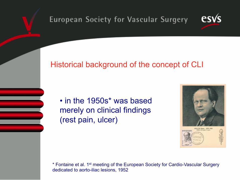
Historical background of the concept of CLI
* Fontaine et al. 1st meeting of the European Society for Cardio-Vascular Surgery dedicated to aorto-iliac lesions, 1952
• in the 1950s* was based merely on clinical findings (rest pain, ulcer)

Historical background of the concept of CLI
• Later, the importance of haemodynamic abnormalities with well defined thresholds of perfusion pressures was emphasize
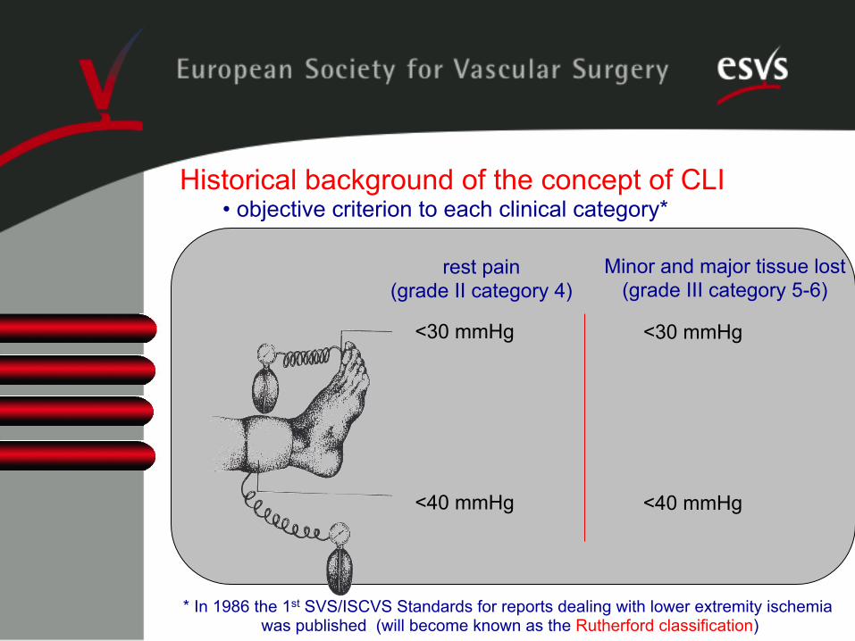
Historical background of the concept of CLI • objective criterion to each clinical category*
* In 1986 the 1st SVS/ISCVS Standards for reports dealing with lower extremity ischemia was published (will become known as the Rutherford classification)
<40 mmHg
<30 mmHg
rest pain (grade II category 4)
<40 mmHg
<30 mmHg
Minor and major tissue lost (grade III category 5-6)

Historical background of the concept of CCLI
• Second Consensus document on CLI (1991)
Recommandation 1 a) persistently recurring ischemic rest pain requiring
regular adequate analgesia (two weeks)
b) or ulceration or gangrene of the foot or toes
≤ 50 mmHg
≤ 30 mmHg

Historical background of the concept of CCLI
• TASC I Ischemic rest pain, ulcers, or gangrene attributable to objectively proven arterial occlusive disease.
thresholds just below the lower limit
values for patients with intermittent
claudication, and even the lower limit of
normality for forefoot TcPO2
≤ 50-70 mmHg
≤ 30-50 mmHg TcPO2 < 30-50 mmHg
Historical background of the concept of CCLI
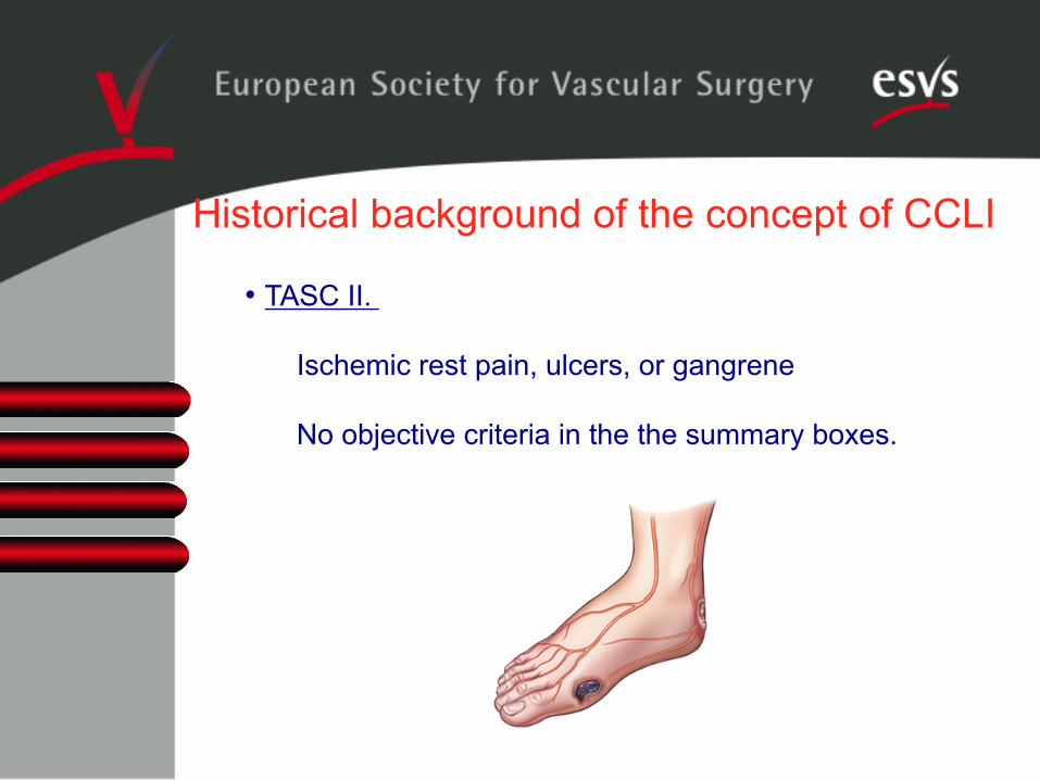
• TASC II. Ischemic rest pain, ulcers, or gangrene
No objective criteria in the the summary boxes.

Historical background of the concept of CLI
• Unfortunately, the evolution over the years has been to ignore the second part of the definition of CLI (i.e. the haemodynamic criteria) and to include all patients with rest pain or trophic changes in clinical studies regardless of the severity of the PAOD .

Haemodynamic criteria - ankle pressure (expressed as absolute value or as ABI) is not a perfect parameter in patients with suspected CLI.
- The measurements providing functional information on tissue perfusion and skin viability, such as forefoot TcPO2, are too rarely used and should be strongly encouraged (èSection: prognosis).

Chapter I
• Definitions - Historical background of the concept of CLI
• Epidemiology - Incidence - Prevalence - Risk factors
• Clinical presentation • Prognosis
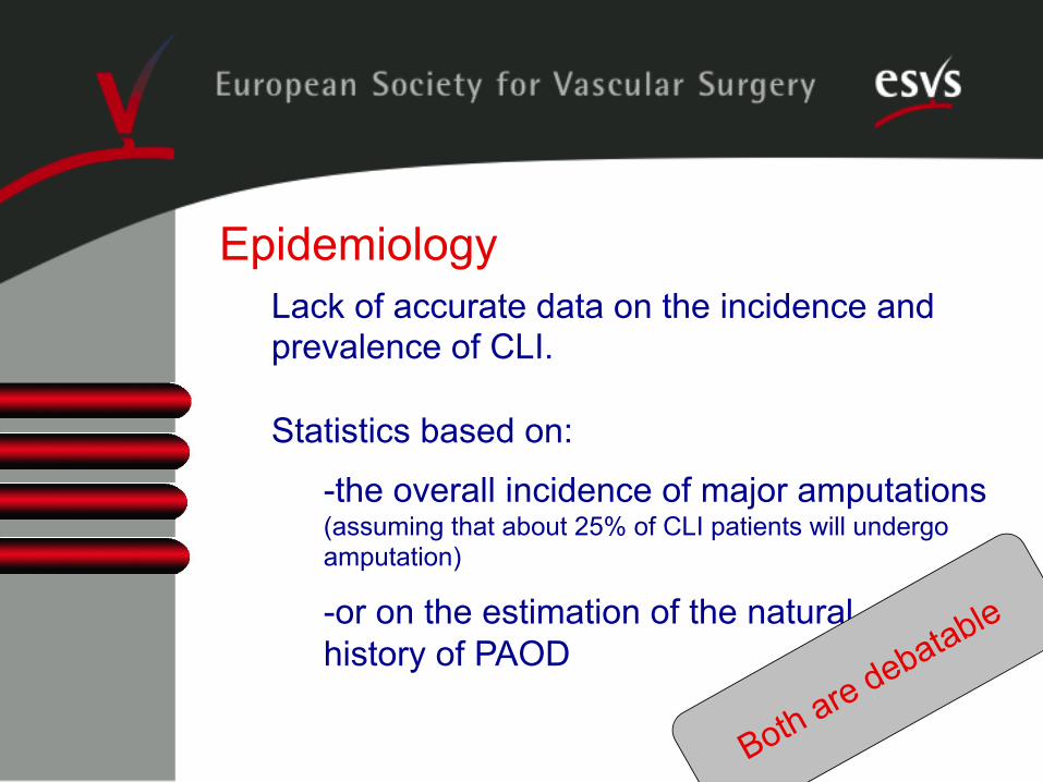
Lack of accurate data on the incidence and prevalence of CLI. Statistics based on:
- the overall incidence of major amputations (assuming that about 25% of CLI patients will undergo amputation)
- or on the estimation of the natural history of PAOD
Epidemiology

It is estimated that - 5 to 10% of patients with PAOD (asymptomatic or with claudication), will progress to CLI at 5 years - 1 to 3% of patients with PAOD are in CLI stage at initial presentation [older and sedentary patients who have limited mobility (and therefore do not claudicate), and by patients with sensitive neuropathy who have impaired pain sensation]
Epidemiology

- for one patient with known asymptomatic PAOD at least three have an unknown asymptomatic PAOD
Hirsch AT, Haskal ZJ, Hertzer NR, Bakal CW, Creager MA, Halperin JL et al. ACC/AHA 2005 Guidelines for the management of patients with peripheral arterial disease. Circulation. 2006 Mar 21;113(11):e463-654.
Epidemiology
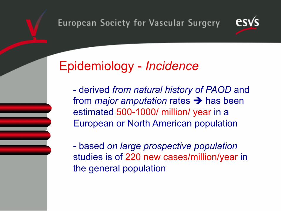
- derived from natural history of PAOD and from major amputation rates è has been estimated 500-1000/ million/ year in a European or North American population
- based on large prospective population studies is of 220 new cases/million/year in the general population
Epidemiology - Incidence

- The estimation of the prevalence of CCLI in the population aged 60 to 90 years ranges from 1% to 2%
Epidemiology - Prevalence
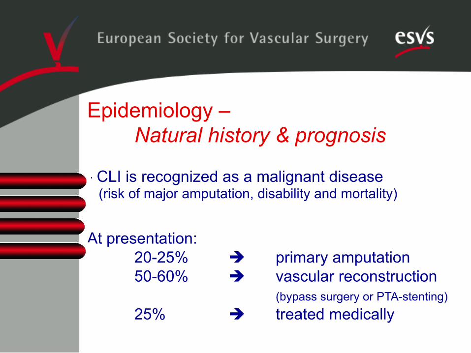
- CLI is recognized as a malignant disease (risk of major amputation, disability and mortality)
Epidemiology – Natural history & prognosis
At presentation:
20-25% è primary amputation 50-60% è vascular reconstruction (bypass surgery or PTA-stenting) 25% è treated medically

- CLI is recognized as a malignant disease (risk of major amputation, disability and mortality) At 1 year:
20-25% è died 25-30% è major amputation 20% è still CLI 20% è cured (no CLI signs)
Epidemiology – Natural history & prognosis

The TAMARIS trial offers probably the most recent reliable data concerning the natural history and prognosis
Epidemiology – Natural history & prognosis
Belch J, Hiatt WR, Baumgartner I, Driver IV, Nikol S, Norgren L, Van Belle E; TAMARIS Committees and Investigators. Effect of fibroblast growth factor NV1FGF on amputation and death: a randomised placebo-controlled trial of gene therapy in critical limb ischaemia. Lancet. 2011 Jun 4;377(9781):1929-37
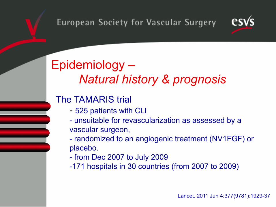
The TAMARIS trial - 525 patients with CLI - unsuitable for revascularization as assessed by a vascular surgeon, - randomized to an angiogenic treatment (NV1FGF) or placebo. - from Dec 2007 to July 2009 - 171 hospitals in 30 countries (from 2007 to 2009)
Epidemiology – Natural history & prognosis
Lancet. 2011 Jun 4;377(9781):1929-37

The TAMARIS trial è natural history (placebo group)
@ 1 year - Major amputation 21% - Death 15% The cause of death - cardio-vascular 49%, - non-cardiovascular 41% - unknown in 10%
Epidemiology – Natural history & prognosis
Lancet. 2011 Jun 4;377(9781):1929-37

Chapter I
• Definitions - Historical background of the concept of CLI
• Epidemiology - Incidence - Prevalence - Risk factors
• Clinical presentation • Prognosis

As inclusion criteria, distal pressure measurements are important (limited precision)
RISK STRATIFICATION
The evaluation of perfusion reserve and foot viability è Forefoot TcPO2

Forefoot TcPO2, if measured according to the methodological rules (particularly avoiding areas of thick or edematous skin) is probably the best non-invasive method in clinical practice to quantify the degree of ischemia and assess prognosis.
RISK STRATIFICATION

Degree 1 : 10 < TcPO2 ≤ 35 mmHg in supine position Degree 2 : forefoot TcPO2 < 10 mmHg in supine position but clear improvement (≥ 40 mmHg) in sitting position or under oxygen inhalation.
RISK STRATIFICATION based of forefoot TcPO2: degree 1-4 Recommendation grade B
Good prognosis
Intermediate prognosis
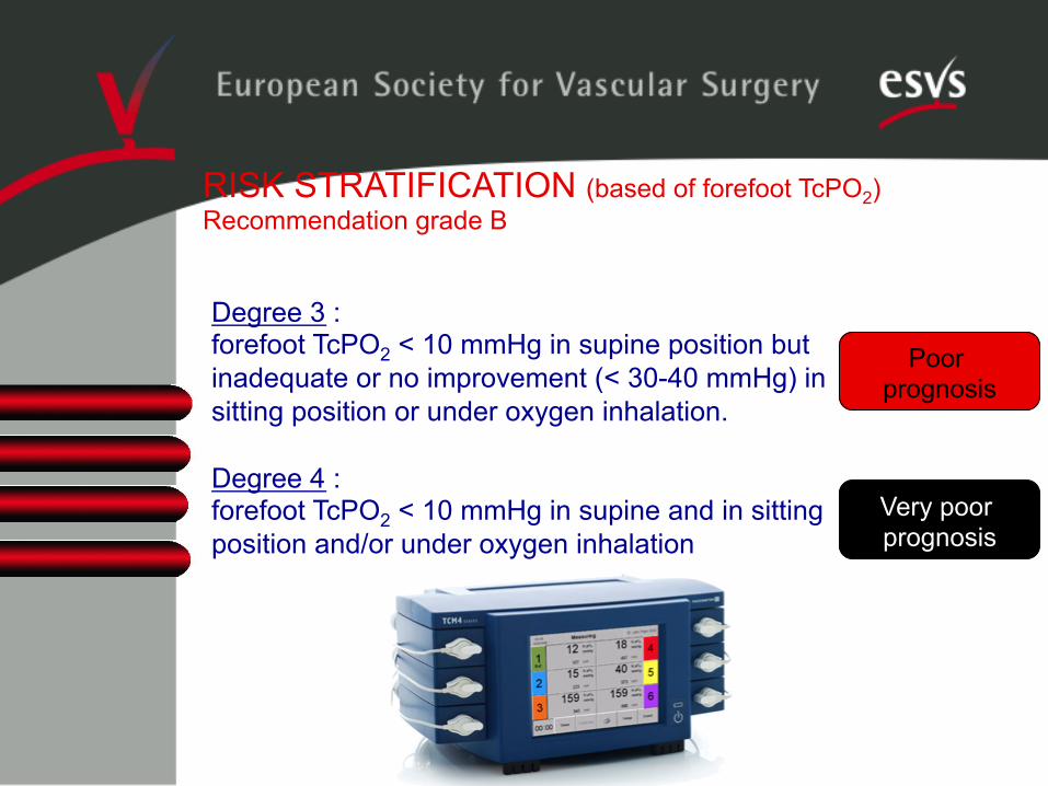
Degree 3 : forefoot TcPO2 < 10 mmHg in supine position but inadequate or no improvement (< 30-40 mmHg) in sitting position or under oxygen inhalation. Degree 4 : forefoot TcPO2 < 10 mmHg in supine and in sitting position and/or under oxygen inhalation
RISK STRATIFICATION (based of forefoot TcPO2) Recommendation grade B
Poor prognosis
Very poor prognosis

Chapter I (Epidemiology): Becker & Ricco
Chapter II (Diagnostic methods): Cao & Eckstein
Chapter III (Medical treatment): Schmidili & Diehm
Chapter IV (Treatment of CLI): Setacci & Moll
Chapter V (Diabetic foot): Lepantalo & Apelqvist
Chapter VI (Follow-up): Ricco & Davies & Dick
Chairman Carlo Setacci
GUIDELINES FOR CRITICAL LIMB ISCHEMIA & DIABETIC FOOT
Co-chairman: Jean-Baptiste Ricco

GUIDELINES FOR CRITICAL LIMB ISCHEMIA
CHAPTER II: DIAGNOSTIC METHODS
Piergiorgio Cao, Hans-Henning Eckstein,
Paola De Rango

In patients with critical limb ischemia (CLI) an accurate diagnosis can be established with modern non-invasive vascular diagnostic techniques to provide adequate information for creation of a therapeutic plan Non-invasive data will be supplemented by the use of more invasive imaging techniques, such as computed tomography angiography (CTA) or magnetic resonance angiography (MRA), and selective use of lower extremity Angiography
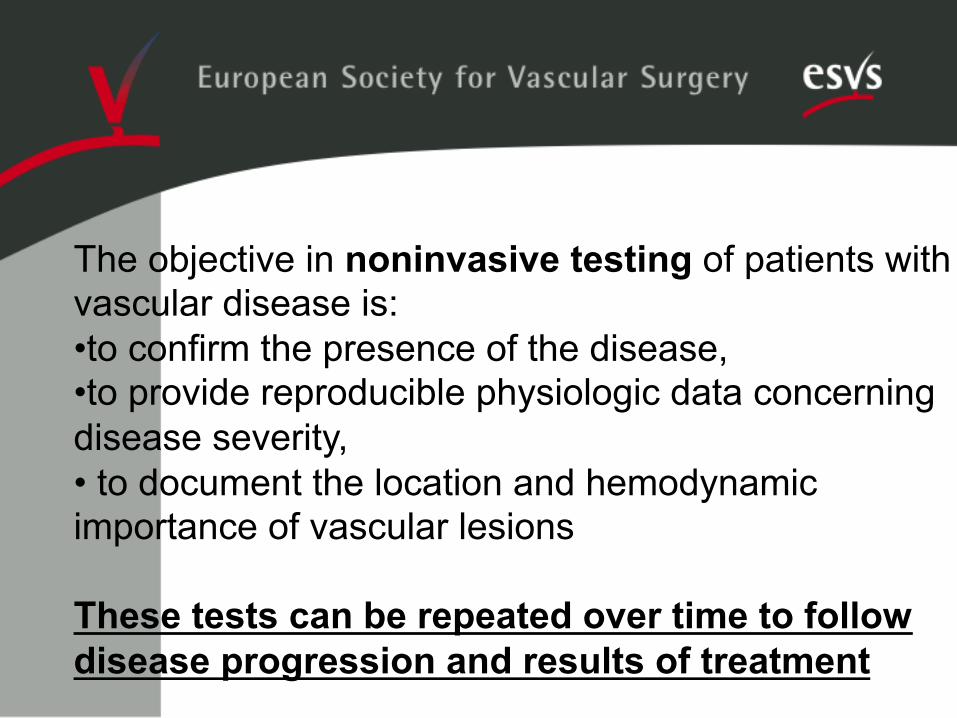
The objective in noninvasive testing of patients with vascular disease is: • to confirm the presence of the disease, • to provide reproducible physiologic data concerning disease severity, • to document the location and hemodynamic importance of vascular lesions These tests can be repeated over time to follow disease progression and results of treatment
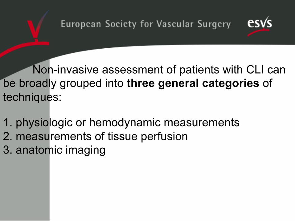
Non-invasive assessment of patients with CLI can be broadly grouped into three general categories of techniques: 1. physiologic or hemodynamic measurements 2. measurements of tissue perfusion 3. anatomic imaging

Non-invasive Vascular Laboratory
- DOPPLER ULTRASONOGRAPHY • Ankle brachial and toe-brachial indices • Segmental limb pressure • Continuous-wave doppler measurements
- PLETHYSMOGRAPHY • Pulse volume recording • Measurements of tissue perfusion

A routine measurement for screening • in 50-69 years old patients with smoking history , • in >70y patients • in all diabetic patients
Ankle brachial index (ABI)
A quick and cost-effective way to establish or refute CLI diagnosis
• provides information on long-term prognosis. • ABI <0.90 is associated with a 3-6 fold increased risk of cardiovascular mortality • may not be accurate in the presence of non compressible infracrural arteries

Recommendations
The resting ABI should be measured on both legs in patients with CLI to confirm diagnosis and establish baseline and should be used in individuals with non-healing ulcer, lower limb rest pain (Level 2c) ABI should be used to assess cardiovascular risk in those patients who are >70 years or >50 years with other cardiovascular risk factors as diabetes and history of smoking (Level 2a)

Toe brachial index Toe-brachial index is a quick way to noninvasively establish or refute the CLI diagnosis in patients with lower limb rest pain or nonhealing ulcers

Recommendation Toe-brachial index should be used in patients in whom CLI is clinically suspected and the ABI test is not reliable due to noncompressible vessels as in patients with diabetes, advanced age or long-standing renal failure (Level 2b, Grade B)
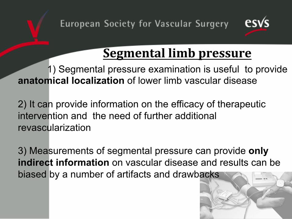
Segmental limb pressure 1) Segmental pressure examination is useful to provide
anatomical localization of lower limb vascular disease 2) It can provide information on the efficacy of therapeutic intervention and the need of further additional revascularization 3) Measurements of segmental pressure can provide only indirect information on vascular disease and results can be biased by a number of artifacts and drawbacks

Recommendation Lower limb segmental pressure measurements are useful to define the CLI initial diagnosis and localization of arterial lesion along lower limb (Level 2b, Grade B)
Continuous-‐wave doppler ultrasound
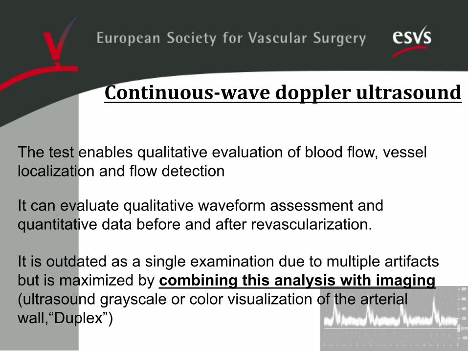
The test enables qualitative evaluation of blood flow, vessel localization and flow detection It can evaluate qualitative waveform assessment and quantitative data before and after revascularization. It is outdated as a single examination due to multiple artifacts but is maximized by combining this analysis with imaging (ultrasound grayscale or color visualization of the arterial wall,“Duplex”)
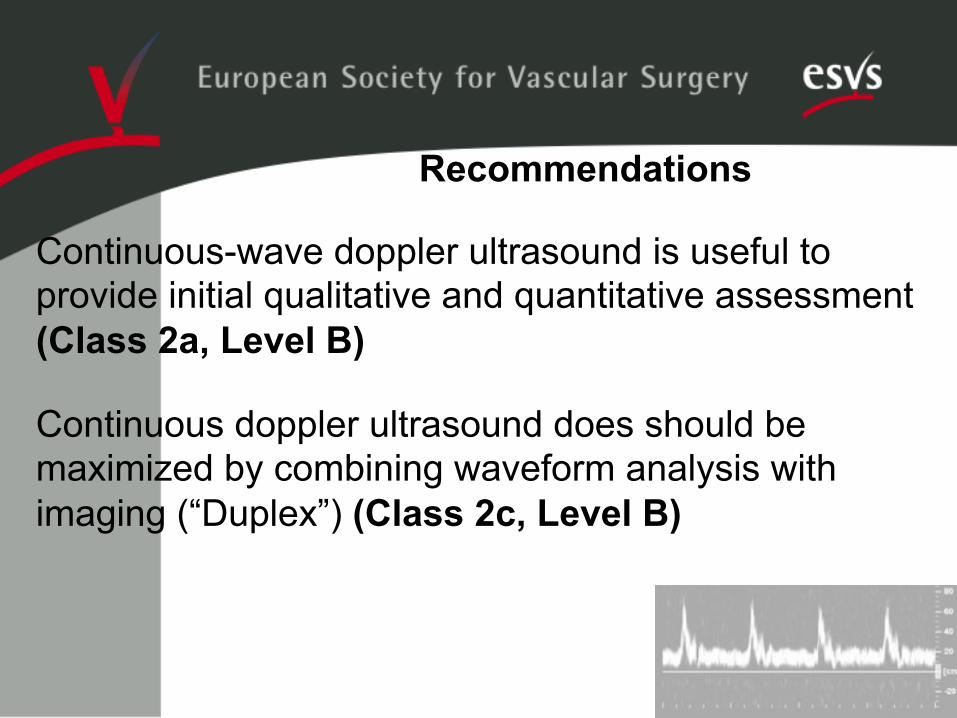
Recommendations Continuous-wave doppler ultrasound is useful to provide initial qualitative and quantitative assessment (Class 2a, Level B) Continuous doppler ultrasound does should be maximized by combining waveform analysis with imaging (“Duplex”) (Class 2c, Level B)

Pulse volume recording (PVR) PVR can provide a tool to evaluate small-vessel disease and CLI in individuals with noncompressible vessels in whom ABI and segmental pressures are spuriously elevated PVR does not allow quantitative measure of perfusion and may not be accurate in more distal segments of the leg. PVR may be abnormal in patients with low cardiac stroke volume
Recommendations
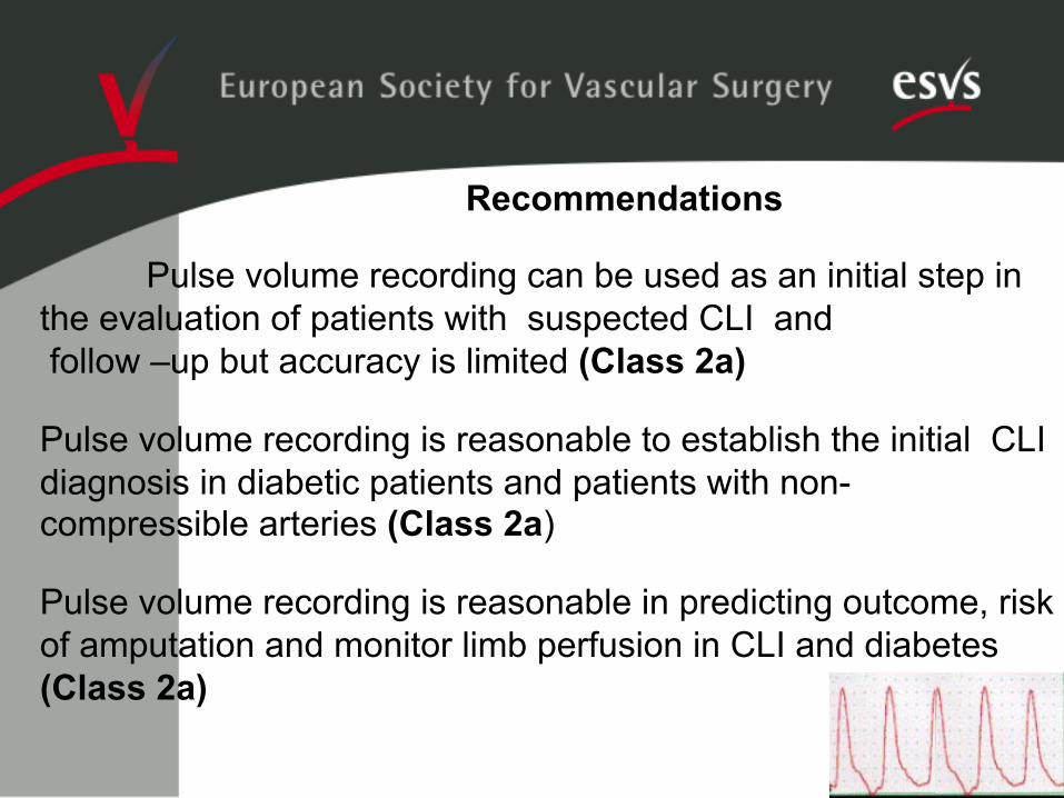
Pulse volume recording can be used as an initial step in the evaluation of patients with suspected CLI and follow –up but accuracy is limited (Class 2a) Pulse volume recording is reasonable to establish the initial CLI diagnosis in diabetic patients and patients with non-compressible arteries (Class 2a) Pulse volume recording is reasonable in predicting outcome, risk of amputation and monitor limb perfusion in CLI and diabetes (Class 2a)

Measurements of tissue perfusion include microcirculation techniques, the most common employed being transcutaneous partial pressure of oxygen (TcPO2) measurements
Measurements of tissue perfusion

Recommendations Patients with ischemic rest pain or foot ulcers should be investigated with objective tests to confirm diagnosis of CLI (Level B), these may include TcPO2, laser doppler and hyperspectral measurements to assess metabolic state of tissue perfusion (Level C) Tissue perfusion tests (TcPO2, laser doppler, spectral imaging) can be used to assess healing potential of ulcers/amputation in patients with CLI (Level C)

CHAPTER II: DIAGNOSTIC METHODS Ø Imaging techniques
• Duplex Ultrasound • Computed Tomography Angiography • Magnetic Resonance Angiography • Digital Subtraction Angiography
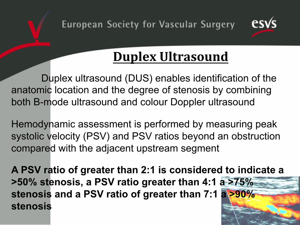
Duplex Ultrasound Duplex ultrasound (DUS) enables identification of the
anatomic location and the degree of stenosis by combining both B-mode ultrasound and colour Doppler ultrasound Hemodynamic assessment is performed by measuring peak systolic velocity (PSV) and PSV ratios beyond an obstruction compared with the adjacent upstream segment A PSV ratio of greater than 2:1 is considered to indicate a >50% stenosis, a PSV ratio greater than 4:1 a >75% stenosis and a PSV ratio of greater than 7:1 a >90% stenosis

Recommendations Grade of recommenda
tion
Level of Evidence
Duplex of the extremities is useful to diagnosis anatomic location and degree of obstruction
A 1a Duplex may be considered for routine surveillance after revascularisation B 2b Duplex can be useful to select patients as candidates for endovascular intervention
B 2b Duplex ultrasound may be considered for routine surveillance after femoro-‐popliteal bypass with a synthetic conduit
B 3b
Duplex Ultrasound

Computed Tomography Angiography Shorter acquisition times, thinner slice thicknesses, higher spatial resolution, and improvement of multidetector computed tomographic (CT) scanners enable scanning of the entire vascular tree CTA offers by comparison to MRA better patient acceptance, a higher speed of examination, a better spatial resolution and the ability to evaluate previously stented arteries Disadvantages of CTA include image interference from calcified arteries and the need for potentially nephrotoxic contrast and radiation exposure

Computed Tomography Angiography
Recommendations Grade of recommend
ation
Level of Evidenc
e CTA of the extremi.es may be considered to diagnose anatomic loca.on and presence of significant stenosis in pa.ents with lower extremity PAD
B 3a
CTA of the extremi.es may be considered as a subs.tute for MRA for those pa.ents with contraindica.ons to MRA
B 3a

Magnetic Resonance Angiography
There have been major technical advances in recent years including 3-D contrast enhanced magnetic resonance angiography (ce-MRA) and the development of moving tabletops which enable whole limb examinations with a single contrast injection Unlike DUS and CTA it is unaffected by arterial calcification Relative disadvantages include a tendency to overestimate stenosis

Recommendations Grade of recommendatio
n
Level of Evidence
MRA of the extremi.es is useful to diagnose anatomic loca.on and degree of stenosis of PAD A 1a MRA of the extremi.es should be performed with gadolinium enhancement A 2a MRA of the extremi.es is useful in selec.ng pa.ents with lower extremity PAD as candidates for endovascular interven.on A 2a
MRA of the extremi.es may be considered for postrevasculariza.on (endovascular and surgical bypass) surveillance in pa.ents with lower extremity PAD
B 3b
Magnetic Resonance Angiography
Recommendations Grade of recommendation
Level of Evidence
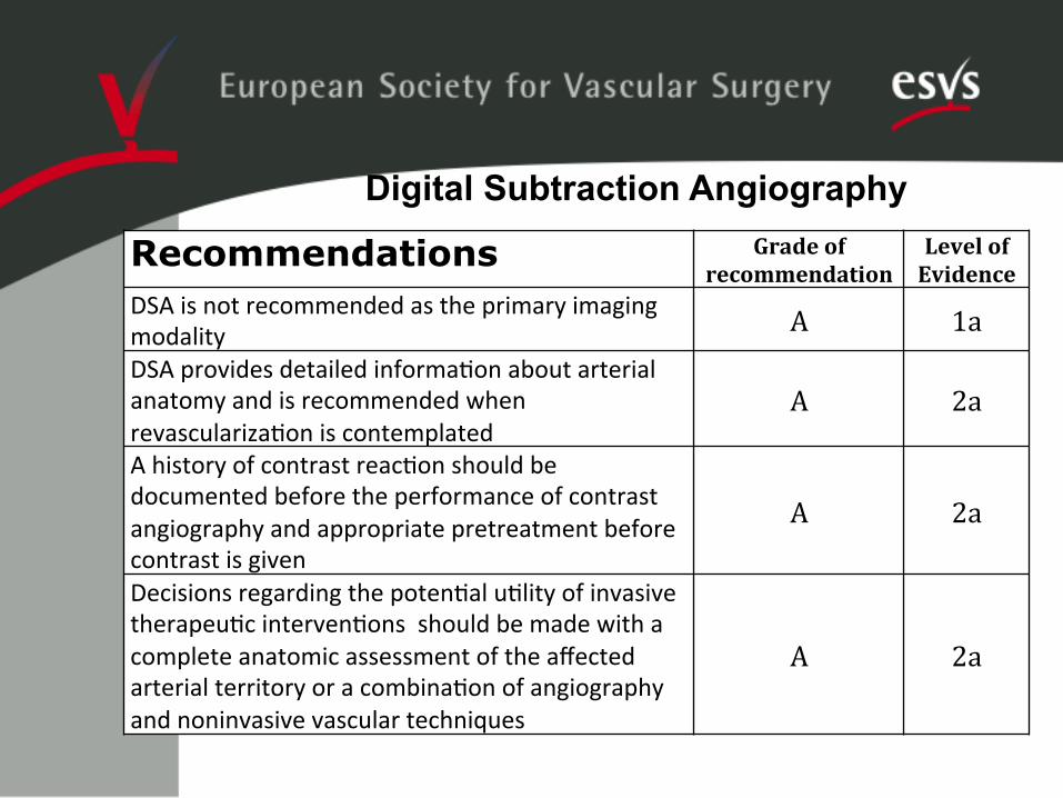
DSA is not recommended as the primary imaging modality A 1a DSA provides detailed informa.on about arterial anatomy and is recommended when revasculariza.on is contemplated
A 2a A history of contrast reac.on should be documented before the performance of contrast angiography and appropriate pretreatment before contrast is given
A 2a
Decisions regarding the poten.al u.lity of invasive therapeu.c interven.ons should be made with a complete anatomic assessment of the affected arterial territory or a combina.on of angiography and noninvasive vascular techniques
A 2a
Digital Subtraction Angiography
Grade of recommendation
Level of Evidence
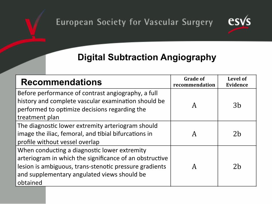
Before performance of contrast angiography, a full history and complete vascular examina.on should be performed to op.mize decisions regarding the treatment plan
A 3b
The diagnos.c lower extremity arteriogram should image the iliac, femoral, and .bial bifurca.ons in profile without vessel overlap
A 2b When conduc.ng a diagnos.c lower extremity arteriogram in which the significance of an obstruc.ve lesion is ambiguous, trans-‐steno.c pressure gradients and supplementary angulated views should be obtained
A 2b
Recommendations
Digital Subtraction Angiography

Recommendations Grade of recommendati
on
Level of Evidence
Pa.ents with baseline renal insufficiency should receive hydra.on before A 2b Follow-‐up clinical evalua.on, including a physical examina.on and measurement of renal func.on, is recommended within 2 weeks aOer contrast angiography to detect the presence of delayed adverse effects, such as atheroembolism, deteriora.on in renal func.on, or access site injury (e.g., pseudoaneurysm or arteriovenous fistula)
A 3a
Noninvasive imaging modali.es, including MRA, CTA, and color flow duplex imaging may be used in advance of invasive imaging to develop an individualized diagnos.c strategic plan
A 2a
Digital Subtraction Angiography

CLI Guidelines Medical Treatment
Jürg Schmidli Nicolas Diehm Bern, Switzerland

Management of Cardiovascular Risk Factors and Medical Therapy
• Critical issue: • Most of the outlined recommendations apply to PAD
patients in general. Thus, it has to be kept in mind that recommendations are frequently extrapolated to the subgroup of PAD with critical limb ischemia.

Cigarette Smoking
• Recommendations: • CLI patients should be strongly and repeatedly
advised to stop smoking (Level 2a, Grade B). • • Smoking cessation rates can be improved by
offering medical advice, group counseling session, nicotine replacement, nicotine receptor partial agonists (varenicline) or antidepressant drug therapy (bupropion) (Level 1a, Grade A).

Hyperlipidemia • Recommendations: • In CLI patients, statins should be the primary agents to
lower LDL cholesterol levels to reduce the risk of cardiovascular events (Level 1a, Grade A).
• For CLI patients, LDL cholesterol should be <100mg/dl • (Level 5, Grade C). • Dietary modification is aimed at controlling body weight
and lipid disorders (Level 5, Grade D). • Statins are indicated for secondary prevention of
cardiovascular events in patients with CLI • (Level 1a, Grade A).
Arterial Hypertension • Recommendations: • CLI patients with arterial hypertension should be
treated with antihypertensive medical therapy aimed at lowering cardiovascular mortality (Level 1c, Grade B).
• Treatment goals for CLI patients with arterial hypertension: arterial blood pressure should be <140/90mmHg and <130/80mmHg in case of concomitant diabetes mellitus or renal insufficiency (Level 1c, Grade B).
• ACE inhibitors are recommended in CLI patients • (Level 5, Grade C). • Beta-adrenergic blocking drugs are not contraindicated
in CLI patients (Level 1a, Grade A) and should be administered to patients undergoing surgical lower limb revascularization (Level 1c, Grade B).

Diabetes Mellitus
• Recommendation: • Blood glucose levels should be monitored in CLI
patients with a hemoglobin A1c (HbA1c) goal of <7.0% (Level 5, Grade D).

Antiplatelet Therapy • Recommendations: • Antiplatelet (aspirin or clopidogrel) therapy is
indicated in patients with symptomatic peripheral arterial disease (Level 1a, Grade A).
• Both aspirin and clopidogrel reduce rates of cardiovascular events in patients with symptomatic peripheral arterial disease (Level 1b, Grade A).
• In line with recommendations for patients with coronary heart disease, intermittent administration of dual antiplatelet therapy (aspirin plus clopdiogrel) may be considered for patients undergoing stent implantation or drug eluting balloon angioplasty of femoro-popliteal or infrapopliteal arteries
• (Level 5, Grade D).
Vasoactive Drugs
• Recommendation: • Parenteral prostanoids can be used in patients with
critical limb ischemia not suitable for arterial revascularization or after unsuccessful revascularization
• (Level 1a, Grade B).
Gene and Stem Cell Therapy
• Recommendation: • Due to conflicting or missing data, neither gene nor
stem cell therapy can be recommended as a treatment for CLI outside clinical trials
• (Level 5, Grade D).

Exercise and Lower Limb Rehabilitation • Recommendation: • Due to the risk of worsening pre-exisiting or causing
new ischemic wounds in the affected lower limb, walking exercise is contraindicated in CLI patients (Level 5, Grade D).

Treatment of Co-Existing Disease • Coronary artery disease (CAD) • Recommendation: • CLI patients with clinical evidence of CAD (angina,
ischemic congestive heart failure) should be evaluated and treated according to current guidelines for coronary revascularization
• (Level 1c, Grade A). • Routine treatment with beta blockers before
vascular surgery is recommended • (Level 1c, Grade B). • Routine coronary revascularization before vascular
surgery is not recommended (Level 1b, Grade A).

Treatment of Co-Existing Disease • Carotid artery disease • Recommendation: • The treatment for both symptomatic and
asymptomatic carotid artery disease in PAD patients should be based on current guidelines
• (Level 1a, Grade A).

Treatment of Co-Existing Disease • Renal artery disease • Recommendation: • In significant renal artery disease, as evidenced by
poorly controlled hypertension or renal insufficiency, patients should be referred to a vascular physician and be treated according to current guidelines (Level 2c, Grade B).

Health economics of risk-factor interventions • Cost-effectiveness • smoking cessetion interventions • pharmacologic interventions
• No literature available !
• No recommendations !

Summary • Besides arterial revascularization, risk factor
modification and administration of antiplatelet therapy is a major goal in the treatment of CLI patients.
• Key elements • smoking cessation, treatment of hyperlipidemia and
arterial hypertension; diabetes mellitus should be adequately adjusted.
• In CLI patients not suitable for arterial revascularization parenteral prostanoids may be considered.
• Gene and cell stem therapy are OUT • Walking exercise is contraindicated • Co-existing comorbidities should be managed according to
current guidelines. • Considering the above-mentioned treatment goals,
interdisciplinary treatment approaches for CLI patients are warranted.

Summary • Besides arterial revascularization, risk factor modification and
administration of antiplatelet therapy is a major goal in the treatment of CLI patients.
• Key elements • smoking cessation, treatment of hyperlipidemia
and arterial hypertension; diabetes mellitus should be adequately adjusted.
• In CLI patients not suitable for arterial revascularization parenteral prostanoids may be considered.
• Gene and cell stem therapy are OUT • Walking exercise is contraindicated • Co-existing comorbidities should be managed according to
current guidelines. • Considering the above-mentioned treatment goals,
interdisciplinary treatment approaches for CLI patients are warranted.
Summary • Besides arterial revascularization, risk factor modification and
administration of antiplatelet therapy is a major goal in the treatment of CLI patients.
• Key elements • smoking cessation, treatment of hyperlipidemia and
arterial hypertension; diabetes mellitus should be adequately adjusted.
• In CLI patients not suitable for arterial revascularization parenteral prostanoids may be considered.
• Gene and cell stem therapy are OUT • Walking exercise is contraindicated • Co-existing comorbidities should be managed according to
current guidelines. • Considering the above-mentioned treatment goals,
interdisciplinary treatment approaches for CLI patients are warranted.

Summary • Besides arterial revascularization, risk factor modification and
administration of antiplatelet therapy is a major goal in the treatment of CLI patients.
• Key elements • smoking cessation, treatment of hyperlipidemia and
arterial hypertension; diabetes mellitus should be adequately adjusted.
• In CLI patients not suitable for arterial revascularization parenteral prostanoids may be considered.
• Gene and cell stem therapy are OUT • Walking exercise is contraindicated • Co-existing comorbidities should be managed according
to current guidelines. • Considering the above-mentioned treatment goals,
interdisciplinary treatment approaches for CLI patients are warranted.
Summary • Besides arterial revascularization, risk factor modification and
administration of antiplatelet therapy is a major goal in the treatment of CLI patients.
• Key elements • smoking cessation, treatment of hyperlipidemia and
arterial hypertension; diabetes mellitus should be adequately adjusted.
• In CLI patients not suitable for arterial revascularization parenteral prostanoids may be considered.
• Gene and cell stem therapy are OUT • Walking exercise is contraindicated • Co-existing comorbidities should be managed according to
current guidelines. • Considering the above-mentioned treatment goals,
interdisciplinary treatment approaches for CLI patients are warranted.

C. Setacci1, G. de Donato1, M. Teraa, F. Moll3 & JB Ricco4
1 Department of Surgery, Unit of Vascular and Endovascular Surgery Unit, University of Siena, Italy 2 Department of Vascular Surgery, University Medical Center Utrecht, The Netherlands 3 Department of Nephrology & Hypertension, University Medical Center Utrecht, The Netherlands 4 Department of Vascular Surgery, University Hospital of Poi.ers, Poi.ers, France
September 18th, 2011
ESVS Guidelines for CLI Surgical Treatment

Surgical treatment of CLI -‐ Introduc.on
Decision making in revasculariza.on strategies in CLI differs substan.ally from that in pa.ents with claudica.on as wound healing, leg salvage and maintained ambula3onare different treatment aims than improved walking ability and there are oOen considerable .me constraints

Surgical treatment of CLI -‐ Introduc.on
Since there are almost no RCT exclusively among CLI pa.ents, most of the lessened recommenda.on are based on prospec.ve evidence from subgroup analyses of “PAOD” trials (extrapola3on from RCT), or from prospec.ve cohorts.

Treatmentop.ons • Pharmacological:
– Prostanoids • Surgical:
– Endarterectomy – Bypass
• Endovascular: – PTA – PTA withstentorstentgraO
• Hybrid • Non-‐reconstruc.ve

Guidelines and Classifica.ons • Classifica.ons and guidelinesaim at:
– Standardized care – Evidencebasedmedicine – Highligh.nggaps in currentknowledge
• TASC-‐classifica.onwidelyused, but: – Complex loco-‐regionalclassifica.on – Quicklyout-‐dateddue to fasbechnicaldevelopments – Poorinter-‐observer consensus
• New and simplifiedclassifica.onbasedonarterial segment and lesionlength is preferred

ESVS Guidelines -‐ Treatmentby segment
• Aortoiliac • Infrainguinal:
– CommonFemoralArtery (CFA) – DeepFemoralArtery (DFA) – SuperficialFemoralArtery (SFA)
• Popliteal • Infrapopliteal

AortoiliacObstruc.veDisease (AIOD) • Endovascularlowerlong-termprimarypatency (PP),
butsimilarsecondarypatency (SP)
• 5-year PP of open procedures in CLI:
• AFB, IFB, and AIE approximately 75-80%
Treatmentchoice: • First-‐line: PTA withprovisionalsten.ng(Level 3a, Grade C)
• Diffuse lesions: Aorto-‐(bi)femoral bypass (Level 2a, Grade B)
• Limitedlesions: Endarterectomyà lowermorbidity andmortality
(Level 4, Grade C)
• Extra-‐anatomical bypass reservedfor high risk pa.entorhos.le
abdomen (Level 4, Grade C)

CommonFemoralArtery (CFA) • CFA steno-‐occlusivedisease:
– Endarterectomy (poten.alforhybrid procedure) – PTA (withstent)
Treatmentchoice: • First choice: endarterectomy (5-‐year PP 91% SP 100%) (Level
4, Grade C) • Provides acces to performhybridrevasculariza.on of parallel
EIA, DFA or SFA pathologywithgoodresults(Level 3b, Grade C)

Deep Femoral Artery (DFA) • Recanaliza.on of the DFA:
– Rarelyperformed as isolated procedure forlimbsalvage – Limbsalvagerates:
• 67%, 49% and 36% at 1, 3, and 5 years – Profundoplastycanbe of value to preserve the knee joint
whenamputa.on is necessary Treatmentchoice: • First choice: surgicalprofundoplasty (Level 3b, Grade C)

SuperficialFemoralArtery (SFA) • SFA steno-‐occlusivedisease:
– Short lesions (<5 cm) – Intermediatelesions (5-‐15 cm) – Long lesions (>15 cm)
• Long-‐termpatency of PTA in CLI is muchlowerthan in claudicants: 20-‐37% 3-‐year PP
• Different abempts to reduce low patencydue to: – Recoil – Dissec.on – In.malhyperplasia
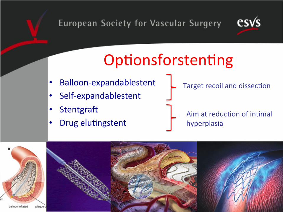
Op.onsforsten.ng • Balloon-‐expandablestent • Self-‐expandablestent • StentgraO • Drug elu.ngstent
Target recoil and dissec.on
Aim at reduc.on of in.mal hyperplasia

EUSC classifica.on Recommenda0on: A new and simplified classifica.on system for peripheral arterial disease lesions is needed to improve inter-‐individual interpreta.on as this is problema.c for the TASC classifica.on. Therefore we would recommend a system based on lesion length to classify lesions for research applica.ons and clinical management (EUSC classifica3on). Future research should prove the applicability and reproducibility of the classifica.on and the addi.onal value of a poten.al subdivision of steno.c versus occlusive lesions. Level 5, Grade D

Supe
rficialFemoralArtery (SFA
)
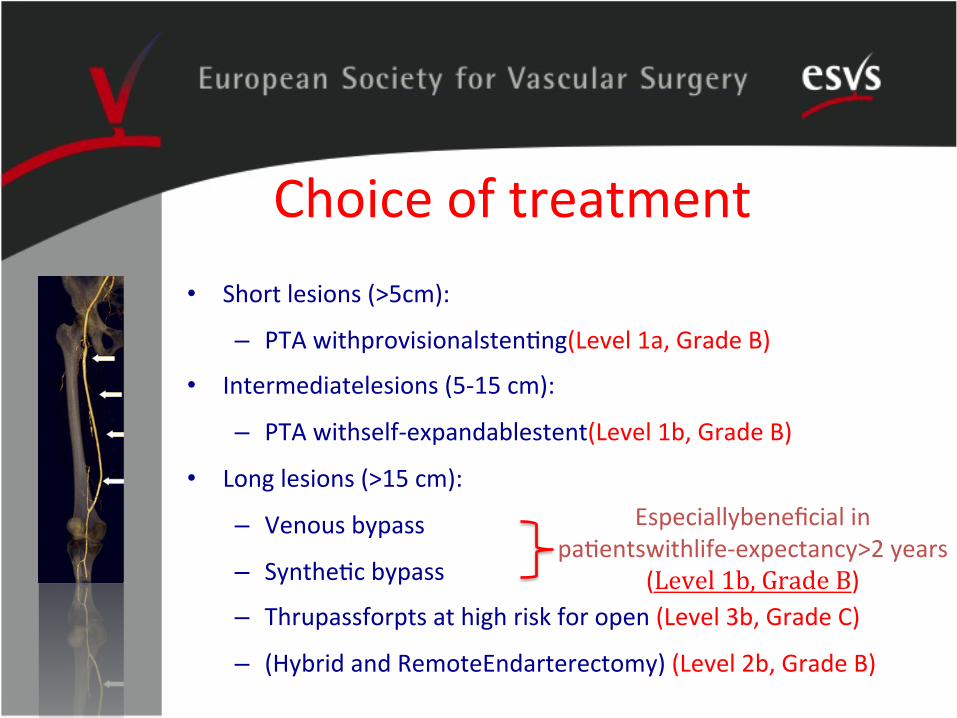
Choice of treatment • Short lesions (>5cm):
– PTA withprovisionalsten.ng(Level 1a, Grade B) • Intermediatelesions (5-‐15 cm):
– PTA withself-‐expandablestent(Level 1b, Grade B) • Long lesions (>15 cm):
– Venous bypass – Synthe.c bypass – Thrupassforpts at high risk for open (Level 3b, Grade C) – (Hybrid and RemoteEndarterectomy) (Level 2b, Grade B)
Especiallybeneficial in pa.entswithlife-‐expectancy>2 years
(Level 1b, Grade B)

Choice of treatment – SFA long lesions (>15 cm)
Recommenda0on:Hybrid procedures are the preferred treatment modality irrespec.ve of lesion length in high-‐risk pa.ents not suitable for open bypass surgery or when no suitable vein is available if minimally open revascularisa.on is mandated, such as CFE. Level 2b, Grade B
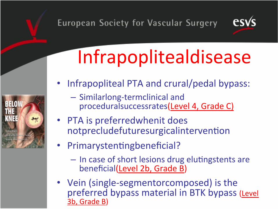
Infrapoplitealdisease • Infrapopliteal PTA and crural/pedal bypass:
– Similarlong-‐termclinical and proceduralsuccessrates(Level 4, Grade C)
• PTA is preferredwhenit does notprecludefuturesurgicalinterven.on
• Primarysten.ngbeneficial? – In case of short lesions drug elu.ngstents are beneficial(Level 2b, Grade B)
• Vein (single-‐segmentorcomposed) is the preferred bypass material in BTK bypass (Level 3b, Grade B)
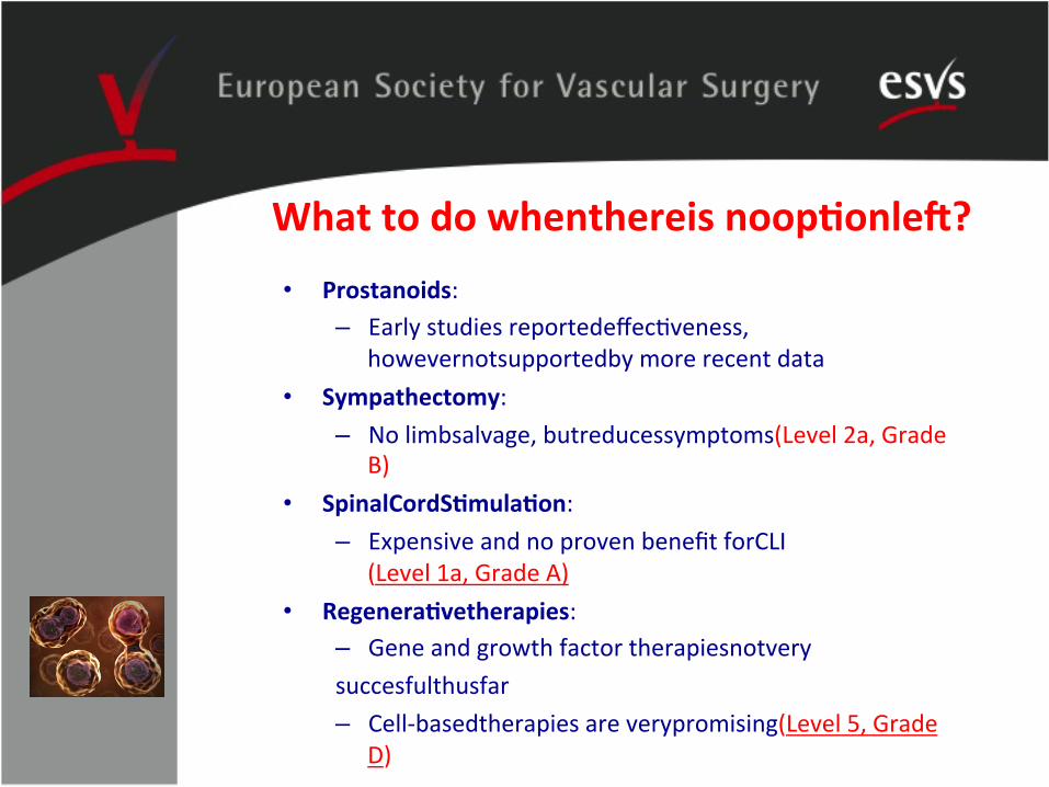
What to do whenthereis noop@onleA? • Prostanoids:
– Early studies reportedeffec.veness, howevernotsupportedby more recent data
• Sympathectomy: – No limbsalvage, butreducessymptoms(Level 2a, Grade
B) • SpinalCordS@mula@on:
– Expensive and no proven benefit forCLI (Level 1a, Grade A)
• Regenera@vetherapies: – Gene and growth factor therapiesnotvery succesfulthusfar – Cell-‐basedtherapies are verypromising(Level 5, Grade
D)

• CLI has a major impact on: – Pa.ent – Physician – Health care system
• A more concise and simplifiedclassifica.on is advocated
• Treatmentconsists of endovascular and surgicalop.onswithanincreasing trend towardsanendovascularfirstapproach
Conclusions

Conclusions
• Principlefirst-‐linetreatment: – AIOD: PTA withprovisionalsten.ng – CFA: Endarterectomy – DFA: Endarterectomy – SFA:
• Short lesion: PTA withprovisionalsten.ng • Intermediate: PTA withself-‐expandablestent • Long lesion: Venous (orsynthe.c) bypass
– Infrapopliteal: PTA (with DES in short lesions)

Ques.ons???
Limbsalvage is the goal, bu.tcan’tbe as nice as it was before…

Chapter V, Follow-up
ESVS CLI guidelines 2011 F. Dick, AH. Davies, JB Ricco, et al.

Follow-up
v sustained treatment success v con.nued best pa.ent care
Follow-up IMPORTANT
v sustained treatment success v con.nued best pa.ent care
v CLI: frail and elderly v ambula.on=independency
nonetheless...
v neglected previously
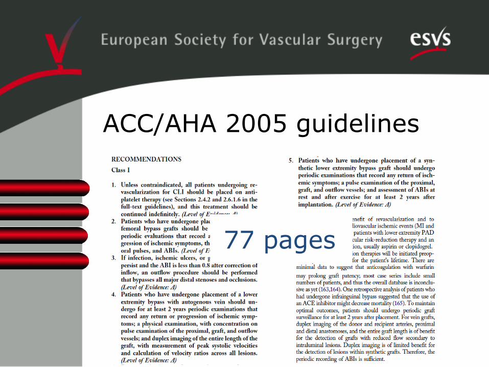
ACC/AHA 2005 guidelines
77 pages

ACC/AHA 2005 guidelines
v class I, level A (B) recommendations
v ...no references presented...

TASC II 2007
63 pages

TASC II 2007
v grade A (C) recommendations
v ...not stratified for CLI...

v ? lack of (stra.fied) studies v ? pessimis.c percep.on
of prognosis
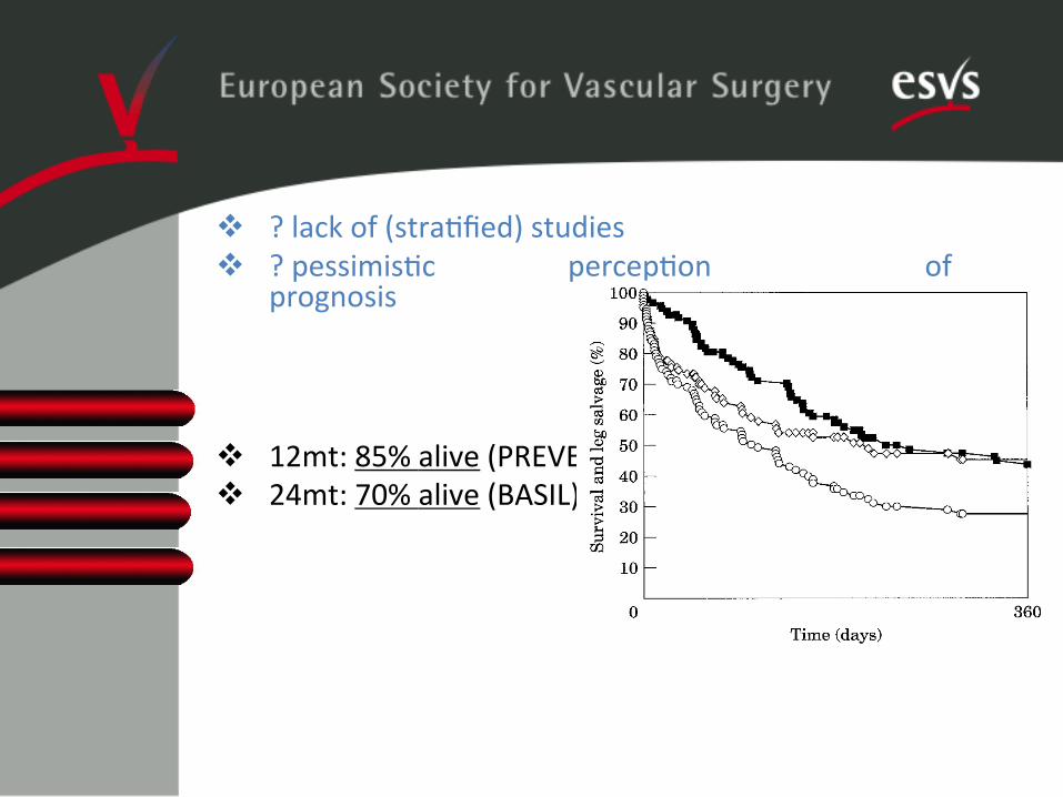
v ? lack of (stra.fied) studies v ? pessimis.c percep.on of
prognosis
v 12mt: 85% alive (PREVENT III) v 24mt: 70% alive (BASIL)

Follow-up: current guidelines
v systematic review v lack of stratified studies v inconsistent CLI definition
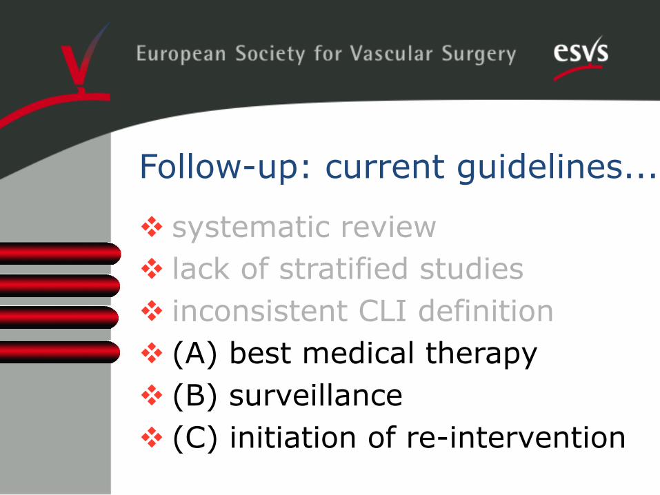
v systematic review v lack of stratified studies v inconsistent CLI definition v (A) best medical therapy v (B) surveillance v (C) initiation of re-intervention
Follow-up: current guidelines...

...based on extrapolations
v grades of recommendation
v degradation of recommendation strength
v critical issue 1: need for well-designed studies
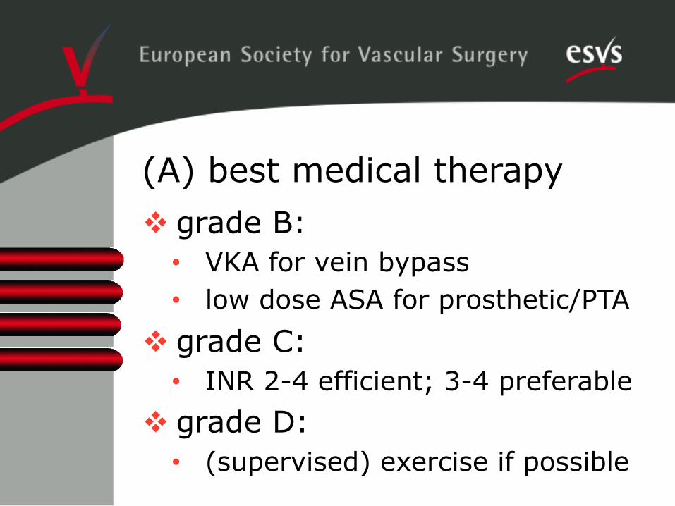
(A) best medical therapy v grade B:
• VKA for vein bypass • low dose ASA for prosthetic/PTA
v grade C: • INR 2-4 efficient; 3-4 preferable
v grade D: • (supervised) exercise if possible

critical issues A: v statin effect on patency?
v systemic benefits of ASA versus local benefits of VKA (vein bp)
v duration of VKA?
v clopidogrel/new antithrombotic agents?

(B) surveillance v grade B:
• early duplex for vein bypass • no routine long-term duplex prgr. • clinical surveillance every 3-6 mts
v grade C: • early duplex after PTA • duplex surveillance for graft at risk

critical issues B: v role of distal landing zone (ie.,
above/below knee)?
v duplex surveillance after use of endovascular adjuncts?
v cost-effectiveness analyses?

(C) repeat interventions v grade B:
• PTA and surgery equivalent for short and late appearing stenosis
v grade C: • early and recurrent stenoses
benefit from surgery • clinical/duplex surveillance after
re-intervention
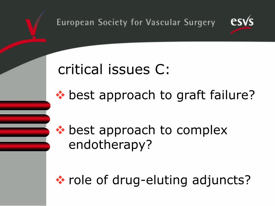
critical issues C:
v best approach to graft failure?
v best approach to complex endotherapy?
v role of drug-eluting adjuncts?
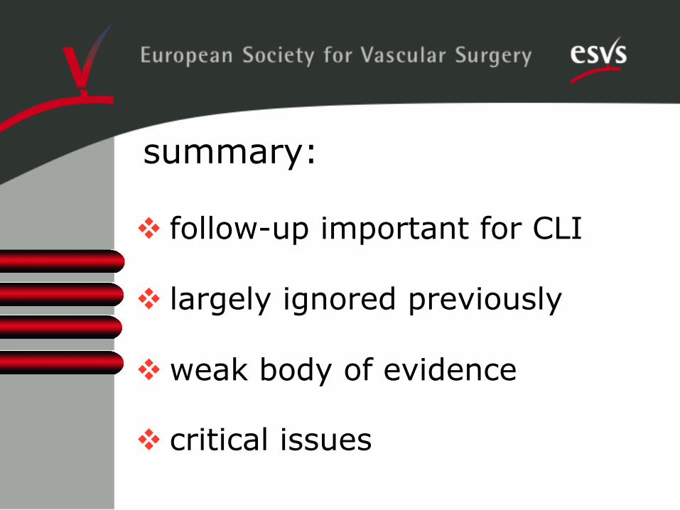
summary:
v follow-up important for CLI
v largely ignored previously
v weak body of evidence
v critical issues

conclusions:
v extrapolated recommendations (no grade A)
v valid evidence to be developed
v important contexts: diabetes, renal failure, functionally impaired

Guidelines for Cri@cal Limb Ischemia & Diabe@c Foot
Mauri Lepäntalo, MD, PhD, Helsinki Jan Apelqvist, MD, PhD, Malmö
The previous work of Interna.onal Working Group on the Diabe.c Foot – Peripheral Arterial Disease is acknowledged

Diabetic foot ulcers • Over 55 million diabe.cs in Europe • Indica.ve annual costs for EU have been es.mated to be
4-‐6 billion Euro’s • Complica.ons of foot ulcers are a leading cause of
hospitaliza.on and amputa.on • 20-‐40% of health care resources for diabetes is related to
diabe.c foot • Annual incidence of foot ulcera.on is over 2-‐5% among
diabe.cs • Major amputa.on will be needed within a year in 5-‐8% of
pa.ents with diabe.c ulcers – 85% preceded by a foot ulcer

Neuropathy and ischemia are the ini@a@ng factors, with a different weight in different pa@ents, and infec@on is mostly a consequence

Ischemia and neuroischemia of the diabe@c foot Underes@ma@on of the role of ischemia
• up to 60% of neuroischemic or ischemic origin
•
• Recommenda0on: Ischemia should not be excluded as a cause of diabe.c foot ulcer unless proven absent. Level 5;Grade D

Inadequate understanding of neuroischemia • Ischemia is caused by peripheral arterial disease, typically
affec.ng infrapopliteal arteries. • The combined effect of diabe.c neuropathy and
ischemia, oOen called neuroischemia, decreases the foot perfusion even further.
• Microvascular dysfunc.on – presence of arterio-‐venous shun.ng – pre-‐capillary sphincter malfunc.on – capillary leakage – venous pooling – hormonal ac.vity in the vessel – inflamma.on in its wall

Inadequate understanding of neuroischemia
• Recommenda0on: • In neuroischemic legs the healing is primarily affected by the severity of ischemia.
• Therefore, from a prac.cal point of view neuroischemic and ischemic lesions should be considered together as both may need revascularisa.on. Level 2b;Grade C

Why CLI criteria for non-‐diabe@cs are not applicable in diabe@cs
• Use of rigid noninvasive methods not good enough – bias due to medial sclerosis, .ssue lesions
• A clear need to introduce and recognize decreased perfusion as indicator for need for revasculariza.on in the diabe.c foot to achieve and maintain healing and to avoid or delay a future amputa.on

Why CLI criteria for non-‐diabe@cs are not applicable in diabe@cs
• Recommenda0on: Interna.onal Working Group for Diabe.c Foot recommends further vascular studies in case ulcer has not healed in proper treatment in six weeks although ini.al diagnos.cs have suggested only ques.onable or mild disease. Level 5;Grade D
•
• Cri0cal issue: Criteria for impaired perfusion should be established
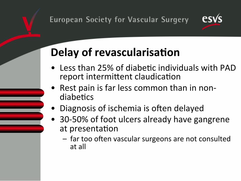
Delay of revascularisa@on • Less than 25% of diabe.c individuals with PAD report intermibent claudica.on
• Rest pain is far less common than in non-‐diabe.cs
• Diagnosis of ischemia is oOen delayed • 30-‐50% of foot ulcers already have gangrene at presenta.on – far too oOen vascular surgeons are not consulted at all

Delay of revasculariza@on
• Recommenda0on: To prevent the delay of vascular consulta.on and revasculariza.on early noninvasive vascular evalua.on is important in iden.fying pa.ents with poor ulcer healing and high risk for amputa.on. Level 2b;Grade B

Neuroischemia, infec@on and @ssue damage
• An infec.on in the diabe.c foot is a limb-‐threatening condi.on – immediate cause for amputa.on in 25-‐50% of diabe.c pa.ents
– feet with a combina.on of ischemia, infec.on and .ssue damage fare even worse

Clinical examina@on
• Recommenda0on: Every foot ulcer should be examined for the presence of ischemia.
• Level 5;Grade 4
• Recommenda0on: Every foot ulcer should be examined for the presence of neuropathy.
• Level 5;Grade4
• Recommenda0on: Every diabe.c foot ulcer should be examined for the presence of infec.on.
• Level 5;Grade D

Non-‐invasive vascular studies • Recommenda0on: Trust ABI when low but not when high. An
ABI <0.6 indicates significant ischemia in respect to wound healing poten.al whereas on ABI >0.6 has lible predic.ve value and therefore at least the toe pressure should be measured. Level 5;Grade D
• Recommenda0on: An ulcera.on of the foot in diabetes will generally heal if the toe pressure is >55 mmHg, whereas healing is usually severely impaired when toe pressure is <30 mmHg. Level 2b;Grade B
• Recommenda0on: Ulcera.on of the foot in diabetes will generally heal if the tcpO2 >50 mmHg. Healing is usually severely impaired when tcpO2 <30 mmHg. Level 2b;Grade B

Vascular imaging and revascularization should be considered irrespective of pressure values if diabetic foot ulcer does not heel in conservative treatment
Probability of healing (%) as related to noninvasive data
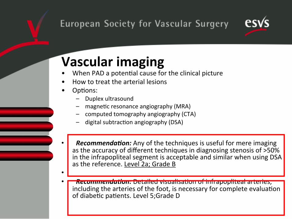
Vascular imaging • When PAD a poten.al cause for the clinical picture • How to treat the arterial lesions • Op.ons:
– Duplex ultrasound – magne.c resonance angiography (MRA) – computed tomography angiography (CTA) – digital subtrac.on angiography (DSA)
• Recommenda0on: Any of the techniques is useful for mere imaging as the accuracy of different techniques in diagnosing stenosis of >50% in the infrapopliteal segment is acceptable and similar when using DSA as the reference. Level 2a; Grade B
• • Recommenda0on: Detailed visualisa.on of infrapopliteal arteries,
including the arteries of the foot, is necessary for complete evalua.on of diabe.c pa.ents. Level 5;Grade D

Specific problems in imaging • Duplex and CT
– extensive calcifica.on of infrapopliteal arterial tree may prevent proper Duplex diagnos.cs and computed tomography angiography
– mul.-‐sliced devices decreases interpreta.on difficul.es caused by arterial wall calcifica.ons
• MRA – limited spa.al resolu.on – its images may be distorted by previous stents, implants and flow
disturbances – use of paramagne.c contrast material gadolinium has been
reported to cause nephrogenic systemic fibrosis typically in pa.ents with renal failure
• Cri0cal issue: The risks of gadolinium-‐enhanced MRA for imaging diabe.c pa.ents with kidney failure should be considered and further evaluated

Mul@factorial approach mandatory I Multifactorial treatment of diabetic foot ulcer Goal Treatment Improvement of perfusion Endovascular revascularization (PTA)
Reconstructive vascular surgery (bypass) Vascular drugs Reduction of edema Hyberbaric oxygen
Treatment of infection Antibiotics (oral or parenteral) Incision, drainage Resection
Reduction of edema External compression therapy Intermittent compression (pumps) Diuretics
Pain control Analgesic drugs (local or systemic) Immobilisation, off loading, relief of anxiety and fear, TNS
Improvement of metabolic control Insulin treatment
Necessary nutritional support

Mul@factorial approach mandatory II Off loading Protective and therapeutic footwear
Insoles, orthosis
Total contact cast, walkers
Crutches, wheelchair, bed rest
Wound bed preparation Debridement, removal of debris
Topical treatment, dressings
Control of exudation, moist wound healing, GCSF infection control, NPWT
Tissue engineering, growth factors, matrix modulation
Removal of dead tissue Incision, drainage, amputation
Correction of foot deformities Corrective foot surgery, skin transplant, amputation
Improvement of general condition
Fluid and nutrition replacement therapy
Aggressive treatment of concomitant disease, antiplatelet drugs, antihypertensive agents, lipid decreasing agents
Cessation of smoking
Physiotherapy
Implementation of systematic care
Patient and staff education
Support and follow up
Multidisciplinary co-ordination, communication, staggered treatment chains
Improvement of concordance process oriented approach
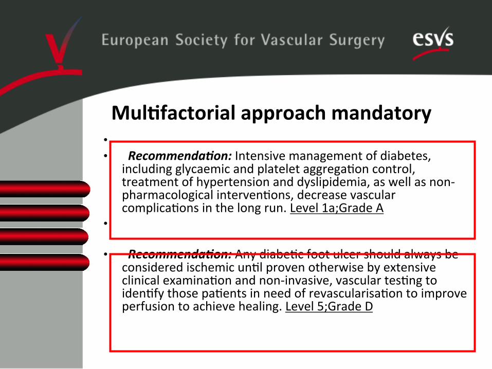
Mul@factorial approach mandatory • • Recommenda0on: Intensive management of diabetes,
including glycaemic and platelet aggrega.on control, treatment of hypertension and dyslipidemia, as well as non-‐pharmacological interven.ons, decrease vascular complica.ons in the long run. Level 1a;Grade A
•
• Recommenda0on: Any diabe.c foot ulcer should always be considered ischemic un.l proven otherwise by extensive clinical examina.on and non-‐invasive, vascular tes.ng to iden.fy those pa.ents in need of revascularisa.on to improve perfusion to achieve healing. Level 5;Grade D

Revasculariza@on • The crucial issue is to decide whether revasculariza.on is needed for a
certain lesion, for a certain pa.ent • Although noninvasive evalua.on is helpful, the decision to intervene
is made due to presen.ng symptoms and clinical findings • Anatomic imaging should be considered only strategic
• Recommenda0on: There are no convincing data that endovascular or open revasculariza.on would give beber outcome in diabe.c pa.ent with ischemic ulcer as the results strongly depend on the severity and distribu.on of peripheral arterial disease. Level 2c;Grade B
• •

Case series on infrainguinal revascularisa@ons for ischemic ulcerated diabe@c foot
Author Patients; N / gender / age (mean/median)
Comorbidity Intervention Infrapopliteal distribution
30-day complications
Follow-up Outcome
Rosenbaum, 1994 39 / M33, F6 / 62,3 yrs
NA infrapopliteal bypass grafts 79 % Major amputation 3%, mortality NA
21,2 months (mean), range 2-64
NA
Wolfle, 2000 125 / NA / 70 yrs
CAD 57%, ESRD 25%
infrapopliteal bypass vein grafts
100 % Major amputation NA, mortality 2%
24 months (mean)
Limb salvage 80% at 1 yr, mortality 51% during fu
74 / NA / 68 yrs CAD 48%, ESRD 42%
infrapopliteal PTA 100 % Major amputation NA, mortality 6%
24 months (mean)
Limb salvage 82% at 1 yr, mortality 35% during fu
Schneider, 2001 110 / M67, F43 / 69 yrs (weighted mean)
CAD 43%, ESRD 69% (weighted mean)
Revascularisation using either fem-distal bypass, combined SFA PTA and distal bypass grafting or short distal bypass graft
100 % NA 23 months (mean)
Limb salvage 89%, patency 78% at 2 yr, mortality NA (weighted mean)
Faglia, 2002 221 / NA / NA CAD 55%, ESRD 4%
femorodistal and infrapopliteal PTA
94 % Major amputation 5%, mortality 0%
12 months (median), range 5-30
Limb salvage NA, mortality 5.3% at 1 yr
Dorweiler, 2006 46 / M36, F10 / 69 yrs
CAD 46%, ESRD 13%
pedal bypass grafts 100 % Major amputation 7%, mortality 2%
28 months (median), range 1-70
NA
Bargellini, 2008 60 / M41, F19 / 69,4 yrs
CAD 42%, CVD 25%
multilevel subintimal PTA in patients unfit for surgery
43 % Major amputation 5%, mortality 5%
23 months (mean), range 0–48
Limb salvage 93.3%, mortality 10% at 1yr
Ferraresi, 2009 101 / M85, F16 / 66 yrs
CAD 28%, ESRD 3%
infrapopliteal PTA 100 % NA 35 months (mean)
Limb salvage 93%, mortality 9% during FU

Management of infec@on • An.bio.c therapy
– necessary for virtually all infected wounds – not beneficial for noninfected ulcers – insufficient without appropriate wound care – Pa.ents with uncontrolled or limb-‐threatening infec.ons require
immediate hospitaliza.on, immobiliza.on and intravenous an.bio.cs
• Recommenda0on: Surgical interven.on for moderate or severe infec.ons is likely to decrease the risk for major amputa.on. Level 2c; Grade B
• Timing of treatment of infec.on vs. revascularisa.on
• Recommenda0on: The severity of infec.on guides the decision whether to debride first or to revascularize first. Level 2c;Grade C

Debridement • Surgical, enzyma.c, biological or autoly.c methods
• Recommenda0on: No single method is outstanding in terms of enhancing diabe@c ulcer healing. Level 1c;Grade A
• Yet, in selected cases, systemic hyperbaric oxygen therapy may be effec.ve in non healing long standing ulcers, nega.ve pressure wound therapy may promote healing of postopera.ve wounds and resec.on of plantar ulcers may be beneficial
• Recommenda0on: Hyperbaric oxygen therapy may be indicated for a selected group of diabe@c ulcers but it is not clear which pa@ents are likely to benefit and what is the op@mal dura@on. Level 1b; Grade A
•
• Recommenda0on: Nega@ve pressure wound therapy appears to be as effec@ve and under certain circumstances more effec@ve than other available local wound treatments in pa@ents without significant infec@on. Level 1a;Grade A
•
• Recommenda0on: Foot surgery to offload pressure areas may be beneficial to prevent ulcer recurrence aAer revasculariza@on for neuroischemic diabe@c foot ulcer. Level 4;Grade 5

Minor amputa@on and removal of necro@c @ssue
• Recommenda0on: Toe, ray and trans-‐metatarsal amputa.ons are preferred whenever possible as they enable broader distribu.on of weight during ambula.on. Level 4;Grade 5
• Recommenda0on: Bedridden pa.ents, poor ambula.on that is not worsened by amputa.on, life expectancy less than one year, and non-‐revascularizable leg are causes to perform major amputa.on, even above the knee when necessary. Level 4;Grade D
•

Outcomes • Wound healing • Revasculariza.ons improve ulcer healing
– number of ulcers, severity of PAD, conges.ve heart failure and renal func.on impairment were associated with poor ulcer healing
• Completeness of revasculariza.on seems important • Complete .ssue healing aOer revasculariza.on very slow • Leg salvage • Leg salvage rates around 80% at one year and around 70% at
three years aOer revascularisa.ons • Risk for major amputa.on
– occlusion of all three crural arteries – ESRD – wound infec.on – mul.ple ulcers – oedema – non-‐compliance to the treatment
•

Mortality • Periopera.ve mortality below 5% • Mortality 10-‐20% at one and 40-‐50% at five years
aOer open surgery, whereas long term data are missing in endovascular series
• Recommenda0on: Co-‐morbidi.es especially renal failure and impaired ambulatory status at presenta.on are major factors for poor outcome in diabe.cs with ischemic ulcers. These co-‐morbidi.es should be taken into considera.on when and when not to revascularize. Level 2a;Grade B

Mul@disciplinary team approach
• Diabe.c foot ulcers need to be treated in a systema.c way by a mul.disclipinary team
• Vascular surgeon should be a part of this team – Urgent need to include vascular diagnos.cs and
interven.on as an integrated part of the strategy to achieve ulcer healing and to avoid major amputa.on
– Otherwise the window of opportunity could be easily missed
• Up to 85% of the amputa.ons may be prevented by a mul.disciplinary approach
Summary • Urgent need for a paradigm shiO in diabe.c foot care, i.e. a new approach and classifica.on of diabe.cs with impaired perfusion with regard to clinical prac.ce and research
• This change will considerably increase the need of distal revasculariza.ons for diabe.c foot ulcers in near future