guidance document for the preparation of … · investigational device ... in the field including a...
TRANSCRIPT

page 1
This guidance was written prior to the February 27, 1997 implementation of FDA’s GoodGuidance Practices, GGP’s. It does not create or confer rights for or on any person and does not
operate to bind FDA or the public. An alternative approach may be used if such approachsatisfies the requirements of the applicable statute, regulations, or both. This guidance will be
updated in the next revision to include the standard elements of GGP’s.
GUIDANCE DOCUMENT FOR THE PREPARATION OFINVESTIGATIONAL DEVICE EXEMPTIONS ANDPREMARKET APPROVAL APPLICATIONS FOR
INTRA-ARTICULAR PROSTHETIC KNEE LIGAMENT DEVICES
September 1, 1987(Revised February 18, 1993)
(reformatted 12/17/97)
This guidance document may contain references to addresses and telephone numbers thatare now obsolete. The following contact information is to be used instead:• While this guidance document represents a final document, comments and suggestions
may be submitted at any time for Agency consideration to the Orthopedic DevicesBranch, 9200 Corporate Blvd., HFZ-410, Rockville, MD 20850.
• For questions regarding the use or interpretation of this guidance, contact theOrthopedic Devices Branch at 301-594-2036.
• To contact the Division of Small Manufacturers Assistance (DSMA), call 800-638-2041or 301-443-6597; fax 301-443-8818; email [email protected]; or write to DSMA(HFZ-200), Food and Drug Administration, 1350 Piccard Drive, Rockville, Maryland20850-4307. FACTS-ON-DEMAND (800-899-0381 or 301-827-0111) and the WorldWide Web (CDRH home page: http://www.fda.gov/cdrh/index.html) also provide easyaccess to the latest information and operating policies and procedures.
U.S. DEPARTMENT OF HEALTH AND HUMAN SERVICESFood and Drug Administration
Center for Devices and Radiological HealthRockville, MD 20850

page 2
PREFACE
The development of a guidance document for intra-articular prosthetic knee ligament devices isbased on the Division of General and Restorative Devices' (DGRD's) evaluation of numerousdevices and the recognition of certain criteria necessary to conduct these evaluations. Thepurpose of this document is to suggest to the device manufacturer or investigation sponsorimportant preclinical and clinical tests that should be performed to generate data that will providereasonable assurance of the safety and effectiveness of these devices for their intended purposes.Suggestions and recommendations written in the document are not mandatory requirements.They reflect methodologies which DGRD has determined to be acceptable and which, iffollowed, will assure well designed and scientifically valid Investigational Device Exemption(IDE) and Premarket Approval (PMA) applications. In this context several points should beremembered:
1. The guidance document is primarily intended to be a scientific position paper. Therefore,it suggests some important evaluation criteria, test procedures, and end points. If thesame objectives can be achieved by other means, the investigator should not refrain fromdoing so.
2. The guidance document should be viewed as a living document. As science changes andscientific techniques are improved, FDA will periodically revise the document.Nonetheless, it should be remembered that the basic objectives may remain the same.
3. The word "should" and "must" have been used frequently in this document to emphasizethe relative merit or importance of a specific aspect of a test or protocol. However, thisverbiage is not used in a regulatory sense and should not be construed as such.
Guidance document preparation was initiated by DSRD (DGRD was formerly known as Divisionof Surgical and Rehabilitation Devices (DSRD)) staff Karen L. Goldenthal, M.D., Janet Guerrin,M.S., and Nirmal K. Mishra, Ph.D., D.V.M., in December of 1985 following a history of activityin the field including a meeting of the Orthopedic and Rehabilitation Devices Panel to discussprosthetic ligament devices in November, 1983. In May of 1986, DSRD completed a first draftof the document containing preclinical and clinical testing recommendations and suggestions forthe preparation of IDEs and PMAs. It was intended that the document be reviewed by Panelmembers with public comment at the June 19 and 20, 1986 Panel meeting so that the draft couldbe revised and made available for public comment. The industry representative to the Panelrequested that the discussion be postponed until he could receive comments from hisconstituents. HIMA undertook the task of soliciting comments from industry and provided theseto DSRD shortly before the October 31, 1986 Panel meeting. At this meeting, an open publicsession preceded the discussion of the document by the Panel. As a result of the October 31,1986 Panel meeting, two working groups were established based on nominations from the Paneland industry:
Working Group for Evaluating Mechanical Test Recommendations:
Chairman (designated by Panel) - Savio Woo, Ph.D.FDA Liaison - Janet Guerrin, M.S.FDA Liaison - Daniel Chwirut, M.S., ~P.E.Panel Member - A. Seth Greenwald, D. Phil(Oxon)

page 3
Panel Member - Peter A. Torzilli, Ph.D.HIMA Representative - William C. Bruchman, B.S.OSMA Representative - Walter P. Spires, Jr., M.S.
Working Group for Evaluating Biological Test Recommendations:
FDA Liaison - Nirmal Mishra, Ph.D., D.V.M.HIMA/OSMA Representative - Vince Mendenhall, Ph.D., D.V.M.HIMA/OSMA Representative - John Willson, Ph.D.
Working groups reviewed draft document recommendations which were revised according toPanel member comments, HIMA comments, and OSMA comments. After meeting with bothworking groups, DSRD again revised the document and accepted further comments from theworking groups. The final draft of the document was then reviewed by the following Panelmembers and consultants at the May 7, 1987 Panel meeting:
Kenneth E. DeHaven, M.D.Victor J. Ferrans, M.D., Ph.D.A. Seth Greenwald, D. Phil(Oxon)F. Joseph Halcomb, M.D.Randall J. Lewis, M.D.Michael B. Mayor, M.D. (Chairman)M. Clinton Miller, III, Ph.D.Kurt M.W. Niemann, M.D.Eric L. Radin, M.D.Kenneth M. Singer, M.D.Peter A. Torzilli, Ph.D.Bertram Zarins, M.D.
The Orthopedic and Rehabilitation Devices Panel voted unanimously to endorse the documentwith several changes at the May 7, 1987 Panel meeting. This document incorporates the changes.According to recommendations made by industry groups and the Panel this document would bereviewed again in 1 year at a meeting of the Orthopedic and Rehabilitation Devices Panel.Revisions would be made according to comments voiced by the public and the Panel at thatmeeting. The document would be reviewed and revised, if necessary, every 2 years thereafter.
After the May 7, 1987 Panel meeting and the document was finalized on September 1, 1987,there were no further formal discussions of this document. This document has been used bymany in industry for conducting new studies with ligaments and has been well received. Sinceits introduction, several new ligaments have been approved for market bringing the currentnumber of approved ligaments in the U.S. to three. The three approved ligaments are the Gore-Tex Cruciate Ligament Prosthesis by W.L. Gore and Associates approved on October 10, 1986,the Stryker Dacrone Ligament Prosthesis by Meadox Medicals, Inc. approved on December 30,1988 and the 3M Kennedy LAD Ligament Augmentation Device by 3M which was firstapproved on May 7, 1987 for the Marshall-MacIntosh procedure and then the indication wasexpanded on December 31, 1992 to include the use of the patellar tendon graft of 10 mm orsmaller in patients more than 3 weeks since injury. The Summaries of Safety and Effectivenessfor these devices can be obtained from the FDA Freedom of Information Office at 5600 FisherLane, HFI-35, Rockville, Maryland 20857 to obtain examples of preclinical and clinical worknecessary for FDA approval.

page 4
INTRODUCTION
Purpose
It has been determined by the Food and Drug Administration (FDA) that all intra-articularprosthetic ligament devices are post-amendment significant risk devices. Therefore, aninvestigational device exemption (IDE) application is required prior to the start of clinical trialsand a premarket approval (PMA) application is required prior to marketing these devices. It isthe purpose of this document to aid in the preparation of IDEs and PMAs for intra-articularprosthetic knee ligament devices. Specifically, this document is intended to inform the devicemanufacturer or investigation sponsor.(hereinafter referred to as the sponsor) of the preclinicaland clinical testing that should be performed to generate data that will provide reasonableassurance of the safety and effectiveness of these devices for their intended use. Due to thecritical nature of anterior cruciate ligament (ACL) and posterior cruciate ligament (PCL) injuriesand because of their strenuous loading and harsh environment, this document is concerned withACL and PCL prosthetic devices rather than prosthetic knee ligament devices in general.However, it is believed that the following requirements and suggestions will be applicable to allsuch devices. The Division of General and Restorative Devices (DGRD) of the Office of DeviceEvaluation, Center for Devices and Radiological Health (CDRH), may be consulted prior to theinitiation of any tests and the submission of an IDE in order to discuss recommendations orspecific requirements for a particular device.
Structure
The document consists of two sections: preclinical and clinical. The preclinical section includessuggestions and requirements for physical and chemical analyses-, biological tests, sterilizationand stability, mechanical tests, and long-term animal studies. The clinical section includes FDAdefinitions and suggestions and requirements for clinical protocol and the presentation of clinicaldata.
Authority
While use of this document to prepare preclinical and clinical protocols will not ensure IDE orPMA approval, following the document will ensure that necessary tests are conducted to enableFDA to determine whether or not an application is approvable. Approval can be expected tofollow if tests are conducted properly, data are adequately analyzed and presented, and the testresults support a conclusion that there is reasonable assurance that the device is safe and effectivefor its intended use. Use of the procedures that differ from those outlined in the documentrequire that the applicant demonstrate to FDA that such procedures provide the requisitereasonable assurance of device safety and effectiveness.

page 5
Pertinent Regulations
FDA regulations relevant to this document can be found in the Code of Federal Regulation Title21 (21 CFR):
General Information• Determination of Safety and Effectiveness (defines valid scientific evidence) (21 CFR
860.7)• Environmental Impact Considerations (21 CFR 25)
Investigational Devices• Protection of Human Subjects; Informed Consent (21 CFR 50)• Standards for Institutional Review Boards for Clinical Investigations (21 CFR 56)• Good Laboratory Practice (GLP) Regulations (21 CFR 58)• Investigational Device Exemptions (21 CFR 812)
Premarket Approval Devices• Premarket Approval Application procedural regulation (Federal Register, July 22, 1986)
and "Premarket Approval (PMA) Manual", October, 1986• Medical Device Reporting (21 CFR 803)• Premarket Approval of Medical Devices (21 CFR 814)• Good Manufacturing Practice (GMP) for Medical Device General (21 CFR 820)
Types of Devices
CDRH recognizes two basic types of intra-articular prosthetic knee ligament devices. These are:1) devices intended as frank replacements and 2) devices intended to augment natural tissue. Theaugmentation type devices include a broad category of prostheses with diverse functions such asprostheses which act as a scaffold for tissue ingrowth, prostheses which give mechanical supportto autogenous reconstruction procedures, prostheses which resorb or degrade with time and areintended to be replaced with ingrown host tissue, and other prostheses whose function isdependent on tissue ingrowth or mechanical support from autogenous structures.
Preclinical and clinical protocols for these two device types will vary according to differences inmaterials, intended function, and risk-benefit considerations. It must also be recognized thatfrank replacement and augmentation type devices made of heterograft will have different risk-benefit considerations. At this time, CDRH does not regulate the use of allograft tissue forligament reconstruction.
Abbreviations Used in the Document
Anatomic terms: Anterior cruciate ligament (ACL), posterior cruciate ligament (PCL), medialcollateral ligament (MCL), lateral collateral ligament (LCL)
Administrative terms: Division of General and Rehabilitation Devices (DGRD), Center forDevices and Radiological Health (CDRH), Food and Drug Administration (FDA), InvestigationalDevice Exemption (IDE), Premarket ApprovaI Application (PMA), Investigational ReviewBoard (IRB)

page 6
PRECLINICAL RECOMMENDATIONS
Introduction
The purpose of the preclinical section of the document is to assist the sponsor in developingadequate preclinical protocols and testing procedures to demonstrate the safety and effectivenessof intra-articular prosthetic knee ligaments.
The preclinical data should include a comprehensive description of the device. The sponsorshould clearly list the device components and materials and state whether or not any have beenused previously for human implantation, and, if so, list these components and/or materials. Forfrequently used materials, several examples of previous use will suffice. If the material has notbeen used for human implantation, but has industrial uses, these uses should be stated and anyadverse data concerning the effect on animals or the environment must be provided to CDRH.
The requirements for preclinical testing will be influenced by the type of material, the type ofprosthesis, and previous use of the material in humans. For example, processed products ofbiological origin will require extensive immunological testing. If a material degrades, then thefate of the material in the body or joint must be determined. Consultation should be made at anearly stage with DGRD to determine what preclinical tests are appropriate.
A comprehensive summary of all preclinical testing should be included in addition to specificdetailed test descriptions. For each test, the sponsor should detail the test procedures includingequipment, protocol, measurement techniques, and test parameters. Test descriptions shouldclearly state what component of the device is being tested. The consequences of test resultsshould be discussed in terms of the expected in-vivo performance of the device in the humanknee.
In general, CDRH requires that all preclinical test data must be provided before an IDE can beapproved prior to the initiation of a clinical trial. The sponsor must state whether or not allpreclinical safety tests were performed in compliance with GLP, 21 CFR Part 58. The GLPregulation is limited to safety studies, i.e., those which can be used to predict adverse effects of,and to establish safe use characteristics for a regulated product. Functionality studies areexcluded. However, all nonclinical tests should be conducted according to good scientificpractice.
PHYSICAL AND CHEMICAL ANALYSES
These procedures are intended to supplement biological testing and are required for all devicetypes. The first objective of physical and chemical analyses is to identify and characterize thedevice in its entirety. If the device is claimed to be reasonably comparable to devices describedin literature, then these tests can be used to demonstrate that data from the literature can beextrapolated in support of the investigational device safety.
The objective of these analyses is also to identify leachable materials per unit weight of finisheddevice material under exhaustive extraction conditions. At present, it is suggested that at leasttwo solvents (one polar, one non-polar) be used for extraction at elevated temperatures (37°C, for5 days) in a ratio of 1 gm of synthetic polymer (shredded, if possible, to maximize surface/wt)

page 7
per 5 milliliters (ml) of extraction media, according to ASTM F619. It is suggested that theextracts should then be re-extracted with a compatible solvent, such as methylene chloride ortetrahydrofuran, to a minimum possible volume in order to achieve maximum sensitivity of theanalytic technique. When possible, and where a potentially leachable substance is known,calibration standards should be prepared and the concentration of the substance in the extractshould be calculated using suitable analytical techniques (GC, HPLC, etc.). For processedmaterials of biological origin, the extraction process may be tailored to identify the extraneousprocessing agents in an optimal fashion, e.g., cross-linking chemicals.
Identification of the extracted material should be performed on extracts concentrated to aconvenient volume. CDRH recommends that a sensitive procedure such as gas chromatographybe used in conjunction with mass spectroscopic Analysis for identification of separated peaks.However, other validated, sensitive analytic methods may also be used.
BIOLOGICAL TESTING
The objective of pre-clinical biologic testing is to establish that the material and processing usedto fabricate the device do not present adverse toxicological effects. The ultimate goal of these-tests is to ensure that the final device does not impose undue risk to the patient. If the materialhas prior clinical usage history, many conclusions regarding device safety can be made byreviewing such data. Similarly, toxicological information, particularly component toxicologyand pharmokinetic information, can often be obtained from a careful literature search. It shouldbe noted, that in order to use data taken from the literature, the sponsor must establish that thechemical and physical characteristics of the investigational device, including the processresiduals, are reasonably comparable to those of the device found in the literature.
The following tests describe methods of worst-case determinations used to identify toxicsubstances. The results of these tests, the so called "hazard identification information," should beprovided. It should be realized that "hazard parameters" are generally utilized in accordance withbasic tenants of toxicology and consist of three distinct phases: identifying the hazard,extrapolating from the dose given to obtain a risk estimate, and evaluating the risk compared tothe benefit of the use of the substance. All testing procedures must conform to acceptabletoxicological principles such as exaggerated dose/response criteria and statistical validity of data.
Pyrogenicity Testing
The goal of pyrogenicity testing is to determine the presence of fever-producing substances. Formost devices, it may be appropriate to conduct a USP rabbit test on a saline extract of the deviceto demonstrate device safety preclinically. An in-vitro limulus amebocyte lysate (LAL) assay, forbacterial endotoxin detection, should be conducted as an end-product test for quality assurance.However, for biological materials, both USP rabbit tests and LAL assays should be conductedand reported in the IDE as part of the preclinical safety testing.
The pyrogen test and the LAL assay should be performed with the sterilized device saline extract.The test extract should be prepared at elevated temperatures (37-40°C) using a high surface areato solution ratio. Additionally, other methods such as sonication may be used. For the LAL

page 8
assay, appropriate sensitivity and inhibition/enhancement tests should also be performedconcurrently, and all results should be expressed in standardized units (nanograms or standardunits of endotoxin per unit weight of the device).
Hemolytic Potential
Contact tests or saline extract, tests should be used for determining the hemolytic potential of thedevice or material. Any standard protocol which uses spectrophotometric analysis forhemoglobin may be used. CDRH recommends using the "Standard Practice for Assessment ofHemolytic Properties of Materials", ASTM F756.
Acute Toxicity And Intracutaneous Irritation Testing
Acute toxicity and intracutaneous irritation tests should be conducted using extracts preparedaccording to USP. One polar and one non-polar solvent, such as water or saline and cottonseedor sesame oil should be evaluated. Two tests should be performed: a USP systemic injection test,and a USP intracutaneous test.
Cytotoxicity Testing
An appropriate cell line such as L929 mouse fibroblasts should be exposed to the device materialand to both the polar and nonpolar USP extracts of the intact device. It may be appropriate toexpose the cell lines to a DMSO extract in addition to an aqueous extract. It should be noted thatDMSO should be used at concentrations below 5% to prevent toxicity to the cell culture. Thebasic purpose of these tests are to detect soluble leachables (primarily low-molecular weightchemicals) during early investigations.
1. Agar diffusion test (Toxicological Evaluation of Biomaterials, 1977); an in-vitro assaythat measures the toxic response of the device in L929 mouse fibroblasts. The assay isdesigned to detect toxic water soluble and diffusionable entities in the product. Inaddition to agar diffusion tests, the sponsor should attempt to conduct direct contactand/or water or minimal essential medium (MEM) elution tests.
2. Direct testing for cytotoxicity. CDRH recommends that the USP extracts be tested forcytolethality by comparing colony forming ability (colony suppression assay) and growthpattern changes at low cellular plating densities. These are simple, inexpensive tests inwhich the cell division time parameters and the ability of individual cells to establishcolonies are measured in both control and treated groups.
Genetic Toxicity Testing
It is recommended that the battery of tests listed below be performed on a minimum of twoextracts, one polar solvent and one non-polar solvent. When evaluating data from this testbattery, equal weight is assigned to each system without preferential weight given to any

page 9
particular system. Substitutions of other accepted genetic toxicity tests may be made for thoselisted below. The sponsor should give justification for any variation in the tests performed.
1. Ames/Salmonella Assay (Methods for Detecting Carcinogens and Mutagens, 1977). Thisassay should be performed with and without metabolic activation in Salmonella strainsTA1535, TA1537, TA1538, TA98, and TA100.
2. Mammalian Mutagenesis Assay (Laboratory Procedures for Assessing Specific LocusMutations, 1975, and Utilization of a Quantitative Mammalian Cell Mutation System,1979). Two mammalian mutagenesis systems are recommended. These are theL5178Y/TK ± assay and the CHO/HGPRT assay. Both systems utilize mammalian cellsin culture and are believed to detect forward mutations at the thymidine kinase (TK) locusin L5178Y mouse lymphoma cells or the hypoxanthine guanine phosphoribosyltransferase (HGPRT) locus in Chinese hamster ovary cells (CHO). Both systems havebeen demonstrated to identify both base pair substitution type and-frame shift typemutagens. Any one of the above assay systems are acceptable to CDRH.
3. Mammalian cell transformation assay (Cell Transformation by Chemical Agents, 1983).This is the only in-vitro. assay that may detect a carcinogenic response, i.e.,transformation of a normal cell to a malignant cell. Two systems are recommended,C3H/10T1/2 assay and Ba1b/C3T3. Any one of the above assay systems is acceptable toCDRH.
4. Unscheduled DNA synthesis in primary rat hepatocytes (UDS Assay) (Unscheduled DNASynthesis Tests, 1983). This is an assay system that can detect damage produced tomolecular DNA in cultures of primary rat hepatocytes. A positive response indicatespotential mutagenic or carcinogenic properties of the test material since the damagedetected is to the genetic material of the cell.
Immunological Potential Testing
The biomaterial used for the fabrication of the ligament should be evaluated for delayed-typecontact sensitization potential by a suitable method (Dermal and Eye Toxicity Tests, 1977).
Immunologic studies other than contact sensitization studies are not required for syntheticpolymers. However, if the ligament is fabricated from materials of biological origin (e.g.,processed heterograft) extensive preclinical testing should be performed in suitable models, suchas the rabbit and guinea pig. These studies should be directed to establish the quantitativebiologic response toward the device material. A sensitive test procedure for circulating antibodyresponse (e.g., competition radioimmune assay or ELISA assay) and for cell-mediated immuneresponse should be utilized. Careful documentation must also be made for histological studies indevice implantation studies in terms of immune response. CDRH recommends that specialstaining techniques be used in addition to standard histological staining.
STERILIZATION AND STABILITY
Sterility information for devices and their packaging must be included in the description ofmanufacturing in IDEs and PMAs. In addition, devices of biological origin should be testedpreclinically to validate the sterilization process and to demonstrate that the process does nothave a deleterious effect on the biological or mechanical properties of the device.

page 10
For devices of biological origin, the method and details of the sterilization process and validationand bioburden level data must be submitted in an IDE. These should conform to AAMIguidelines. Validation data should include mechanical testing performed on the sterilized device.Products sterilized by ethylene oxide gas must be analyzed to determine residual EtOlevels.
The shelf life of the sterilized device should also be stated. Data should be submitted whichdemonstrate that device properties are not compromised by prolonged storage.
For products not marketed sterile, labeling must recommend the method and details of thesterilization process. Data must be submitted to assure that the process will reasonably achievethe desired sterility levels.
MECHANICAL TESTING
The following mechanical tests should be conducted to assure acceptable strength, stiffness,elongation due to creep, and fatigue life. CDRH does not require that all tests be completed priorto submission of an IDE. However, there must be adequate data and information for CDRH tomake a reasonable assumption concerning the safety and effectiveness of the device.
Tensile Testing
For all types of devices, the sponsor must determine the structural properties, and if possible, thematerial properties of the finished ligament prosthesis. Tensile testing should be conducted tofailure in order to provide data as indicated in Appendix 1. Mean load-deformation curvesshould be provided with standard deviations. where appropriate, mean stress-strain curves withstandard deviations should be provided for the constituent material in order to characterize thematerial.
Tensile tests should be conducted on the finished device as manufactured in a preconditionedstate. Preconditioning should involve the introduction of factors such as sterilization andpreloading that are present prior to implantation of the device. Tests should be conducted at aminimum of three different strain rates representing slow to rapid loading (for example, 2%/secto 100%/sec) in order to determine any strain rate dependency of the material. For tests ofstructural properties, grip configurations and gage lengths should simulate in-vivo loadingconditions as closely as possible. If necessary, gage lengths less than that of in-vivo loadingconditions may be used for rapid loading. Tests should be conducted at 37°C in a normalphysiological fluid unless the material and structural properties are demonstrated to beindependent of the effects of these variables. It may be appropriate to determine the effects ofprolonged soaking in physiological solution and prolonged exposure to body temperature. Aminimum of six devices should be tested at each strain rate.
Experimental and analytical techniques used for the measurement or calculation of load, stress,displacement, and strain should be described. Data for each test should be presented as a meanvalue with the standard deviation and coefficient of variance given. An in-depth discussion ofthe data should be provided which includes a comparison between the device properties andknown properties of the human ligament in the population in which the device will be implanted.

page 11
Fatigue Testing
Fatigue testing must be conducted in order to determine the fatigue life of the device and theelongation due to creep. Fatigue life may be determined by developing a load-cycle curve andestimating the cycles to onset of rupture due to fatigue under simulated in-vivo conditions.Excessive elongation due to creep may also be a mode of failure and may be determined bydeveloping an elongation-cycle curve and estimating the cycles to onset of rupture due to creepor excessive elongation. Both potential failure mechanisms should be characterized in this test.Data should be gathered and presented as shown in Appendix 2.
Augmentation devices which are designed to degrade with time and which are not expected toretain any of their original properties in-vivo may be excluded from long-term tensile fatiguetesting. For these devices, the intended function must be described in detail and demonstratedwith animal data. The length of time the device is expected to carry a significant portion of theload imposed on the knee should be stated. Abbreviated tensile fatigue testing should be done asdescribed below in which the fatigue life and elongation due to creep are determined within thistime period. In addition data concerning in-vivo device strength reduction with time must beprovided.
Tensile fatigue testing should be conducted on the finished device. Tests should be conducted innormal physiological fluids at 37°C unless the effects of prolonged exposure to fluids andtemperatures generated during testing are known and accounted for in the calculation of devicelife. The cycling rate should be representative of normal activities. A justification for the choiceof cycling rate should be provided including a demonstration that the rate is compatible with thetesting machine, temperature effects, and material properties. The cycle profile should attempt todescribe in-vivo loading.
Fatigue life should be established by determining the number of cycles to failure under cyclicpeak loads ranging from high loads which will produce failure in less than one million cycles tolow loads typical of normal activities. A minimum of three load levels should be used andjustification should be provided for those chosen. A sufficient number of devices should betested to characterize the variability of the material at each load level; it is suggested that sixdevices may be adequate. Fatigue data should be fit to a regression model in order to predict thenumber of cycles required to produce device failure at these loads. It is suggested that elongationbe measured throughout the fatigue process. Permanent elongation due to creep should bedetermined as a function of cycles. An SN type curve and an elongation-cycle curve should beincluded. if failure does not occur by 1x10⁷ cycles, fatigue tests may be discontinued. Totalpermanent elongation at zero load should be measured. The device should then be tested intension to failure. Residual failure load and stiffness should be recorded.
A description of equipment, test protocol including cycle profile, and measurement techniquesused for the determination of load and elongation due to creep, including any calculations, shouldbe provided. The expected life of the device, in-vivo, under loading conditions endured by thepopulation in which the device will be implanted should be presented. A discussion of theresults should also include the significance of the device creep properties in terms of deviceperformance. Static creep may be conducted in support of the tensile fatigue data.

page 12
Bending Fatigue Testing
For frank replacement type devices and most augmentation devices, bending fatigue tests must beconducted to demonstrate adequate bending fatigue life under conditions simulating in-vivo use.CDRH recognizes that there does not exist a standard for bending fatigue testing. However,there has been clinical evidence, with investigational devices, that bending and/or abrasionoccurs and can be related to device failure. Therefore, careful consideration should be given tothis potential made of failure. CDRH suggests that the following test may provide valuable datafor a worst case determination of device failure due to bending and/or fatigue. Appendix 3indicates the required data.
Bending tests should be conducted on the finished device. Tests should be conducted in normalphysiological fluids, preferably at 37°C. CDRH suggests that a special purpose test apparatus. beconstructed in order to conduct tests with the device in bending. Tests should be conducted atthree angles with a load typical of normal activity or at three loads with an angle representative ofnormal bending. The angle of bending should be representative of normal bending in-vivo andwill vary depending upon the method of implantation, i.e., around bone tunnels or "over-the-top.”However, CDRH recognizes that angles could be in excess of 90’. Tests should be conducted tofailure or to 1x10⁷ cycles. At the completion of the test, if rupture has not occurred permanentelongation, the residual failure load, elongation, and stiffness should be determined. A minimumof three devices should be tested at each load or angle, i.e., a total of nine samples.
A description of equipment, test protocol, and measurement techniques should be provided. Themethod used to produce bending, cycling rate and profile, and any calculations should beincluded. A discussion of the bend fatigue life of the device under conditions simulating in-vivouse should be given with conclusive data.
Fixation Strength
Testing must be conducted in order to determine the fixation anchorage strength and the potentialfor abrasion for the device. The ultimate tensile pull-out strength of the device from itsattachment site should be determined under loading conditions simulating normal physiologicalconditions. If the ligament portion of the device attaches directly to bone or soft tissue, such thatingrowth is its primary mode of fixation, animal studies should be used to determine fixationstrength as a function of implant time. Initial fixation, and fixation of devices not intended tohave biological ingrowth at the attachment sites, can be tested without animal implantation.However, CDRH suggests using animal studies to evaluate device fixation no matter what typefixation is used.
In lieu of animal data, the device with the fixation system should be implanted and tested incadaverous bone to demonstrate device safety with respect to fixation pull-out strength andfixation loosening. For devices whose primary mode of fixation is not by ingrowth, that is,through a non-biological fixator or interface between the ligament substrate and the bone or softtissue attachment site, pull-out strength should be determined for (1) the entire device from its in-situ bone or soft tissue attachment site, (2) the ligament substrate itself from its fixator to bone orsoft tissue, for example, staple, metal plug, cement, etc., and (3) the fixator itself from its bone orsoft tissue attachment site. Ultimate pull-out strength and mode of failure should be reported.

page 13
Abrasion Testing
CDRH recognizes that failure due to abrasion and complications due to the release of particulatematter into the joint have been observed clinically with investigational devices. Therefore,careful consideration should be given to the potential of device abrasion. However, at this time,CDRH is not aware of a predictive mechanical test to measure this phenomenon. It is suggestedthat careful examination be made in animal studies and attempts be made to characterize therelease of particulate. It may be appropriate upon finding evidence of particulate clinically, in asignificant number of patients, to conduct mechanical abrasion tests. Abrasion tests could beperformed by simulating physiological conditions for abrasion at the attachment sites and at otherplaces along the device where rubbing on bone might occur. CDRH suggests utilizing cadaverbone, or, if not appropriate, different grades of abrasive material. While CDRH realizes thatthese tests will probably produce premature device failure, well before that occurring in actualuse, the data will provide meaningful information for the prediction of wear life and particulatematerial accumulation. Attempts should be made to perform separate tests incorporated into thebending fatigue tests described above, with the purpose of characterizing particles generated dueto abrasion and determining the maximum volume of particles that could be released.
LONG-TERM ANIMAL STUDIES
Device Implantation in Animal Stifles
Pre-clinical in-vivo testing should include chronic (1 year or more) device implantation in animalstifles in a loaded configuration to characterize the type and time course of the post-implantationbiological and mechanical events. While CDRH realizes that the unique properties of the humanknee, including its large range of motion, make it difficult to extrapolate data from an animalstudy in support of device effectiveness, much data can be obtained from such a study in supportof device safety. Therefore, in-vivo test data will be relied on heavily as evidence of:
1. the histological reaction to the device and device particulate;2. the immunological reaction to the device;3. device material degradation leading to a loss of desired properties;4. device abrasion and/or damage;5. the migration of particulate matter; and6. the strength of fixation.
CDRH may not require that all tests be completed prior to the submission of an IDE. However,there must be adequate test data and information for CDRH to make a reasonable assumptionconcerning the safety of the device as outlined above.
CDRH has not identified an ideal animal model which should be used for in-vivo testing.However, CDRH notes that studies conducted on sheep, goats and dogs have been successful.Animals should be determined to have closed epiphyses prior to study. The testing shouldinclude the same device and, preferably, the same fixation system intended for humanapplication, although a different size may be necessary. Any difference between the device orfixation system used for human implantation versus that used for animal studies should beclarified by the sponsor.

page 14
Interim animal sacrifices should be scheduled to reflect the histological and mechanical responseat acute and subchronic time points. CDRH recommends that evaluations be conducted at 3-months and 12-months post-implantation at a minimum. If pathology studies and mechanicaltests are performed on separate groups of animals, then CDRH estimates that pathology studieson three control animals (sham operated) and three device implanted animals at an early and alate time point and mechanical tests on six animals at each time point, or 24 total animals, is theminimum study design that should be adequate. However, it should be noted that these areminimum recommendations and do not take into account the possible premature loss of animals.Careful consideration should be placed on the type of device and the purpose of the animal studyparticular to that device type. For example, investigators of devices intended to achieve tissueingrowth must demonstrate the nature of ingrowth with animal studies and must demonstratewhether or not device strength is increasing with time due to ingrowth.
The surgical implantation technique and postoperative care should be described in detail.Specifics of the surgery such as graft isometry, measured graft tension and joint position at thetime of graft fixation, as well as joint position with the limb immobilized should be discussed.Clinical evaluation of the stifle-stability and usage must be performed on all animals. A measureof clinical functionality and/or x-rays should be obtained from anesthetized animals for thepurpose of suggesting effectiveness.
Pathology Studies
At sacrifice, each implanted stifle should be examined and described in detail and in situphotographs of the prosthesis and surrounding joint components should be taken, whetherthe stifle will be used for mechanical or histologic studies. For the pathology studyanimals, the gross and microscopic pathology of the tissue surrounding the device, theamount of fibrous ingrowth into the device, and the various joint components such as themenisci should be reported. Synovial fluid should be analyzed. Abraded particles in thejoint should be evaluated for size distribution, quantity, and type of reaction elicited.Gross necropsy examinations should be conducted on all animals and conventionalhistologic studies of major organs (e.g., liver, kidney, lungs, spleen) should be performed.In addition, any areas of grossly evident pathology should be evaluated histologically.Lymph nodes, particularly regional lymph nodes, should be examined histologically indetail for migrated particulates. It may also be possible to examine the synovium andlymph nodes of mechanical test animals grossly and microscopically. Raw histologicaldata in addition to summarized data should be submitted.
Mechanical Testing
At sacrifice, the device itself should be examined and the gross and, if possible, themicroscopic findings should be described. The amount of fibrous ingrowth and anyabraded or damaged material should be reported. Normal control ligament laxity shouldbe documented with the ligament intact and after sectioning. Laxity should be measuredafter implantation of the device, and at sacrifice. Mechanical testing should be conductedon the entire device with the fixation system intact and on the intra-articular devicematerial in order to test the ligament strength and the integrity of the attachment site as afunction of time. If ingrowth is intended to supplement fixation strength, this must be

page 15
demonstrated in tensile tests with the initial fixation removed. The fixation strength andstiffness and the intra-articular material strength and stiffness of the device and normalcontrol ligament should be compared at implantation and at sacrifice at various timeintervals. A regression analysis of strength and stiffness versus implantation time shouldbe provided for both the device and the normal control ligament.
Particulate Migration Studies
The purpose of particulate migration studies is to obtain worse-case information for possiblefuture corroboration with clinical results. For devices in which abrasion may cause the release ofmaterial particles into the knee joint, preclinical in-vivo testing should include a study of themigration of particulate matter. The sponsor should estimate the worse case situation for intra-articular abraded particles and inject that amount of material into the joints of an appropriateanimal model.
Detailed justification should be provided for the doses and size/geometry of the particles used.Some animals should be kept for a minimum of 1 year to estimate the long-term effects. Thetype of histologic reaction elicited by abraded particles, and the effect on intra-articular structuresshould be documented with gross and microscopic pathology. Regional lymph nodes of theanimals should be examined for migration of particulate matter. CDRH notes that it may beappropriate to evaluate these data only in conjunction with the clinical results.
Carcinogenesis Bioassay
If the sponsor cannot demonstrate that the device materials) has been previously used for humanimplantation for a significant period of time, CDRH will consider it a new biomaterial. For allnew implant materials, the carcinogenic risk to humans must be addressed. For a newbiomaterial CDRH requires that a life-time (2 year) implant bioassay be performed. An IDE canbe approved with an ongoing bioassay provided the results of the genetic toxicity battery arenegative. However, a PMA cannot be approved without acceptable final results from thebioassay.
The bioassay should be performed as follows. The maximum implantable dose (MID) of thedevice should be implanted in the paravertebral muscle of rats. The MID should be expressed asa multiple of the actual "worse case" exposure with detailed justification of the calculation given.The MID may be introduced in either a solid or a ground/shredded form, again, with justificationgiven for the chosen method. CDRH suggests that a ground/shredded material be used in orderto maximize the available surface area and to minimize the possibility of solid-statecarcinogenesis. Rats with a reasonable natural background occurrence of tumors, such as aFischers, rat, should be chosen. There should be 100 animals (50 male and 50 female) receivinga suitable negative control material and 100 animals (50 male and 50 female) receiving theinvestigated implant material. Animals should be examined regularly. (Interim sacrifice can bemade; however, at least 50 percent of the animals per sex, per group, should be available for finalsacrifice.) Detailed gross pathology and microscopy must be presented on animals that dieduring the interim. Complete accounting and postmortem examination, with microscopicpathology, must be performed on all animals. In general, up-to-date methodologies andguidelines issued by the National Toxicology Program should be adhered to for all aspects ofconducting the assay.

page 16
CLINICAL RECOMMENDATIONS
Introduction
The purpose of the clinical section of the document is to assist the sponsor in developing anadequate protocol for use during the clinical investigation of prosthetic intra-articular kneeligaments and in presenting the data obtained from this investigation. The clinical protocol ispart of the investigational plan that must be presented to an IRB and CDRH to give reasonableassurance that the clinical trial conducted under the IDE will accrue useful information. The dataobtained under the IDE must be presented in order to establish reasonable assurance of devicesafety and effectiveness and subsequently to obtain PMA. approval. Part 812.25 of 21 CFR and"Premarket Approval (PMA) Manual" include the required elements of an investigational planand the required clinical data to be included in an IDE and PMA, respectively.
The IDE investigational plan should state the purpose for the study. The protocol should clearlylist the major study characteristics (number of patients, number of investigators, number ofinvestigational sites, study period, patient selection criteria, and success/failure criteria) andinclude the data collection and reporting procedures that will be used to determine whether thedevice is safe and effective for its intended use. It is also important that the study be designedand conducted in a manner that provides data which will constitute valid scientific evidencewithin the meaning of 21 CFR 860.7. The investigation is a clinical trial, not a compilation ofavailable patient records. Proper monitoring of the study, accountability for all patients, anddocumentation and evaluation of reasons for patient discontinuation are essential.
The following is a discussion of the major elements of a typical clinical study and suggestedmethodologies to be included in the protocol. Appendices 4 through 12 of the clinical section ofthe document include sample visitation observation forms and patient complication forms.Situations and issues to be addressed will vary with different device types and intended uses; theclinical investigations must be tailored to meet specific needs. When questions remainconcerning the protocol or content and form of an IDE each sponsor should consult with DGRDprior to finalizing their clinical protocol and initiating the investigation.
OVERVIEW OF CRITICAL CLINICAL TRIAL ELEMENTS
CDRH requires PMA data on statistically justified number of study patients receiving aninvestigational device (device patients) in a prospective multicenter clinical study with 2-yearfollow-up for each injury class (e.g., 50 chronic ACL patients with 2-year follow-up, etc.). Injuryclasses are defined below. These are the minimum data necessary to evaluate device safety andeffectiveness. Longer follow-up (up to 5 years) may be required. The rationale for the number ofpatients in each category must take into account the pooling of data from multiple investigators,with an adequate number of patients per investigator, as discussed below, and also to allow thedetection of low incident complications. It is necessary for the sponsor to provide a statisticaljustification for the number of study patients requested based on the ability to detect differencesbetween the device patients and control group with a given power, the expected failure/explantrate, and the expected lost to follow-up rate. FDA in the past has used 100 device patients and100 control patients as a rule of thumb but the statistical calculations could justify a request forsignificantly more than 100 device patients or less.

page 17
The sponsor must provide PMA data on prospective concurrent control patients who havereceived accepted medical treatment. This control group should consist of patients withautogenous reconstructions performed by surgeons experienced in these techniques.Randomization of patients into the control and device groups is strongly encouraged. CDRH isrecommending a 1:1 entry of control and device patients into the study. Each investigator musthave control and device patients. Furthermore, follow-up by evaluators not knowing thetreatment status can provide a more objective evaluation of the patients. Using the patient ashis/her own control presents many problems. The exclusive use of historical and/or retrospectivecomparative data is not acceptable. However, CDRH recognizes that problems may arise inproviding a control group for certain patients, i.e., "salvage" patients (defined below).
It is advisable to subdivide a clinical study into two phases. A two phase study is beneficial forsignificant risk devices of new materials or new intended uses. Phase I should be a single centerpilot or feasibility study. A pilot study can help identify device related problems and user relatedproblems with risk to a minimum number of patients. Phase II should be the multicenter clinicaltrial.
In the prospective multicenter clinical trial of ACL repairs, CDRH recommends at least 6surgeons/investigators with a minimum of 10 to 15 device implant procedures per surgeon. Toofew patients per investigator per treatment category will decrease the probability of a giveninvestigator having representative patients of a given injury type, and will make difficult theanalysis of failure rate versus experience gained. However, concentration of patients with asingle investigator or at a single investigational site should be avoided. The Orthopedic andRehabilitation Devices Advisory Panel has recommended that each investigator's relationship tothe sponsor (e.g., paid clinical consultant, company owner, etc.) be disclosed.
DGRD has defined an acute injury as one which has occurred less than or equal to 3 weeks priorto surgery. A subchronic injury is one which has occurred more than 3 weeks and up to 6 monthsprior to surgery. Lastly, a chronic injury is defined as one which has occurred more than 6months prior to surgery. "Salvage" ACL patients are defined as patients having a previous failedintra-articular autogenous ACL reconstruction. CDRH recommends that any ACL deviceclinical trial should contain a chronic cohort.
PCL injuries are much less common than ACL injuries. Difficulties may arise in obtaining 10 to15 patients per investigator per treatment category within a reasonable time frame and also inobtaining 100 patients in both the device and control groups. These problems will be consideredby CDRH when reviewing IDE and PMA applications for PCL repairs.
SUGGESTED CLINICAL PROTOCOLS
Patient Characteristics
Patients entered in the clinical trial should have the following characteristics.
1. There must be a well documented ligament deficiency by history and physicalexamination. The actual appearance of the ligament should be documented at the time of

page 18
surgery. If combined ligamentous injuries are present, the sponsor must justify poolingthe data.
2. Patients must be old enough to give informed consent.
3. Both tibial and femoral epiphyses must be closed.
4. There are no upper age limitations; however, it is anticipated that patients over 45 will berare and the median patient age will be 25 to 30 years for chronic ACL first timereconstructions.
The following patients should be excluded from the study:
1. patients with active articular infections;
2. patients with metabolic bone disease, e.g., osteoporosis, rickets;
3. patients with crystal deposition disease, e.g., gout;
4. patients with inflammatory joint disease, e.g., rheumatoid arthritis;
5. patients with severe degenerative joint disease (CDRH recognizes that many chronicpatients will have minimal osteoarthritic changes, which should be well documented bythe sponsor);
6. patients with known neoplastic disease;
7. patients with a medical condition that interferes with their ability to participate in arehabilitation program; and
8. patients who the physician thinks are unlikely to comply with, or participate in, therehabilitation program and return for follow-up visits. Geographic location should be aconsideration.
In addition, DGRD believes that patients with local circulatory problems such asthrombophlebitis and lymphedema may be at higher risk for complications. Furthermore,. theinclusion of patients with contralateral knee pathology, particularly ligamentous instability, maypresent problems both for side-by-side comparisons (e.g., Lachman score) and also for poolingpatients' data (e.g., the rehabilitation may be more difficult for the patient with bilateral kneepathology).
Initial Evaluation
An initial visit examination form (in the form of Appendix 4, 5, and 6) should be filled out andsigned at the time of the initial visit by the investigator or co-investigator performing theexamination. Please note that the ACL evaluation format presented in Clinical Orthopedics andRelated Research 218: 167, 1987, Lukianov, et al, is an acceptable. alternative. If the sponsorwishes to use a different scale for the pivot shift than that shown in Appendix 6 (e.g., a 0 to 3

page 19
scale instead of a 0 to 4), the chosen scale should be fully explained. Patients should be informedthat up to 5 years of follow-up may be required. In addition to routine clinical chemistry, thesponsor may find it useful to freeze preoperative sera to help evaluate later unexpected clinicalfindings.
Operative Evaluation
The operative evaluation form (Appendix 7) and physical examination form (Appendix 6) shouldbe completed.
Follow-Up Evaluation
All patients (both in the device and control groups) should be on the same follow-up visitschedule. The schedule for. patient evaluation should be as follows: preoperative, intraoperative(pre-repair), 3, 6, 12, 18, and 24 months. Data collected for patients at greater than 2 years post-implantation should be presented at 12-month increments.
For each of these visits a follow-up examination form (in the form of Appendix 5 and Appendix 6)is to be filled out and signed by the investigator or co-investigator performing the examination.However, the laxity testing portion of Appendix 6 is optional at the 3-month follow-up. Inaddition, arthrometer testing to measure laxity at 20° of flexion and isokinetic testing isrecommended.
A comparison of the rehabilitation, including the milestones of rehabilitation, between the studyand control groups is one of the most important aspects of the study. This comparison ofrehabilitation must include a detailed time course for the following: immobilization, protection(e.g., partial weight bearing), type of exercise, and activity.
Other Medical Data
The IDE and PMA should contain a detailed operative illustration. Also, a description of therevision procedure in the event of device explantation should be provided. A demonstration"model" knee with the device in place should be provided to CDRH.
CDRH recommends that a uniform study protocol be used for post-implantation antibioticprophylaxis for patients undergoing dental work, instrumentation, etc. If antibiotic prophylaxiswill be used in the study, this information must be included in the informed consent. Any specialoperative room measures to improve sterility should be discussed. The study protocol of anyclinical immunological evaluations (antibody titers, sensitivity testing) should be provided in theIDE.
PRESENTATION OF CLINICAL DATA
Safety Data
Presentation of data for the device group should include, but not be limited to, the followingcomplication and failure analysis information. These data must be submitted in IDE progressand annual reports as well as in the PMA. In the PMA, data from original study device

page 20
implantations must be presented and analyzed separately from subsequent device implantationsat the same anatomic site (e.g., if the first study device falls and is replaced with a seconddevice). Also, data from foreign investigation sites must be presented and analyzed separatelyfrom U.S. data. In addition to the separate presentation of the U.S. and foreign data, the sponsormay pool such data with justification.
If any of the following are observed, the occurrence must be fully documented and should bereported as shown in Appendix 8. This is a patient by patient listing of complication details.
1. any evidence (clinical or physical exam) that the device has ruptured; data on devicerupture to include detailed pathological evaluation of all explanted devices and anydiagnostic pathology relevant to a complication;
2. the occurrence of a poor clinical outcome, including instability/laxity;
3. joint swelling or tenderness that is persistent beyond the initial postoperative period; dataon joint effusions to include volume and appearance of fluid, cell count with differential,examination for particulate matter, and any other relevant data;
4. joint infection or systemic infection; data on infections to include culture results; and
5. synovitis (if a biopsy is performed, diagnostic pathology reports must be submitted).
Any other complications, or significant intercurrent medical events, device related or otherwise,must also be reported in the IDE progress and annual reports as well as in the PMA.
To facilitate failure analysis, the patients with the following results should be identified (list thestudy number) in the PMA clinical summary (whether or not the event is considered to be device-related). This should be done for both the entire population and also the 2-year (or longer)follow-up cohort.
1. explantation for any reason, including infection; providing the reason for explantation(for example, 10 explants for device rupture, 5 explants for infection, etc.);
2. device rupture, whether or not related to trauma;
3. any resurgery of the reconstruction, including retensioning;
4. any other resurgery of the knee, including total knee replacement and amputation;
5. cases of synovitis and effusion;
6. cases of local infection and any serious systemic infection; separately, identify patientswith intra-articular infections;
7. cases of laxity; all patients with a Lachman of >2+;

page 21
8. a regression of successive laxity scores (taken under the same conditions) over at least 1year with no improvement by the final examination, for any score;
9. no improvement of > 2 grades on the Lachman scores, for ACL patients;
10. cases of instability; all patients experiencing giving way with activities of daily living, itmay be useful to identify other subsets of patients (giving way with sports, etc.);
11. any pain with activities of daily living that is no better at the latest examination than pre-operatively/post-injury; and
12. no improvement in function at the latest examination compared to pre-operatively/post-injury.
In addition, the number of patients in each of the above categories should be tabulated (e.g., 10device ruptures, 12 cases of synovitis, etc.) and patients in more than one category should beidentified. This tabulation should be performed for both the entire group and also the 2-year (orlonger) follow-up cohort. For each complication (e.g., device rupture) or event (e.g., deviceexplant), the number of complications/ events should be plotted as a function of time post-implantation of initial occurrence, using 1- or 2-month increments. At a minimum, such graphs(see Appendix 11 B) should be prepared for each of the following complications/events: devicerupture, device explant, synovitis/effusion, intra-articular infections, and instability withactivities of daily living. The number of patients having explants, device ruptures, and cases ofinstability for both the entire populations and the 2-year (or longer) follow-up cohort, should alsobe tabulated. The sponsor may find it useful to tabulate the number of patients in variouscombinations of items (1) through (12) of the failure analysis data. Please note that patients withcomplications will not necessarily be considered failures. For items such as synovitis, theseverity of the problem will be considered.
A summary and analysis of complications and significant intercurrent medical events notmentioned in failure analysis data (1) through (12) above, should include the incidence of eachtype of complication/event for both the entire study population and the 2-year (or longer) cohort.In some instances, complications/events will be considered failures.
Effectiveness Data
Presentation of PMA data for both the autogenous control and also the device group shouldinclude, but not be limited to, the following effectiveness information. Data from original studydevice implantations must be presented and analyzed separately from subsequent deviceimplantations at the same anatomical site (e.g., if the first study device fails and is replaced witha second device) for items 1 through 10 below.
1. The PMA should clearly state the number of patients eligible for the cohort at the timepoint used for final evaluation. In the PMA, the term "cohort" should refer to the post-implantation group. For example, if the sponsor presents data and statistical analysis on150 device patients at 2 years, but an additional 15 patients who are 2 years post-implantation were lost to follow-up and an additional 6 patients who were 2 years post-implantation had the device explanted before 2 years due to complications, the document

page 22
should state that a total of 171 device patients were in the ~2-year cohort (2 years or morepost-implantation). The sponsor must continue to follow device patients whose devicehas been explanted.
A summary patient accountability table as shown in Appendix 12 should be completed.
2. A list and an analysis of the lost to follow-up patients, including the informationrequested in Appendix 9 should be provided.
3. Pooling data from different investigators and different centers must be justified. Thisjustification should include an investigator by investigator listing of a) the incidence ofeach complication and also incidence of other problems associated with a poor clinicaloutcome, such as laxity/instability and b) the number of patients lost to follow-up. Thesedata can be compared to the overall incidence for each item. Also, there should be a listof investigators with the number of patients per investigator.
4. Pooling data from patients with different characteristics such as different activity levels(e.g., sedentary versus competitive athlete), different degrees of initial knee pathology(e.g., radiographic degenerative changes versus no radiographic changes), and differentprevious surgeries (e.g., previous ACL reconstruction versus no previous knee surgery)must be justified.
5. Distribution of scores for each objective item from Appendix 6 and subjective assessmentfrom Appendix 5 for the entire population, at each time point of data collection, for alldevice patients in a group (e.g., study device group) should be presented according toAppendix 11.
6. Distribution of scores for each evaluation item for only the 2-year or more cohort (orother cohort) at each time point of data collection, for patients in a given group should bepresented according to Appendix 11.
7. A patient by patient listing of the Lachman and pivot shift laxity scores in a separatetable, for the ACL patients should be presented according to Appendix 11 A.
8 . Follow-up arthroscopic and surgical data must be included and should include adescription of the condition/appearance of the device and intra-articular structures, with acomparison to that seen at initial implantation.
9. Stratification of success/failure versus patient characteristics should be provided.
10. Analysis and statistical tests of significance should be provided (see "Guidance forClinical Investigations for a PMA” in the Premarket Approval (PMA) Manual). Thisshould include a statistical comparison of the study patients with the control patients forall evaluation items, including rehabilitation, and complications. A life table projectionof device failures at 5 and 10 years, with details of the calculation, should also beprovided. For these life table analyses, device failures can include device breakage, andinstability/laxity, and explantation for any reason. Statistical evidence of the device'seffectiveness is essential for PMA approval.
11. A separate volume(s), with the patient by patient data, as suggested in Appendix 10 mustbe included. This does not substitute for a presentation and discussion of complicationsin the clinical summary.

page 23
APPENDIX 1
TENSILE TEST DATA
Strain Yield Yield Failure Failure StiffnessRate Load Elongation or or (N/mm)(%/sec) (N) (%) Ultimate Ultimate
Load Elongation(N) (%)
PreconditionedDevice
Device SoakedFor 1 Month In37°C Saline
Strain Yield Yield Failure Failure ModulusRate Stress Strain or or (N/m2)(%/sec) (MPa) (%) Ultimate Ultimate
Stress Strain(MPa) (%)
PreconditionedDevice
Device SoakedFor 1 Month In37°C Saline

page 24
APPENDIX 2
TENSILE FATIGUE TEST DATA
Cycle Peak Number Total Failure Failure StiffnessRate Load of Elongation* Load Elongation* (N/mm)(Hz) (N) Cycles (%) (N) (%)
Device
*Permanent elongation at zero load; relaxed length.
Note: Failure Load, Failure Elongation, and Stiffness are to be determined in tensile tests iffailure does not occur by 1x10⁷ cycles.

page 25
APPENDIX 3
BEND FATIGUE TEST DATA
Cycle Peak Angle Number Total Failure Failure StiffnessRate Axial of of Elongation* Load Elongation* N/mm)(Hz) Load Bending Cycles (%) (N) (%)
(N)
Device
*Permanent elongation at zero load; relaxed length.
Note: Failure Load, Failure Elongation, and Stiffness are to be determined in tensile tests iffailure does not occur by the predetermined design life.

page 26
APPENDIX 4
STUDY ENROLLMENT / ELIGIBILITY / PATIENT HISTORY
Patient Name: Study #: Sex: Address: Telephone Number: Birthday:
Name Of Closest Friend/Relative Not Living With Patient: Address: Telephone Number:
Investigation Site: Physician: Patient Classification: Date Of Original Knee Injury: Date Of Entry Into Study: Patient Age At Injury:
Patient Eligible for Study Based on Criteria Below: YES NONote: a "YES” answer to any questions below means that the patient is not eligible for the study.
HISTORY OF: YES NO
Metabolic Bone Disease (e.g.., Osteoporosis, Rickets)
Joint Infection Or Systemic Infection
Crystal Deposition Disease (E.G., Gout)
Inflammatory Joint Disease (E.G., Rheumatoid Arthritis)
Periarticular or Patella Fracture
Known Neoplastic Disease
Epiphyses That Have Not Yet Closed
Medical Condition That Interferes With Ability To Participate In A RehabilitationProgram
Other Reason (Please Specify) That Patient Is Unlikely To Participate In RehabilitationOr Return For Follow-Up Visits

page 27
Cause Of Injury (Athletic, Traffic Accident, Etc.): Name Sport If Applicable
Symptoms At Injury (Yes or No Response):Pop ______ Pain __________ Swelling __________If Sports Related, Able To Continue Activity Immediately After Injury _____________
Time After Injury Before Evaluation By Physician: _______________
Previous Diagnostic Arthroscopy: Yes _____ No ______Findings and Date: ________________
PREVIOUS TREATMENT:YES NO If YES, Give Date(s)
SurgeryPrimary ACL RepairIntra-Articular ACL ReconstructionPrevious Prosthetic ACL LigamentExtra-Articular ReconstructionMCL RepairMCL ReconstructionLCL RepairLCL ReconstructionPCL ReconstructionMeniscus SurgeryConservativeComplication, if any
Details of any previous treatment to include exact type of previous surgery (e.g., partial medialmeniscectomy) and description of autogenous or allograft tissue used (e.g., autogenous patellartendon ACL reconstruction). If applicable, complications, etc.____________________________________________________
Tegner Activity Level (See Appendix 5A) Prior To Injury:Radiographic Findings (Degenerative Changes, etc.):Comments:
Date:Investigator Completing Report (Print):Investigator Completing Report (Signature):

page 28
APPENDIX 5
PRE AND POSTOPERATIVE SYMPTOMS AND FUNCTION LEVEL
Patient Name: Study Number:
PAIN:LEFT RIGHT
No Pain, Normal Knee, Performs 100%Occasional Pain With Strenuous Sports Or Heavy Work, Knee Not Entirely Normal,
Some Limitation But Minor And LivableOccasional Pain With Light Recreational Sports Or Moderate Work Activities,
Frequently Brought On By Vigorous Activities, Running, Heavy Labor, StrenuousSports
Pain, Usually Brought On By Sports, Light Recreational Activities Or Moderate Work.Occasionally Occurs With Walking, Standing Or Light Work.
Pain Is A Significant Problem With Activities As Simple As Walking. Relieved By Rest.Unable To Do Sports.
Pain Present All The Time, Occurs With Walking, Standing And At Night-Time. NotRelieved With Rest.
Not Known. I Have Not Tested My Knee.
Intensity of Pain:Right: Mild Moderate SevereLeft: Mild Moderate Severe
Location of Pain:Right: Medial Lateral Anterior-Patellar Posterior DiffuseLeft: Medial Lateral Anterior-Patellar Posterior Diffuse
Pain Occurs On:Right: Stairs Sitting Kneeling StandingLeft: Stairs Sitting Kneeling Standing
Type Of Pain:Right: Sharp Aching Throbbing BurningLeft: Sharp Aching Throbbing Burning
GIVING WAY:RIGHT LEFT
No Giving Way, Normal Knee, Performs 100%Occasional Giving Way With Strenuous Sports Or Heavy Work. Can Participate In All
Sports But Some Guarding Or Limitations Are Still Present.Occasional Giving Way With Light Recreational Activities Or Moderate Work. Able To
Compensate, Limits Vigorous Activities, Sports Or Heavy Work; Not Able ToCut Or Twist Suddenly.
Giving Way Limits Sports And Moderate Work, Occurs Infrequently With Walking OrLight Work (About 3 Times/Year)
Giving Way With Simple Walking Activities And Light Work. Occurs Once Per Month.Requires Guarding.

page 29
Severe Problem With Simple Walking Activities Cannot Turn Or Twist Without GivingWay
Not Known. I Have Not Tested My Knee.
SWELLING:RIGHT LEFT
No Swelling, Normal Knee, 100% ActivityOccasional Swelling With Strenuous Sports Or Heavy Work. Some Limitations But
Minor And Liveable.Occasional Swelling With Light Recreational Sports Or Moderate Work Activities,
Frequently Brought On By Vigorous Activities, Running, Heavy Labor, StrenuousSports
Swelling Limits Sports And Moderate Work. Occurs Infrequently With Simple WalkingActivities Or Light Work (About 3 Times Per Year)
Swelling Brought On By Simple Walking Activities And Light Work. Relieved WithRest.
Severe Problem All Of The Time, With Simple Walking ActivitiesNot Known. I Have Not Tested My Knee.
Intensity:Right: Mild Moderate SevereLeft: Mild Moderate Severe
Frequency:Right: Intermittent ConstantLeft: Intermittent Constant
STIFFNESS:RIGHT LEFT
No Stiffness, Normal Knee, 100% ActivityOccasional Stiffness With Strenuous Sports Or Heavy Work.Occasional Stiffness With Light Recreational Sports Or Moderate Work Activities.
Frequently Brought On By Vigorous Activities.Stiffness Limits Sports And Moderate Work. Occurs Infrequently With Simple Walking
Activities Or Light WorkStiffness Brought On By Simple Walking Activities And Light Work. Relieved With
Rest.Severe Problem All Of The Time, With Simple Walking ActivitiesNot Known. Knee Not Tested.

page 30
FUNCTIONAL ACTIVITYRIGHT LEFT
No Limitation, Normal Knee, Able To Do Everything Including Strenuous Sports OrHeavy Labor If Desired.
Perform Sports Including Vigorous Activities, But At A Lower Performance Level,Involves Guarding Or Some Limits To Heavy Labor Activity
Light Recreational Activities Possible With Rare Symptoms, More Strenuous ActivitiesCause Problems. Active But In Different Sports, Limited To Moderate Work.
No Sports Or Recreational Activities Possible. Walking Activities Possible With RareSymptoms, Limited To Light Work.
Walking, Activities Of Daily Living Cause Moderate Symptoms, Frequent LimitationWalking, Activities Of Daily Living Cause Severe Problems, Persistent SymptomsNot Known. I Have Not Tested My Knee Or I Have Given Up Strenuous Sports.
FUNCTIONS (5 items)RIGHT LEFT
WalkingNormal, UnlimitedSlight, Mild ProblemModerate Problem: Smooth Surface OK Up To 1/2 MileSevere Problem: Only 2-3 Blocks PossibleSevere Problem: Requires Cane, Crutches
Climbing StairsNormal, UnlimitedSlight, Mild ProblemModerate Problem: Only 10 To 15 Steps PossibleSevere Problem: Requires Banister, SupportSevere Problem: Only 1-5 Steps Possible
Descending StairsNormal, UnlimitedSlight, Mild ProblemModerate Problem: Only 10 To 15 Steps PossibleSevere Problem: Requires Banister, SupportSevere Problem: Only 1-5 Steps Possible

page 31
Running ActivityNormal, Unlimited Fully Competitive, StrenuousSlight, Mild Problem: Run Half SpeedModerate Problem: Only 1-2 Miles PossibleSevere Problem: Only 1-2 Blocks PossibleSevere Problem: Only A Few Steps
Jumping Or Twisting ActivitiesNormal, Unlimited, Fully Competitive, StrenuousSlight, Mild Problem: Some Guarding, But Sports OKModerate Problem: Gave Up Strenuous Sports But Recreational Sports OKSevere Problem: Affects All Sports, Must Constantly GuardSevere Problem: Only Light Activity Possible (Golf, Swimming)
Modified from:Noyes, Frank R., McGinniss, George H., and Grood, Edward S. The Variable FunctionalDisability of the Anterior Cruciate Ligament-Deficient knee. The Orthopedic Clinics ofNorth America. 16: 60, 1985
SUPPORT/ACTIVITIES OF DAILY LIVINGKnee Brace: Yes ______ No ______ Type ______________Cane: Yes ______ No ______Other Support: Yes _____ No ______,Comment
SUPPORT/ATHLETICSKnee Brace: Yes ______ No ______ Type ______________Cane: Yes ______ No ______Other Support: Yes _____ No ______,Comment

page 32
APPENDIX 5A
TEGNER ACTIVITY LEVEL SCALE
Level 10. Competitive sports - soccer, football, rugby (national elite)
Level 9. Competitive sports - soccer, football, rugby (lower divisions); ice-hockey,wrestling, gymnastics, basketball
Level 8. Competitive sports - racquetball or bandy, squash or badminton, track and fieldathletics (jumping, etc.), Down-hill skiing
Level 7. Competitive sports - tennis, running, motorcross speedway, handball
Recreational sports - soccer, football, rugby, bandy and ice-hockey, basketball,squash, racquetball, running
Level 6. Recreational sports - tennis and badminton, handball, racquetball, down-hillskiing, jogging at least 5 times per week
Level 5. Work - heavy labor (construction, etc.)
Competitive sports - cycling, cross-country skiingRecreational sports - jogging on uneven ground at least twice weekly
Level 4. Work - moderately heavy labor (e.g. Truck driving, etc.)
Recreational sports - cycling, cross-country skiing, jogging on even ground atleast twice weekly.
Level 3. Work - light labor (nursing, etc.)
Backpacking or hiking, swimming
Level 2. Work - light labor
Walking on uneven ground possible but impossible to backpack or hike
Level 1. Work - sedentary (secretarial, etc.)
Walking on even ground possible
Level 0. Sick-leave or disability pension because of knee problems
Y. Tegner and J. Lysolm. Rating Systems. in the Evaluation of Knee Ligament Injuries. ClinicalOrthopedics and Related Research. Vol. 198: 43-49, 1985.

page 33
APPENDIX 6
PRE AND POSTOPERATIVE PHYSICAL EXAM
Patient Name: Study #:Patient Status:Height:Weight:
Lachman (20° Flexion): 0, +1, +2, +3, +4 (RIGHT)0, +1, +2, +3, +4 (LEFT)side to side difference in mm ______
Anterior Drawer, Neutral Rotation (90° Flexion):0, +1, +2, +3, +4 (RIGHT)0, +1, +2, +3, +4 (LEFT)side to side difference in mm ______
*Pivot Shift: 0, +1, +2, +3, +4 (RIGHT)0, +1, +2, +3, +4 (LEFT)
Valgus Laxity At 25° Flexion: 0, +1, +2, +3, +4 (RIGHT)0, +1, +2, +3, +4 (LEFT)
Valgus Laxity At 0° Flexion: 0, +1, +2, +3, +4 (RIGHT)0, +1, +2, +3, +4 (LEFT)
Varus Laxity At 25° Flexion: 0, +1, +2, +3, +4 (RIGHT)0, +1, +2, +3, +4 (LEFT)
Varus Laxity At 0° Flexion: 0, +1, +2, +3, +4 (RIGHT)0, +1, +2, +3, +4 (LEFT)
Posterior Drawer (90° Flexion): 0, +1, +2, +3, +4 (RIGHT)0, +1, +2, +3, +4 (LEFT)side to side difference in mm ______
Posterior Sag (90° Flexion): 0, +1, +2, +3, +4 (RIGHT)0, +1, +2, +3, +4 (LEFT)
Thigh Circumference: 5cm Above Patella Right _____ Left ________15cm Above Patella Right _____ Left ________
Valgus And Varus Alignment (Clinical, X-Ray, Etc.): ___________________
Range Of Motion: From ______° to ______°

page 34
Patellofemoral Joint:Patellofemoral Crepitus:Patellofemoral Pain:Relative Height Of Patella:Apprehension To Lateralward Pressure:Radiographic Evaluation Of Patellofemoral Joint:
Effusion Present (0-3): None ____ Mild ____ Moderate ____ Severe_____Lab Results (WBC With Differential On Aspirated Fluid, Etc.): ___
Comment: ___________
Meniscus Test (Could Include: Palpation Of Both Menisci At Joint Line To Locate Tenderness,Detection Of Catch Or Pop, McMurray Test):
Neurovascular, Status (To Include Palpation Of Popliteal, Posterior Tibial, And Dorsalis PedisPulses):
LAXITY GRADING SYSTEM:0 No Excess Laxity+1 Up To 5mm Excess Laxity+2 6 To 10mm Excess Laxity+3 11 To 15mm Excess Laxity+4 Greater Than 15mm Excess Laxity
*PIVOT SHIFT GRADING SYSTEM:0 Negative+1 (Mild) Flexion-Rotation Drawer +1, Can Be Physiologic Laxity+2 (Moderate) Subtle Subluxation. Reduction That Is Detected As A "Slide" Or
“Slip" Rather Than An Obvious "Jump”, “Thud" Or "Jerk”+3 (Severe) Gross Subluxation. Reduction With Obvious “Jump”, “Thud”, Or "Jerk”+4 (Gross) Gross Impingement Of The Lateral Femoral Condyle In Front Of The
Tibia During Subluxation, Requiring "Backing Off” To AchieveReduction
*F.R. Noyes, E.S. Grood, W.J. Suntay, and D.L. Butler. The Three Dimensional Laxity Of TheAnterior Cruciate Deficient Knee As Determined By Clinical Laxity Tests. The Iowa OrthopedicJournal Vol. 3: 32-44, 1983.
*B. Zarins and C.R. Rowe. Combined Anterior Cruciate-Ligament Reconstruction UsingSemitendinosus Tendon And Iliotibial Tract. The Journal Of Bone And Joint Surgery. Vol. 68-A: 160-177, 1986
Date:Physician (Printed):Physician (Signature):

page 35
APPENDIX 7
OPERATIVE FORM
Patient Name:Patient Study Number:Patient Classification (Acute ACL, Chronic ACL, Control PCL, etc.):
Investigational Site:Date of Surgery:Operative Chronology For This Physician (1st Implant, etc.):Preoperative Diagnoses:
Presurgical Examination Under Anesthesia - Use Appendix 6.
Complete Description Of All Intra-Articular Structures:
Surgery Performed, Including Exact Description Of Autogenous Tissue Used.
Intraoperative Complications:
Postoperative Diagnosis:
Tournique Time:
Antibiotics, Intraoperatively (State if Topical, IV, etc.):
Antibiotics, Postoperatively:
Implant Serial Number:
Date:Surgeon's Name (Printed):Surgeon’s Name (Signature):

page 36
APPENDIX 8
REPORT OF DEVICE FAILURES / RUPTURES OR “CLINICAL FAILURES”
1. Patient Study Number
2. Investigator
3. Investigation Site
4. Surgical Procedure
5. Diagnosis With Initial Classification (Chronic Injury, etc.)
6. Time Interval In Months From Implant Surgery To Onset Of Complication, IncludingActual Dates (e.g., 12 Months, 1/12/86-1/87).
7. Type Of Complication.
8. Pertinent Laboratory Data (Microbiology Results, etc.).
9. If Complication Was Activity/Sport Related State What Activity/Sport.
10. Treatment Of Complication.
11. Time From Original Surgery That Second Device Was Implanted, If Applicable.
12. Latest Status Of Patient (Should State Time Post-Implantation And Should IncludeEvaluation Of Pain, Limitation Of Range Of Motion, Function, And Stability).
13. State If Complication Is Device Related. Give Justification.
14. State Site Where Device Ruptured (At Entrance To Tibial Tunnel, etc.), If Applicable.
15. Additional Information.
16. Attached Pathology Report, If Available, And Commentary.

page 37
APPENDIX 9
PATIENT LOST TO FOLLOW-UP
Patient Name:
Study Number:
Patient Age:
Investigator:
Classification Of Injury (E.G., Chronic ACL, etc.):
Date Of Implantation:
Time Post-Implantation Of Recorded Follow-Ups, Including Time Post-Implantation Of TheLast Follow-Up:
Clinical Status At Last Follow-Up (Include Stability Assessment, Range Of Motion,Complications):
Time Post-Implantation Of Follow-Up Visit That Was Due And Missed:
Reason For Lost To Follow-Up And Documentation:
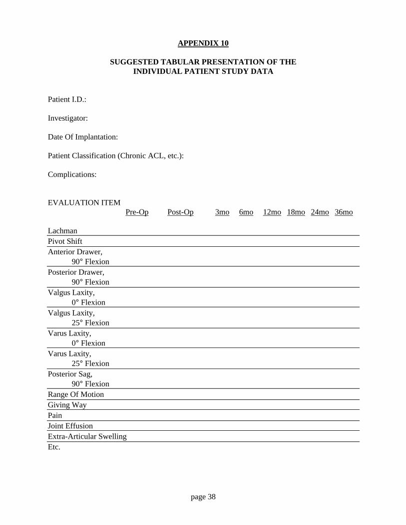
page 38
APPENDIX 10
SUGGESTED TABULAR PRESENTATION OF THEINDIVIDUAL PATIENT STUDY DATA
Patient I.D.:
Investigator:
Date Of Implantation:
Patient Classification (Chronic ACL, etc.):
Complications:
EVALUATION ITEMPre-Op Post-Op 3mo 6mo 12mo 18mo 24mo 36mo
LachmanPivot ShiftAnterior Drawer,
90° FlexionPosterior Drawer,
90° FlexionValgus Laxity,
0° FlexionValgus Laxity,
25° FlexionVarus Laxity,
0° FlexionVarus Laxity,
25° FlexionPosterior Sag,
90° FlexionRange Of MotionGiving WayPainJoint EffusionExtra-Articular SwellingEtc.

page 39
APPENDIX 11
PRESENTATION OF INDIVIDUAL EVALUATION ITEMSFOR THE PATIENT POPULATION
Item Time Point
LACHMAN SCOREPre-Op Post-Op 3mo 6mo 12mo 18mo 24mo 36mo
0+1+2+3+4Total #PatientsAt Each Time

page 40
APPENDIX 11 A
PRESENTATION OF PATIENT BY PATIENTLAXITY SCORES IN THE CLINICAL SUMMARY
Item: Lachman Score Time Point
PATIENT IDENTIFICATIONPre-Op Post-Op 3mo 6mo 12mo 18mo 24mo 36mo
Patient #11Patient #2Patient #3Etc.
Designate Patients Lost To Follow-Up.
APPENDIX 11 B
NUMBER OF PATIENTS AS A FUNCTION OF TIME OF COMPLICATION/EVENT*
Complication: Device Ruptures, 2 Yr Cohort
Total Number: N
# of Patientsetc. |5 |4 |3 |2 |1 |
|__________________________________________0 2 4 6 8 10 12 14 16 18 20 22 24 26, etc.
Months Post-Implant, Time That Complication First Occurred
* May Be Presented As A Bar Graph

page 41
APPENDIX 12
PATIENT ACCOUNTABILITY TABLE
Patients In A Given Class Of Injury (Chronic ACL Patients With First Implant Of Study Device,Etc.)
1. *Patients 2 Years Or More Post-Implant
1a. _____ Patients With 2 Yrs Follow-Up Available, Device In Place
1b. _____ Patients With 2 Yrs Follow-Up Available, Device Explanted
1c. _____ Patients Lost To Follow-Up Prior To 2 Yrs, Device In Place At LastFollow-Up
1d. _____ Patients Lost To Follow-Up Prior To 2 Yrs, Device Explanted By LastFollow-Up
2. Patients Less Than 2 Years Post-Implant
2a. _____ Patients Still Being Followed, Device In Place
2b. _____ Patients Still Being Followed, Device Explanted
2c. _____ Patients Lost To Follow-Up, Device In Place At Last Follow-Up
2d. _____ Patients Lost To Follow-Up, Device Explanted By Last Follow-Up
*Sponsor may also use a longer than 2-year cohort.
In the example given above, the sum of patients 1a through 1d must equal the number of 2 yearcohort patients (1 above) and the sum of patient 2a through 2d must equal the number of patientsless than 2 years post-implant (2 above).

page 42
REFERENCES
Ames, B.N., McCann, J., and E. Yamasaki, Methods for Detecting Carcinogens and Mutagenswith the Salmonella/Mammalian-Microsome Mutagenicity Test, Handbook of Mutagenicity TestProcedures, Kilbey, Legator, Nichols, and Ramel (Ed), Elsevier Scientific Publishing Co.,Amsterdam, pp. 1-18, 1977.
J. Autian, Toxicological Evaluation of Biomaterials. Primary Acute Toxicity ScreeningProgramme, Artificial Organs, 1: 53-60, 1977.
Biological Tests - Plastics, United States Pharmocopeia, 20th Edition, p. 950, Rockville, MD(1980).
Chen, E.H., and J. Black, Material Design Analysis of the Prosthetic Anterior Cruciate Ligament,Journal of Biomedical Materials Research, 14: 567-586, 1980.
Clive, D., and J.F.S. Spector, Laboratory Procedure for Assessing Specific Locus Mutations atthe TK Locus in Cultures of L5178Y Mouse Lymphoma Cells, Mutation Research, 31: 17-29,1975.
Dermal and Eye Toxicity Tests, Principles and Procedures for Evaluating the Toxicity ofHousehold Substances, National Academy of Science, Washington D.C., pp. 23-59, 1977
Grood, E., and F. Noyes, Cruciate Ligament Prosthesis: Strength, Creep, and Fatigue Properties,Journal of Bone and Joint Surgery, 58A: No 8, 1083-1088, 1976.
Heidelberger, C., et al., Cell Transformation by Chemical Agents - A Review and Analysis of theLiterature. A Report of the U.S. Environmental Protection Agency Gene-Tox Program, MutationResearch, 114: 283-385, 1983.
Hsie, A.W., Couch, D.B., O'Neill, J.P., et al., Utilization of a Quantitative Mammalian CellMutation System, CHO/HGPRT, in Experimental Mutagenesis and Genetic Toxicology.Strategies for Short-Term Testing for Mutagens/Carcinogens. Butterworth, B.E. (Ed), CRCPress, West Palm Beach Florida, pp. 39-54, 1979.
M.W. King, R. Guidoin, P. Blais, A. Garton, and K.R. Gunasekera. Degradation of PolyesterArterial Prosthesis: A Physical or Chemical Mechanism? Special Technical Testing Publication859. Copyright American Society for Testing and Materials. 1916 Race Street, Philadelphia, PA19103, pp.294-307, 1985.
J.C. Kennedy, R.J. Hawkins, R.B. Willis and K.D. Danylchuk. Tension Studies of Human KneeLigaments. The Journal of Bone and Joint Surgery, Vol. 58-A, pp.350-355, 1976.
A.V. Lukianov, J. Gillquist, W.A. Grana, K.E. DeHaven. An Anterior Cruciate Ligament (ACL)Evaluation Format for Assessment of Artificial or Autologous Anterior Cruciate ReconstructionResults. Clinical Orthopedics and Related Research, 218: 167-180, 1987.

page 43
Mitchell, A.D., et al., Unscheduled DNA Synthesis Tests. A Report of the U.S. EnvironmentalProtection Agency Gene-Tox Program, Mutation Research, 123: 363-410, 1983.
F.R. Noyes and E.S. Grood. The Strength of the Anterior Cruciate Ligament in Humans andRhesus Monkeys. The Journal of Bone and Joint Surgery, Vol. 58-A: 1074-1082, 1976.
F.R. Noyes, D.L. Butler, E.S. Grood, R.F. Zernicke, and M.S. Hefzy. Biomechanical Analysis ofHuman Ligament Grafts used in Knee-Ligament Repairs and Reconstructions. The Journal ofBone and Joint Surgery, Vol. 66-A, 344-352, 1984.
F.R. Noyes, P.A. Mooar, D.S. Matthews, and D.L. Butler. The Symptomatic Anterior Cruciate-Deficient Knee. Part 1: The Long-term Functional Disability in Athletically ActiveIndividuals. The Journal of Bone and Joint Surgery, Vol. 65-A: 154-162, 1983.
F.R. Noyes, D.S. Matthews, P.A. Mooar, and E.S. Grood. The Symptomatic Anterior Cruciate-Deficient Knee. Part II: The Results of Rehabilitation, Activity Modification, and Counseling onFunctional Disability. The Journal of Bone and Joint Surgery, Vol. 65-A: 163-174, 1983.
The Orthopedic Clinics of North America: Symposium on The Anterior Cruciate Ligament, PartI. W.B. Saunders Co., J.R. Andrews and W.G. Carson, Jr., Guest Editors. Vol. 16, no.1 1985.
The Orthopedic Clinics of North America: Symposium on The Anterior Cruciate Ligament, PartII. W.B. Saunders Co., J.R. Andrews and W.G. Carson, Jr., Guest Editors. Vol. 16, no.2 1985.
Symposium: Application of Cell Culture in Toxicology, Fundamentals of Applied Toxicology, 6:579-617, 1986.
Y. Tegner and J. Lysolm. Rating Systems in the Evaluation of Knee Ligament Injuries. ClinicalOrthopedics and Related Research, 198: 43-49, 1985.
B. Zarins and C.R. Rowe. Combined Anterior Cruciate-Ligament Reconstruction usingSemitendinosus Tendon and Iliotibial Tract. The Journal of Bone and Joint Surgery, 68-A: 160-177, 1986.