gst gene fusion system
TRANSCRIPT
Third Edition,Revision 2
18-1123-20
GSTGene Fusion System
PROTEIN DATA BANK ADVISORY NOTICE
The Protein Data Bank (PDB) database, com-piled at Brookhaven National Laboratory(BNL), is supported by a combination of FederalGovernment Agency funds and user fees.Support is provided by the United StatesNational Science Foundation, the United StatesPublic Health Service, the National Institutes ofHealth, the National Center for ResearchResources, the National Institute for GeneralMedical Sciences, the National Library ofMedicine, and the United States Department ofEnergy under Contract No. DE-AC02-76CH00016.
Any opinion, findings and conclusions, or rec-ommendations expressed in the PDB databaseare those of the authors/contributors and do not
necessarily reflect the views of the NationalScience Foundation, the National Institutes ofHealth, the Department of Energy, the U.S.Government, BNL or its Operator.
It is a common goal of the National ScienceFoundation, the National Institutes of Health,and the Department of Energy to have theresults of work which they support made avail-able to the public and disseminated as widely aspossible. The PDB database is being made avail-able to the public in furtherance of that com-mon objective. It should be noted, however, thatsuch availability and dissemination are precondi-tioned upon any recipient or end user of thePDB database agreeing not to offer said comput-er files, media, or any further PDB product orcopies thereof for sale as a commercial item.
Copyright © 1997 by Pharmacia Biotech, Inc
On the cover:Structure of recombinant glutathione S-transferase from Schistosoma japonicum as expressedfrom pGEX-3X Expression Vector in E. coli. Cover figure was rendered using the Persistance ofVision RayTracer. Atomic coordinates were converted to POV objects using David Richardson'sMolPOV program from Protein Data Bank entry 1GTA of February 1995 from M. A. McTigueand J. A. Tainer. Any publications resulting from the use of this information must cite ProteinData Bank as well as the primary depositor(s) as the source of the data.

Table of Contents
Table of Contents
Overview ....................................................... 2
pGEX Vectors ............................................. 2
Purification Modules................................... 4
Site-Specific Proteases ............................... 5
Detection Module........................................ 5
Procedures for Use .......................................... 7
Manipulation of pGEX Vectors........... 7
1. Restriction Digestion of pGEX Vectors...................................... 7
2. Dephosphorylation of Linearized pGEX Vector ....................................... 7
3. Ligation of Insert to pGEX DNA .......... 8
4. Preparation of Competent Cells andTransformation with pGEX DNA .......... 8
5. Small-Scale Isolation of pGEX DNA .... 9
6. Large-Scale Isolation of pGEX DNA .... 9
Notes on Sequencing pGEX Vectors ... 9
Notes on Mutagenesis of pGEX Vectors...................................... 10
7. Screening pGEX Vectors with PCR ..... 10
Purification of GST Fusion Proteins .. 11
8. Preparation of Glutathione Sepharose 4B ..................................... 12
9. Screening of pGEX Recombinants for Fusion Protein Expression .................. 13
10. High Throughput Screening of pGEXRecombinants usingMicroSpin Columns ............................ 14
11. Preparation of Large-Scale Bacterial Sonicates ............................. 15
12. Batch Purification of Fusion Proteins Using Bulk Glutathione Sepharose 4B.. 16
13. Column Purification of Fusion Proteins Using Glutathione Sepharose 4BRediPack or Disposable Columns ...... 17
14. PreScission Protease Cleavage ........... 17
15. Thrombin Cleavage ............................. 18
16. Factor Xa Cleavage ............................. 19
Detection of GST Fusion Proteins .... 20
17. Measurement of GST Activity by CDNB Assay........................................ 21
18. Detection of GST Fusion Protein by Western Blot .................................. 22
19. Radiolabelling of GST Fusion ProteinsProduced Using pGEX-2TK ................. 23
Troubleshooting Guide ..................................... 24
Appendices .................................................... 29
1. Reagents for Manipulation of pGEXVectors................................................. 29
2. Electroporation..................................... 30
3. Reagents for Purification of GSTFusion Proteins .................................... 31
4. Reagents for Detection of GST FusionProteins ............................................... 31
5. Cross-adsorption of Anti-GST Anti-serum with E. coli proteins................... 32
6. Characteristics and Regeneration ofGlutathione Sepharose 4B .................... 33
7. E. coli BL21 Strain Information ............ 33
8. Characteristics of GST.......................... 33
9. pGEX Vector Control Regions............... 34
References...................................................... 36
Ordering Information ....................................... 38
1

Overview
OverviewThe Glutathione S-transferase (GST) Gene Fusion System isan integrated system for the expression, purification anddetection of fusion proteins produced in E. coli. The systemconsists of three major components: pGEX plasmid vectors*,two GST Purification Modules and the GST DetectionModule. A series of site-specific proteases complements thesystem. The pGEX plasmids are designed for inducible, high-level intracellular expression of genes or gene fragments asfusions with Schistosoma japonicum GST (1).
GST occurs naturally as a 26 kDa protein that can be expressedin E. coli with full enzymatic activity. Fusion proteins that pos-sess the complete amino acid sequence of GST also demonstrateGST enzymatic activity and can undergo dimerization similarto that observed in nature (2, 3, 4). The crystal structure ofrecombinant Schistosoma japonicum GST from pGEX vectorshas been determined (5) and matches that of the native protein.
Fusion proteins are easily purified from bacterial lysates by affin-ity chromatography using Glutathione Sepharose 4B containedin the GST Purification Modules. Cleavage of the desired proteinfrom GST is achieved using a site-specific protease whose recog-nition sequence is located immediately upstream from the multi-ple cloning site on the pGEX plasmids. Fusion proteins can bedetected using a colorimetric assay or immunoassay provided inthe GST Detection Module. The system has been used success-fully in many applications such as molecular immunology (6),the production of vaccines (7, 8) and studies involving protein-protein (9) and DNA-protein (10) interactions.
pGEX Vectors*
All of the GST gene fusion vectors offer:
■ A tac promoter for chemically inducible, high-level expression.
■ An internal lac Iq gene for use in any E. coli host.
■ Very mild elution conditions for release of fusion proteinsfrom the affinity matrix, thus minimizing effects on anti-genicity and functional activity.
■ PreScission, thrombin or factor Xa protease recognition sitesfor cleaving the desired protein from the fusion product.
Thirteen pGEX vectors are available (Figure 1). Nine of the vec-tors have an expanded multiple cloning site (MCS) that containssix restriction sites. The expanded MCS facilitates the unidirec-tional cloning of cDNA inserts obtained from libraries con-structed using many available lambda vectors including λ ExCellCloning Vector (27-5013-01 or 27-5011-01; see page 39 formore details) and Lambda ZAP®. pGEX-6P-1, pGEX-6P-2 and pGEX-6P-3 each encode the recognition sequence for site-specific cleavage by PreScission Protease (27-0843-01) betweenthe GST domain and the multiple cloning site. pGEX-4T-1,pGEX-4T-2, and pGEX-4T-3 are derived from pGEX-2T and
2
*Note: For research use only. Not for diagnostic or therapeutic purposes.A license for commercial use of GST gene fusion vectors must be obtainedfrom AMRAD Corporation Ltd., 17-27 Cotham Road, Kew, Victoria3101 Australia.
Figure 1. Map of the glutathione S-transferase fusion vectors showing thereading frames and main features.
pGEX-1λT (27-4805-01)
pGEX-6P-1 (27-4597-01)
EcoR I Sma I Sal I Xho I Not IBamH I
PreScission™ Protease
Leu Glu Val Leu Phe Gln Gly Pro Leu Gly Ser Pro Glu Phe Pro Gly Arg Leu Glu Arg Pro HisCTG GAA GTT CTG TTC CAG GGG CCC CTG GGA TCC CCG GAA TTC CCG GGT CGA CTC GAG CGG CCG CAT
↓
pGEX-6P-2 (27-4598-01)
BamH I
PreScission™ ProteaseLeu Glu Val Leu Phe Gln Gly Pro Leu Gly Ser Pro Gly Ile Pro Gly Ser Thr Arg Ala Ala Ala SerCTG GAA GTT CTG TTC CAG GGG CCC CTG GGA TCC CCA GGA ATT CCC GGG TCG ACT CGA GCG GCC GCA TCG
↓
EcoR I Sma I Sal I Xho I Not I
pGEX-6P-3 (27-4599-01)
BamH I
PreScission™ Protease
Leu Glu Val Leu Phe Gln Gly Pro Leu Gly Ser Pro Asn Ser Arg Val Asp Ser Ser Gly ArgCTG GAA GTT CTG TTC CAG GGG CCC CTG GGA TCC CCG AAT TCC CGG GTC GAC TCG AGC GGC CGC
↓
EcoR I Sma I Sal I Xho I Not I
BamH I EcoR I Sma I Sal I Xho I Not I
BamH I EcoR I Sma I Sal I Xho I Not I
BamH I EcoR I Sma I Sal I Xho I Not I
BamH I EcoR I Sma I Sal I Xho I Not I
BamH I EcoR I Sma I Sal I Xho I Not I
BamH I EcoR I Sma I Sal I Xho I Not I
EcoR ICTG GTT CCG CGT GGA TCC CCG GAA TTC ATC GTG ACT GAC TGA CGA
BamH I
Leu Val Pro Arg Gly Ser Pro Glu Phe Ile Val Thr Asp
Thrombin
Stop codons
↓
pGEX~4900 bp
pBR322ori
Bal I
BspM I
Ptac glutathioneS-transferase
Amp r
lacI q
Nar I
EcoR V
BssH II
BstE II Mlu I
Apa I
Tth111 I Aat II
Pst I
p4.5AlwN I
pSj10 Bam7Stop7∆
pGEX-4T-2 (27-4581-01)
pGEX-5X-1 (27-4584-01)
pGEX-5X-2 (27-4585-01)
pGEX-5X-3 (27-4586-01)
pGEX-4T-1 (27-4580-01)
pGEX-4T-3 (27-4583-01)
pGEX-3X (27-4803-01)
pGEX-2TK (27-4587-01)
Leu Val Pro Arg Gly Ser Pro Gly Ile Pro Gly Ser Thr Arg Ala Ala Ala SerCTG GTT CCG CGT GGA TCC CCA GGA ATT CCC GGG TCG ACT CGA GCG GCC GCA TCG TGA
Stop codon
Ile Glu Gly Arg Gly Ile Pro Glu Phe Pro Gly Arg Leu Glu Arg Pro His Arg AspATC GAA GGT CGT GGG ATC CCC GAA TTC CCG GGT CGA CTC GAG CGG CCG CAT CGT GAC TGA
Stop codons
Ile Glu Gly Arg Gly Ile Pro Gly Ile Pro Gly Ser Thr Arg Ala Ala Ala SerATC GAA GGT CGT GGG ATC CCC GGA ATT CCC GGG TCG ACT CGA GCG GCC GCA TCG TGA
Stop codon
Ile Glu Gly Arg Gly Ile Pro Arg Asn Ser Arg Val Asp Ser Ser Gly Arg Ile Val Thr AspATC GAA GGT CGT GGG ATC CCC AGG AAT TCC CGG GTC GAC TCG AGC GGC CGC ATC GTG ACT GAC TGA
Stop codons
Leu Val Pro Arg Gly Ser Pro Glu Phe Pro Gly Arg Leu Glu Arg Pro His Arg AspCTG GTT CCG CGT GGA TCC CCG GAA TTC CCG GGT CGA CTC GAG CGG CCG CAT CGT GAC TGA
Stop codons
Leu Val Pro Arg Gly Ser Pro Asn Ser Arg Val Asp Ser Ser Gly Arg Ile Val Thr AspCTG GTT CCG CGT GGA TCC CCG AAT TCC CGG GTC GAC TCG AGC GGC CGC ATC GTG ACT GAC TGA
Stop codons
ATC GAA GGT CGT GGG ATC CCC GGG AAT TCA TCG TGA CTG ACT GACIle Glu Gly Arg Gly Ile Pro Gly Asn Ser Ser
Stop codons
Leu Val Pro Arg Gly Ser Arg Arg Ala Ser Val
Kinase
CTG GTT CCG CGT GGA TCT CGT CGT GCA TCT GTT GGA TCC CCG GGA ATT CAT CGT GAC TGAStop codons
Thrombin↓
Thrombin↓
Thrombin↓
Thrombin↓
Factor Xa↓
Factor Xa↓
Factor Xa↓
Factor Xa↓
EcoR IBamH I Sma I
EcoR IBamH I Sma I
pGEX-2T (27-4801-01)
CTG GTT CCG CGT GGA TCC CCG GGA ATT CAT CGT GAC TGA CTG ACGLeu Val Pro Arg Gly Ser Pro Gly Ile His Arg Asp
Stop codonsEcoR I
Thrombin↓
BamH I Sma I
Overview
3
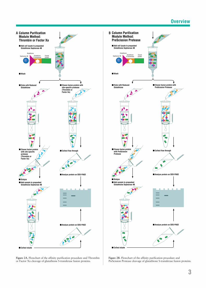
Figure 2A. Flowchart of the affinity purification procedure and Thrombinor Factor Xa cleavage of glutathione S-transferase fusion proteins.
Figure 2B. Flowchart of the affinity purification procedure and PreScission Protease cleavage of glutathione S-transferase fusion proteins.
Column Purification Module Method:Thrombin or Factor Xa
■ Cleave fusion protein with site-specific protease (Thrombin or Factor Xa)
■ Analyze protein on SDS-PAGE
■ Collect flow through
■ Analyze protein on SDS-PAGE
■ Add cell lysate to prepacked Glutathione Sepharose 4B
■ Elute with Reduced Glutathione
■ Cleave fusion protein with site-specific protease (Thrombin or Factor Xa)
■ Wash
■ Add sample to prepacked Glutathione Sepharose 4B
■ Dialyze
■ Collect eluate
Glutathione S-transferase
Clonedprotein
Glutathione
Sepharose 4B
Column Purification Module Method:PreScission Protease
■ Cleave fusion protein with PreScission Protease
■ Analyze protein on SDS-PAGE
■ Collect flow through
■ Analyze protein on SDS-PAGE
■ Add cell lysate to prepacked Glutathione Sepharose 4B
■ Elute with Reduced Glutathione
■ Cleave fusion protein with PreScission Protease
■ Wash
■ Add sample to prepacked Glutathione Sepharose 4B
■ Dialyze
■ Collect eluate
Glutathione S-transferase
Clonedprotein
Glutathione
Sepharose 4B
A B

contain a thrombin recognition site. pGEX-5X-1, pGEX-5X-2,and pGEX-5X-3 are derivatives of pGEX-3X and possess a fac-tor Xa recognition site.
pGEX-2TK is uniquely designed to allow the detection ofexpressed proteins by directly labelling the fusion products in vitro (11). This vector contains the recognition sequence for the catalytic subunit of cAMP-dependent protein kinaseobtained from heart muscle. The protein kinase site is locatedbetween the GST domain and the MCS. Expressed proteinscan be directly labelled using protein kinase and [γ-32P]ATPand readily detected using standard radiometric or autoradi-ographic techniques. pGEX-2TK is a derivative of pGEX-2T;its fusion proteins can be cleaved with thrombin.
Collectively, the pGEX vectors provide all three translationalreading frames beginning with the EcoR I restriction site. pGEX-1λT, pGEX-6P-1, pGEX-4T-1 and pGEX-5X-1 can directlyaccept and express cDNA inserts isolated from λ gt11 libraries.
Purification Modules
The GST Purification Modules feature the GST-glutathioneaffinity system for rapid, mild affinity-purification of GSTfusion proteins (Figure 2). Using Glutathione Sepharose 4B(Figure 3), fusion proteins can be purified to >90% in a singlechromatographic step. Fusion proteins are recovered from thematrix under mild elution conditions (10 mM glutathione),which preserve antigenicity and functionality of the proteins.
GST fusion proteins are produced in E. coli cells containing arecombinant pGEX plasmid. Protein expression from a pGEXplasmid is under the control of the tac promoter, which isinduced using the lactose analog isopropyl β-D-thiogalactoside(IPTG). Induced cultures are allowed to express GST fusionproteins for several hours, after which time cells are harvestedand then lysed by mild sonication. The bacterial lysate is clearedof cellular debris by centrifugation and the cleared lysate isready to be applied directly to Glutathione Sepharose 4B.
After the fusion proteins are bound to the matrix, it is washedwith buffer to remove non-specifically bound proteins. BoundGST fusion proteins can then be eluted with the mild elutionbuffer and used in a variety of applications.
The Bulk GST Purification Module (27-4570-01) provides a10 ml bulk pack of Glutathione Sepharose 4B and five dispos-
able columns. With this module, fusion proteins can be puri-fied using either column chromatography or a batch method.The batch method is more flexible, as purification can be per-formed with 50 µl to 10 ml of Glutathione Sepharose 4B. TheBulk Module contains enough reagents for five, 10 mg purifi-cations. Protocols for using Glutathione Sepharose in this for-mat are given in Procedure 12 (page 16), and a timetable isgiven on page 12.
The RediPack GST Purification Module (27-4570-02) pro-vides convenient disposable columns prepacked with 2 ml ofGlutathione Sepharose 4B, enough for two, 10 mg purifica-tions. A protocol for use of the prepacked columns is given inProcedure 13 (page 17), and a timetable is given on page 12.
If separation of the cloned protein from the GST affinity handleis desired, the fusion protein can be digested with the appropri-ate site-specific protease while the fusion protein is bound toGlutathione Sepharose 4B. Alternatively, the fusion protein canbe digested following elution from the matrix. Cleavage of thebound fusion protein eliminates the extra step of separating the released protein from GST since the GST moiety remainsbound to the matrix while the cloned protein is eluted usingwash buffer (12). Both purification modules include thrombinfor cleavage of GST fusion proteins containing the thrombinrecognition sequence.
4
Overview
Figure 3. Glutathione is attached to Sepharose 4B by coupling to the oxiranegroup using epoxy-activation. The structure of glutathione is complementaryto the binding site of the glutathione S-transferase binding site.
CH2
C
CH2
H OH
SC O
NH
C
OO
N H
CO
C
O
NH3+
O
O

Site-Specific Proteases
PreScission Protease (27-0843-01) is a fusion protein of GSTand human rhinovirus 3C protease (13). The protease specifi-cally recognizes the amino acid sequence Leu-Glu-Val-Leu-Phe-Gln ↓ Gly-Pro cleaving between the Gln and Gly residues(14). Since the protease is fused to GST, it is easily removedfrom cleavage reactions using Glutathione Sepharose (Figure4). Also available for site-specific cleavage are Thrombin (27-0846-01), which cleaves following Arg in Pro-Arg ↓ Lys, andFactor Xa (27-0849-01), which cleaves after the first aminoacid that follows the tetrapeptide Ile-Glu-Gly-Arg ↓, for fusionproteins that contain these recognition sites. See Procedure 14(page 17) for performing digests with PreScission Protease,Procedure 15 (page 18) for Thrombin digests or Procedure 16(page 19) for Factor Xa digests.
5
Overview
Figure 4. Flowchart of the affinity purification and PreScission Proteasecleavage procedure for isolating glutathione S-transferase fusion partners.
GlutathioneSepharose® 4B
E. coli contaminant protein
PreScission™ Protease
glutathioneS-transferase fusion partner
■ Addition of PreScission Protease
■ Elution of fusion partner with wash buffer
■ Binding and washing of GST-fusion protein to Glutathione Sepharose 4B
Detection Module
The GST Detection Module is designed to identify GST fusionproteins using either a biochemical or immunological assay.GST fusion proteins produced using a pGEX plasmid in E. colimay be assayed using the GST substrate 1-chloro-2,4-dinitro-benzene (CDNB). This assay can be used to optimize condi-tions for expression or to trace steps in the purification of aGST fusion protein. GST from many sources, including theschistosomal form used in the pGEX system, has a high affinityfor CDNB (15, 16). The enzyme catalyzes the conjugation of
Enzymatic Assay
Glutathione S-transferase
■ Add fusion protein extract to cuvette containing CDNB and glutathione in reaction buffer.
1-chloro-2,4-dinitrobenzene (CDNB)
Glutathione
GST Fusion Protein
Cloned protein
GST activity
Cl-H+
340 nm
■ Monitor change in absorbance at 340 nm with a UV spectrophotometer.
γ-Glu
Cys
Gly
HS
γ-Glu
Cys
Gly
HS
Cl
NO2
Cl
NO2
O2N
O2N
NO2
γ-Glu
Cys
Gly
SO2N
NO2
γ-Glu
Cys
Gly
SO2N
Figure 5. Diagram of the enzymatic assay for glutathione S-transferase.
CDNB with glutathione and results in a CDNB-glutathioneproduct with a strong molar absorptivity at 340 nm (ε = 9.6 mM-1 cm-1). The reaction is relatively insensitive to reactionconditions and, as such, the assay can be used with crude bac-terial sonicates or with purified GST fusion protein.
In this assay, a sample containing a GST fusion protein is incu-bated in a reaction buffer containing CDNB and glutathione(Figure 5). The change in absorbance at 340 nm is monitoredfor 5 minutes and, based on these results, the relative amountof GST fusion protein in the sample is calculated. This protocolis given in Procedure 17 (page 21). This assay can easily beused with multiple samples and is ideal for screening E. colistrains for expression, optimizing culture conditions for expres-sion, and for tracing the GST fusion protein during the purifi-cation process.
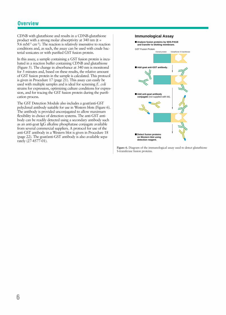
The GST Detection Module also includes a goat/anti-GSTpolyclonal antibody suitable for use in Western blots (Figure 6).The antibody is provided unconjugated to allow maximumflexibility in choice of detection systems. The anti-GST anti-body can be readily detected using a secondary antibody suchas an anti-goat IgG alkaline phosphatase conjugate availablefrom several commercial suppliers. A protocol for use of theanti-GST antibody in a Western blot is given in Procedure 18(page 22). The goat/anti-GST antibody is also available sepa-rately (27-4577-01).
6
Figure 6. Diagram of the immunological assay used to detect glutathione S-transferase fusion proteins.
Immunological Assay
Glutathione S-transferase
■ Analyze fusion proteins by SDS-PAGE and transfer to blotting membrane.
■ Add goat anti-GST antibody.
Cloned protein
■ Add anti-goat antibody conjugate (not supplied with kit).
-SS-
-SS-
-SS-
-SS-
-SS-
-SS-
-SS--SS-
-SS-
-SS-
-SS-
-SS-
-SS--SS
-
-SS-
-SS-
■ Detect fusion proteins on Western blot using detection reagent.
GST Fusion Protein
-SS--SS--SS
-
-SS-
-SS-
-SS-
-SS-
-SS-
-SS-
-SS-
-SS-
-SS-
-SS-
-SS-
-SS-
-SS-
-SS-
-SS-
-SS-
-SS-
-SS-
-SS-
-SS-
-SS-
Overview
Procedures for UseManipulation of pGEX Vectors
Procedures for UseManipulation of pGEX VectorsPlease note , it is important to select the proper vector tomaintain the GST reading frame through the cloned insert.
The locations of control regions for the pGEX vectors canbe found in Appendix 9 (pages 34-35). Complete DNAsequences and restriction site tables for all of the pGEX vec-tors are available through the Pharmacia Biotech WorldWide Web site: www.biotech.pharmacia.se and also fromGenBank®. See Appendix 9 for GenBank accession numbers.
Procedure 1: Restriction Digestion of pGEX Vectors
Reagents RequiredRecipes are provided in Appendix 1, page 29.
pGEX DNA10X One-Phor-All Buffer PLUS*Restriction enzyme
1. Prepare the following reaction mixture. Volumes mayvary depending on the amount of pGEX DNA to bedigested. We recommend that the final DNA concentra-tion in the reaction mixture be 0.1 µg/µl.
pGEX DNA 5 µg10X One-Phor-All Buffer PLUS (OPA+) 5-10 µlOptional components(e.g., BSA, Triton X-100, NaCl, etc.) 5-10 µlRestriction enzyme 10-25 unitsWater to 50 µl
2. Incubate at the appropriate temperature for 2-16 hours.
3. Examine a small aliquot of the reaction by agarose gelelectrophoresis to determine if the pGEX DNA has beendigested to completion.
4. If digestion with a second enzyme is required, adjust theOPA+ concentration and reaction volume as appropriate,add new enzyme and continue incubation.
5. Dephosphorylate the pGEX DNA with an alkaline phosphatase if it is to be used following digestion with asingle restriction enzyme (Procedure 2). If using OPA+,dephosphorylation can be performed in the same tubeimmediately following digestion.
6. If the pGEX DNA was digested with two restrictionenzymes, the linearized vector should be agarose gel-purified. This can be conveniently accomplished withSephaglas BandPrep Kit from Pharmacia Biotech.
Procedure 2: Dephosphorylation of Linearized pGEX Vector
Reagents RequiredRecipes are provided in Appendix 1, page 29.
Alkaline phosphatase10X One-Phor-All Buffer PLUS*PhenolChloroform/isoamyl alcohol3 M Sodium acetateEthanol (70%, 95%)TE buffer
1. Dilute sufficient Calf Intestinal Alkaline Phosphatase (27-0620-01) 20-fold for all dephosphorylations to beperformed. For dilution, use 10X OPA+ and water togive a final buffer concentration of 1X.
2. Add 0.1 unit (1-2 µl of diluted enzyme) of alkaline phos-phatase to the digested pGEX DNA and incubate for 30 minutes at 37°C. (In radiolabel and transformationstudies, dephosphorylation appears complete within 5 minutes when using 0.5X or 1X OPA+. When 2XOPA+ is used, an incubation period of 15-30 minutes isrequired for complete dephosphorylation.)
3. Heat inactivate the alkaline phosphatase at 85°C for 15 minutes. (Heat inactivation is complete for concentra-tions of alkaline phosphatase of 0.1 unit or less, but isnot effective for concentrations greater than 1 unit.)
4. Add an equal volume of phenol to the aqueous sample.Vortex for 1 minute and centrifuge for 5 minutes at fullspeed to separate the phases.
5. Transfer the upper aqueous phase to a fresh tube andadd an equal volume of chloroform/isoamyl alcohol.Vortex for 1 minute, then centrifuge for 5 minutes at fullspeed to separate the phases.
6. Transfer the upper aqueous phase to a fresh tube and add 0.1 volume of 3 M sodium acetate (pH 5.4) and 2.5 volumes of 95% ethanol. Mix and place at -20°C for 15 minutes.
7. Centrifuge at 4°C for 15 minutes, remove the superna-tant, and wash the pellet with 1 ml of 70% ethanol.
8. Recentrifuge for 2 minutes, drain thoroughly, and drythe DNA pellet under vacuum.
9. Dissolve the DNA pellet in 10-20 µl of TE buffer. pGEXDNA can be stored at -20°C for later use.
7*OPA+ is our unique buffer that is compatible with virtually all restriction enzymes and many modifying enzymes. It is supplied with most restriction enzymes from Pharmacia Biotech. See Appendix 1, page 29, for the composition of OPA+.

Procedures for UseManipulation of pGEX Vectors
Procedure 3: Ligation of Insert to pGEX DNA
Please note , it is important to select the proper vector tomaintain the GST reading frame through the cloned insert.
Ready-To-Go T4 DNA Ligase (27-0361-01) can be used to achieve ligations in 30-45 minutes. Contact your localPharmacia Biotech representative for more information. Analternate procedure is described below.
Reagents RequiredRecipes are provided in Appendix 1, page 29.
Insert DNAATP, 100 mM10X One-Phor-All Buffer PLUS*T4 DNA Ligase
1. The linearized pGEX DNA and insert DNA should be ata vector to insert molar ratio of 1:5. The moles of ends oflinear DNA can be calculated with the following formula:
moles of ends = 2 x (g of DNA)÷[(# of bp) x (649 Daltons/bp)]
Example: 100 ng of pGEX DNA (0.06 pmol of ends)would require 100 ng of a 1 kb insert (0.3 pmol of ends).
2. For ligation of cohesive ends, the final reaction mixshould contain 1 mM ATP (27-2056-01, diluted) and0.5-5 units of FPLCpure® T4 DNA Ligase (27-0870-03, -04), and should be incubated for 1-4 hours at 10°C.
3. For ligation of blunt ends, the final reaction mix shouldcontain 0.1-1 mM ATP (27-2056-01, diluted) and 10-15 units of FPLCpure T4 DNA Ligase (27-0870-03, -04) and should be incubated for 2-16 hours at 4-16°C.
4. Based upon the above considerations, prepare thefollowing reaction mixture specific for your application:
Linearized pGEX DNA 1-5 µlInsert DNA 1-5 µl10X One-Phor-All Buffer PLUS (OPA+) 2 µl100 mM ATP 0.2 µlT4 DNA Ligase 0.5-15 unitsWater to 20 µl
5. Incubate as described in step 2 or 3.
6. Terminate the reaction by heating at 65°C for 10 minutes.
7. The ligation reaction can be used directly to transformcompetent cells. Otherwise, it can be stored at -20ºC until needed.
Procedure 4: Preparation of Competent Cells andTransformation with pGEX DNA
pGEX vectors carry the lacIq gene, so there are no specifichost requirements for propagation of the plasmids or forexpression of fusion proteins. However, E. coli BL21 is
provided with the pGEX vectors and this strain is recom-mended for expression of GST fusion proteins (see Appendix7, page 33 for BL21 strain information). E. coli BL21 cellsare also available separately (27-1542-01). BL21 does nottransform well and an alternate strain (e.g., JM105) isrecommended for cloning and maintenance of the plasmid.For some applications, it may be desirable to use a protease-deficient host (e.g., BL21) if proteolytic degradation of thefusion protein is a concern.
We do not recommend the use of E. coli strains carrying therecA1 allelle for propagation of pGEX plasmids. There havebeen reports that these strains can cause rearrangements ordeletions within plasmid DNA.
In these procedures, E. coli host cells are made competentand are transformed with either uncut pGEX DNA orpGEX DNA containing an insert. (If electroporation is usedto transform the cells, see Appendix 2, page 30. Otherwise,proceed as described below). We recommend that 1 ng ofuncut (supercoiled) vector DNA be transformed in parallelwith insert/pGEX ligations to determine the efficiency ofeach competent cell preparation.
Preparation of Competent CellsThis protocol is based on the procedure of Chung et al. (17).
IMPORTANT: All steps in this procedure should be carriedout aseptically.
Reagents RequiredRecipes are provided in Appendix 1, page 29.
Glycerol stock of E. coli host strainLB medium platesLB mediumTSS buffer (ice-cold)
1. Using sterile technique, streak an E. coli host strain (e.g., JM105, BL21, etc.) from a glycerol stock onto a LB medium plate. Grow overnight at 37°C.
2. Isolate a single colony and inoculate 50-100 ml of LBbroth. Incubate at 37°C with shaking at 250 rpm. Growcells to an A600 of 0.4 - 0.5. (It is critical that the absor-bance is not more than 0.5). This will take approximately3-6 hours.
3. Sediment the cells at approximately 2500 x g for 15 minutes at 4°C, then gently resuspend in 1/10 volume(5-10 ml) of ice-cold TSS buffer and place on ice. Cellsmust be used for transformations within 2-3 hours.
Transformation of Competent CellsReagents RequiredRecipes are provided in Appendix 1, page 29.
LBG mediumLBAG medium platesGlycerol, 80%
1. For each ligation reaction and the uncut vector control,add 1 ml of freshly prepared competent E. coli host cellsto separate pre-chilled 50 ml sterile disposable centrifugetubes. Store on ice.
8 *OPA+ is our unique buffer that is compatible with virtually all restriction enzymes and many modifying enzymes. It is supplied with most restriction enzymes from Pharmacia Biotech. See Appendix 1, page 29, for the composition of OPA+.
Procedures for UseManipulation of pGEX Vectors
2. Add 20 µl of each ligation reaction or 1 ng of uncut vec-tor to the competent cells, swirl gently to mix, and placeon ice for 45 minutes.
3. Incubate the tubes in a 42°C water bath for 2 minutes,then chill briefly on ice.
4. For each sample, immediately transfer 100 µl of thetransformed cells to a 17 x 100 mm tube (Falcon®) con-taining 900 µl of LBG medium (prewarmed to 37°C) andincubate for 1 hour at 37°C with shaking (250 rpm).
5. Plate 100 µl of the diluted, transformed cells from theligated samples and 10 µl of the diluted, transformedcells from the uncut vector sample onto separate LBAGplates. Also plate 100 µl of untransformed, competent E. coli host cells as a negative control. Incubate theplates at 37°C overnight.
6. To prepare a frozen stock culture, add 100 µl of thediluted, transformed cells containing the pGEX DNA to1 ml of LBAG medium and incubate for 30 minutes at37°C with shaking at 250 rpm. After incubation, add200 µl of sterile 80% glycerol and mix with a pipet tip.Store at -70°C.
Procedure 5: Small-Scale Isolation of pGEX DNA
Rapid isolation of plasmid DNA is greatly simplified by theuse of FlexiPrep Kit (27-9281-01). An alternate procedure isdescribed below.
Reagents RequiredRecipes are provided in Appendix 1, page 29.
Solution ISolution IISolution IIIIsopropanolTE bufferPhenolChloroform/isoamyl alcoholSodium acetate, 3 MEthanol (70%, 95%)
1. Transfer 1.5 ml of an overnight culture of E. coli to amicrocentrifuge tube and centrifuge at full speed for 30 seconds to pellet the cells.
2. Remove the supernatant by aspiration without disturb-ing the cell pellet, leaving the pellet as dry as possible.
3. Resuspend the pellet in 200 µl of Solution I by vigor-ously vortexing.
4. Add 200 µl of Solution II and mix by inverting the tubeseveral times. Incubate at room temperature for 5 minutes.
5. Add 200 µl of Solution III and mix by inverting the tubeseveral times. Place on ice for 5 minutes.
6. Centrifuge at full speed for 5 minutes at room temperature.
7. Carefully decant the supernatant into a clean centrifuge tube.
8. Add 420 µl (0.7 volume) of ambient-temperature iso-propanol to the supernatant and vortex to mix. Incubatefor 5 minutes at room temperature.
9. Centrifuge at full speed for 10 minutes. Decant the super-natant and invert the tube to drain.
10. Resuspend the DNA pellet in 200 µl of TE buffer by vortexing.
11. Add 200 µl of phenol to the aqueous sample. Vortex for1 minute and centrifuge for 5 minutes at full speed toseparate the phases.
12. Transfer the upper aqueous phase to a fresh tube and add 200 µl of chloroform/isoamyl alcohol. Vortex for 1 minute, then centrifuge for 5 minutes at full speed toseparate the phases.
13. Transfer the upper aqueous phase to a fresh tube and add 20 µl of 3 M sodium acetate and 500 µl of 95% ethanol. Mix and place at -20°C for 15 minutes.
14. Centrifuge at 4°C for 15 minutes, remove the super-natant, and wash the pellet with 1 ml of 70% ethanol.
15. Recentrifuge for 2 minutes, drain thoroughly, and dry the DNA pellet under vacuum.
16. Dissolve the DNA pellet in 20 µl of TE buffer. pGEXDNA can be stored at -20°C for later use.
Procedure 6: Large-Scale Isolation of pGEX DNA
Use a large-scale isolation technique to prepare a stock ofplasmid DNA that can be used repeatedly in various proce-dures (e.g., sequencing, cloning, etc). Rapid, large-scale isola-tion of plasmid DNA from cultures up to 500 ml is greatlysimplified by the use of FlexiPrep Kit (27-9281-01).
1. Grow an appropriate volume of pGEX-containing E. coli in 2X YTA medium overnight.
2. Dilute an inoculum of the overnight culture at least1:100 into the desired volume of the same medium prewarmed to the growth temperature.
3. Grow with aeration to an A600 of 1.0 or greater.
4. Isolate plasmid DNA using FlexiPrep Kit (27-9281-01)from Pharmacia Biotech, or protocols from reference 18.
Notes on Sequencing pGEX VectorspGEX vectors can be sequenced using the 5′ pGEX Sequen-cing Primer (27-1410-01) and the 3′ pGEX Sequencing Primer(27-1411-01). The sequences and the binding regions of theseprimers are given below:
5′ pGEX Sequencing Primer
5′-d[GGGCTGGCAAGCCACGTTTGGTG]-3′
The 5′ pGEX Sequencing Primer binds at nucleotides 869-891 on all 13 pGEX vectors.
9
Procedures for UseManipulation of pGEX Vectors
3′ pGEX Sequencing Primer
5′-d[CCGGGAGCTGCATGTGTCAGAGG]-3′
The 3′ pGEX Sequencing Primer binds at the following loca-tions on the pGEX vectors:
Vector Binding Site
pGEX-1λT 1019-997pGEX-2T 1020-998
pGEX-2TK 1041-1019pGEX-3X 1024-1002
pGEX-4T-1 1041-1019pGEX-4T-2 1042-1020pGEX-4T-3 1040-1018pGEX-5X-1 1044-1022pGEX-5X-2 1045-1023pGEX-5X-3 1046-1024pGEX-6P-1 1056-1034pGEX-6P-2 1057-1035pGEX-6P-3 1055-1033
See also Appendix 9, page 34-35.
Notes on Mutagenesis of pGEX VectorsSite-specific mutagenesis of protein-coding sequences cloned in pGEX vectors is readily accomplished with the U.S.E.Mutagenesis Kit (27-1699-01) in conjunction with pGEXU.S.E. Primers (see below). The U.S.E. Mutagenesis Kit uti-lizes a two-primer system to generate site-specific mutations.The first primer, or target mutagenic primer, is directed tothe gene of interest and carries the desired mutation. Targetmutagenic primers are custom-designed by the user. The sec-ond primer, called the U.S.E. selection primer, carries a muta-tion in a unique, nonessential restriction site which is ulti-mately used as the basis for selection of mutated plasmids.These two primers bind to the same strand of DNA, but thegeneration of single-stranded DNA is not required.
Two sets of pGEX U.S.E. Primers are available. The Nar I/Nhe IpGEX U.S.E. Primers (27-1661-01) feature a Nar I selectionprimer which anneals to the Nar I site in the lacIq gene and con-verts this site to an Nhe I site. The Nhe I toggle primer convertsthe Nhe I site back to the Nar I site. The Pst I/Sac II pGEXU.S.E. Primers (27-1662-01) work in a similar manner to togglethe Pst I site in the ampicillin resistance gene with a Sac II site.The use of the latter primer to prepare a series of 37 pointmutations at 25 different positions in a GST fusion partner isdescribed by Kazanietz and Blumberg in Science Tools, Vol. 1,No. 1 (Pharmacia Biotech 1996). For more information on theU.S.E. Mutagenesis Kit and the pGEX U.S.E. Primers, contactyour local Pharmacia Biotech representative.
Procedure 7: Screening pGEX Vectors with PCR✧
The 5´ pGEX Sequencing Primer (27-1410-01) and the 3´pGEX Sequencing Primer (27-1411-01) can be used in therapid screening of transformants by PCR.
Ready-To-Go PCR Beads▲ (27-9555-01)1. Resuspend bead in 25 µl of water per standard instructions.
2. Add 10 pmol each of 5 ́pGEX Sequencing Primer (27-1410-01) and 3´ pGEX Sequencing Primer (27-1411-01).
3. Gently touch a sterile micropipet tip to the bacterialcolony to be screened and then to resuspended PCR bead.Pipet gently to disperse bacterial cells.
– NOTE: Avoid transferring too much of the bacterialcolony. Results are better with very few bacterial cells.
4. Overlay the reaction mixture with 50 µl of mineral oil.
5. Amplify in a licensed thermal cycler with the followingcycle parameters:
35 cycles:95°C 1 minute58°C 1 minute72°C 2 minutes
6. Transfer aqueous phase from under oil to a clean tube.Analyze 10-20 µl by agarose gel electrophoresis.
Standard PCR1. Mix the following components in a 0.65 ml tube:
10 X Taq Polymerase Buffer 10 µl1.25 mM each dNTP in water 16 µl5´ pGEX Sequencing Primer (27-1410-01) 5 µl3´ pGEX Sequencing Primer (27-1411-01) 5 µlWater to 99.5 µl
2. Gently touch a sterile micropipet tip to the bacterialcolony to be screened and then to above PCR mixture.Pipet gently to disperse bacterial cells.
– NOTE: Avoid transferring too much of the bacterialcolony. Results are better with very few bacterial cells.
3. Add 0.5 µl of 5 U/µl Taq DNA Polymerase (27-0799-01).
4. Overlay the reaction mixture with 50 µl of mineral oil.
5. Amplify in a licensed thermal cycler with the followingcycle parameters:
25-35 cycles:94°C 1 minute55°C 1 minute72°C 2 minutes
6. Transfer aqueous phase from under oil to a clean tube.Analyze 20-40 µl by agarose gel electrophoresis.
10
Procedure 6: Large-Scale Isolation of pGEX DNA (cont).
▲Purchase of Ready-To-Go® PCR Beads is accompanied by a limited license to use it in the Polymerase Chain Reaction (PCR) process solelyfor the research and development activities of the purchaser in conjunction with a thermal cycler whose use in the automated performance ofthe PCR process is covered by the up-front license fee, either by payment to Perkin-Elmer or as purchased, i.e. an authorized thermal cycler.
✧PCR (polymerase chain reaction) is covered by U.S.Patents 4,683,195 and 4,683,202 owned by Hoffman-LaRoche Inc. Use of the PCR process requires a license.
Procedures for UsePurification of GST Fusion Proteins
11
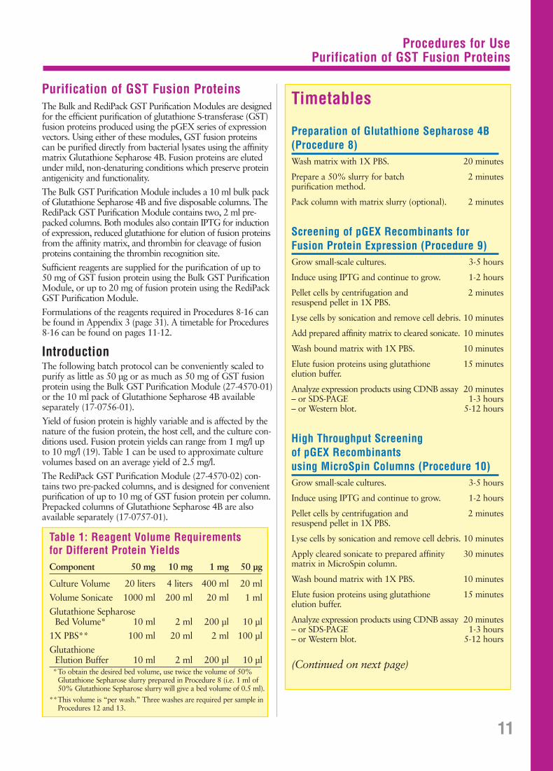
Table 1: Reagent Volume Requirements for Different Protein YieldsComponent 50 mg 10 mg 1 mg 50 µg
Culture Volume 20 liters 4 liters 400 ml 20 ml
Volume Sonicate 1000 ml 200 ml 20 ml 1 ml
Glutathione SepharoseBed Volume* 10 ml 2 ml 200 µl 10 µl
1X PBS** 100 ml 20 ml 2 ml 100 µl
GlutathioneElution Buffer 10 ml 2 ml 200 µl 10 µl*To obtain the desired bed volume, use twice the volume of 50%
Glutathione Sepharose slurry prepared in Procedure 8 (i.e. 1 ml of 50% Glutathione Sepharose slurry will give a bed volume of 0.5 ml).
**This volume is “per wash.” Three washes are required per sample inProcedures 12 and 13.
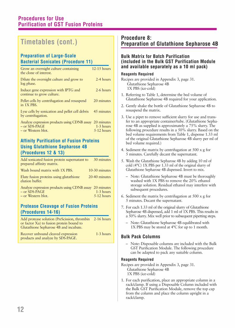
Timetables
Preparation of Glutathione Sepharose 4B(Procedure 8)Wash matrix with 1X PBS. 20 minutes
Prepare a 50% slurry for batch 2 minutespurification method.
Pack column with matrix slurry (optional). 2 minutes
Screening of pGEX Recombinants forFusion Protein Expression (Procedure 9)Grow small-scale cultures. 3-5 hours
Induce using IPTG and continue to grow. 1-2 hours
Pellet cells by centrifugation and 2 minutesresuspend pellet in 1X PBS.
Lyse cells by sonication and remove cell debris. 10 minutes
Add prepared affinity matrix to cleared sonicate. 10 minutes
Wash bound matrix with 1X PBS. 10 minutes
Elute fusion proteins using glutathione 15 minuteselution buffer.
Analyze expression products using CDNB assay 20 minutes– or SDS-PAGE 1-3 hours– or Western blot. 5-12 hours
High Throughput Screening of pGEX Recombinants using MicroSpin Columns (Procedure 10)Grow small-scale cultures. 3-5 hours
Induce using IPTG and continue to grow. 1-2 hours
Pellet cells by centrifugation and 2 minutesresuspend pellet in 1X PBS.
Lyse cells by sonication and remove cell debris. 10 minutes
Apply cleared sonicate to prepared affinity 30 minutesmatrix in MicroSpin column.
Wash bound matrix with 1X PBS. 10 minutes
Elute fusion proteins using glutathione 15 minuteselution buffer.
Analyze expression products using CDNB assay 20 minutes– or SDS-PAGE 1-3 hours– or Western blot. 5-12 hours
(Continued on next page)
Purification of GST Fusion ProteinsThe Bulk and RediPack GST Purification Modules are designedfor the efficient purification of glutathione S-transferase (GST)fusion proteins produced using the pGEX series of expressionvectors. Using either of these modules, GST fusion proteins can be purified directly from bacterial lysates using the affinitymatrix Glutathione Sepharose 4B. Fusion proteins are elutedunder mild, non-denaturing conditions which preserve proteinantigenicity and functionality.
The Bulk GST Purification Module includes a 10 ml bulk packof Glutathione Sepharose 4B and five disposable columns. TheRediPack GST Purification Module contains two, 2 ml pre-packed columns. Both modules also contain IPTG for inductionof expression, reduced glutathione for elution of fusion proteinsfrom the affinity matrix, and thrombin for cleavage of fusionproteins containing the thrombin recognition site.
Sufficient reagents are supplied for the purification of up to 50 mg of GST fusion protein using the Bulk GST PurificationModule, or up to 20 mg of fusion protein using the RediPackGST Purification Module.
Formulations of the reagents required in Procedures 8-16 canbe found in Appendix 3 (page 31). A timetable for Procedures8-16 can be found on pages 11-12.
IntroductionThe following batch protocol can be conveniently scaled topurify as little as 50 µg or as much as 50 mg of GST fusionprotein using the Bulk GST Purification Module (27-4570-01)or the 10 ml pack of Glutathione Sepharose 4B available separately (17-0756-01).
Yield of fusion protein is highly variable and is affected by thenature of the fusion protein, the host cell, and the culture con-ditions used. Fusion protein yields can range from 1 mg/l upto 10 mg/l (19). Table 1 can be used to approximate culturevolumes based on an average yield of 2.5 mg/l.
The RediPack GST Purification Module (27-4570-02) con-tains two pre-packed columns, and is designed for convenientpurification of up to 10 mg of GST fusion protein per column.Prepacked columns of Glutathione Sepharose 4B are alsoavailable separately (17-0757-01).
Procedures for UsePurification of GST Fusion Proteins
Procedure 8:Preparation of Glutathione Sepharose 4B
Bulk Matrix for Batch Purification (included in the Bulk GST Purification Moduleand available separately as a 10 ml pack)Reagents RequiredRecipes are provided in Appendix 3, page 31.
Glutathione Sepharose 4B1X PBS (ice-cold)
1. Referring to Table 1, determine the bed volume of Glutathione Sepharose 4B required for your application.
2. Gently shake the bottle of Glutathione Sepharose 4B toresuspend the matrix.
3. Use a pipet to remove sufficient slurry for use and trans-fer to an appropriate container/tube. (Glutathione Sepha-rose 4B as supplied is approximately a 75% slurry. Thefollowing procedure results in a 50% slurry. Based on thebed volume requirements from Table 1, dispense 1.33 mlof the original Glutathione Sepharose 4B slurry per ml ofbed volume required.)
4. Sediment the matrix by centrifugation at 500 x g for 5 minutes. Carefully decant the supernatant.
5. Wash the Glutathione Sepharose 4B by adding 10 ml of cold (4°C) 1X PBS per 1.33 ml of the original slurry ofGlutathione Sepharose 4B dispensed. Invert to mix.
– Note: Glutathione Sepharose 4B must be thoroughlywashed with 1X PBS to remove the 20% ethanol storage solution. Residual ethanol may interfere withsubsequent procedures.
6. Sediment the matrix by centrifugation at 500 x g for 5 minutes. Decant the supernatant.
7. For each 1.33 ml of the original slurry of GlutathioneSepharose 4B dispensed, add 1 ml of 1X PBS. This results ina 50% slurry. Mix well prior to subsequent pipetting steps.
– Note: Glutathione Sepharose 4B equilibrated with 1X PBS may be stored at 4°C for up to 1 month.
Bulk Pack Columns– Note: Disposable columns are included with the Bulk
GST Purification Module. The following procedurecan be adapted to pack any suitable column.
Reagents RequiredRecipes are provided in Appendix 3, page 31.
Glutathione Sepharose 4B1X PBS (ice-cold)
1. For each purification, place an appropriate column in arack/clamp. If using a Disposable Column included withthe Bulk GST Purification Module, remove the top capfrom the column and place the column upright in arack/clamp.
12
Timetables (cont.)
Preparation of Large-Scale Bacterial Sonicates (Procedure 11)Grow an overnight culture containing 12-15 hoursthe clone of interest.
Dilute the overnight culture and grow to 2-4 hourslog phase.
Induce gene expression with IPTG and 2-6 hourscontinue to grow culture.
Pellet cells by centrifugation and resuspend 20 minutesin 1X PBS.
Lyse cells by sonication and pellet cell debris 45 minutesby centrifugation.
Analyze expression products using CDNB assay 20 minutes– or SDS-PAGE 1-3 hours– or Western blot. 5-12 hours
Affinity Purification of Fusion ProteinsUsing Glutathione Sepharose 4B (Procedures 12 & 13)Add sonicated fusion protein supernatant to 30 minutesprepared affinity matrix.
Wash bound matrix with 1X PBS. 10-30 minutes
Elute fusion proteins using glutathione 20-40 minuteselution buffer.
Analyze expression products using CDNB assay 20 minutes– or SDS-PAGE 1-3 hours– or Western blot. 5-12 hours
Protease Cleavage of Fusion Proteins(Procedures 14-16)Add protease solution (PreScission, thrombin 2-16 hoursor factor Xa) to fusion protein bound to Glutathione Sepharose 4B and incubate.
Recover unbound cleaved expression 1-3 hoursproducts and analyze by SDS-PAGE.
Procedures for UsePurification of GST Fusion Proteins
2. Referring to Table 1, determine the bed volume of Glutathione Sepharose 4B required for your application.
3. Gently shake the bottle of Glutathione Sepharose 4B toresuspend the matrix.
4. Use a pipet to remove sufficient slurry for use and transferto the column. (Glutathione Sepharose 4B as supplied isapproximately a 75% slurry. Based on the bed volumerequirements from Table 1, dispense 1.33 ml of the origi-nal Glutathione Sepharose 4B slurry per ml of bed volumerequired.)
5. Tap the column to dislodge any trapped air bubbles in the matrix bed. Allow to settle.
6. If using a Disposable Column included with the Bulk GSTPurification Module, remove the bottom cap and save forlater use. Allow the column to drain.
– Note: Gentle pressure with a gloved thumb over the topof the column may be required to start the flow of liquid.
7. Wash the Glutathione Sepharose 4B by adding 10 ml ofcold (4°C) 1X PBS per 1.33 ml of the original slurry ofGlutathione Sepharose 4B dispensed. Allow the column to drain.
– Note: Glutathione Sepharose 4B must be thoroughlywashed with 1X PBS to remove the 20% ethanol stor-age solution. Residual ethanol may interfere with sub-sequent procedures.
– Note: Glutathione Sepharose 4B equilibrated with 1X PBS may be stored at 4°C for up to 1 month.
RediPack Columns (included in the RediPack GST Purification Module and available separately as 2 ml prepacked columns)Reagents RequiredRecipes are provided in Appendix 3, page 31.
1X PBS (ice-cold)
1. Remove the top cap from the RediPack Column andpour off the excess liquid.
2. Remove the bottom cap and cut the tip off of the column.Save the cap for later use. Allow the column to drain.
– Note: Gentle pressure with a gloved thumb over the topof the column may be required to start the flow of liquid.
3. Wash the Glutathione Sepharose 4B by adding 20 ml ofcold (4°C) 1X PBS. Allow the column to drain.
– Note: Glutathione Sepharose 4B must be thoroughlywashed with 1X PBS to remove the 20% ethanol stor-age solution. Residual ethanol may interfere with sub-sequent procedures.
– Note: Glutathione Sepharose 4B equilibrated with 1X PBS may be stored at 4°C for up to 1 month.
Procedure 9: Screening of pGEX Recombinants forFusion Protein Expression
Sections of this procedure have been adapted with permissionfrom Current Protocols in Molecular Biology, Vol. 2, Supple-ment 10, Unit 16.7. Copyright © 1990 by Current Protocols.
The following steps may be used prior to large-scale purifica-tion to check clones for expression of the desired fusion pro-tein. Due to the small scale of the screening process (~5 µg offusion protein), affinity purification should only be performedin a batch method using bed volumes for the 50 µg scale (seeTable 1, page 11).
Reagents RequiredRecipes are provided in Appendix 3, page 31.
2X YTA Medium100 mM IPTG1X PBS (ice-cold)Glutathione Sepharose 4B (50% slurry)Glutathione Elution Buffer
1. Pick several colonies of E coli transformed with the pGEXrecombinants into separate tubes containing 2 ml of 2X YTA medium.
– Note: For comparison, it is advisable to inoculate acontrol tube with bacteria transformed with theparental pGEX plasmid.
2. Grow liquid cultures to an A600 of 0.6-0.8 (3-5 hours)with vigorous agitation at 20-37°C.
3. Induce fusion protein expression by adding 2 µl of 100 mM IPTG (final concentration 0.1 mM).
4. Continue incubation for an additional 1-2 hours.
5. Transfer 1.5 ml of the liquid cultures to labelled micro-centrifuge tubes.
6. Centrifuge in a microcentrifuge for 5 seconds and discardthe supernatants.
7. Resuspend each pellet in 300 µl of ice-cold 1X PBS.Remove 10 µl of these resuspended cells into labelledtubes (for later use in SDS-PAGE analysis).
– Note: Except where noted, keep all samples and tubes on ice.
8. Lyse the cells using a sonicator equipped with an appropriate probe.
– Note: Lysis is complete when the cloudy cell suspensionbecomes translucent. The frequency and intensity ofsonication should be adjusted such that complete lysisoccurs in 10 seconds, without frothing (frothing maydenature proteins).
– Note: Crude sonicates can be screened for the relativelevel of expression of GST fusion proteins using theGST substrate CDNB (1-chloro-2,4-dinitrobenzene).See Procedure 17 (page 21).
13

Procedures for UsePurification of GST Fusion Proteins
9. Centrifuge in a microcentrifuge for 5 minutes to removeinsoluble material. Save a 10 µl aliquot of the insolublematerial for analysis by SDS-PAGE. Transfer the super-natants to fresh tubes.
10. Add 20 µl of a 50% slurry of Glutathione Sepharose 4B(prepared as described in Procedure 8) to each superna-tant and mix gently for 5 minutes at room temperature.
11. Add 100 µl of 1X PBS, vortex briefly, and centrifuge for 5 seconds to sediment the Sepharose beads.
12. Discard the supernatants. Repeat this 1X PBS washtwice for a total of three washes.
13. Elute the fusion protein by adding 10 µl of GlutathioneElution Buffer. Suspend the Sepharose beads and incubateat room temperature for 5 minutes.
14. Centrifuge in a microcentrifuge for 5 minutes to sedimentthe Sepharose beads, then transfer the supernatants tofresh tubes.
SDS-PAGE AnalysisReagents RequiredRecipes are provided in Appendix 3, page 31.
6X SDS Loading Buffer
1. Remove 10 µl of each supernatant from Step 14 (above)to fresh tubes.
2. To these aliquots, and to the 10 µl samples retained following steps 7 and 9 (above), add 2 µl of 6X SDSLoading Buffer.
3. Vortex briefly and heat for 5 minutes at 90-100°C.
4. Load the samples onto a 10-12.5% SDS-polyacrylamide gel.
5. Run the gel for the appropriate length of time and stainwith Coomassie Blue to visualize the parental GST (madein control cells carrying the parental pGEX vector) andthe fusion protein.
– Note: Transformants expressing the desired fusion pro-tein will be identified by the absence from total cellularproteins of the parental 29 kDa GST and by the pres-ence of a novel, larger fusion protein. Parental pGEXvectors produce a 29 kDa GST fusion protein contain-ing amino acids coded for by the pGEX MCS.
If the above analysis indicates that the fusion protein hasadsorbed to the Glutathione Sepharose 4B, proceed to large-scale purification as described in Procedure 11. If, on theother hand, the fusion protein is absent from the purifiedmaterial, it may be insoluble or expressed at very low levels;refer to the Troubleshooting Guide (page 24) for a discussion
of this problem. Interpretation is sometimes complicatedwhen fusion proteins break down and release the 26 kDaGST moiety. Such cases are usually recognized by the reducedlevel of the 26 kDa species, and by the series of larger, partialproteolytic fragments above it.
CDNB AssayIn addition to SDS-PAGE analysis of recombinants, the rela-tive level of expression of GST fusion protein can be estimatedusing the GST substrate CDNB (1-chloro-2,4-dinitrobenzene)as described in Procedure 17 (page 21).
Additional AnalysesIf recombinants expressing fusion proteins cannot be identi-fied using the methods described above, clones can also beidentified by Western blot analysis using the anti-GST anti-body contained in the GST Detection Module (Procedure18, page 22). Another alternative is to perform a functionalassay, if available, specific for the protein of interest.
DNA SequencingTwo primers specific for the pGEX vector series are availablefor DNA sequencing of inserts (5′ pGEX Sequencing Primer,27-1410-01; and 3′ pGEX Sequencing Primer, 27-1411-01;see page 9). These may be used, particularly in the event thatno protein expression is detected, to verify that the clonedprotein-coding sequences are in the proper orientation andtranslation frame relative to GST.
Procedure 10High Throughput Screening of pGEXRecombinants using MicroSpin Columns
Rapid screening of pGEX recombinants can easily be accom-plished using MicroSpin columns. This permits the analysisof many putative clones simultaneously so that those with thehighest expression levels can be used for large-scale prepara-tions (Procedure 11). The procedure is also useful for pro-cessing numerous samples during the optimization of expres-sion conditions. For example, samples from evaluations ofmedia, growth temperature, culture density, induction condi-tions and other variables can be processed rapidly with theprocedure below. The procedure can be further adapted forprocessing sets of 24 MicroSpin columns in a multi-well for-mat using MicroPlex 24 (27-3564-01).
Reagents RequiredRecipes are provided in Appendix 3, page 31.
2X YTA Medium100 mM IPTG1X PBS (ice-cold)Glutathione Sepharose 4B (50% slurry)Glutathione Elution Buffer
14
Procedure 9: Screening of pGEX Recombinants forFusion Protein Expression (cont.)
Procedures for UsePurification of GST Fusion Proteins
1. Grow, induce and lyse E. coli pGEX recombinants following Procedure 9, steps 1-9.
2. For each recombinant to be screened, add 20 µl of a50% slurry of Glutathione Sepharose 4B (prepared as described in Procedure 9) to an empty MicroSpin column (27-3565-01).
3. Remove (and save) the bottom cap from the MicroSpincolumn and place each column into a clean 1.5 ml micro-centrifuge tube. Spin the column/tube in a microcentrifugefor 1 minute at 3000 rpm to remove excess buffer.
4. Discard the buffer and replace the bottom cap on theMicroSpin column. Add the supernatant from the appro-priate culture sonicate to the MicroSpin column and screwon the cap. Mix gently and incubate at room temperaturefor 30 minutes.
5. Remove top and bottom caps from the MicroSpin col-umn, place the column in a clean 1.5 ml microcentrifugetube. Spin in a microcentrifuge for 1 minute at 3000 rpmto collect flow-through. Save for analysis.
6. Apply 100 µl of 1X PBS to MicroSpin column, place column in a clean 1.5 ml microcentrifuge tube, and spinas above to wash matrix. Repeat 1X PBS washes twicefor a total of three washes collecting all in the samemicrocentrifuge tube.
7. Elute fusion protein by applying 20 µl of GlutathioneElution Buffer and replace top and bottom caps. Incu-bate at room temperature for 5 minutes.
8. Remove caps, place column in a clean 1.5 ml microcen-trifuge tube, and collect eluted fusion protein by spinningin a microcentrifuge as above.
9. Analyze by SDS-PAGE as described in Procedure 9.
Procedure 11: Preparation of Large-Scale Bacterial Sonicates
pGEX vectors carry the lacIq gene, so there are no specifichost requirements for propagation of the plasmids or forexpression of fusion proteins. However, E. coli BL21 (Appen-dix 7) is provided with the pGEX vectors and this strain isrecommended for expression of GST fusion proteins. BL21does not transform well and an alternate strain (e.g., JM105)is recommended for maintenance of the plasmid.
Before performing a large-scale purification, it is advisable tocheck protein expression in the culture or to do a small pilotexperiment to establish optimal conditions for expression.Fusion protein expression can be monitored during growthand induction by SDS-PAGE or by measuring GST activitywith the CDNB assay (Procedure 17, page 21).
Small samples should be retained at key steps in the proce-dure for analysis of the purification method.
Reagents RequiredRecipes are provided in Appendix 3, page 31.
2X YTA Medium100 mM IPTG1X PBS (ice-cold)Triton® X-100, 20%
1. Use a single colony of E. coli cells containing a recombinant pGEX plasmid to inoculate 2-100 ml of 2X YTA medium.
2. Incubate for 12-15 hours at 37°C with vigorous shaking.
3. Dilute the culture 1:100 into fresh pre-warmed 2X YTAmedium, and grow at 20-30°C with shaking until theA600 reaches 0.5-2.
– Note: To ensure adequate aeration, fill flasks to only20-25% capacity (e.g., 20 ml in a 100 ml flask).
– Note: Optimize the growth temperature and A600 forinduction as these will vary with each fusion protein.
4. Add 100 mM IPTG to a final concentration of 0.1 -1.0 mM and continue incubation for an additional2-6 hours.
5. Transfer the culture to appropriate centrifuge containersand centrifuge at 7,700 x g (e.g., 8,000 rpm in a BeckmanJA20 rotor) for 10 minutes at 4°C to sediment the cells.
6. Discard the supernatant and drain the pellet. Place on ice.
7. Using a pipet, completely suspend the cell pellet byadding 50 µl of ice-cold 1X PBS per ml of culture.
8. Disrupt suspended cells using an appropriately equippedsonicator for the suspended volume. Sonicate on ice inshort bursts. Save an aliquot of the sonicate for analysisby SDS-PAGE as described in Procedure 9, page 14.
– Note: Cell disruption is evidenced by partial clearing ofthe suspension or may be checked by microscopic exam-ination. Avoid frothing as this may denature the fusionprotein. Over-sonication can also lead to co-purificationof host proteins with the GST fusion protein.
– Note: Detection of GST activity can be performedusing the CDNB assay at this stage. This can be a use-ful screening technique and provides confidence thatprotein expression is sufficient to justify subsequentsteps (see Procedure 17, page 21).
9. Add 20% Triton X-100 to a final concentration of 1%.Mix gently for 30 minutes to aid in solubilization of thefusion protein.
10. Centrifuge at 12,000 x g (e.g., 10,000 rpm in a BeckmanJA20 rotor) for 10 minutes at 4°C. Transfer the superna-tant to a fresh container. Save aliquots of the supernatantand the cell debris pellet for analysis by SDS-PAGE asdescribed in Procedure 9, page 14. These samples can beused to identify the fraction in which the fusion protein is located.
11. Proceed with either Procedure 12 or Procedure 13.
15

Procedures for UsePurification of GST Fusion Proteins
Procedure 12: Batch Purification of Fusion Proteins Using Bulk Glutathione Sepharose 4B
Batch Binding/Column WashReagents RequiredRecipes are provided in Appendix 3, page 31.
Glutathione Sepharose 4B, 50% slurry (Procedure 8)1X PBS
1. Add 2 ml of the 50% slurry of Glutathione Sepharose 4Bequilibrated with 1X PBS to each 100 ml of sonicate fromProcedure 11 (i.e. use a 1 ml bed volume* per 100 ml of sonicate).
2. Incubate with gentle agitation at room temperature for 30 minutes.
3. Use a pipet to transfer the matrix to a DisposableColumn provided in the Bulk GST Purification Module.
– Note: If maintenence of the sample in batch format isdesired, do not transfer the matrix to the column. Allcentrifugations for washing and elution may be per-formed at 500 x g for 5 minutes.
4. Tap the column to dislodge any trapped air bubbles inthe matrix bed. Allow to settle.
5. Remove the bottom cap and save for later use. Allow thecolumn to drain.
– Note: Gentle pressure with a gloved thumb over the topof the column may be required to start the flow of liquid.
– Note: The majority of the flow-through can be dis-carded. However, a sample should be retained for anal-ysis by SDS-PAGE (see Figure 7) or by CDNB assay(Procedure 17, page 21) to measure the efficiency ofbinding to the matrix.
6. Wash the matrix by the addition of 10 bed volumes* of1X PBS. Allow the column to drain. Repeat twice morefor a total of three washes.
– Note: Bound fusion protein may be eluted directly atthis stage using Glutathione Elution Buffer (see Appen-dix 3, page 31 for recipe), or the protein may be cleavedon the matrix to liberate the protein of interest fromthe GST moiety. [Refer to Procedure 14, PreScissionProtease Cleavage (page 17), Procedure 15, ThrombinCleavage (page 18), or to Procedure 16, Factor XaCleavage (page 19).]
ElutionReagents RequiredRecipes are provided in Appendix 3, page 31.
Glutathione Elution Buffer
1. Once the column with bound protein has been washedand drained, replace the bottom cap.
2. Elute the fusion protein by the addition of 1 ml of Gluta-thione Elution Buffer per ml bed volume*. Incubate thecolumn at room temperature (22-25°C) for 10 minutesto elute the fusion protein.
3. Remove the end cap and collect the eluate. This containsthe fusion protein.
4. Repeat the elution and collection steps twice more. Poolthe three eluates.
– Note: Following the elution steps, a significant amountof fusion protein may remain bound to the matrix.Volumes and times used for elution may vary amongfusion proteins. Additional elutions may be required.Eluates should be monitored for GST fusion protein bySDS-PAGE (see Figure 7; see also Figure 8, page 19) orby CDNB assay (Procedure 17, page 21).
– Note: The yield of fusion protein can be estimated bymeasuring the absorbance at 280 nm. The GST affinitytag can be approximated by 1 A280 ≈ 0.5 mg/ml (Thisis based on the extinction coefficient of the GST mono-mer using a Bradford protein assay. Other proteindetermination methods may result in different extinc-tion coefficients.).
– Note: The yield of protein may also be determined by standard chromogenic methods (e.g., Lowry, BCA, Bradford, etc.). If a Lowry or BCA type method is to beused, the sample must first be dialyzed against 2000 vol-umes of 1X PBS to remove glutathione, which can inter-fere with protein measurement. The Bradford methodcan be performed in the presence of glutathione.
Typical results for this procedure are shown in Figure 7.
16
M 1 2 3 4 5 6
– GST-luc
– luc
– GST
kDa
94.0 –
67.0 –
43.0 –
30.0 –
20.1 –
14.4 –
Figure 7. Expression of a GST-luciferase fusion protein in pGEX-6P-1 anddigestion by PreScission™ Protease while bound to Glutathione Sepharose. M = LMW Electrophoresis Calibration Kit (17-0446-01); lane 1, sonicateof E. coli BL21 cells containing a pGEX-6P-1 plasmid that codes for GST-luciferase; lane 2, eluate following purification of sonicate on GlutathioneSepharose and elution with buffer containing 10 mM reduced glutathione;lane 3, flow-through following PreScission Protease digestion (4 hours, 5°C,80 units/ml gel bed) of GST-luciferase fusion protein bound to GlutathioneSepharose; lane 4, flow-through following PreScission Protease digestion(16 hours, 5°C, 80 units/ml gel bed) of GST-luciferase fusion protein boundto Glutathione Sepharose; lane 5, eluate following PreScission Protease digestof GST-luciferase fusion protein bound to Glutathione Sepharose and elutionwith buffer containing 10 mM reduced glutathione; lane 6, purified GST.
*Bed volume is equal to 0.5 x the volume of the 50% Glutathione Sepharose slurry used or 0.75 x the volume of the originalGlutathione Sepharose slurry. RediPack or prepacked columns of Glutathione Sepharose 4B contain a 2 ml bed volume.
Procedures for UsePurification of GST Fusion Proteins
Procedure 13: Column Purification of Fusion ProteinsUsing Glutathione Sepharose 4B RediPack or Disposable Columns
Column BindingReagents RequiredRecipes are provided in Appendix 3, page 31.
Glutathione Sepharose 4B, Column (Procedure 8)1X PBS
1. Use a pipet to apply the bacterial sonicate (Procedure 11,page 15) to a column of drained and washed GlutathioneSepharose 4B RediPack or Disposable Column (Proce-dure 8, page 13).
– Note: If needed, the sonicate may be clarified by filtra-tion through a 0.45 µm filter before applying it to the column.
2. Remove the end cap and allow the sonicate to flowthrough the column.
– Note: The majority of the eluate can be discarded.However, a sample should be retained for analysis bySDS-PAGE (see Figure 7; see also Figure 8, page 19) or CDNB assay (Procedure 17, page 21) to measurethe efficiency of binding to the matrix.
3. Wash the matrix by the addition of 10 bed volumes* of1X PBS. Allow the column to drain. Repeat twice morefor a total of three washes.
– Note: Fusion protein bound to the matrix may beeluted directly at this stage using Glutathione ElutionBuffer (see “Column Elution”), or the protein may becleaved on the matrix with PreScission Protease, throm-bin or factor Xa to liberate the protein of interest fromthe GST moiety. [Refer to Procedure 14, PreScissionProtease Cleavage (page 17), Procedure 15, ThrombinCleavage (page 18), or to Procedure 16, Factor XaCleavage (page 19).]
Column ElutionReagents RequiredRecipes are provided in Appendix 3, page 31.
Glutathione Elution Buffer
1. Once the column with bound protein has been washedand drained, replace the bottom cap.
2. Elute the fusion protein by the addition of 1 ml of Gluta-thione Elution Buffer per ml bed volume*. Incubate thecolumn at room temperature (22-25°C) for 10 minutesto elute the fusion protein.
3. Remove the bottom cap and collect the eluate. This con-tains the fusion protein.
4. Repeat the elution and collection steps twice more. Poolthe three eluates.
– Note: Following the elution steps, a significant amountof fusion protein may remain bound to the matrix.Volumes and times used for elution may vary amongfusion proteins. Additional elutions may be required.Eluates should be monitored for GST fusion proteinby SDS-PAGE (see Figure 7, page 16; see also Figure 8,page 19) or by CDNB assay (Procedure 17, page 21).
– Note: The yield of fusion protein can be estimated bymeasuring the absorbance at 280 nm. The GST affinitytag can be approximated by 1 A280 ≈ 0.5 mg/ml. (Thisis based on the extinction coefficient of the GST mono-mer using a Bradford protein assay. Other proteindetermination methods may result in different extinc-tion coefficients.).
– Note: The yield of protein may also be determined by standard chromogenic methods (e.g., Lowry, BCA, Bradford, etc.). If a Lowry or BCA type method is to beused, the sample must first be dialyzed against 2000 vol-umes of 1X PBS to remove glutathione, which can inter-fere with protein measurement. The Bradford methodcan be performed in the presence of glutathione.
Results of a typical column purification procedure are similarto those shown in Figure 7, page 16.
Procedure 14:PreScission Protease Cleavage
In most cases, functional tests can be performed using theintact fusion with GST. If removal of the GST affinity tail isnecessary, fusion proteins containing a PreScission Proteaserecognition site may be cleaved either while bound to Gluta-thione Sepharose 4B or in solution after elution.
During the following procedures, samples should be removedat various time points and analyzed by SDS-PAGE to estimatethe yield, purity and extent of PreScission Protease digestion.(The approximate molecular weight for PreScission Proteaseis 46 kDa.) The amount of PreScission Protease, temperatureand length of incubation required for complete digestionmay vary depending on the fusion partner. Generally, cleav-age should be complete following a 4 hour treatment with≤10 cleavage units/mg of fusion protein. Optimal conditionsfor each fusion should be determined in pilot experiments.
The effectiveness of PreScission Protease cleavage is demon-strated in Figure 7, page 16.
PreScission Protease Cleavage of Fusion Protein Bound to Column/Bulk MatrixReagents RequiredRecipes are provided in Appendix 3, page 31.
PreScission Protease (27-0843-01)PreScission Cleavage Buffer
1. Wash the fusion protein-bound Glutathione Sepharosematrix from Procedure 12, step 6 or Procedure 13, step3 with 10 bed volumes* of PreScission Cleavage Buffer.
2. Prepare the PreScission Protease reaction mixture as fol-lows: For each ml of Glutathione Sepharose bed volume*,
17*Bed volume is equal to 0.5 x the volume of the 50% Glutathione Sepharose slurry used or 0.75 x the volume of the originalGlutathione Sepharose slurry. RediPack or prepacked columns of Glutathione Sepharose 4B contain a 2 ml bed volume.

Procedures for UsePurification of GST Fusion Proteins
mix 40 µl (80 units) of PreScission Protease with 960 µlof PreScission Cleavage Buffer at 5°C.
3. Replace the bottom cap on the washed column and addthe PreScission Protease mixture. If batch format is used,add PreScission Protease mixture to Glutathione Sepha-rose pellet. Gently shake or rotate the suspension at 5°Cfor 4 hours.
– Note: Incubation times may be reduced by adding agreater amount of PreScission Protease.
4. Following incubation, remove the bottom cap and collectthe eluate in a clean tube. If batch format is used, centri-fuge the suspension at 500 x g for 5 minutes to pellet theSepharose beads and carefully transfer the eluate to aclean tube. The eluate will contain the protein of inter-est, while the GST portion of the fusion protein and thePreScission Protease remain bound to the GlutathioneSepharose matrix.
PreScission Protease Cleavage of Eluted Fusion ProteinReagents RequiredRecipes are provided in Appendix 3, page 31.
PreScission Protease (27-0843-01)PreScission Cleavage Buffer
1. Following the elution of the GST fusion protein fromeither batch or column purification, dialyze the eluateextensively against PreScission Cleavage Buffer in orderto remove reduced glutathione from the sample.
2. Add 1 µl (2 U) of PreScission Protease for each 100 µg of fusion protein in the eluate. If the amount of fusionprotein in the eluate has not been determined, add 40 µl(80 U) of PreScission Protease for each ml of GlutathioneSepharose bed volume* from which the fusion proteinwas eluted. Incubate at 5°C for 4 hours.
– Note: Incubation times may be reduced by adding agreater amount of PreScission Protease.
3. Once digestion is complete, apply the sample to washedand equilibrated Glutathione Sepharose to remove theGST portion of the fusion protein and the PreScissionProtease from the protein of interest (see Procedures 12or 13 for detailed protocols).
Procedure 15: Thrombin Cleavage
In most cases, functional tests can be performed using theintact fusion with GST. If removal of the GST affinity tail isnecessary, fusion proteins containing a thrombin recognitionsite may be cleaved either while bound to GlutathioneSepharose 4B or in solution after elution.
During the following procedures, samples should be removed
at various time points and analyzed by SDS-PAGE to esti-mate the yield, purity and extent of thrombin digestion. (Theapproximate molecular weight for bovine thrombin is 37 kDa.) The amount of thrombin, temperature and lengthof incubation required for complete digestion of a given GSTfusion protein varies. Generally, cleavage should be completefollowing overnight treatment with ≤10 cleavage units/mg offusion protein. Optimal conditions for each fusion should bedetermined in pilot experiments. For some applications,thrombin should be subsequently removed from the sampleby chromatography.
The effectiveness of thrombin cleavage is demonstrated inFigure 8A, page 19.
Thrombin Cleavage of Fusion Protein Bound to Column/Bulk MatrixReagents RequiredRecipes are provided in Appendix 3, page 31.
Thrombin Solution (27-0846-01)1X PBS
1. Prepare the thrombin reaction mixture as follows: Foreach ml of Glutathione Sepharose bed volume*, mix 50 µl of thrombin solution and 950 µl of 1X PBS.
2. Replace the bottom cap on the washed column fromProcedure 12, step 6 or Procedure 13, step 3 and add theThrombin Protease mixture. If batch format is used, addThrombin Protease mixture to Glutathione Sepharosepellet. Gently shake or rotate the the suspension at roomtemperature for 2-16 hours.
3. Following incubation, remove the bottom cap and collectthe eluate in a clean tube. If batch format is used, cen-trifuge the suspension at 500 x g for 5 minutes to pelletthe Sepharose beads and carefully transfer the eluate to aclean tube. The eluate will contain the protein of interestwhile the GST portion of the fusion protein remainsbound to the Glutathione Sepharose matrix.
Thrombin Cleavage of Eluted Fusion ProteinReagents RequiredRecipes are provided in Appendix 3, page 31.
Thrombin Solution (27-0846-01)1X PBS
1. To the eluate from either batch or column purification,add 10 µl of thrombin solution (10 cleavage units) permg fusion protein. If the amount of fusion protein in theeluate has not been determined, add 80 µl (80 U) ofthrombin for each ml of Glutathione Sepharose bed vol-ume* from which the fusion protein was eluted.
2. Mix gently and incubate at room temperature (22-25°C)for 2-16 hours.
3. Once digestion is complete, GST can be removed by firstremoving glutathione by extensive dialysis (e.g., 2000 vol-umes) against 1X PBS followed by batch or column puri-fication on Glutathione Sepharose 4B (see Procedures 12or 13 for detailed protocols). The purified protein of inter-est will be found in the flow-through.
18 *Bed volume is equal to 0.5 x the volume of the 50% Glutathione Sepharose slurry used or 0.75 x the volume of the originalGlutathione Sepharose slurry. RediPack or prepacked columns of Glutathione Sepharose 4B contain a 2 ml bed volume.
Procedure 14:PreScission Protease Cleavage (cont.)

Procedures for UsePurification of GST Fusion Proteins
Procedure 16: Factor Xa Cleavage
In most cases, functional tests can be performed using theintact fusion with GST. If removal of the GST affinity tail is necessary, fusion proteins containing a Factor Xa recogni-tion site may be cleaved either while bound to GlutathioneSepharose 4B or in solution after elution.
During the following procedures, samples should be removedat various time points and analyzed by SDS-PAGE to esti-mate the yield, purity and extent of factor Xa digestion.(The approximate molecular weight for bovine factor Xa is48 kDa.) The amount of factor Xa, temperature and lengthof incubation required for complete digestion of a given GSTfusion protein varies. Generally, cleavage should be completefollowing overnight treatment with a factor Xa to substrateratio of 1% (w/w). Optimal conditions for each fusion
should be determined in pilot experiments. For some appli-cations, factor Xa should be subsequently removed from thesample by chromatography.
Factor Xa consists of two subunits linked by disulfide bridges.Since glutathione can disrupt disulfide bridges, it should beremoved from the sample by dialysis against 2000 volumes ofFactor Xa Cleavage Buffer prior to the cleavage reaction.
The effectiveness of Factor Xa cleavage is demonstrated inFigure 8B.
Factor Xa Cleavage of Fusion Protein Bound to Column/Bulk MatrixReagents RequiredRecipes are provided in Appendix 3, page 31.
Factor Xa Solution (27-0849-01)Factor Xa Cleavage Buffer
1. Wash the fusion protein-bound matrix from Procedure12, step 6 or Procedure 13, step 3 with 10 bed volumes*of Factor Xa Cleavage Buffer.
2. Prepare the Factor Xa reaction mixture as follows: Foreach ml of Glutathione Sepharose bed volume, mix 50 µl of Factor Xa Solution with 950 µl of 1X PBS orFactor Xa Cleavage Buffer.
3. Replace the bottom cap on the washed column and addthe Factor Xa mixture. If batch format is used, addFactor Xa mixture to Glutathione Sepharose pellet.Gently shake or rotate the suspension at room tempera-ture for 2-16 hours.
4. Following incubation, remove the bottom cap and col-lect the eluate in a clean tube. If batch format is used,centrifuge the suspension at 500 x g for 5 minutes to pel-let the Sepharose beads and carefully transfer the eluateto a clean tube. The eluate will contain the protein ofinterest while the GST portion of the fusion proteinremains bound to the Glutathione Sepharose matrix.
Factor Xa Cleavage of Eluted Fusion ProteinReagents RequiredRecipes are provided in Appendix 3, page 31.
Factor Xa Solution (27-0849-01)Factor Xa Cleavage Buffer
1. To the eluate from either batch or column purification,add 10 cleavage units of Factor Xa solution per mgfusion protein. If the amount of protein in the eluate hasnot been determined, add 80 µl (80 U) of Factor Xa foreach ml of Glutathione Sepharose bed volume* fromwhich the fusion protein was eluted.
2. Mix gently and incubate at room temperature (22-25°C)for 2-16 hours.
3. Once digestion is complete, GST can be removed by batchor column purification on Glutathione Sepharose 4B (seeProcedures 12 or 13 for detailed protocols). The purifiedprotein of interest will be found in the flow-through.
19*Bed volume is equal to 0.5 x the volume of the 50% Glutathione Sepharose slurry used or 0.75 x the volume of the originalGlutathione Sepharose slurry. RediPack or prepacked columns of Glutathione Sepharose 4B contain a 2 ml bed volume.
Figure 8. Cleavage of GST Fusion Proteins with Site-Specific Proteases.
A. Thrombin Protease. 17.5 µg of GST-luciferase fusion protein wasdigested overnight (~16 hours) with 0.2 units of Thrombin Protease.After digestion, the samples were separated using PhastSystem (18-1018-23). 8 µg of total protein was loaded onto a 12.5% homogeneousSDS PhastGel (17-0623-01). M, LMW Electrophoresis Calibration Kit(17-0446-01); lane 1, E. coli TG1/pGEX-4T-Luc sonicate; lane 2, eluatefrom Glutathione Sepharose 4B; lane 3, thrombin digest of eluate fromGlutathione Sepharose 4B; lane 4, flow-through following thrombindigest of GST-luciferase fusion protein bound to Glutathione Sepharose4B; lane 5, purified GST.
B. Factor Xa. 25.6 µg of GST-E7 fusion protein was digested overnight(~16 hours) with 0.25 µg of Factor Xa. After digestion, the samples wereseparated on a 12%acrylamide SDS-PAGE gel. M, LMW ElectrophoresisCalibration Kit (17-0446-01); lane 1, E. coli TG1/pGEX-5X-E7 sonicate;lane 2, eluate from Glutathione Sepharose 4B; lane 3, Factor Xa digest ofeluate from Glutathione Sepharose 4B; lane 4, flow-through followingfactor Xa digest of GST-E7 fusion protein bound to GlutathioneSepharose 4B; lane 5, purified GST.
M 1 2 3 4 5
M 1 2 3 4 5
– GST-luc– luc
– GST
– GST-E7
– GST
kDa
94.0 –67.0 –
43.0 –
30.0 –
20.1 –14.4 –
kDa
94.0 –67.0 –
43.0 –
30.0 –
20.1 –
A
B

Procedures for UseDetection of GST Fusion Proteins
Detection of GST Fusion ProteinsThe GST Detection Module is designed for the rapid detec-tion of GST fusion proteins produced using the pGEX seriesof expression vectors. The Detection Module includesreagents for both enzymatic and immunological tests.Enzymatic detection uses the GST substrate 1-chloro-2,4-dinitrobenzene (CDNB) (16). The GST-mediated reaction ofCDNB with glutathione produces a conjugate that can bemeasured by absorbance at 340 nm using a standard UVspectrophotometer or plate reader. The CDNB assay may beperformed in less than 10 minutes using crude bacterial soni-cates or purified GST fusion protein. In addition, the modulecontains goat/anti-GST antiserum for use in Western blots.The utility of these two assay methods in tracing GST fusionproteins during the purification process is demonstrated inFigures 9 and 11.
The GST Detection Module contains sufficient materials for50 detection experiments using either method. The goat/anti-GST antibody is also available separately (27-4577-01). Theprotocol presented in Procedure 18 can be adapted for usewith antibodies from other sources.
Formulations of the required reagents can be found inAppendix 4, page 31.
20
Timetables
CDNB Assay (Procedure 17):Mix CDNB assay solution. 5 minutes
Pipette CDNB solution and protein extract 1 minuteinto a UV-transparent cuvette, and place cuvette into spectrophotometer (340 nm).
Record absorbance readings at 1-minute 5 minutesintervals for 5 minutes.
Calculate relative concentration of GST 5 minutesfusion protein.
Western Blot Analysis (Procedure 18):Fractionate proteins by – SDS-PAGE PhastGel® 30 minutes– or conventional gel. 1-2 hours
Transfer proteins from gel to membrane – using PhastTransfer® 30 minutes– or conventional transfer. 1 hour
Incubate membrane with blocking 1-16 hoursbuffer solution.
Incubate membrane with diluted Anti-GST 1 hourantibody solution.
Wash membrane. 30 minutes
Incubate membrane with prepared secondary 1 hourantibody conjugate (if applicable).
Wash membrane three times. 30 minutes
Develop Western blot by reacting conjugate 30 minuteswith appropriate substrate solutions.
1 2 3 4
5010
015
020
0
Spe
cific
Act
ivity
(A
340/
min
/mg
prot
ein)
Sample
Figure 9. Results of CDNB assay for samples collected during the purifica-tion of a GST-luciferase fusion protein. Assays were performed accordingto the instructions for the GST Detection Module. Sample 1, E. coli TG1/pGEX-4T-Luc sonicate; sample 2, eluate from Glutathione Sepharose 4B;sample 3, thrombin digest of eluate from Glutathione Sepharose 4B; sam-ple 4, flow-through following thrombin digest of GST-luciferase fusion pro-tein bound to Glutathione Sepharose 4B.
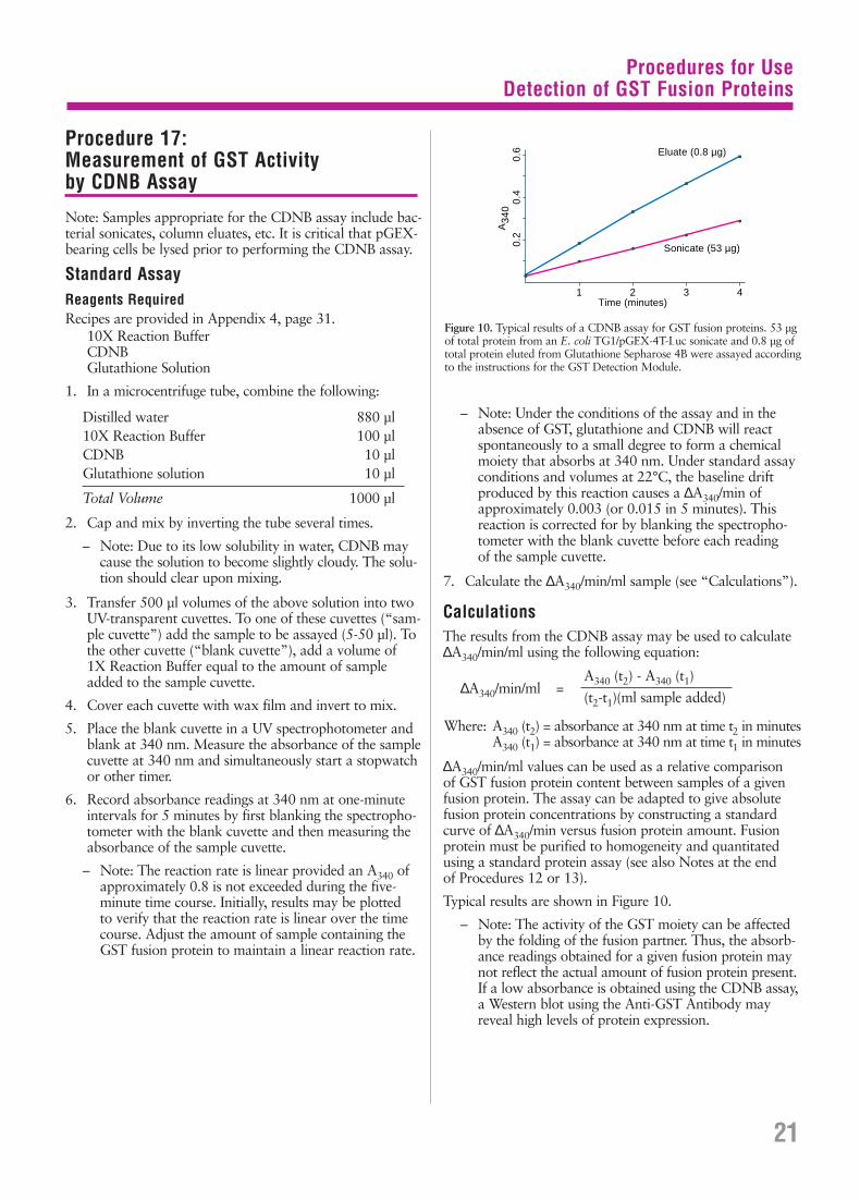
Procedures for UseDetection of GST Fusion Proteins
Procedure 17: Measurement of GST Activity by CDNB Assay
Note: Samples appropriate for the CDNB assay include bac-terial sonicates, column eluates, etc. It is critical that pGEX-bearing cells be lysed prior to performing the CDNB assay.
Standard AssayReagents RequiredRecipes are provided in Appendix 4, page 31.
10X Reaction BufferCDNBGlutathione Solution
1. In a microcentrifuge tube, combine the following:
Distilled water 880 µl10X Reaction Buffer 100 µlCDNB 10 µlGlutathione solution 10 µl
Total Volume 1000 µl
2. Cap and mix by inverting the tube several times.
– Note: Due to its low solubility in water, CDNB maycause the solution to become slightly cloudy. The solu-tion should clear upon mixing.
3. Transfer 500 µl volumes of the above solution into twoUV-transparent cuvettes. To one of these cuvettes (“sam-ple cuvette”) add the sample to be assayed (5-50 µl). Tothe other cuvette (“blank cuvette”), add a volume of 1X Reaction Buffer equal to the amount of sampleadded to the sample cuvette.
4. Cover each cuvette with wax film and invert to mix.
5. Place the blank cuvette in a UV spectrophotometer andblank at 340 nm. Measure the absorbance of the samplecuvette at 340 nm and simultaneously start a stopwatchor other timer.
6. Record absorbance readings at 340 nm at one-minuteintervals for 5 minutes by first blanking the spectropho-tometer with the blank cuvette and then measuring theabsorbance of the sample cuvette.
– Note: The reaction rate is linear provided an A340 ofapproximately 0.8 is not exceeded during the five-minute time course. Initially, results may be plotted to verify that the reaction rate is linear over the timecourse. Adjust the amount of sample containing theGST fusion protein to maintain a linear reaction rate.
– Note: Under the conditions of the assay and in theabsence of GST, glutathione and CDNB will reactspontaneously to a small degree to form a chemicalmoiety that absorbs at 340 nm. Under standard assayconditions and volumes at 22°C, the baseline drift produced by this reaction causes a ∆A340/min ofapproximately 0.003 (or 0.015 in 5 minutes). Thisreaction is corrected for by blanking the spectropho-tometer with the blank cuvette before each reading of the sample cuvette.
7. Calculate the ∆A340/min/ml sample (see “Calculations”).
CalculationsThe results from the CDNB assay may be used to calculate∆A340/min/ml using the following equation:
∆A340/min/ml =
Where: A340 (t2) = absorbance at 340 nm at time t2 in minutesA340 (t1) = absorbance at 340 nm at time t1 in minutes
∆A340/min/ml values can be used as a relative comparison of GST fusion protein content between samples of a givenfusion protein. The assay can be adapted to give absolutefusion protein concentrations by constructing a standardcurve of ∆A340/min versus fusion protein amount. Fusionprotein must be purified to homogeneity and quantitatedusing a standard protein assay (see also Notes at the end of Procedures 12 or 13).
Typical results are shown in Figure 10.
– Note: The activity of the GST moiety can be affectedby the folding of the fusion partner. Thus, the absorb-ance readings obtained for a given fusion protein maynot reflect the actual amount of fusion protein present.If a low absorbance is obtained using the CDNB assay,a Western blot using the Anti-GST Antibody mayreveal high levels of protein expression.
A340 (t2) - A340 (t1)
(t2-t1)(ml sample added)
21
1 2 3 4Time (minutes)
0.2
0.4
0.6
A34
0
Sonicate (53 µg)
Eluate (0.8 µg)
Figure 10. Typical results of a CDNB assay for GST fusion proteins. 53 µgof total protein from an E. coli TG1/pGEX-4T-Luc sonicate and 0.8 µg oftotal protein eluted from Glutathione Sepharose 4B were assayed accordingto the instructions for the GST Detection Module.
Procedures for UseDetection of GST Fusion Proteins
Procedure 18: Detection of GST Fusion Protein by Western Blot
1. Fractionate the protein samples by SDS-PAGE.
– Note: Depending on the source of the anti-GST anti-body, it may contain antibodies that react with variousE. coli proteins that may be present in your fusion protein sample. The anti-GST antibody should becross-adsorbed with E. coli proteins prior to use (seeAppendix 5, page 32). However, this process may notremove all cross-reacting antibodies. We suggest that asample of an E. coli sonicate made from a culture thatdoes not contain a recombinant pGEX plasmid be runas a control. The antibody from Pharmacia Biotechhas been cross-adsorbed with E. coli proteins.
2. Transfer the separated proteins from the gel to an appro-priate membrane (i.e. nitrocellulose, nylon, etc.).
– Note: Electrophoresis and protein transfer may beaccomplished using a variety of equipment and rea-gents (20-22). Using PhastSystem (18-1018-23) andPhastTransfer (18-1001-23) from Pharmacia Biotech,protein blots may be prepared in less than 1 hour (22).
Blocking of MembraneReagents RequiredRecipes are provided in Appendix 4, page 31.
Blocking/Incubation Buffer
1. Transfer the membrane onto which the proteins havebeen blotted to an appropriate container (e.g., Petri dishor small baking dish).
2. Add 50-200 ml of blocking/incubation buffer.
3. Incubate for 1-16 hours at ambient temperature withgentle shaking.
– Note: Longer incubation times (up to 16 hours) withblocking buffer may reduce background signal.
4. Decant and discard the buffer.
Anti-GST AntibodyReagents RequiredRecipes are provided in Appendix 4, page 31.
Anti-GST Antibody (27-4577-01)Blocking/Incubation BufferWash Buffer
1. Prepare an appropriate dilution of anti-GST antibodywith blocking/incubation buffer. If using the anti-GSTantibody from Pharmacia Biotech, dilute the antibody1:1000-1:2000, e.g., 5-10 µl of antibody to 10 ml of buffer.
2. Pour the antibody-buffer mixture into the container withthe membrane.
3. Incubate for 1 hour at ambient temperature with gentle shaking.
4. Decant and discard the antibody-buffer.
5. Rinse twice with 20-30 ml of wash buffer to remove themajority of unbound antibody.
6. Decant and discard the rinses.
7. Wash the membrane with 20-30 ml of wash buffer for 10 minutes at ambient temperature with gentle shaking.
8. Discard the wash and repeat.
Secondary AntibodyReagents RequiredRecipes are provided in Appendix 4, page 31.
Secondary Antibody (e.g., Sigma catalog number A 7650)Blocking/Incubation BufferWash Buffer
1. Dilute an appropriate secondary antibody with blocking/incubation buffer according to the manufacturer’s recom-mendation. (A secondary antibody, such as an anti-goatIgG alkaline phosphatase conjugate, Sigma catalog number A 7650, is required to detect the anti-GST antibody supplied with the GST Detection Module.)
2. Pour the antibody-buffer mixture into the container withthe membrane.
3. Incubate for 1 hour at ambient temperature with gentle shaking.
4. Decant and discard the antibody-buffer.
5. Rinse twice with 20-30 ml of wash buffer to remove themajority of unbound antibody.
6. Decant and discard the rinses.
7. Wash the membrane with 20-30 ml of wash buffer for 10 minutes at ambient temperature with gentle shaking.
8. Discard the wash and repeat.
9. Develop the blot with the appropriate substrate for theconjugated secondary antibody.
Typical results for detection of GST fusion proteins on aWestern blot are shown in Figure 11.
22
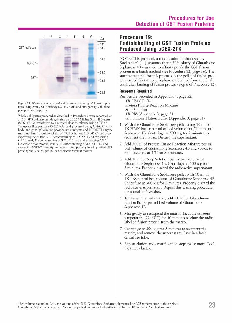
Procedure 19: Radiolabelling of GST Fusion ProteinsProduced Using pGEX-2TK
NOTE: This protocol, a modification of that used by Kaelin et al. (11), assumes that a 50% slurry of GlutathioneSepharose 4B was used to affinity purify the GST fusionprotein in a batch method (see Procedure 12, page 16). Thestarting material for this protocol is the pellet of fusion-pro-tein-loaded Glutathione Sepharose obtained from the finalwash after binding of fusion protein (Step 6 of Procedure 12).
Reagents RequiredRecipes are provided in Appendix 4, page 32.
1X HMK BufferProtein Kinase Reaction MixtureStop Solution1X PBS (Appendix 3, page 31)Glutathione Elution Buffer (Appendix 3, page 31)
1. Wash the Glutathione Sepharose pellet using 10 ml of 1X HMK buffer per ml of bed volume* of GlutathioneSepharose 4B. Centrifuge at 500 x g for 2 minutes to sediment the matrix. Discard the supernatant.
2. Add 300 µl of Protein Kinase Reaction Mixture per mlbed volume of Glutathione Sepharose 4B and vortex tomix. Incubate at 4°C for 30 minutes.
3. Add 10 ml of Stop Solution per ml bed volume of Glutathione Sepharose 4B. Centrifuge at 500 x g for 2 minutes. Properly discard the radioactive supernatant.
4. Wash the Glutathione Sepharose pellet with 10 ml of 1X PBS per ml bed volume of Glutathione Sepharose 4B.Centrifuge at 500 x g for 2 minutes. Properly discard theradioactive supernatant. Repeat this washing procedurefor a total of 5 washes.
5. To the sedimented matrix, add 1.0 ml of Glutathione Elution Buffer per ml bed volume of GlutathioneSepharose 4B.
6. Mix gently to resuspend the matrix. Incubate at roomtemperature (22-25ºC) for 10 minutes to elute the radio-labelled fusion protein from the matrix.
7. Centrifuge at 500 x g for 5 minutes to sediment thematrix, and remove the supernatant. Save in a fresh centrifuge tube.
8. Repeat elution and centrifugation steps twice more. Poolthe three eluates.
23
Figure 11. Western blot of E. coli cell lysates containing GST fusion pro-teins using Anti-GST Antibody (27-4577-01) and anti-goat IgG alkalinephosphatase conjugate.
Whole cell lysates prepared as described in Procedure 9 were separated ona 12% SDS polyacrylamide gel using an SE 250 Mighty Small II System(80-6147-45), transferred to a nitrocellulose membrane using a TE 62Transphor II apparatus (80-6209-58) and processed using Anti-GST Anti-body, anti-goat IgG alkaline phosphatase conjugate and BCIP/NBT enzymesubstrate; lane 1, sonicate of E. coli TG1 cells; lane 2, KL45 (DnaK over-expressing) cells; lane 3, E. coli containing pGEX-5X-1 and expressingGST; lane 4, E. coli containing pGEX-5X-2-Luc and expressing GST-luciferase fusion protein; lane 5, E. coli containing pGEX-4T-1-E7 andexpressing GST-E7 transcription factor fusion protein; lane 6, purified GSTprotein; and lane M, pre-stained molecular weight marker.
1 2 3 4 5 6 M
GST-luciferase –
GST-E7 –
GST –
kDa
– 101– 83.0
– 50.6
– 35.5
– 29.1
– 20.9
Procedures for UseDetection of GST Fusion Proteins
*Bed volume is equal to 0.5 x the volume of the 50% Glutathione Sepharose slurry used or 0.75 x the volume of the originalGlutathione Sepharose slurry. RediPack or prepacked columns of Glutathione Sepharose 4B contain a 2 ml bed volume.
Troubleshooting Guide
Troubleshooting Guide
A Low Yield of Vector Is Obtainedafter Plasmid Isolation
Amplify plasmid copy number: Increased yields of vectorDNA may require amplification of plasmid copy number. To amplify, grow a pGEX-containing E. coli culture to anA600 of 1.0. Add spectinomycin to a final concentration of300 µg/ml. After overnight incubation, isolate amplifiedplasmid DNA using FlexiPrep Kit.
High Basal Level of Expression
Basal level expression (i.e. expression in the absence of aninducer, such as IPTG), present with most inducible promot-ers, can affect the outcome of cloning experiments for toxicinserts; it can select against inserts cloned in the proper orien-tation. Basal level expression can be minimized by cataboliterepression (e.g., growth in the presence of glucose). The tacpromoter is not subject to catabolite repression. However,there is a lac promoter located upstream between the 3′-endof the lacIq gene and the tac promoter. This lac promotermay contribute to the basal level of expression of insertscloned into the pGEX multiple cloning site, and it is subjectto catabolite repression.
If the basal level of expression is too high for your applica-tion, addition of 2% glucose to the growth medium mayhelp. Growth in the presence of 2% glucose will decrease thebasal expression level associated with the upstream lac pro-moter but will not affect basal level expression from the tacpromoter. The presence of glucose should not significantlyaffect overall expression following induction with IPTG.
No Protein Is Detected by Coomassie-Stained SDS Gel of Bacterial Sonicate
Optimize expression conditions: By optimizing expressionconditions, the yield of fusion protein may be greatly improved. Investigate the effect of cell strain, medium com-position, incubation temperature and induction conditionson yield of fusion protein. Exact conditions will vary foreach fusion protein expressed.
Check DNA sequences: It is essential that protein-codingDNA sequences be cloned in the proper translation frame inpGEX vectors. Cloning junctions should be sequenced using 5′pGEX Sequencing Primer (27-1410-01) and 3′ pGEX Sequenc-ing Primer (27-1411-01) to verify that inserts are in-frame withthe GST gene. The reading frame of the multiple cloning sitefor each pGEX vector is shown in Figure 1 (page 2).
Analyze a small aliquot of an overnight culture by SDS-PAGE: Generally, a highly expressed protein will bevisible by Coomassie staining when 5-10 µl of an inducedculture whose A600 is ~ 1.0 is loaded on the gel. Non-transformed host E. coli cells and cells transformed with theparental pGEX vector should be run in parallel as negativeand positive controls, respectively. The presence of the fusionprotein in this total cell preparation and its absence from a clarified sonicate may indicate the presence of inclusionbodies (see below).
Check for expression by immunoblotting: Some fusionproteins may be masked on an SDS-polyacrylamide gel by a bacterial protein of approximately the same molecularweight. Immunoblotting can be used to identify fusion proteins in these cases. Run an SDS-polyacrylamide gel of induced cells as above and transfer the proteins to a nitrocellulose or PVDF membrane. Detect fusion proteinusing Anti-GST Antibody (27-4577-01 or included in theGST Detection Module, 27-4590-01).
Majority of Protein Is Found in Post-Sonicate Pellet
SDS-PAGE analysis of samples collected during the prepara-tion of the bacterial sonicate may indicate that the majorityof the GST fusion protein is located in the post-sonicate pellet(Step 10, Procedure 11). Possible causes and solutions arediscussed below.
Sonication may be insufficient: Cell disruption is evidencedby partial clearing of the suspension or may be checked bymicroscopic examination. Addition of lysozyme (0.1 volumeof a 10 mg/ml lysozyme solution in 25 mM Tris-HCl, pH 8.0)prior to sonication may improve results. Avoid frothing asthis may denature the fusion protein. Over-sonication canalso lead to co-purification of host proteins with the GSTfusion protein.
Fusion protein may be insoluble (inclusion bodies):If insufficient protein is found in the soluble fraction fol-lowing centrifugation of the sonicate, it may be necessaryto alter the growth conditions:
1. Fusion protein solubility can be dramatically increased by lowering the growth temperature. Experiment withgrowth temperatures in the range of 20-30°C (23, 24).
2. Alter level of induction by decreasing IPTG concentrationto <0.1 mM.
3. Alter timing of induction.
4. Induce for a shorter period of time.
5. Induce at a higher cell density for a short period of time.
6. Increase aeration. High oxygen transport can help prevent the formation of inclusion bodies (25).
It may be necessary to combine the above approaches. Exact conditions must be determined empirically for eachfusion protein.
24
Troubleshooting Guide
If the above techniques do not significantly improve expres-sion of soluble fusion protein, protein can be solubilizedfrom inclusion bodies using common denaturants such as 4-8 M guanidinium hydrochloride, 4-8 M urea, detergents,alkaline pH (> 9), organic solvents (26, 27), or N-lauroyl-sarcosine (Sarkosyl) (28, 29). Other variables that affect solubilization include time, temperature, ionic strength, theratio of denaturant to protein and the presence of thiol rea-gents (26, 27). For reviews see references 23, 26, 27, 30.
Following solubilization, proteins must be properly refoldedto regain function. Denaturant can be removed by dialysis,dilution, or gel filtration to allow refolding of the protein andformation of the correct intramolecular associations. Criticalparameters during refolding include pH, presence of thiolreagents and the speed of denaturant removal (26, 27, 31).Once refolded, protein may be purified by ion-exchange, gelfiltration or affinity chromatography.
Fusion proteins can be purified to some extent while dena-tured. In some instances where GST fusion proteins formedinclusion bodies, solubilization and binding to GlutathioneSepharose 4B were achieved in the presence of 2-3 M guan-idinium HCl or urea. Success has also been achieved usingup to 2% Tween® 20 for solubilization and binding. Bindingto Glutathione Sepharose 4B can also be achieved in thepresence of 1% CTAB, 10 mM DTT or 0.03% SDS (19).Success of affinity purification in the presence of these agentswill depend on the nature of the fusion protein.
An approach which might be of general applicability forworking with insoluble fusion proteins is described by Frangioni and Neel (29). Their method was developed whileworking with GST fusions of chicken muscle pyruvatekinase and protein tyrosine phosphatases. This method issummarized below.
Cultures of E. coli expressing GST fusion proteins were har-vested, washed once with STE (10 mM Tris, pH 8.0, 150 mMNaCl, 1 mM EDTA) and resuspended in STE containing 100 µg/ml of lysozyme. Following a 15 minute incubationon ice, dithiothreitol (DTT) was added to a final concentra-tion of 5 mM. Sarkosyl was added to a final concentrationof 1.5% (from a 10% stock in STE) and the cells were vor-texed for 5 seconds. Cells were then sonicated on ice using a“Cup Horn” sonicated water bath. Following centrifugation,the supernatant was transferred to a new tube and Triton X-100 was added to a final concentration of 2 or 4%. Thispreparation was added to prepared glutathione agarose beads (50% suspension in PBS) for affinity purification ofGST fusion proteins. Fusion proteins were eluted using 75 mM HEPES, pH 7.4, 150 mM NaCl, 10 mM reducedglutathione, 5 mM DTT and 2% N-octyl glucoside.
Important points to this procedure are:
• The concentration of sarkosyl must be optimized for eachfusion protein isolated (29).
• EDTA must be used with sarkosyl since this detergent isnot soluble in the presence of divalent cations (29).
• Cells were gently sonicated on ice by immersing the tubecontaining the lysate in a sonicated water bath for 1 minute.Sonication using a microtip probe usually resulted in adenatured fusion protein that would not bind to glutathi-one agarose (29).
• The addition of 5 mM DTT after lysozyme treatment but before the addition of sarkosyl significantly increasedGST binding to glutathione agarose (29).
• The addition of Triton X-100 prior to binding to glutathi-one agarose sequesters the sarkosyl and enables the bind-ing of the fusion proteins to the glutathione agarose (29). Triton X-100 also prevents the precipitation of sarkosylupon the addition of calcium and magnesium. The sarkosyl:Triton X-100 molar ratio must be optimized for eachfusion protein (29).
• The addition of a non-ionic detergent significantly improvedelution of fusion proteins from glutathione agarose. N-octylglucoside was used because it is easier to remove by dialysisthan Triton X-100 (29).
Fusion Protein Does Not Bind to Glutathione Sepharose 4B
Fusion protein denatured by sonication: Over-sonicationcan denature the fusion protein and prevent its binding toGlutathione Sepharose 4B (29). Use mild sonication condi-tions during cell lysis.
Add DTT prior to cell lysis: The addition of DTT to a finalconcentration of 5 mM prior to cell lysis can significantlyincrease binding of some GST fusion proteins to GlutathioneSepharose 4B (29).
Test binding of GST from parental pGEX: Prepare a soni-cate of cells harboring the parental pGEX plasmid and checkbinding to the matrix. If GST produced from the parentalplasmid binds with high affinity, then the fusion partner mayhave altered the conformation of GST, thereby reducing itsaffinity. Adequate results may be obtained by reducing thetemperature used for binding to 4°C, and by limiting thenumber of washes.
Use fresh Glutathione Sepharose 4B: If the GlutathioneSepharose 4B has already been used several times, it may be necessary to use fresh matrix. Refer also to Appendix 6,Regeneration of Glutathione Sepharose 4B (page 33).
Equilibrate Glutathione Sepharose 4B before use:Binding of GST fusion proteins to Glutathione Sepharose isnot efficient at pH less than 6.5 or greater than 8. Check toensure that the matrix has been equilibrated with a buffer6.5 < pH < 8.0 (e.g., 1X PBS) before the fusion protein hasbeen applied.
25
Troubleshooting Guide
Fusion Protein Is Not Efficiently Elutedfrom Glutathione Sepharose 4B
Increase the time used for elution: In some instances, over-night elution at room temperature or 4°C is most effective.
Increase the volume of elution buffer: Keep in mind thatGlutathione Sepharose 4B will also function as a gel filtrationmedium with an approximate molecular weight exclusionlimit of 2 x 10⁷ Daltons. Small proteins (especially thoseliberated following cleavage with a site-specific protease)may require large elution volumes. In these cases, the batchpurification method may be preferred (Procedure 12, page16). Proteins eluted in a large volume may require concen-tration by ultrafiltration.
Increase the concentration of glutathione in the elution buffer: General protocols use 5 mM glutathione forelution. Note that the concentration of glutathione used inthe GST Purification Modules is 10 mM, which should besufficient for most applications. Keep in mind that if the glutathione concentration is significantly increased above 15 mM, the buffer concentration will have to be increasedto maintain proper pH (32).
Increase the pH of the elution buffer: A low pH may limitelution from Glutathione Sepharose. Increasing the pH ofthe elution buffer to 8-9 may improve elution withoutrequiring an increase in the concentration of glutathioneused for elution. Note that the pH of elution buffer usedwith the GST Purification Modules is 8.
Increase the ionic strength of the elution buffer: Theaddition of 0.1-0.2 M NaCl to the elution buffer may alsoimprove results. Keep in mind that very hydrophobic pro-teins may precipitate under high salt conditions. If this is thecase, addition of a non-ionic detergent may improve results(see below).
One researcher obtained satisfactory results using the fol-lowing elution buffer: 20 mM glutathione, 100 mM Tris-HCl (pH 8.0), 120 mM NaCl (32).
Add a non-ionic detergent to the elution buffer: Nonspe-cific hydrophobic interactions may prevent solubilizationand elution of fusion proteins from Glutathione Sepharose4B (29). Addition of a non-ionic detergent can improveresults. The addition of 0.1% Triton X-100 or 2% N-octylglucoside can significantly improve elution of some GSTfusion proteins (29).
Multiple Bands Are Observed on Gel Following Elution from Glutathione Sepharose 4B
Add a protease inhibitor: Multiple bands may be a result ofpartial degradation of fusion proteins by proteases. Additionof 1 mM PMSF to the lysis solution may improve results. Anon-toxic, water soluble alternative to PMSF is 4-(2-amino-ethyl)-benzenesulfonyl fluoride hydrochloride (AEBSF) (33),commercially available as Pefabloc® SC from Boehringer
Mannheim. Serine protease inhibitors must be removed priorto cleavage by PreScission Protease, thrombin or factor Xa.
Use a protease-deficient host: Multiple bands may be theresult of proteolysis in the host bacteria. If this is the case, theuse of a protease-deficient strain may be required (e.g., lon-
or ompT). E. coli BL21 (27-1542-01) is provided with thepGEX vectors. This strain is ompT (Appendix 7, page 33).
Decrease sonication: Cell disruption is evidenced by partialclearing of the suspension or may be checked by microscopicexamination. Addition of lysozyme (0.1 volume of a 10 mg/mllysozyme solution in 25 mM Tris-HCl pH 8.0) prior to soni-cation may improve results. Avoid frothing as this may denature the fusion protein. Over-sonication can also lead to co-purification of host proteins with the GST fusion protein.
Include an additional purification step: Additional bandsmay be caused by the copurification of a variety of proteinsknown as chaperonins which are involved in the proper fold-ing of nascent proteins in E. coli (34). These include, but may not be limited to: DnaK (~70 kDa, reference 35), DnaJ(~37 kDa, ref. 36), GrpE (~40 kDa, ref. 37), GroEL (~57 kDa,ref. 38), and GroES (~10 kDa, ref. 39). Several methods forthe purification of GST fusion proteins free of these copurify-ing proteins have been described (see below for DnaK andref. 40 for GroEL).
70 kDa Protein Co-purifies with the Recombinant Protein
Disrupt association using ATP and Mg++: A 70 kDa proteinis probably a protein product of the E. coli gene dnaK. Thisprotein is involved in the degradation of “abnormal” proteinsin E. coli (41). It has been reported that this association can be disrupted by incubating the recombinant protein solutionin 2 mM ATP, 10 mM MgSO4, 50 mM Tris (pH 7.4) for 10 minutes at 37°C prior to affinity chromatography (41).
Alternatively, the DnaK protein can also be removed by pass-ing the recombinant protein solution over ATP-agarose (ATPlinked to agarose by C-8 of ATP) (42).
Separate DnaK from pGEX-GST by ion exchange chromatography: DnaK can be removed from GST by ion-exchange chromatography using a method similar to one usedin the purification of DnaK (43). The method, described inPharmacia Biotech Analects & Separations, page 24 (1996),was developed using DEAE Sepharose Fast Flow (17-0709-01)on an FPLC System equipped with FPLCdirector software.Glutathione Sepharose affinity-purified GST was successfullypurified free of detectable DnaK using a 400 mM NaCl stepgradient elution. Although this procedure was developed forGST expressed from a pGEX vector, without a fusion partner,it should provide a good starting point for working with GSTfusion proteins. The presence of a fusion partner may alter theconditions under which the GST fusion protein will bind toand elute from DEAE Sepharose Fast Flow. The method canbe optimized for a particular fusion protein by using a differentstep gradient or by using a linear gradient. Provided that thefusion protein elutes under conditions different from those forDnaK, separation of the two proteins can be achieved.
26
Troubleshooting Guide
Multiple Bands Are Observed on Western Blot Following Detection with an Anti-GST Antibody
Add a protease inhibitor: Multiple bands may be a result ofpartial degradation of fusion proteins by proteases. Additionof protease inhibitors to the lysis solution may improveresults. Serine protease inhibitors must be removed prior tocleavage by PreScission Protease, thrombin or factor Xa.
Cross-adsorb antibody with E. coli proteins: Dependingon the source of the anti-GST antibody, it may contain anti-bodies that react with various E. coli proteins that may bepresent in your fusion protein sample. Cross-adsorb the anti-body with an E. coli sonicate to remove anti-E. coli antibodiesfrom the preparation (see Appendix 5, page 32). PharmaciaBiotech Anti-GST Antibody (27-4577-01) has been cross-absorbed against E. coli proteins and tested for its lack ofnon-specific background binding in a Western blot.
PreScission Protease Cleavage Is Incomplete
PreScission Protease to fusion protein ratio is incorrect:Check the amount of fusion protein in the digestion. Adjustthe amount of PreScission Protease added to at least 10 cleav-age units/mg of fusion protein. If performing on-columncleavage, note that the capacity of Glutathione Sepharose forGST is typically ≥8 mg/ml. In most purifications, however,the resin is not saturated with fusion protein.
Increase incubation time and enzyme concentration:If the fusion protein is not degraded by extensive incubationwith PreScission Protease, increase the reaction time to 20 hours or longer and increase the amount of PreScissionProtease used in the reaction.
Verify presence of PreScission Protease cleavage site:Compare the DNA sequence of the construct with theknown PreScission Protease cleavage sequence. Verify that the optimal PreScission Protease recognition site, Leu-Glu-Val-Leu-Phe-Gln ↓ Gly-Pro, has not been alteredduring the course of cloning your fusion protein.
Ensure absence of PreScission Protease inhibitors:Dialyze the fusion protein against PreScission ProteaseCleavage Buffer (Appendix 3, page, 31) before cleaving with PreScission Protease. The presence of Zn2+ as well asPefabloc® SC or chymostatin has been shown to interferewith the activity of PreScission Protease.
Multiple Bands Are Observed on SDS Gel Following PreScission Protease Cleavage
Determine when bands appear: Test to be certain thatadditional bands are not present prior to PreScission Protease cleavage. These may be the result of proteolysis inthe host bacteria. If this is the case, the use of a protease-deficient strain (e.g., lon- or ompT) such as E. coli BL21 (27-1524-01) may be required.
Fusion partner may contain recognition sequences forPreScission Protease: PreScission Protease optimally recog-nizes the sequence Leu-Glu-Val-Leu-Phe-Gln ↓ Gly-Pro andcleaves between the Gln and Gly residues (14), howeverother similar secondary sites may exhibit some propensityfor cleavage. Check the sequence of the fusion partner (ifpossible) for the presence of additional PreScission Proteaserecognition sites. Adjusting reaction conditions (e.g., time,temperature, salt concentration) may result in selective cleav-age at the desired site. If adjustment of the conditions doesnot correct the problem, reclone the insert into a pGEX T(Thrombin) or pGEX X (factor Xa) expression vector.
The Fusion Partner is Contaminated withPreScission Protease after Purificationwith Glutathione Sepharose
Pass the sample over fresh Glutathione Sepharose:If the Glutathione Sepharose was saturated with GST fusionprotein, not all of the PreScission Protease may have beenremoved by the matrix. Use of a second round of Gluta-thione Sepharose chromatography should remove the resid-ual PreScission Protease.
Thrombin Cleavage Is Incomplete
Thrombin to fusion protein ratio is incorrect: Check the amount of fusion protein in the digestion. Adjust theamount of thrombin added to 10 cleavage units/mg of fusionprotein. One cleavage unit of thrombin protease from Pharmacia Biotech digests ≥ 90% of 100 µg of a test fusionprotein in 16 hours at 22°C in Glutathione Elution Buffer(Appendix 3, page 31). A cleavage unit is approximatelyequal to 0.2 NIH units.
Increase incubation time and enzyme concentration:If the fusion protein is not degraded by extensive incubationwith thrombin, increase the reaction time to 20 hours andincrease the amount of thrombin used in the reaction.
Verify presence of thrombin site: Check DNA sequence ofconstruct. Compare with known sequence, and verify thatthe thrombin recognition site has not been altered during thecourse of cloning your fusion protein.
27
Troubleshooting Guide
Multiple Bands Are Observed on SDSGel Following Thrombin Cleavage
Determine when bands appear: Test to be certain thatadditional bands are not present prior to thrombin cleavage.If bands are present prior to cleavage, see discussions above.
Fusion partner may contain recognition sequences for thrombin: The optimum cleavage site for thrombin canhave the following two structures (44):
1) P4-P3-Pro-Arg/Lys ↓P1′-P2′ where P3 and P4 are hydro-phobic amino acids and P1′ and P2′ are nonacidic aminoacids. The Arg/Lys ↓P1′ bond is cleaved.
Examples: P4 P3 Pro R/K↓ P1′ P2′
A) Met Tyr Pro Arg ↓ Gly AsnB) Ile Arg Pro Lys ↓ Leu LysC) Leu Val Pro Arg ↓ Gly Ser
In A, the Arg ↓Gly bond is cleaved very fast by thrombin. InB, the Lys ↓Leu bond is cleaved. C is the recognition sequencefound on the thrombin series of pGEX plasmids and the Arg ↓Gly bond is cleaved.
2) P2-Arg/Lys ↓P1′, where P2 or P1′ are Gly. The Arg/Lys ↓P1′ bond is cleaved.
Examples:P2 R/K↓ P1′
A) Ala Arg ↓ GlyB) Gly Lys ↓ Ala
In A, the Arg ↓Gly bond is cleaved efficiently. In B, the Lys ↓Ala bond is cleaved.
Check the sequence of the fusion partner (if possible) for thepresence of thrombin recognition sites. Adjusting time andtemperature of digestion can result in selective scission at thedesired thrombin site. If adjustment of conditions does notcorrect the problem, reclone the insert into a pGEX-6P(PreScission) or pGEX X expression vector.
Factor Xa Cleavage Is Incomplete
Factor Xa is not properly activated: Functional factor Xarequires activation of factor X with Russell’s viper venom.Activation conditions are a ratio of Russell’s viper venom tofactor X of 1% (w/w) in 8 mM Tris-HCl (pH 8.0), 70 mMNaCl, 8 mM CaCl2. The reaction is incubated at 37°C for 5 minutes (45). Pharmacia Biotech Factor Xa (27-0849-01)has been preactivated by this procedure.
The first amino acid after the Factor Xa recognitionsequence is Arg or Pro: The recognition sequence for fac-tor Xa is Ile-Glu-Gly-Arg ↓X, where X can be any amino acidexcept Arg or Pro (46, 47). Check the sequence of the fusionpartner to be sure that the first three nucleotides after thefactor Xa recognition sequence do not code for Arg or Pro.
Factor Xa to fusion protein ratio is incorrect: Check theamount of fusion protein in the digestion. Adjust the amountof factor Xa added to at least 1% (w/w) of fusion protein.For some fusion proteins, up to 5% factor Xa can be used.The optimum amount of factor Xa for a given fusion proteinmust be determined empirically.
Remove glutathione: Factor Xa consists of two subunitslinked by disulfide bridges. Since glutathione can disruptdisulfide bridges, it should be removed from the sample bydialysis against 2000 volumes of factor Xa cleavage buffer(Appendix 3, page 31). It has been observed that removal ofglutathione from the factor Xa cleavage reaction improvesresults (48).
Increase substrate concentration: In some cases, a fusionprotein concentration of 1 mg/ml has been found to yieldoptimal results (48).
Add a small amount of SDS to the reaction buffer: Theaddition of SDS (≤0.5% w/v) to the reaction buffer cansignificantly improve factor Xa cleavage with some fusionproteins (49). Various concentrations of SDS should betested to find the optimum concentration.
Increase incubation time and enzyme concentration:If the fusion protein is not degraded by extensive incubationwith factor Xa, increase the reaction time to 20 hours or longerand increase the amount of factor Xa used in the reaction.
Verify presence of factor Xa site: Check DNA sequence ofconstruct. Compare with known sequence, and verify thatthe factor Xa recognition site has not been altered during thecourse of cloning your fusion protein.
Multiple Bands Are Observed on SDSGel Following Factor Xa Cleavage
Determine when bands appear: Test to be certain thatadditional bands are not present prior to factor Xa cleavage.If bands are present prior to cleavage, see discussions above.
Fusion partner may contain recognition sequences forfactor Xa: Factor Xa is very specific for the recognitionsequence Ile-Glu-Gly-Arg ↓ (50). The bond following the Arg residue is cleaved.
Check the sequence of the fusion partner (if possible) for thepresence of factor Xa recognition sites. Adjusting time andtemperature of digestion can result in selective scission at the desired factor Xa site. If adjustment of conditions doesnot correct the problem, reclone the insert into a pGEX-6P(PreScission) or pGEX T (Thrombin) expression vector.
28
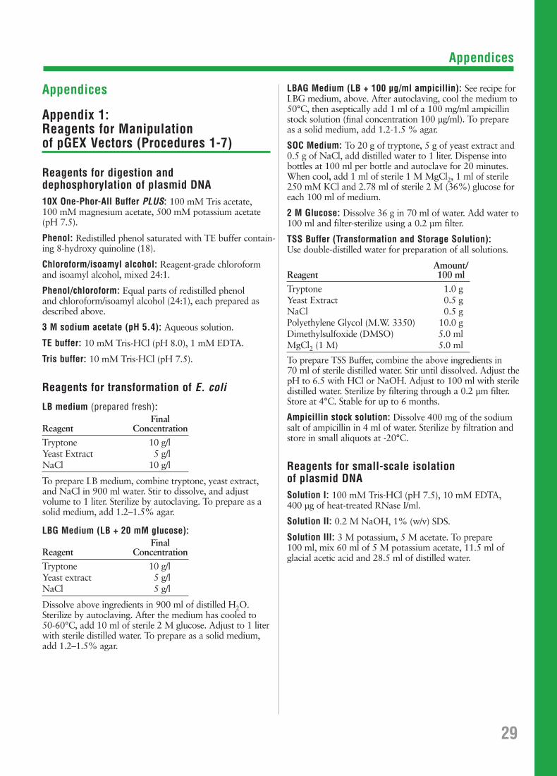
Appendices
Appendices
Appendix 1: Reagents for Manipulation of pGEX Vectors (Procedures 1-7)
Reagents for digestion and dephosphorylation of plasmid DNA10X One-Phor-All Buffer PLUS: 100 mM Tris acetate, 100 mM magnesium acetate, 500 mM potassium acetate(pH 7.5).
Phenol: Redistilled phenol saturated with TE buffer contain-ing 8-hydroxy quinoline (18).
Chloroform/isoamyl alcohol: Reagent-grade chloroformand isoamyl alcohol, mixed 24:1.
Phenol/chloroform: Equal parts of redistilled phenol and chloroform/isoamyl alcohol (24:1), each prepared asdescribed above.
3 M sodium acetate (pH 5.4): Aqueous solution.
TE buffer: 10 mM Tris-HCl (pH 8.0), 1 mM EDTA.
Tris buffer: 10 mM Tris-HCl (pH 7.5).
Reagents for transformation of E. coli
LB medium (prepared fresh):Final
Reagent Concentration
Tryptone 10 g/lYeast Extract 5 g/lNaCl 10 g/l
To prepare LB medium, combine tryptone, yeast extract,and NaCl in 900 ml water. Stir to dissolve, and adjust volume to 1 liter. Sterilize by autoclaving. To prepare as asolid medium, add 1.2–1.5% agar.
LBG Medium (LB + 20 mM glucose):Final
Reagent Concentration
Tryptone 10 g/lYeast extract 5 g/lNaCl 5 g/l
Dissolve above ingredients in 900 ml of distilled H2O. Sterilize by autoclaving. After the medium has cooled to 50-60°C, add 10 ml of sterile 2 M glucose. Adjust to 1 literwith sterile distilled water. To prepare as a solid medium,add 1.2–1.5% agar.
LBAG Medium (LB + 100 µg/ml ampicillin): See recipe forLBG medium, above. After autoclaving, cool the medium to50°C, then aseptically add 1 ml of a 100 mg/ml ampicillinstock solution (final concentration 100 µg/ml). To prepare as a solid medium, add 1.2-1.5 % agar.
SOC Medium: To 20 g of tryptone, 5 g of yeast extract and0.5 g of NaCl, add distilled water to 1 liter. Dispense intobottles at 100 ml per bottle and autoclave for 20 minutes.When cool, add 1 ml of sterile 1 M MgCl2, 1 ml of sterile250 mM KCl and 2.78 ml of sterile 2 M (36%) glucose foreach 100 ml of medium.
2 M Glucose: Dissolve 36 g in 70 ml of water. Add water to100 ml and filter-sterilize using a 0.2 µm filter.
TSS Buffer (Transformation and Storage Solution):Use double-distilled water for preparation of all solutions.
Amount/Reagent 100 ml
Tryptone 1.0 gYeast Extract 0.5 gNaCl 0.5 gPolyethylene Glycol (M.W. 3350) 10.0 gDimethylsulfoxide (DMSO) 5.0 mlMgCl2 (1 M) 5.0 ml
To prepare TSS Buffer, combine the above ingredients in 70 ml of sterile distilled water. Stir until dissolved. Adjust thepH to 6.5 with HCl or NaOH. Adjust to 100 ml with steriledistilled water. Sterilize by filtering through a 0.2 µm filter.Store at 4°C. Stable for up to 6 months.
Ampicillin stock solution: Dissolve 400 mg of the sodiumsalt of ampicillin in 4 ml of water. Sterilize by filtration andstore in small aliquots at -20°C.
Reagents for small-scale isolation of plasmid DNASolution I: 100 mM Tris-HCl (pH 7.5), 10 mM EDTA, 400 µg of heat-treated RNase I/ml.
Solution II: 0.2 M NaOH, 1% (w/v) SDS.
Solution III: 3 M potassium, 5 M acetate. To prepare 100 ml, mix 60 ml of 5 M potassium acetate, 11.5 ml ofglacial acetic acid and 28.5 ml of distilled water.
29
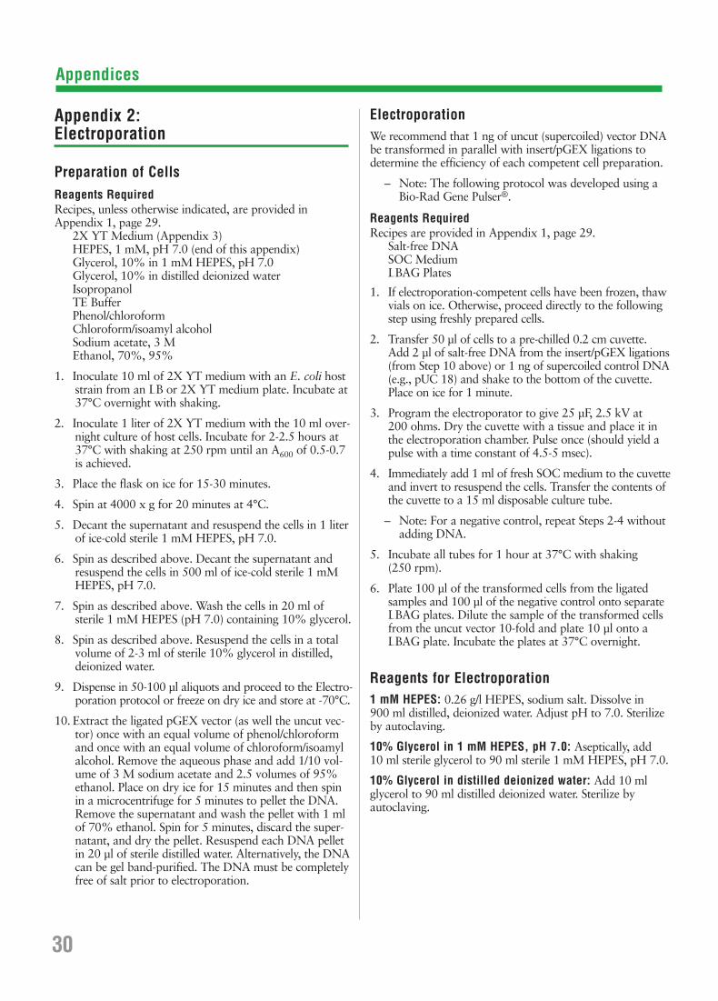
Appendices
Appendix 2: Electroporation
Preparation of CellsReagents RequiredRecipes, unless otherwise indicated, are provided in Appendix 1, page 29.
2X YT Medium (Appendix 3)HEPES, 1 mM, pH 7.0 (end of this appendix)Glycerol, 10% in 1 mM HEPES, pH 7.0 Glycerol, 10% in distilled deionized water IsopropanolTE BufferPhenol/chloroformChloroform/isoamyl alcoholSodium acetate, 3 MEthanol, 70%, 95%
1. Inoculate 10 ml of 2X YT medium with an E. coli hoststrain from an LB or 2X YT medium plate. Incubate at37°C overnight with shaking.
2. Inoculate 1 liter of 2X YT medium with the 10 ml over-night culture of host cells. Incubate for 2-2.5 hours at37°C with shaking at 250 rpm until an A600 of 0.5-0.7 is achieved.
3. Place the flask on ice for 15-30 minutes.
4. Spin at 4000 x g for 20 minutes at 4°C.
5. Decant the supernatant and resuspend the cells in 1 literof ice-cold sterile 1 mM HEPES, pH 7.0.
6. Spin as described above. Decant the supernatant andresuspend the cells in 500 ml of ice-cold sterile 1 mMHEPES, pH 7.0.
7. Spin as described above. Wash the cells in 20 ml of sterile 1 mM HEPES (pH 7.0) containing 10% glycerol.
8. Spin as described above. Resuspend the cells in a totalvolume of 2-3 ml of sterile 10% glycerol in distilled,deionized water.
9. Dispense in 50-100 µl aliquots and proceed to the Electro-poration protocol or freeze on dry ice and store at -70°C.
10. Extract the ligated pGEX vector (as well the uncut vec-tor) once with an equal volume of phenol/chloroformand once with an equal volume of chloroform/isoamylalcohol. Remove the aqueous phase and add 1/10 vol-ume of 3 M sodium acetate and 2.5 volumes of 95%ethanol. Place on dry ice for 15 minutes and then spin in a microcentrifuge for 5 minutes to pellet the DNA.Remove the supernatant and wash the pellet with 1 mlof 70% ethanol. Spin for 5 minutes, discard the super-natant, and dry the pellet. Resuspend each DNA pellet in 20 µl of sterile distilled water. Alternatively, the DNAcan be gel band-purified. The DNA must be completelyfree of salt prior to electroporation.
ElectroporationWe recommend that 1 ng of uncut (supercoiled) vector DNAbe transformed in parallel with insert/pGEX ligations todetermine the efficiency of each competent cell preparation.
– Note: The following protocol was developed using aBio-Rad Gene Pulser®.
Reagents RequiredRecipes are provided in Appendix 1, page 29.
Salt-free DNASOC MediumLBAG Plates
1. If electroporation-competent cells have been frozen, thawvials on ice. Otherwise, proceed directly to the followingstep using freshly prepared cells.
2. Transfer 50 µl of cells to a pre-chilled 0.2 cm cuvette.Add 2 µl of salt-free DNA from the insert/pGEX ligations(from Step 10 above) or 1 ng of supercoiled control DNA(e.g., pUC 18) and shake to the bottom of the cuvette.Place on ice for 1 minute.
3. Program the electroporator to give 25 µF, 2.5 kV at 200 ohms. Dry the cuvette with a tissue and place it inthe electroporation chamber. Pulse once (should yield apulse with a time constant of 4.5-5 msec).
4. Immediately add 1 ml of fresh SOC medium to the cuvetteand invert to resuspend the cells. Transfer the contents ofthe cuvette to a 15 ml disposable culture tube.
– Note: For a negative control, repeat Steps 2-4 withoutadding DNA.
5. Incubate all tubes for 1 hour at 37°C with shaking (250 rpm).
6. Plate 100 µl of the transformed cells from the ligatedsamples and 100 µl of the negative control onto separateLBAG plates. Dilute the sample of the transformed cellsfrom the uncut vector 10-fold and plate 10 µl onto aLBAG plate. Incubate the plates at 37°C overnight.
Reagents for Electroporation1 mM HEPES: 0.26 g/l HEPES, sodium salt. Dissolve in 900 ml distilled, deionized water. Adjust pH to 7.0. Sterilizeby autoclaving.
10% Glycerol in 1 mM HEPES, pH 7.0: Aseptically, add 10 ml sterile glycerol to 90 ml sterile 1 mM HEPES, pH 7.0.
10% Glycerol in distilled deionized water: Add 10 mlglycerol to 90 ml distilled deionized water. Sterilize by autoclaving.
30
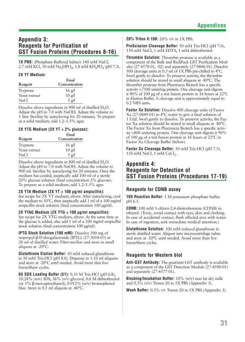
Appendices
Appendix 3: Reagents for Purification of GST Fusion Proteins (Procedures 8-16)
1X PBS: (Phosphate-Buffered Saline): 140 mM NaCl, 2.7 mM KCl, 10 mM Na2HPO4, 1.8 mM KH2PO4 (pH 7.3).
2X YT Medium:Final
Reagent Concentration
Tryptone 16 g/lYeast extract 10 g/lNaCl 5 g/l
Dissolve above ingredients in 900 ml of distilled H2O.Adjust the pH to 7.0 with NaOH. Adjust the volume to 1 liter. Sterilize by autoclaving for 20 minutes. To prepare as a solid medium, add 1.2–1.5% agar.
2X YTG Medium (2X YT + 2% glucose):Final
Reagent Concentration
Tryptone 16 g/lYeast extract 10 g/lNaCl 5 g/l
Dissolve above ingredients in 800 ml of distilled H2O.Adjust the pH to 7.0 with NaOH. Adjust the volume to 900 ml. Sterilize by autoclaving for 20 minutes. Once themedium has cooled, aseptically add 100 ml of a sterile 20% glucose solution (final concentration 2% glucose). To prepare as a solid medium, add 1.2–1.5% agar.
2X YTA Medium (2X YT + 100 µg/ml ampicillin): See recipe for 2X YT medium, above. After autoclaving, coolthe medium to 50°C, then aseptically add 1 ml of a 100 mg/mlampicillin stock solution (final concentration 100 µg/ml).
2X YTAG Medium (2X YTG + 100 µg/ml ampicillin): See recipe for 2X YTG medium, above. At the same time asthe glucose is added, also add 1 ml of a 100 mg/ml ampicillinstock solution (final concentration 100 µg/ml).
IPTG Stock Solution (100 mM): Dissolve 500 mg of isopropyl-β-D-thiogalactoside (IPTG) (27-3054-03) in 20 ml of distilled water. Filter-sterilize and store in smallaliquots at -20°C.
Glutathione Elution Buffer: 10 mM reduced glutathione in 50 mM Tris-HCl (pH 8.0). Dispense in 1-10 ml aliquotsand store at -20°C until needed. Avoid more that fivefreeze/thaw cycles.
6X SDS Loading Buffer (51): 0.35 M Tris-HCl (pH 6.8),10.28% (w/v) SDS, 36% (v/v) glycerol, 0.6 M dithiothreitol(or 5% β-mercaptoethanol), 0.012% (w/v) bromophenolblue. Store in 0.5 ml aliquots at -80°C.
20% Triton X-100: 20% v/v in 1X PBS.
PreScission Cleavage Buffer: 50 mM Tris-HCl (pH 7.0),150 mM NaCl, 1 mM EDTA, 1 mM dithiothreitol.
Thrombin Solution: Thrombin protease is available as acomponent of the Bulk and RediPack GST Purification Mod-ules (27-4570-01, -02) and separately (27-0846-01). Dissolve500 cleavage units in 0.5 ml of 1X PBS pre-chilled to 4°C.Swirl gently to dissolve. To preserve activity, the thrombinsolution should be stored in small aliquots at -80°C. Thethrombin protease from Pharmacia Biotech has a specificactivity >7500 units/mg protein. One cleavage unit digests ≥ 90% of 100 µg of a test fusion protein in 16 hours at 22°Cin Elution Buffer. A cleavage unit is approximately equal to0.2 NIH units.
Factor Xa Solution: Dissolve 400 cleavage units of FactorXa (27-0849-01) in 4°C water to give a final solution of 1 U/µl. Swirl gently to dissolve. To preserve activity, the Fac-tor Xa solution should be stored in small aliquots at -80°C.The Factor Xa from Pharmacia Biotech has a specific activ-ity >800 units/mg protein. One cleavage unit digests ≥ 90%of 100 µg of a test fusion protein in 16 hours at 22°C inFactor Xa Cleavage Buffer (below).
Factor Xa Cleavage Buffer: 50 mM Tris-HCl (pH 7.5), 150 mM NaCl, 1 mM CaCl2.
Appendix 4: Reagents for Detection of GST Fusion Proteins (Procedures 17-19)
Reagents for CDNB assay10X Reaction Buffer: 1 M potassium phosphate buffer, pH 6.5.
CDNB: 100 mM 1-chloro-2,4-dinitrobenzene (CDNB) inethanol. (Toxic, avoid contact with eyes, skin and clothing.In case of accidental contact, flush affected area with water.In case of ingestion, seek immediate medical attention.)
Glutathione Solution: 100 mM reduced glutathione in sterile distilled water. Aliquot into microcentrifuge tubes and store at -20°C until needed. Avoid more than fivefreeze/thaw cycles.
Reagents for Western blotAnti-GST Antibody: The goat/anti-GST antibody is availableas a component of the GST Detection Module (27-4590-01)and separately (27-4577-01).
Blocking/Incubation Buffer: 10% (w/v) non-fat dry milkand 0.3% (v/v) Tween-20 in 1X PBS (Appendix 3).
Wash Buffer: 0.3% v/v Tween-20 in 1X PBS (Appendix 3).
31

Appendices
Reagents for radiolabelling of GST fusion proteins produced with pGEX-2TK10X HMK (Heart Muscle Kinase) Buffer: 200 mM Tris (pH7.5), 1 M NaCl, 120 mM MgCl2. Dilute as needed for use.
Bovine Heart Kinase: 10 U/µl in 40 mM dithiothreitol, available from Sigma Chemical Co. as a lyophilized powder,catalog number P 2645.
[γ-32P]ATP: 3000 Ci/mmol, 10 mCi/ml.
Protein Kinase Reaction Mixture: 15 µl 10X HMK Buffer,5 µl bovine heart kinase solution, 5 µl [γ-32P]ATP, 125 µl distilled H2O.
Stop Solution: 10 mM sodium phosphate (pH 8.0), 10 mMsodium pyrophosphate, 10 mM EDTA, 1 mg/ml bovineserum albumin.
Appendix 5: Cross-adsorption of Anti-GST AntiserumWith E. coli Proteins
Depending on the source of your anti-GST antibody, it maycontain anti-E. coli antibodies that will react with E. coliproteins that may be present in your fusion protein sample.Use the following protocols to prepare an immobilized E.coli sonicate that can be used to cross-adsorb anti-E. coliantibodies. The antibody available from Pharmacia Biotechhas been cross-adsorbed with E. coli proteins. The followingprotocols will treat 240 ml of anti-GST antiserum.
Preparation of SonicateReagents RequiredRecipes are provided at the end of Appendix 5.
2X YT Medium (see Appendix 3, page 31)Coupling Buffer
1. Use a single colony of non-transformed E. coli cells toinoculate 30 ml of 2X YT medium.
2. Incubate for 12-15 hours at 37°C with vigorous shaking.
3. Transfer 25 ml of the culture into 2 liters of prewarmed2X YT medium contained in a 4 liter flask. Incubate at37°C with vigorous shaking until the A600 reaches 2.5.
4. Transfer the culture to 42°C and continue incubating foran additional hour.
5. Transfer the culture to appropriate centrifuge bottles andcentrifuge at 7,700 x g for 10 minutes (or 5,000 x g for30 minutes) at 4°C to sediment the cells.
6. Discard the supernatant and resuspend the cells in Cou-pling Buffer to an A600 of 80. The final volume shouldbe approximately 50-75 ml.
7. Transfer the cell suspension to an appropriate container.
8. Place the container on ice and disrupt the cells using anappropriately equipped sonicator. Sonication should achieve>90% cell lysis as determined by microscopic examination.
9. Store the sonicate at -70°C until needed.
Preparation of Immobilized SonicateReagents RequiredRecipes are provided at the end of Appendix 5.
CNBr-activated Sepharose 4B (see page 39)Coupling BufferAcetate BufferTris Buffer1X PBS (see Appendix 3, page 31)
1. Prepare 15 g of CNBr-activated Sepharose 4B accordingto manufacturer’s instructions.
2. To the 15 g of prepared CNBr-activated Sepharose 4B,add 45 ml of thawed E. coli sonicate. Close the con-tainer securely and incubate at 4°C for 12-16 hours withgentle shaking (do not use magnetic stirring).
3. Transfer the suspension to an appropriately sized sin-tered filter funnel attached to a vacuum source. Apply alight vacuum to remove the solution.
4. Wash the matrix with 500 ml of coupling buffer byslowly pouring the buffer into the funnel while stirringthe matrix with a glass stir rod. Apply a light vacuum toremove the solution.
5. Turn off the vacuum. Add 40 ml of acetate buffer. Stirfor 5 minutes with a glass stir rod. Apply a light vacuumto remove the solution.
6. Turn off the vacuum. Add 40 ml of Tris buffer. Stir for 5 minutes with a glass stir rod. Apply a light vacuum toremove the solution.
7. Repeat Steps 5 and 6 for a total of 3 times.
8. Add 100 ml of 1X PBS to the matrix. Stir to suspend.Split the suspension equally into four 100 ml centrifugebottles. Add 50 ml of 1X PBS to each of the four bottles.Centrifuge at 500 x g for 10 minutes at 4°C. Aspirate offthe supernatant taking care not to disturb the matrix.
Cross-adsorption of Anti-GST Antiserum With Immobilized E. coli Sonicate1. Add 60 ml of crude anti-GST antiserum to each of the
four bottles containing the immobilized E. coli sonicate.
2. Incubate at room temperature (22-25°C) for 1 hour withgentle shaking (do not use magnetic stirring).
3. Remove the matrix by filtering the serum-matrix suspen-sion over Whatman® 40 ashless filter paper.
4. Store the cross-adsorbed anti-GST antiserum at 4°C.
32
Appendix 4: Reagents for Detection of GST FusionProteins (Procedures 17-19) (cont.)

Appendices
Reagents for Cross-adsorption of Anti-GST AntiserumCoupling Buffer: 29 g/l NaCl, 8.4 g/l NaHCO3. Dissolve in 800 ml distilled, deionized water. Adjust pH to 8.3 withHCl. Bring final volume to 1 liter. Store at room temperaturefor no longer than 1 week.
Acetate Buffer: 8.2 g/l sodium acetate, 29 g/l NaCl. Dissolve in 800 ml distilled, deionized water. Adjust pH to4.0 with acetic acid. Bring final volume to 1 liter. Store atroom temperature.
Tris Buffer: 12.1 g/l Tris-base, 29 g/l NaCl. Dissolve in 800 ml distilled deionized water. Adjust pH to 8.0 with HCl.Bring final volume to 1 liter. Store at room temperature.
Appendix 6: Characteristics and Regeneration of Glutathione Sepharose 4B
Characteristics:Ligand density: 7-15 µmol glutathione per ml gel.
Capacity: >8.0 mg recombinant glutathione S-transferase per ml drained gel.
Bead form: Spherical, 45-165 µm wet bead diameter.
Spacer arm: 12 atoms (10 carbons).
Chemical stability: No significant loss of the capacity is detected when GlutathioneSepharose 4B is exposed to 0.1 Mcitrate (pH 4.0), 0.1 M NaOH,70% ethanol or 6 M guanidinehydrochloride for 2 hours at roomtemperature. No significant loss ofbinding capacity is observed afterexposure to 1% SDS for 14 days.
Molecular weight ~ 2 x 10⁷ daltons.exclusion limit:
Maximum operating 80 cm H2O.pressure:
Maximum volumetric 0.96 ml/min based on a flow rate: 2.5 x 30 cm column.
Maximum linear 11.5 cm/hr based on a flow rate: 2.5 x 30 cm column.
RegenerationGlutathione Sepharose 4B may be regenerated for re-use bywashing the gel with 2-3 bed volumes* of alternating highpH (0.1 M Tris-HCl + 0.5 M NaCl, pH 8.5) and low pH(0.1 M sodium acetate + 0.5 M NaCl, pH 4.5) buffers. Thiscycle should be repeated 3 times followed by re-equilibrationwith 3-5 bed volumes of 1X PBS.
If the gel appears to be losing binding capacity, it may be dueto an accumulation of precipitated, denatured or non-specifi-cally bound proteins.
To remove precipitated or denatured substances, wash thematrix with 2 bed volumes of 6 M guanidine hydrochloride,immediately followed by a wash with 5 bed volumes of 1X PBS.
To remove hydrophobically bound substances, wash thematrix with 3-4 bed volumes of 70% ethanol or with 2 bedvolumes of a non-ionic detergent (conc. 0.1%), immediatelyfollowed by a wash with 5 bed volumes of 1X PBS.
For long-term storage (>1 month), the following procedureof additional washes is recommended:
1. Wash the gel twice with 10 bed volumes of 1X PBS.
2. Repeat washes using 20% ethanol.
3. Store at +4°C.
4. Re-equilibrate the gel with 1X PBS before re-use.
Appendix 7: E. coli BL21 (27-1542-01)Strain Information
Genotype: F–, ompT, hsdS (rB-, mB
-), gal (52, 53).
Growth conditions: Resuspend lyophilized cultures obtainedfrom Pharmacia Biotech in 1 ml of L-broth. Grow overnight before platingonto L-broth media plates.
Long term storage: Mix equal volumes of stationaryphase culture (grown in L-broth) andglycerol. Store at -70°C. Revive frozenglycerol stocks of BL21 by streakingonto L-broth media plates.
Recommended usage: Use BL21 for expression of GST fusionproteins. Since BL21 does not trans-form well, use an alternate strain forcloning and maintenance of the vector.
Appendix 8: Characteristics of GST
The following biochemical properties where determined forGST expressed from pGEX-1N (54).
Dimer Molecular Weight (3): 58.5 kDa
Km (glutathione): 0.43 ± 0.07 mM
Km (CDNB): 2.68 ± 0.77 mM
pI (chromatofocusing): 5.0
GST class: hybrid of Alpha and Mu characteristics.
33*Bed volume is equal to 0.5 x the volume of the 50% Glutathione Sepharose slurry used or 0.75 x the volume of the originalGlutathione Sepharose slurry. RediPack or prepacked columns of Glutathione Sepharose 4B contain a 2 ml bed volume.

Appendices
34
Appendix 9: pGEX Vector Control Regions
Complete DNA sequences and restriction site data are available at the Pharmacia Biotech Web Site (http://www.biotech.pharmacia.se).
Glutathione S-Transferase Regiontac promoter -10 205 - 211 205 - 211 205 - 211 205 - 211 205 - 211tac promoter -35 183 - 188 183 - 188 183 - 188 183 - 188 183 - 188lac operator 217 - 237 217 - 237 217 - 237 217 - 237 217 - 237Ribosome binding site for GST 244 244 244 244 244Start codon (ATG) for GST 258 258 258 258 258
Coding region for thrombin cleavage 918 - 935 918 - 935 918 - 935 918 - 935
Coding region for factor Xa cleavage 921 - 932
Coding region for PreScission protease cleavage
Coding for kinase recognition site 936 - 950
Multiple Cloning Site 930 - 945 951 - 966 934 - 949 930 - 944 930 - 966
ββ-lactamase (Ampr) Gene RegionPromoter -10 1309 - 1314 1330 - 1335 1313 - 1318 1308 - 1313 1330 - 1335Promoter -35 1286 - 1291 1307 - 1312 1290 - 1295 1285 - 1290 1307 - 1312Start codon (ATG) 1356 1377 1360 1355 1377Stop codon (TAA) 2214 2235 2218 2213 2235
lacIq gene regionStart codon (GTG) 3297 3318 3301 3296 3318Stop codon (TGA) 4377 4398 4381 4376 4398
Plasmid replication regionSite of replication initiation 2974 2995 2978 2973 2995Region necessary for replication 2281 - 2977 2302 - 2998 2285 - 2981 2280 - 2976 2302 - 2998
Sequencing primers5´ pGEX Sequencing Primer binding 869 - 891 869 - 891 869 - 891 869 - 891 869 - 8913´ pGEX Sequencing Primer binding 1020 - 998 1041 - 1019 1024 - 1002 1019 - 997 1041 - 1019
GenBank Accession Number U13850 U13851 U13852 U13849 U13853
pGEX
-2T
27-4
801-
01
pGEX
-2TK
27-4
587-
01
pGEX
-3X
27-4
803-
01
pGEX
-1λλT
27-4
805-
01
pGEX
-4T-1
27-4
580-
01
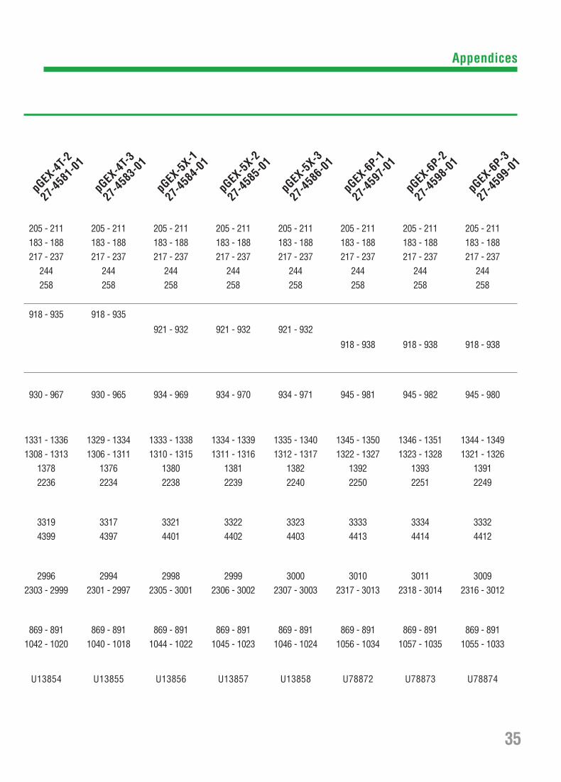
Appendices
35
205 - 211 205 - 211 205 - 211 205 - 211 205 - 211 205 - 211 205 - 211 205 - 211183 - 188 183 - 188 183 - 188 183 - 188 183 - 188 183 - 188 183 - 188 183 - 188217 - 237 217 - 237 217 - 237 217 - 237 217 - 237 217 - 237 217 - 237 217 - 237
244 244 244 244 244 244 244 244258 258 258 258 258 258 258 258
918 - 935 918 - 935
921 - 932 921 - 932 921 - 932
918 - 938 918 - 938 918 - 938
930 - 967 930 - 965 934 - 969 934 - 970 934 - 971 945 - 981 945 - 982 945 - 980
1331 - 1336 1329 - 1334 1333 - 1338 1334 - 1339 1335 - 1340 1345 - 1350 1346 - 1351 1344 - 13491308 - 1313 1306 - 1311 1310 - 1315 1311 - 1316 1312 - 1317 1322 - 1327 1323 - 1328 1321 - 1326
1378 1376 1380 1381 1382 1392 1393 13912236 2234 2238 2239 2240 2250 2251 2249
3319 3317 3321 3322 3323 3333 3334 33324399 4397 4401 4402 4403 4413 4414 4412
2996 2994 2998 2999 3000 3010 3011 30092303 - 2999 2301 - 2997 2305 - 3001 2306 - 3002 2307 - 3003 2317 - 3013 2318 - 3014 2316 - 3012
869 - 891 869 - 891 869 - 891 869 - 891 869 - 891 869 - 891 869 - 891 869 - 8911042 - 1020 1040 - 1018 1044 - 1022 1045 - 1023 1046 - 1024 1056 - 1034 1057 - 1035 1055 - 1033
U13854 U13855 U13856 U13857 U13858 U78872 U78873 U78874
pGEX
-4T-2
27-4
581-
01
pGEX
-4T-3
27-4
583-
01
pGEX
-5X-1
27-4
584-
01
pGEX
-5X-2
27-4
585-
01
pGEX
-5X-3
27-4
586-
01
pGEX
-6P-2
27-4
598-
01
pGEX
-6P-1
27-4
597-
01
pGEX
-6P-3
27-4
599-
01

References
References1. Smith, D. B. and Johnson, K. S., Gene 67, 31 (1988).
2. Parker, M. W. et al., J. Mol. Biol. 213, 221 (1990).
3. Ji, X. et al., Biochemistry 31, 10169 (1992).
4. Maru, Y. et al., J. Biol. Chem. 271, 15353 (1996).
5. McTigue, M. A. et al., J. A., J. Mol. Biol. 246, 21(1995).
6. Toye, B. et al., Infect. Immun. 58, 3909 (1990).
7. Fikrig, E. et al., Science 250, 553 (1990).
8. Johnson, K. S. et al., Nature 338, 585 (1989).
9. Kaelin, W. G. et al., Cell 64, 521 (1991).
10. Kaelin, W. G. et al., Cell 65, 1073 (1991).
11. Kaelin, W. G. et al., Cell 70, 351 (1992).
12. Gearing, D. P. et al., Bio/Technology 7, 1157 (1989).
13. Walker, P. A. et al., Bio/Technology 12, 601 (1994).
14. Cordingley, M. G. et al., J. Biol. Chem. 265, 9062(1990).
15. Habig, W. H. et al., J. Biol. Chem. 249, 7130 (1974).
16. Mannervik, B. and Danielson, U. H., CRC Crit. Rev.Biochem. 23, 283 (1988).
17. Chung, C. T. et al., Proc. Natl. Acad. Sci. USA 86, 2172(1989).
18. Sambrook, J., Fritsch, E. F. and Maniatis, T., MolecularCloning, A Laboratory Manual, Second Edition, ColdSpring Harbor Laboratory Press, Cold Spring Harbor(1989).
19. Smith, D. B. and Corcoran, L. M., in Current Protocolsin Molecular Biology Vol. 2 (Ausubel, F. M. et al. eds.),John Wiley & Sons, New York, p. 16.7.1 (1990).
20. Towbin, H. H. et al., Proc. Natl. Acad. Sci. USA 76,4350 (1979).
21. Tsang, V. C. W. et al., Methods Enzymol. 92, 377(1983).
22. PhastSystem Manual, Pharmacia Biotech.
23. Schein, C. H., Bio/Technology 7, 1141 (1989).
24. Schein, C. H. and Noteborn, M. H. M., Bio/Technology6, 291 (1988).
25. Schein, C. H., personal communication.
26. Schein, C. H., Bio/Technology 8, 308 (1990).
27. Marston, F. A. O., Biochem J. 240, 1 (1986).
28. Gentry, D. R. and Burgess, R. R., Protein ExpressionPurification 1, 81 (1990).
29. Frangioni, J. V. and Neel, B. G., Anal. Biochem. 210,179 (1993).
30. Kelley, R. F. and Winkler, M. E., in Genetic EngineeringVol. 12 (Setlow, J. K., ed.), Plenum Press, New York, pp.1-19 (1990).
31. Pigiet, V. P. and Schuster, B. J., Proc. Natl. Acad. Sci.USA 83, 7643 (1986).
32. Kaelin, W., personal communication.
33. Mintz, G. R., BioPharm 6, No. 2, 34 (1993).
34. Buchberger, A. et al., J. Mol. Biol. 261, 328 (1996).
35. Buchberger, A. et al., EMBO J. 13, 1687 (1994).
36. Zylicz, M. et al., J. Biol. Chem. 260, 7591 (1985).
37. Schönfeld, H.-J. et al., J. Biol. Chem. 270, 2183 (1995).
38. Ellis, R. J. and van der Vies, S. M., Ann. Rev. Biochem.60, 321 (1991).
39. Jackson, J. G. et al., Biochemistry 32, 2554 (1993).
40. Thain, A. et al., Trends in Genetics 12, 209 (1996).
41. Yu-Sherman, M. and Goldberg, A. L., EMBO J. 11, 71(1992).
42. Myers, M., BIOSCI posting (7 July 1993).
43. Zylicz, M. and Georgopoulos, C., J. Biol. Chem. 259,8820 (1984).
44. Chang, J-Y., Eur. J. Biochem. 151, 217 (1985).
45. Fujikawa, K. et al., Biochem. 11, 4892 (1972).
46. Nagai, K. and Thøgersen, H. C., Nature 309, 810(1984).
47. Lauritzen, C. et al., Protein Expression Purification 2,372 (1991).
48. Knudsen, C. R. et al., Biochem. International 28, 353(1992).
49. Ellinger, S. et al., Virology 180, 811 (1991).
50. Nagai, K. and Thøgersen, H. C., Methods Enzymol.153, 461 (1987).
51. Gallagher, S. R., in Current Protocols in MolecularBiology Vol. 2 (Ausubel, F. M. et al. eds.), John Wiley &Sons, New York, p. 10.2.2 (1995).
52. Studier, F. W. and Moffatt, B. A., J. Mol. Biol. 189, 113(1986).
53. Grodberg, J. and Dunn, J. J., J. Bact. 170, 1245 (1988).
54. Walker, J. et al., Molec. Biochem. Parasitol. 61, 255(1993).
36

Notes
37
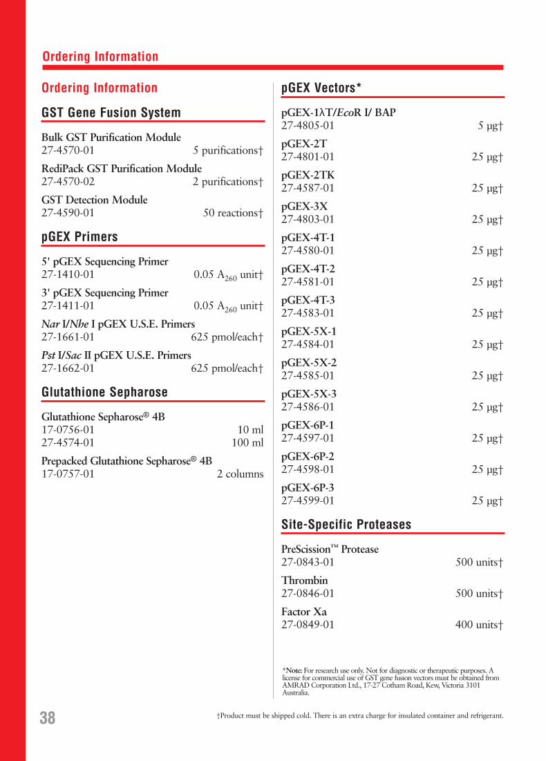
Ordering Information
Ordering Information
GST Gene Fusion System
Bulk GST Purification Module 27-4570-01 5 purifications†
RediPack GST Purification Module27-4570-02 2 purifications†
GST Detection Module27-4590-01 50 reactions†
pGEX Primers
5' pGEX Sequencing Primer27-1410-01 0.05 A260 unit†
3' pGEX Sequencing Primer27-1411-01 0.05 A260 unit†
Nar I/Nhe I pGEX U.S.E. Primers27-1661-01 625 pmol/each†
Pst I/Sac II pGEX U.S.E. Primers27-1662-01 625 pmol/each†
Glutathione Sepharose
Glutathione Sepharose® 4B17-0756-01 10 ml27-4574-01 100 ml
Prepacked Glutathione Sepharose® 4B17-0757-01 2 columns
pGEX Vectors*
pGEX-1λT/EcoR I/ BAP27-4805-01 5 µg†
pGEX-2T27-4801-01 25 µg†
pGEX-2TK27-4587-01 25 µg†
pGEX-3X27-4803-01 25 µg†
pGEX-4T-127-4580-01 25 µg†
pGEX-4T-227-4581-01 25 µg†
pGEX-4T-327-4583-01 25 µg†
pGEX-5X-127-4584-01 25 µg†
pGEX-5X-227-4585-01 25 µg†
pGEX-5X-327-4586-01 25 µg†
pGEX-6P-127-4597-01 25 µg†
pGEX-6P-227-4598-01 25 µg†
pGEX-6P-327-4599-01 25 µg†
Site-Specific Proteases
PreScission™ Protease27-0843-01 500 units†
Thrombin27-0846-01 500 units†
Factor Xa27-0849-01 400 units†
38 †Product must be shipped cold. There is an extra charge for insulated container and refrigerant.
*Note: For research use only. Not for diagnostic or therapeutic purposes. Alicense for commercial use of GST gene fusion vectors must be obtained fromAMRAD Corporation Ltd., 17-27 Cotham Road, Kew, Victoria 3101Australia.

Ordering Information
Companion Products
Adenosine 5'-Triphosphate, 100 mM Solution (ATP)27-2056-01 25 µmol†
Alkaline Phosphatase (Calf Intestinal Mucosa)27-0620-01 500 units†
Anti-GST Antibody27-4577-01 5 ml†
CNBr-activated Sepharose 4B17-0430-01 15 g
E. coli BL2127-1542-01 1 vial
FlexiPrep Kit27-9281-01 1 kit†
GFX™ PCR DNA and Gel Band Purification Kit27-9602-01 100 purifications
Isopropyl ββ-D-Thiogalactoside (IPTG)27-3054-03 1 g27-3054-04 5 g27-3054-05 10 g
λ ExCell EcoR I/CIP27-5011-01 10 µg†
λ ExCell Not I/EcoR I/CIP27-5013-01 10 µg†
LMW Electrophoresis Calibration Kit17-0446-01 200 µg/vial†
MicroPlex® 2427-3564-01 1 set
PhastGel® Homogeneous 12.517-0623-01 10/pk
PhastSystem®
18-1018-23 1 units
PhastTransfer®
18-1001-23 1 kit
Ready-To-Go® PCR Beads (0.2 ml tubes/plate)27-9553-01 96 reactions27-9553-02 5 x 96 reactions
Ready-To-Go® PCR Beads (0.5 ml tubes)27-9555-01 100 reactions
Ready-To-Go® T4 DNA Ligase27-0361-01 50 reactions
rTaq DNA Polymerase27-0798-01 250 units†27-0798-02 4 x 250 units†27-0798-03 10 x 250 units†
Sephaglas™ BandPrep Kit27-9285-01 100 average purifications
Taq DNA Polymerase (Thermus aquaticus)27-0799-01 250 units†27-0799-02 4 x 250 units†27-0799-03 10 x 250 units†
T4 DNA Ligase, FPLCpure®, Cloned27-0870-03 200 units27-0870-04 1000 units
U.S.E. Mutagenesis Kit (25 reactions)27-1699-01 1 kit
39†Product must be shipped cold. There is an extra charge for insulated container and refrigerant.
Licensing Information
All materials for research use only. Not for diagnostic or therapeutic purposes.
A license for commercial use of pGEX vectors must be obtained fromAMRAD Corporation Ltd., 17-27 Cotham Road, Kew, Victoria 3101,Australia.
PreScission Protease is licensed from the National University of Singapore,10 Kent Ridge Crescent, Singapore 0511.
PreScission Protease is produced under commercial license from AMRADCorporation Ltd., 17-27 Cotham Road, Kew, Victoria 3101, Australia.
PCR (polymerase chain reaction) is covered by U.S. Patents 4,683,195 and4,683,202 owned by Hoffman-LaRoche Inc. Use of the PCR processrequires a license.
Purchase of Ready-To-Go® PCR Beads is accompanied by a limited licenseto use it in the Polymerase Chain Reaction (PCR) process solely for theresearch and development activities of the purchaser in conjunction with a thermal cycler whose use in the automated performance of the PCRprocess is covered by the up-front license fee, either by payment to Perkin-Elmer or as purchased, i.e. an authorized thermal cycler.
Trademarks
The following designations are trademarks owned by Pharmacia Biotech AB:FPLC director®, FPLCpure®, GFX™, MicroPlex®, Mighty Small™,Pharmacia®, PhastGel®, PhastSystem®, PhastTransfer®, PreScission™,Ready-To-Go®, Sephaglas™, Sepharose®, and TransPhor™.
Falcon® is a registered trademark of Becton Dickinson Company.
GenBank® is a registered trademark of the National Institutes of Health.
Gene Pulser® is a registered trademark of Bio-Rad Laboratories.
Lambda ZAP® is a registered trademark of Stratagene Cloning Systems.
MicroSpin™ is a trademark of Lida Manufacturing Corp.
Pefabloc® is a registered trademark of Pentapharm AG.
Triton® is a registered trademark of Rohm & Haas Company.
Tween® is a registered trademark of ICI Americas, Inc.
Whatman® is a registered trademark of Whatman Paper Ltd.