group effort in toxin synthesis
TRANSCRIPT
bubbles had dispersed them, because the linear distance they travel would be muchsmaller.
But there is one major problem with anybottom-up theory of magnetic-field forma-tion, which has to do with the existence ofpositive and negative fields. Because fielddirections in different fossils vary randomly,whereas positive and negative fields withineach magnetized bubble presumably canceleach other out, opposing fields could bedragged into proximity and annihilate oneanother, leaving behind almost nothing. Butthe overall rate of field destruction is difficultto estimate — the true picture could rangefrom the survival of isolated bubbles, to partial mergers between magnetic systems in which the fields wander over vast regions of
space, to the nearly field-free situation inwhich total cancellation has occurred. In anyevent, active galaxies continue to spew outmagnetic fields into the intergalactic medi-um, and astronomers continue to extend theirobservations and models, so one day we maybe able to confirm the bottom-up picture. ■
Ellen G. Zweibel is at JILA, University of Colorado,Boulder, Colorado 80309, USA.e-mail: [email protected]. Zweibel, E. G. & Heiles, C. Nature 385, 131–136 (1997).
2. Furlanetto, S. R. & Loeb, A. Astrophys. J. 556, 619–634
(2001).
3. Kronberg, P. P., Dufton, Q. W., Li, H. & Colgate, S. A.
Astrophys. J. 560, 178–186 (2001).
4. Rees, M. J. & Setti, G. Nature 219, 127–128 (1968).
5. Begelman, M. C., Blandford, R. D. & Rees, M. J. Rev. Mod. Phys.
56, 255–351 (1984).
6. Colgate, S. A. & Li, H. Astrophys. Space Sci. 264, 357 (1999).
7. Hawley, J. Astrophys. J. 528, 462–479 (2000).
regulators that control bacterial responses to a variety of environmental signals5. In asecond type of pathway, the peptide signal is imported into the responder cell, where its interaction with an intracellular recep-tor generates the response (Fig. 1b). Thedetails are not known, but it seems that theimported peptide can act in various ways inthe target cell4.
The results of Haas et al.1 are based onanalysis of a toxin called cytolysin, which thisgroup has studied for several years6–8. Thetoxin has two components, LL and LS. Whenreleased from the cell, these components act in concert to attack a variety of bacterialand mammalian target cells. But before theyare released, both are processed by a varietyof accessory components.
A cluster of at least half-a-dozen genes isrequired to produce cytolysin and to protectthe enterococcal cell against its effects. Thecluster is generally encoded by accessorymobile genetic elements, in this case a plas-mid called pAD1 (refs 9, 10). Interestingly,expression of the pAD1 transfer genes is regulated by donor–recipient cell signallingmediated by the peptide cAD1 (ref. 8). Pro-duction of cytolysin increases enterococcalvirulence in several experimental systems,and cytolysin is commonly, but not univer-sally, found in enterococcal strains isolatedfrom hospital patients11,12.
Haas et al.1 analysed a pair of genes, cylR1and cylR2, that are located next to the genecluster responsible for cytolysin biosynthesisbut are transcribed separately. Using a care-fully constructed series of gene knockouts,and fusions between ‘reporters’ of gene transcription, they found that the cylR1 andcylR2 gene products repress expression of thebiosynthetic genes. During growth in cul-ture, this repression is overcome in a density-dependent fashion by a secreted bacterialproduct, which the authors identified as thecylS-encoded component (LS) of the toxin.
Computer analysis1 of the predictedamino-acid sequences of the cylR1 and cylR2 products was particularly illuminating. The first product is predicted to be a smalltransmembrane protein; the second, a cyto-plasmic protein with a DNA-binding motif.Neither molecule has much resemblance tothe proteins of the two-component family ofsignal transducers, or to any other proteinsin the databases. The authors propose that,in the default state of the system, interactionbetween a cytoplasmic segment of R1 and R2 holds R2 in a repressing conformationand biosynthesis occurs only at a low level. Ascell density reaches about 107 cells per milli-litre, the concentration of LS in the mediumbecomes sufficient to cause induction ofcytolysin biosynthetic genes, presumably by direct interaction with an extracellulardomain of R1 and a conformational shiftwhich disrupts the R1–R2 interaction(Fig. 1c). These molecular interactions have
news and views
NATURE | VOL 415 | 3 JANUARY 2002 | www.nature.com 33
Hospital patients with diseases thatimpair their natural defences are sus-ceptible to opportunistic infections.
This is a serious medical problem. Opportu-nistic bacteria, such as Enterococcus faecalis,are often resistant to antimicrobial drugs.And because these bacteria do not generallycause disease in healthy people, little isknown about why they infect and cause disease in hospital patients. On page 84 of this issue1, Haas et al. reveal a previouslyunknown way in which enterococcal cellscommunicate with one another to coordi-nate toxin production. The results shed lighton bacterial cell–cell signalling mechanisms,and may also provide insights into entero-coccal pathogenicity and open up newapproaches to drug discovery.
In the past, microbial life was largelyviewed as individual organisms single-mindedly pursuing growth by cell division.But it is now clear that many microbial activities — including genetic exchange,growth in biofilms, expression of virulencefactors (toxins) and antibiotic production —result from well-coordinated multicellularprocesses controlled by cell–cell signalling2.This applies to both of the two main groups ofbacteria, Gram-negative and Gram-positive(this division is made on the basis of how theircell walls absorb a specific stain).
In Gram-negative bacteria — Pseudo-monas, Agrobacterium and Vibrio, forinstance — the best-studied signal moleculesare acylhomoserine lactones that diffuse intothe bacterial cell and interact with a gene-transcription factor to increase synthesis of
messenger RNA from the relevant targetgenes. Lactone synthesis occurs at a low levelin bacterial cultures, but when bacteria aregrowing exponentially lactone concentrationincreases with cell density. When a sufficientconcentration is reached, the target genes(often including the synthetase gene for acyl-homoserine lactone) are transcribed at highlevels, causing a dramatic ‘auto-induction’ of the same activity throughout the popula-tion. Such cell-density-dependent microbialbehaviour, mediated by signal molecules, iscalled ‘quorum sensing’3.
Gram-positive bacteria, such as strepto-cocci, bacilli, staphylococci and enterococci,also carry out quorum sensing, as well asrelated forms of cell–cell communicationbetween different cell types (for instance,DNA transfer between donor and recipientcells). In these organisms, however, the signal molecules are peptides of about5–20 amino acids in length4. To date, twotypes of signalling pathway have been found.
In most cases, processing of the peptidesignal involves a membrane sensor protein,whose interaction with the signal moleculeresults in autophosphorylation of a histidineresidue in the protein (Fig. 1a, overleaf). Thephosphorylated sensor (one component)communicates with a response-regulatorprotein (a second component) in the cyto-plasm, transferring the phosphate to the regulator and converting it to an active transcription factor that interacts with targetpromoters to activate gene transcription.These two-component signal-transductionsystems are members of a large family of
Microbiology
Group effort in toxin synthesisGary M. Dunny
It is increasingly evident that bacterial cells cooperate for many purposes.New results show that the bacterium Enterococcus uses cell–cell signallingto coordinate toxin production.
© 2002 Macmillan Magazines Ltd

not been demonstrated directly, but they areconsistent with the data and can be testedexperimentally.
These results will affect our view of cell–cell communication in bacteria, and how wedeal with the Enterococcusas an opportunisticpathogen. Clearly, peptide-mediated cell–cellsignalling in bacteria can occur by any of at least three distinct mechanisms (Fig. 1). A search of the nearly completed E. faecalisgenome sequence13 suggests that there may be a dozen or more two-component signal-transduction systems, some of which couldmediate cell–cell communication. In otherwords, in E. faecalis alone, many additionalprocesses could be regulated in this fashion.
The new results1 could also help indeveloping drugs to combat enterococcalinfections. In some infections, productionof cytolysin or other virulence factors (regu-lated, perhaps, by similar mechanisms) maybe required to cause disease but may not benecessary for the organism’s survival andgrowth in its normal habitat, the intestine.A drug that targeted the signal pathway to block virulence could prevent or cureinfection. But because the bacteria would
not be directly killed, the drug would notexert a strong selective pressure for devel-opment of drug resistance — one of thebanes of modern medicine. ■
Gary M. Dunny is in the Department ofMicrobiology, University of Minnesota MedicalSchool, 428 Delaware Street, SE, Minneapolis,Minnesota 55455-0312, USA.e-mail: [email protected]. Haas, W., Shepard, B. D. & Gilmore, M. S. Nature 415, 84–87
(2002).
2. Dunny, G. M. & Winans, S. C. Cell–Cell Signaling in Bacteria
(Am. Soc. Microbiol., Washington, DC, 1999).
3. Fuqua, W. C., Winans, S. C. & Greenberg, E. P. J. Bacteriol.
176, 269–275 (1994).
4. Dunny, G. M. & Leonard, B. A. B. Annu. Rev. Microbiol.
51, 527–564 (1997).
5. Hoch, J. A. & Silhavy, T. J. (eds) Two-Component Signal
Transduction (Am. Soc. Microbiol., Washington, DC, 1995).
6. Ike, Y., Clewell, D. B., Segarra, R. A. & Gilmore, M. S.
J. Bacteriol. 172, 155–163 (1990).
7. Gilmore, M. S., Segarra, R. A. & Booth, M. C. Infect. Immunol.
58, 3914–3923 (1990).
8. Segarra, R. A., Booth, M. C., Morales, D. A., Huycke, M. M. &
Gilmore, M. S. Infect. Immunol. 59, 1239–1246 (1991).
9. Clewell, D. B. et al. J. Bacteriol. 152, 1220–1230 (1982).
10.Gilmore, M. S. et al. J. Bacteriol. 176, 7335–7344 (1994).
11.Huycke, M. M. & Gilmore, M. S. Plasmid 34, 152–156
(1995).
12.Coque, T. M., Patterson, J. E., Steckelberg, J. M. & Murray, B. E.
J. Infect. Dis. 171, 1223–1229 (1995).
13.http//www.tigr.org
news and views
34 NATURE | VOL 415 | 3 JANUARY 2002 | www.nature.com
Daedalus
Atmospheric chargeLightning, says Daedalus, is universal. Not only are there about a thousandthunderstorms going on around the Earthat any one time, but lightning has beenobserved in the atmospheres of otherplanets, with different chemistry fromthat of Earth. The complex organicmolecules built up by earthly lightning,precursors of life itself, should thereforeexist on other planets. They may havegiven life a start there too.
Daedalus can think of two bases forlightning. First, phase changes, like that of water going to ice, must extrude charge. No solution or melt can have thesame electron affinity as the solid, so asolidifying droplet must push chargeahead of the solid front. Second, anycharged gas must expand, by self-repulsion. A big enough voltage (150megavolts for air) would give a vacuum;smaller charges, of just a few megavolts,would merely provide strong lift. Sosolidification of any sort, coupled withcharge sharing to an atmosphere, mustgive rise to convection. Couple this with afeedback mechanism, such as the return of charge in falling raindrops that partlyfreeze, and you soon build up enoughcharge separation for lightning.
To test these ideas, DREADCOphysicists are building a large-scalelightning machine. They are starting with a hollow cooling tower. At the top, waterwill be sprayed down it from perforatedpipes. It will lose heat and should partlyfreeze. The charge thus generated will beexpelled into the remaining liquid. Thisshould pass on charge to the surroundingair, which will expand and rise in its turn.At the top of the tower, copper electrodeswill capture charge from the rising air andreturn it to the falling drops. In this waythe tower will build a charge separation. It may also be done with brine. Brine is a conductor, but its freezing expels solid,insulating salt. Again, the physicists willlook for the accumulation of charge. Anysurplus high-voltage electricity from thetower will be bled from its top for use.
Sadly, Daedalus’s tower may notgenerate much power. Even a full-scalethunderstorm only creates some 20gigawatts. But he hopes to identify theaeroelectric processes that are importantin lightning, on Earth or on otherplanets. The expansion of charged gas,and its resulting speed of uplift, are wellworth knowing; so is the feedback ofcharge by falling drops. Both may help us to understand the biochemistry ofother planets. David Jones
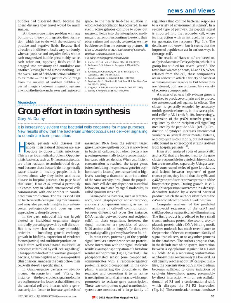
Figure 1 Three mechanisms of cell–cell communication between bacteria. The signalling moleculesare peptides, 5–25 amino acids in length, but are different for each pathway. a, A two-componentsignal-transduction system, in which binding of the peptide to a histidine kinase (HK) receptor onthe cell membrane results in autophosphorylation (~P) of the kinase. The phosphate is transferred to a response regulator (RR), which can then bind to target DNA and activate a gene promoter andtranscription. b, A binding protein (BP) on the cell membrane takes the peptide signal from theenvironment and uses an oligopeptide permease (OPP) to import the peptide into the cell. Peptidebinding to a cytoplasmic receptor (CR) causes a conformational shift and inhibits receptor function(some CRs negatively regulate transcription; others act as phosphatases, with phosphorylatedregulatory proteins as substrates). c, The pathway described by Haas et al.1. In the absence of thepeptide signal (in this case, the LS component of the secreted cytolysin toxin), two proteins, R1 andR2, appear to act in concert to repress the cytolysin biosynthetic genes. Peptide-signal binding to R1is predicted to disrupt the repressive R1–R2 conformation and result in increased gene transcription.Squiggly arrows indicate the effects of interactions; other arrows indicate direct molecularinteractions. Bacterial activities controlled by each pathway are listed in the box.
RR
ATP
ATP ATPADP
Conformationalshift; CR inhibited
DNA binding;promoter activation
Release of R2 frompromoter; derepression
ADP
ADP
OPP OPP
OPPOPP
HK R1
R2~P
CR
BP
a b c
Peptide signal
Cellular response
a Activates genetic transformation (streptococci, bacilli) Activates virulence gene expression (staphylococci) Activates bacteriocin production (lactic acid bacteria)b Activates conjugation (enterococci) Regulates transformation and sporulation (bacilli)c Activates toxin production (enterococci)
© 2002 Macmillan Magazines Ltd