goosecoid and the organizer -...
TRANSCRIPT
Development 1992 Supplement, 167-171 (1992)Printed in Great Britain © The Company of Biologists Limited 1992
167
goosecoid and the organizer
EDDY M. DE ROBERTIS, MARTIN BLUM, CHRISTOF NIEHRS and HERBERT STEINBEISSER
Molecular Biology Institute and Department of Biological Chemistry, University of California, Los Angeles, CA 90024-1737, USA
Summary
The molecular nature of Spemann's organizer phenom-enon has long attracted the attention of embryologists.goosecoid is a homeobox gene with a DNA-binding speci-ficity similar to that of Drosophila bicoid. Xenopusgoosecoid is expressed on the dorsal side of the embryobefore the dorsal lip is formed. Cells expressing goose-coid are fated to become pharyngeal endoderm, headmesoderm and notochord. Transplantation of goosecoidmRNA to the ventral side of Xenopus embryos bymicroinjection mimics the properties of Spemann'sorganizer, leading to the formation of twinned bodyaxes, goosecoid is activated by dorsal inducers and not
affected by ventral inducers. In the mouse, goosecoid isexpressed in the anterior tip of the primitive streak. Theavailability of two early markers, goosecoid andBrachyury, opens the way for the comparative analysisof the vertebrate gastrula. The results suggest that thegoosecoid homeodomain protein is an integral compo-nent of the biochemical pathway leading to Spemann'sorganizer phenomenon.
Key words: goosecoid, Spemann's organizer, gastrulation,Drosophila, Xenopus.
Introduction
It is now generally agreed that development results from aseries of cell-cell interaction events. The experiment thatcontributed more than any other to this view was that ofHans Spemann and Hilde Mangold (1924), showing that asmall fragment of the gastrula, the dorsal lip, had the abil-ity, after transplantation, to form a twinned body axis in theopposite (ventral) side of a host embryo. The transplantedtissue contributed only a small part of this secondary axis,and thus was able to recruit, or organize, cells into com-plex anatomical structures. The quest to understand theinductive properties of the organizer still provides greatimpetus to experimental embryologists (reviewed by Spe-mann 1938; Nakamura and Toivonen, 1978; Hamburger,1988; Marx, 1991).
In the present paper, we will discuss how microinjectionof purified goosecoid homeobox-containing mRNA canmimic most of the properties of Spemann's organizer.
Homeobox genes expressed in the organizer
Homeobox genes encode DNA-binding proteins, which fre-quently are involved in the specification of positional infor-mation in the embryo (reviewed by Gehring 1987; Kesseland Gruss, 1990; De Robertis et al., 1991; McGinnis andKrumlauf, 1992). It is therefore of interest that several suchgenes are expressed in the dorsal lip region of the Xenopusembryo. By screening a cDNA library derived from man-ually dissected Xenopus dorsal lips, four different home-obox genes were isolated by Blumberg et al. (1991). Three
were related to genes isolated previously (X. caudal 1 and2, X. labial), but the most abundant clone (23 out of 30 iso-lates) contained a novel type of homeobox. Because itshomeobox contained similarities to the Drosophila genesgooseberry in the initial Vs of the homeodomain and tobicoid in the DNA recognition helix, this gene was chris-tened goosecoid. Additional genes expressed in the dorsallip have been isolated by other groups from Xenopusembryo cDNA libraries by screening with homologousprobes. X-UM-J contains a homeobox as well as a con-served cysteine-rich domain (Taira et al., 1992), and whileXFKH-1 lacks a homeobox, it has sequence similarities tothe DNA-binding domain of the Drosophila fork-headhomeotic gene (Dirksen and Jamrich, 1992).
The expression of goosecoid in the Xenopusgastrula
All the genes mentioned above are surely important inbuilding the body axis, but we will deal here only withgoosecoid, which is particularly interesting because it hassome degree of functional similarity to the Drosophila ante-rior morphogen bicoid (Niisslein-Volhard, 1991). In an invitro assay a goosecoid recombinant protein was shown tobind target sequences with a DNA-binding specificity sim-ilar to that of Drosophila bicoid on target sequences derivedfrom the promoter of the gap gene hunchback (Blumberget al., 1991). In addition, goosecoid is expressed very earlyin Xenopus embryos. As can be seen in Fig. IB, goosecoidexpression is detectable by whole-mount in situ hybridiz-ation as a crescent on the dorsal marginal zone of the
168 E. M. De Robertis and others
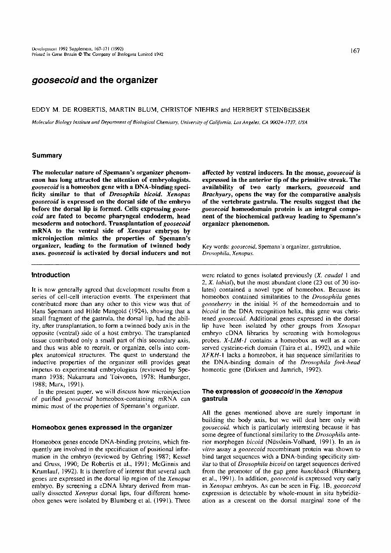
Fig. 1. Time course of goosecoid expression in Xenopus embryos. Whole-mount in situ hybridization (Harland, 1991) at (A) stage 8. midblastula; (B) stage 9. late blastula one hour before the onset of gastrulation; (C) stage 10. early gastrula in which ihe dorsal lip is formed;(D) stage 11. mid gastrula with a circular blastopore. Note that the patch of goosecoid expression in the marginal zone is present beforegastrulation starts (B), and that it invaginates into the interior of the embryo during gastrulation (D). Unfortunately the black and whitereproduction of this figure, which was originally in color, does not have enough contrast.
embryo at least one hour before the start of gastrulation. Atthe start of gastrulation (Fig. IC) goosecoid is most intensein an arc of about 60° just above the dorsal lip (Cho et al.,1991). This corresponds to the region where Spemann'sorganizer, as defined by transplantation experiments, islocated (Cooke, 1972; Gerhart et al., 1989).
In sagittal sections, goosecoid is seen to be expressed inthe internal layer of the dorsal lip region. As shown in Fig.2, cells close to the incipient indentation of the dorsal lip(indicated by an arrow) contain mostly nuclear transcripts,while cells located further away from the dorsal lip containincreasing amounts of cytoplasmic goosecoid RNA. Thecells that express goosecoid correspond to the dorsal-mostinvaginating cells. Their normal fate is to become pharyn-geal endoderm, head endoderm and notochord (Keller,1976. 1991).
Mlcroinjection of goosecoid mRNA gives rise to atwinned body axis
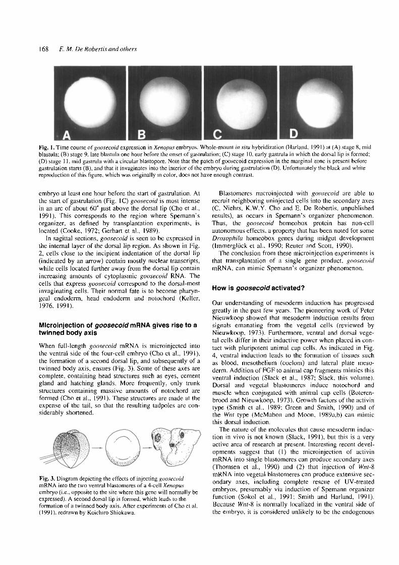
When full-length goosecoid mRNA is microinjected intothe ventral side of the four-cell embryo (Cho et al., 1991),the formation of a second dorsal lip, and subsequently of atwinned body axis, ensues (Fig. 3). Some of these axes arecomplete, containing head structures such as eyes, cementgland and hatching glands. More frequently, only trunkstructures containing massive amounts of notochord areformed (Cho et al., 1991). These structures are made at theexpense of the tail, so that the resulting tadpoles are con-siderably shortened.
Fig. 3. Diagram depicting the effects of injecting goosecoidmRNA into the two ventral blastomeres of a 4-cell Xenopusembryo (i.e.. opposite to the site where this gene will normally beexpressed). A second dorsal lip is formed, which leads to theformation of a twinned body axis. After experiments of Cho et al.(1991), redrawn by Koichiro Shiokawa.
Blastomeres microinjected with goosecoid are able torecruit neighboring uninjected cells into the secondary axes(C. Niehrs, K.W.Y. Cho and E. De Robertis, unpublishedresults), as occurs in Spemann's organizer phenomenon.Thus, the goosecoid homeobox protein has non-cellautonomous effects, a property that has been noted for someDrosophila homeobox genes during midgut development(Immergluck et al., 1990; Reuler and Scott, 1990).
The conclusion from these microinjection experiments isthat transplantation of a single gene product, goosecoidmRNA, can mimic Spemann's organizer phenomenon.
How is goosecoid activated?
Our understanding of mesoderm induction has progressedgreatly in the past few years. The pioneering work of PeterNieuwkoop showed that mesoderm induction results fromsignals emanating from the vegetal cells (reviewed byNieuwkoop, 1973). Furthermore, ventral and dorsal vege-tal cells differ in their inductive power when placed in con-tact with pluripotent animal cap cells. As indicated in Fig.4, ventral induction leads to the formation of tissues suchas blood, mesothelium (coelom) and lateral plate meso-derm. Addition of FGF to animal cap fragments mimics thisventral induction (Slack et al., 1987; Slack, this volume).Dorsal and vegetal blastomeres induce notochord andmuscle when conjugated with animal cap cells (Boteren-brood and Nieuwkoop, 1973). Growth factors of the activintype (Smith et al., 1989: Green and Smith, 1990) and ofthe Wnt type (McMahon and Moon, 1989a,b) can mimicthis dorsal induction.
The nature of the molecules that cause mesoderm induc-tion in vivo is not known (Slack, 1991), but this is a veryactive area of research at present. Interesting recent devel-opments suggest that (1) the microinjection of activinmRNA into single blastomeres can produce secondary axes(Thomsen et al., 1990) and (2) that injection of Wnt-8mRNA into vegetal blastomeres can produce extensive sec-ondary axes, including complete rescue of UV-treatedembryos, presumably via induction of Spemann organizerfunction (Sokol et al., 1991; Smith and Harland, 1991).Because Wnt-8 is nonnally localized in the ventral side ofthe embryo, it is considered unlikely to be the endogenous
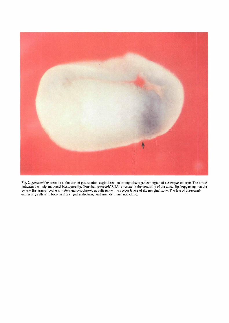
Fig. 2. goosecoid expression at the start of gastrulation, sagittal section through the organizer region of a Xenopus embryo. The arrowindicates the incipient dorsal blastopore lip. Note that goosecoid RNA is nuclear in the proximity of the dorsal lip (suggesting that thegene is first transcribed at this site) and cytoplasmic as cells move into deeper layers of the marginal zone. The fate of goosecoid-expressing cells is to become pharyngeal endoderm, head mesoderm and notochord.
Role of goosecoid in gastrulation 169
Ventral
Metodsrm
Dorsal
Mesoderm
Blood
Coelom
Lateral Plate
Kidney
(muscle)
Notochord
Muscle
F6F
Act I v in
Wnt
Fig. 4. Mesoderm induction can be of a ventral or dorsal nature.Explanted animal caps conjugated and cultured with either ventralor dorsal vegetal blastomeres form different types of mesoderm(after experiments by Boterenbrood and Nieuwkoop, 1973). Theseinductions can be mimicked by treating animal cap cells with FGF(ventral mesoderm) or with activin or Wnt (dorsal signals).
dorsal inducer, but a similar substance, as yet uncloned,could be secreted by dorsal and vegetal blastomeres.
The current model, summarized in Fig. 5, is thereforethat there exists a radial signal (or signals) that induces aring of cells in the overlying marginal zone to become ven-tral mesoderm. On the dorsal side, an additional signal (orsignals) is released from the vegetal cells (Boterenbroodand Nieuwkoop, 1973; Gimlich and Gerhart, 1984), origi-nating in a region that has been designated the Nieuwkoopcenter (Gerhart et al., 1989). The signal from theNieuwkoop center acts upon the overlying marginal zonecells and induces Spemann's organizer tissue, goosecoid isexpressed in the latter region.
As expected, goosecoid is induced by activin but not by
SPEMANN'SORGANIZER
VentralSignal
( F 6 F )
Dorsal Signal(Act iv in)(Wnt)
NieuwkoopCenter
Fig. 5. A current view of mesoderm induction in Xenopus (seeGerhart et al., 1989). A ventral signal is released radially by thevegetal cells (e.g., FGF), inducing the entire marginal zone tobecome mesoderm. On the dorsal side an additional signal(perhaps activin- or wnt-Yike) is released by vegetal cells of theNieuwkoop center, inducing Spemann organizer tissue in theoverlying marginal zone cells.
the ventral inducer FGF (Cho et al., 1991). The effect ofWnt-type factors is presently under investigation, goosecoidis a primary response gene to activin induction, i.e., it'stranscripts will accumulate even in the absence of proteinsynthesis. We have previously argued that this result wouldplace goosecoid high up in the hierarchy of genetic eventsleading to axis formation (Cho et al., 1991). This interpre-tation should now be reassessed in view of recent findingsthat a great many genes are primary targets for activin inXenopus animal cap explants. These include Mix-1 (anendoderm-specific homeobox gene isolated by screening foractivin-induced transcripts; Rosa, 1989), Myo D (Rupp andWeintraub, 1991), Brachyury (a gene expressed as a ringin the marginal zone of the Xenopus embryo as well as inthe organizer region; Smith et al. (1991) and known to berequired for notochord and posterior axis formation in themouse, Rasbash et al., 1991), X-UM-1 (Taira et al., 1992)and XFKH1 (Dirksen and Jamrich, 1992). In fact, tran-scripts for many of these genes are present at low levelsalready at the time when the animal cap are excised at themid blastula stage. This applies to Myo D (Rupp and Wein-traub, 1991), X-LIM-J (Taira et al., 1992), goosecoid (Choet al., 1991) and presumably other inducible genes. Lowlevels of goosecoid mRNA are present in the Xenopusunfertilized egg and early cleavage stages (H. Steinbeisser,unpublished observations) but went unnoticed in our initialstudy (Blumberg et al., 1991). These maternal transcriptsare not detectable by whole-mount in situ hybridization(Fig. 1A).
In order to dissect the hierarchy of these genes in axisformation, if indeed one exists, it will be necessary to carryout loss-of-function studies. For example, one would liketo know whether goosecoid is expressed in a Brachyurymutant, and vice versa. Such studies should be possible inthe mouse embryo.
goosecoid in mouse gastrulation
As is well known, all vertebrate embryos appear very sim-ilar to each other at mid-embryogenesis (the so-called phy-lotypic stage of the vertebrate embryo, Wolpert, 1991;Feduccia and McCrady, 1991). On the other hand, embryosfrom the various vertebrate classes appear to differ greatlyat the gastrula stage. For example, in teleosts the main gas-trulation movement is epiboly, in which the embryo properenvelops the yolk mass; in amphibians, which in generalhave holoblastic cleavage, the main morphogenetic move-ment is the invagination of the endomesoderm through thecircular blastopore; while in birds and mammals (anmiotes)the main morphogenetic movement is the delamination ofthe future endodermal and mesodermal cells through thelinear primitive streak. One of the main lessons that wehave learned in the past few years, in particular from theextraordinary conservation of the workings of Hox genes(reviewed by De Robertis et al., 1990; Kessel and Grass,1990; McGinnis and Krumlauf, 1992), is that whileembryogenesis may appear to differ greatly, the molecularmechanisms involved are universal. Southern blot analysisindicates that all classes of vertebrates contain a goosecoidhomologue (Blum et al., 1992). Thus the goosecoid marker
170 E. M. De Robertis and others
B
En
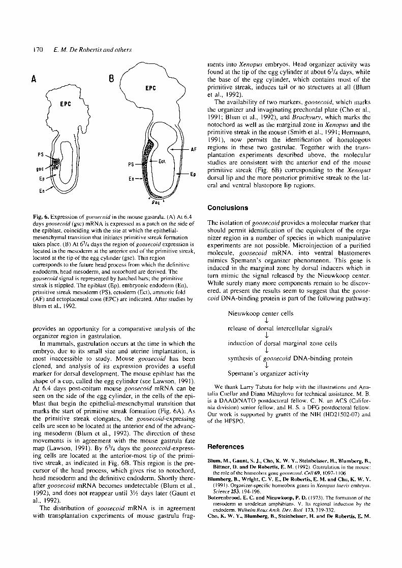
Fig. 6. Expression of goosecoid in the mouse gastrula. (A) At 6.4days goosecoid (gsc) mRNA is expressed as a patch on the side ofthe epiblast, coinciding with the site at which the epithelial-mesenchymal transition that initiates primitive streak formationtakes place. (B) At 63Ai days the region of goosecoid expression islocated in the mesoderm at the anterior end of the primitive streak,located at the tip of the egg cylinder (gsc). This regioncorresponds to the future head process from which the definitiveendoderm. head mesoderm, and notochord are derived. Thegoosecoid signal is represented by hatched bars; the primitivestreak is stippled. The epiblast (Ep), embryonic endoderm (En),primitive streak mesoderm (PS), ectoderm (Ect), amniotic fold(AF) and ectoplacental cone (EPC) are indicated. After studies byBlumetal., 1992.
provides an opportunity for a comparative analysis of theorganizer region in gastrulation.
In mammals, gastrulation occurs at the time in which theembryo, due to its small size and uterine implantation, ismost inaccessible to study. Mouse goosecoid has beencloned, and analysis of its expression provides a usefulmarker for dorsal development. The mouse epiblast has theshape of a cup, called the egg cylinder (see Lawson, 1991).At 6.4 days post-coitum mouse goosecoid mRNA can beseen on the side of the egg cylinder, in the cells of the epi-blast that begin the epithelial-mesenchymal transition thatmarks the start of primitive streak formation (Fig. 6A). Asthe primitive streak elongates, the goosecoid-expressingcells are seen to be located at the anterior end of the advanc-ing mesoderm (Blum et al., 1992). The direction of thesemovements is in agreement with the mouse gastrula fatemap (Lawson, 1991). By 63/4 days the goosecoid-express-ing cells are located at the anterior-most tip of the primi-tive streak, as indicated in Fig. 6B. This region is the pre-cursor of the head process, which gives rise to notochord,head mesoderm and the definitive endoderm. Shortly there-after goosecoid mRNA becomes undetectable (Blum et al.,1992), and does not reappear until 3'A days later (Gaunt etal., 1992).
The distribution of goosecoid mRNA is in agreementwith transplantation experiments of mouse gastrula frag-
ments into Xenopus embryos. Head organizer activity wasfound at the tip of the egg cylinder at about 63/4 days, whilethe base of the egg cylinder, which contains most of theprimitive streak, induces tail or no structures at all (Blumet al., 1992).
The availability of two markers, goosecoid, which marksthe organizer and invaginating prechordal plate (Cho et al.,1991; Blum et al., 1992), and Brachyury, which marks thenotochord as well as the marginal zone in Xenopus and theprimitive streak in the mouse (Smith et al., 1991; Herrmann,1991), now permits the identification of homologousregions in these two gastrulae. Together with the trans-plantation experiments described above, the molecularstudies are consistent with the anterior end of the mouseprimitive streak (Fig. 6B) corresponding to the Xenopusdorsal lip and the more posterior primitive streak to the lat-eral and ventral blastopore lip regions.
Conclusions
The isolation of goosecoid provides a molecular marker thatshould permit identification of the equivalent of the orga-nizer region in a number of species in which manipulativeexperiments are not possible. Microinjection of a purifiedmolecule, goosecoid mRNA, into ventral blastomeresmimics Spemann's organizer phenomenon. This gene isinduced in the marginal zone by dorsal inducers which inturn mimic the signal released by the Nieuwkoop center.While surely many more components remain to be discov-ered, at present the results seem to suggest that the goose-coid DNA-binding protein is part of the following pathway:
Nieuwkoop center cells
release of dorsal intercellular signal/s
induction of dorsal marginal zone cells•I*
synthesis of goosecoid DNA-binding protein•I'
Spemann's organizer activity
We thank Larry Tabata for help with the illustrations and Ana-talia Cuellar and Diana Mihaylova for technical assistance. M. B.is a DAAD/NATO postdoctoral fellow, C. N. an ACS (Califor-nia division) senior fellow, and H. S. a DFG postdoctoral fellow.Our work is supported by grants of the NIH (HD21502-07) andof the HFSPO.
References
Blum, M., Gaunt, S. J., Cho, K. W. Y., Stelnbclsser, H., Blumberg, B.,Bittner, D. and De Robertis, E. M. (1992). Gastrulation in Ihe mouse:the role of the homeobox gene goosecoid. Cell 69, 1097-1 106.
Blumberg, B., Wright, C. V. EM De Robertis, E. M. and Cho, K. W. Y.(1991). Organizer-specific homeobox genes in Xenopus laevis embryos.Science 253. 194-1%.
Boterenbrood, E. C. and Nieuwkoop, P. D. (1973). The formation of themesoderm in urodelean amphibians. V. Its regional induction by iheendoderm. Wilhelm ROIL* Arch. Dev. Biol 173. 319-332.
Cho, K. W. YM Blumberg, B., Steinbeisscr, H. and De Robertis, E. M.
Role o/goosecoid in gastrulation 171
(1991). Molecular nature of Spemann's organizer the role of the Xenopushomeobox gene goosecoid. Cell 67, 1111 -1120.
Cooke, J. (1972). Properties of the primary organization field in the embryoof Xenopus laevis. II. Positional information for axial organizationin embryos with two head organizers. J.Embryol. E\p. Morph. 28, 27-46.
De Robertls, E. M., Oliver, G. and Wright, C. V. E, (1990). Homeoboxgenes and the vertebrate body plan. Scientific American 263,46-52.
De Robertis, E. M., Morita, E. A. and Cho, K. W. Y. (1991). Gradientfields and homeobox genes. Development 112, 669-678.
Dirksen, M. L. and Jamrlch, M. (1992). A novel, activin-inducible,blustopore lip-specific gene of Xenopus laevis contains afork head DNA-binding domain. Genes Dev. 6, 599-608.
Feduccia, A. and McCrady, E. (1991). Torrey's Morphogenesis of theVertebrates, Fifth Edition. New York: John Wiley.
Gaunt, S. J., Blum, M. and De Robertls, E. M. (1992). Expression of themouse goosecoid gene during mid-embryogenesis may markmesenchymal cell lineages in the developing head, limbs and body wall.Development, in press.
Gehring, W. J. (1987) Homeo boxes in the study of development. Science236, 1245-1252.
Gerhart, J., Danilchik, M., Doniach, T., Roberts, S., Rownlng, B. andStewart, R. (1989). Cortical rotation of the Xenopus egg: consequencesfor the anteroposterior pattern of embryonic dorsal development.Development 107 Supplement, 37-51.
Gimlich, R. L. and Gerhart, J. C. (1984). Early cellular interactionspromote embryonic axis formation in Xenopus laevis. Dev. Biol. 104,117-130.
Green, J. B. A. and Smith, J. C. (1990). Graded changes in dose of aXenopus activin A homologue elicit stepwise transitions in embryoniccell fate. Nature 347, 391-394.
Hamburger, V. (1988). The Heritage of Experimental Embryology.Oxford: Oxford University Press.
Harlund, R. M. (1991). In situ hybridization; an improved whole mountmethod for Xenopus embryos. Meth. in Cell Biol. 36, in press.
Herrmann, B. G. (1991). Expression pattern of the Brachyury genein whole-mount fi'ST*"" mutant embryos. Development 113, 913-917.
ImmerglUck, K., Lawrence, P. A. and Bienz, M. (1990). Induction acrossgerm layers in Drosophila mediated by a genetic cascade. Cell 62, 261-268.
Keller, R. E. (1976). Vital dye mapping of the gastrula and neurula ofXenopus laevis. II. Prospective areas and morphogenic movements of thedeep layer. Dev. Biol. 51, 118-137.
Keller, R. E. (1991). Early embryonic development of Xenopus laevis.Meth. in Cell Biol. 36,61-113.
Kessel, M. and Gruss, P. (1990). Murine developmental control genes.Science 249, 374-379.
Lawson, K. A., Meneses, J. J. and Pedersen, R. A. (1991) Clonal analysisof epiblast fate during germ layer formation in the mouse. Development113,891-911.
Marx, J. (1991). How embryos tell heads from tails. Science 254, 1586-1588.
McGlnnis, W. and Krumlauf, R. (1992). Homeobox genes and axialpatterning. Cell 68, 283-302.
McMahon, A. P. and Moon, R. T. (1989a). int-l - a proto-
oncogene involved in cell signalling. Development 107 Supplement,161-167.
McMahon, A. P. and Moon, R. T. (1989b). Ectopic expression of theproto-oncogene int-l in Xenopus embryos leads to a duplication of theembryonic axis. Cell 5$, 1075-1084.
Nakamura, O. and Toivonen, S., Eds. (1978). Organizer • a Milestone of aHalf-Century from Spemann, Amsterdam: Elsevier/North-Holland Press.
Nieuwkoop, P. D. (1973). The "organization center" of the amphibianembryo: its spatial organization and morphogenic action.Adv.Morphogen. 10, 1-39.
Niisslcln-Volhard, C. (1991). Determination of the embryonic axes ofDrosophila. Development Supplement 1, 1-10
Rasbash, P., Cooke, L. A., Herrmann, B. G. and Beddington, R. S. P.(1991). A cell autonomous function of Brachyury in T/T embryonicchimaeras. Nature 353, 348-350.
Reuter, R. and Scott, M. P. (1990). Expression and function of thehomeotic genes Anlennapedia and Sex combs reduced in the embryonicmidgut of Drosophila. Development 109, 289-303.
Rosa, F. M. (1989). Mix.I, a homeobox mRNA inducible by mesoderminducers, is expressed mostly in the presumptive endodermal cells ofXenopus embryos. Cell 57,965-974.
Rupp, R. A. and Weintraub, H. (1991). Ubiquitous Myo D transcription atthe midblastula transition precedes induction-dependent Myo Dexpression in presumptive mesoderm of X.laevis. Cell 65,927-937.
Slack, J. M. W. (1991). The nature of the mesoderm-inducing signal inXenopus: a transfilter induction study. Development 113, 661-669.
Slack, J. M. W., Darlington, B. G., Heath, J. K. and Godsave, S. F.(1987). Mesoderm induction in early Xenopus embryos by heparin-binding growth factors. Nature 326, 197-200.
Smith, J. C , Cooke, J., Green, J. B. A., Howes, G., and Symes, K. (1989).Inducing factors and the control of mesodermal pattern in Xenopus laevis.Development 107, 149-159.
Smith, J. C , Price, B. M. J., Green, J. B. A., Welgel, D. and Herrmann,B. G. (1991). Expression of a Xenopus homolog of Brachyury (T) is animmediate-early response to mesoderm-induction. Cell 67, 79-87.
Smith, W. C. and Hariand, R. M. (1991). Injected Xwnt-% RNA acts earlyin Xenopus embryos to promote formation of a vegetal dorsalizing center.Cell 67,753-765.
Sokol, S., Christian, J. C , Moon, R. T. and Melton, D. A. (1991). InjectedWnt RNA induces a complete body axis in Xenopus embryos. Cell 67,741-752
Spemann, H. (1938). Embryonic Development and Induction New Haven,Connecticut: Yale University Press.
Spemann, H. and Mangold, H. (1924). Uber Induktion vonEmbryonalanlagen durch Implantation artfremder Organisatoren. Roux'Arch.f. Entwtcklungsmcch. Org. 100, 599-638.
Taira, M., Jamrich, M., Good, P. J. and Dawid, 1. B. (1992). The LIMdomain-containing homeobox gene XLIM-1 is expressed specifically inthe organizer region of Xenopus gastrula embryos. Genes Dev. 6, 356-366.
Thomsen, G., Woolf, T., Whitman, M., Sokol, S., Vaughan, J., Vale, W.and Melton, D. A. (1990). Activins are expressed early in Xenopusembryogenesis and can induce axial mesoderm and anterior structures.Cell 63,485^93.
Wolpert, L. (1991). 77ie Triumph of the Embryo, pp. 183-187. Oxford:Oxford University Press.