gold on carbon sem resolution test specimens product no… tn.pdf · gold particles. raise the...
TRANSCRIPT
617 TN V5 08242009 Page 1 of 4
GOLD ON CARBON SEM RESOLUTION TEST SPECIMENS Product No. 617, 617-2, 617-3, 617-4
GENERAL INFORMATION:
These resolution test specimens provide a means of testing scanning electron beam microscopes. The
various sizes of the gaps between gold crystals grown on a graphite substrate allow tests for the resolution
attainable under real operating conditions. At the same time, the samples can be used to assess the quality
of gray-level reproduction at high resolution. High-quality microscope instrumentation gives good results
in the gap test combined with good gray-level reproductions. Medium quality instruments may achieve a
chosen gap resolution, but the gray-level production may be quite poor. Typically, for example, only 4 or
5 gray levels may appear. Gray levels arise in the SE mode due to differential signal collection. This
originates from geometric heterogeneities on the test specimen. For this reason, the angular crystal faces
in the larger gold crystals can be used for the gray level assessment.
As an aid in use, there is an outline image of a square mesh on the surface of the specimen. This is useful
for preliminary focusing at magnifications below 150x. In addition, if the user wishes to preserve the
specimen, then tests can be done on known areas, leaving other areas unirradiated. For a demanding
assessment of the imaging qualities of the microscope, the microscopist may wish to view the very fine
array of particles present in the boundary region between evaporated gold in the grid squares and uncoated
graphite in the grid bars.
When assessing the SE imaging chain, the sample is best viewed using a specimen tilt of 30 degrees to the
SE collector. The stage tilt used in BSE testing will depend on the position of the detector. It is better not
to view the sample in any mode with a tilt greater than 35 degrees since the height of the larger crystals
may be such that the small crystals become shielded from view.
If gap measurements are to be made it should be remembered that the magnification is not constant
throughout the image when the sample is tilted.
USING THE SPECIMEN:
1. Setting up the SEM. The gold crystals are difficult to visualize on the monitor when the SEM is
working below 40000x magnifications. Other than giving this simple prescription, recommending
conditions for SEM resolution and gray level testing is somewhat difficult since there are many levels of
sophistication available in the instruments on the market. As a general guide, the operator will be aiming
to use the test specimen at a fairly short working distance of say 7 or 8mm. The best probe sizes are
available at higher probe energies, so a gun potential of 20kV or above should be chosen for the initial
testing. Subsequently, it may be of interest to examine the performance of the SEM at lower gun
potentials; however, unless the SEM in question has a dedicated facility for high quality imaging below
10keV, there will not be much point in attempting work at this level. It is important that the filament be
correctly saturated and that the gun be working efficiently in terms of beam brightness and stability; that
the apertures be carefully centered; and that the astigmatism and fine focus be carefully checked
immediately prior to recording the image. For ultimate performance the stage should be mechanically
stable, there should be a good chamber vacuum (you may have to wait some 3 or 4 hours to achieve this),
617 TN V5 08242009 Page 2 of 4
the Gauss Maus. Note that the Gauss Maus makes peak measurements and not peak-to-peak. Some
manufacturers quote peak-to-peak criteria for the minimum field requirements in the SEM environment,
but the peak-to-peak figure is twice that measured using the Maus. For recording, if the variables are
available, the largest number of scan lines per frame resolvable on the recording screen should be used. As
well, a long recording time is recommended (up to 10 minutes may be available on some instruments). If
the operator is looking for good gray level reproduction, it is very important that the recording camera lens
be well focused and that the photographic processing be carefully controlled for the best gray range
available. Measurements of gaps can, of course, only be made after a suitably exacting magnification
calibration of the SEM. The microscopist must be satisfied that the calibration is rigorously applicable to
the micrograph recorded of the Au/C test specimen.
2. A suggested imaging strategy for the 617. Set up the SEM, then insert the sample and await a good
vacuum. Switch on the gun and saturate the filament carefully. Starting at a low magnification (below
150x), focus on the edges of the dark grid bars and search for a suitable square of gold. Raise the
magnification to 500x and focus on a heterogeneous portion of the gold film. Increase the magnification
to 40,000x, keeping the specimen in focus. Shift the specimen to image an area well within a grid square
(if the stage is tilted, use X-shift for convenience so that you don’t lose focus); now focus carefully on the
gold particles. Raise the magnification to 80,000x (or above) and make final adjustments to the stigmators
and the fine-focus controls. The latter adjustments should be performed quickly if the vacuum is
contaminated in your SEM, since a heavy layer of contamination can be deposited even within a minute.
Record the image using a very slow probe scanning rate.
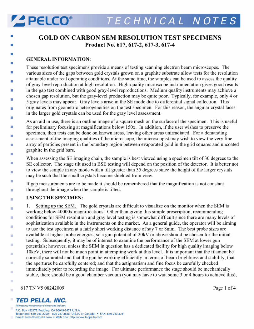
15.0kV x100K 200nm
Particle size range: 5-150nm
Resolution Test Specimen Gold on Carbon
Product No. 617
Each specimen has a square grid pattern with large crystals in the center of each square and very fine crystals
at the edges of each grid (as illustrated). Thus, medium and high resolution gap tests are performed on the
same specimen. The larger crystals show facets which allow assessment of the gray level reproduction
available at high resolution.

617 TN V5 08242009 Page 3 of 4
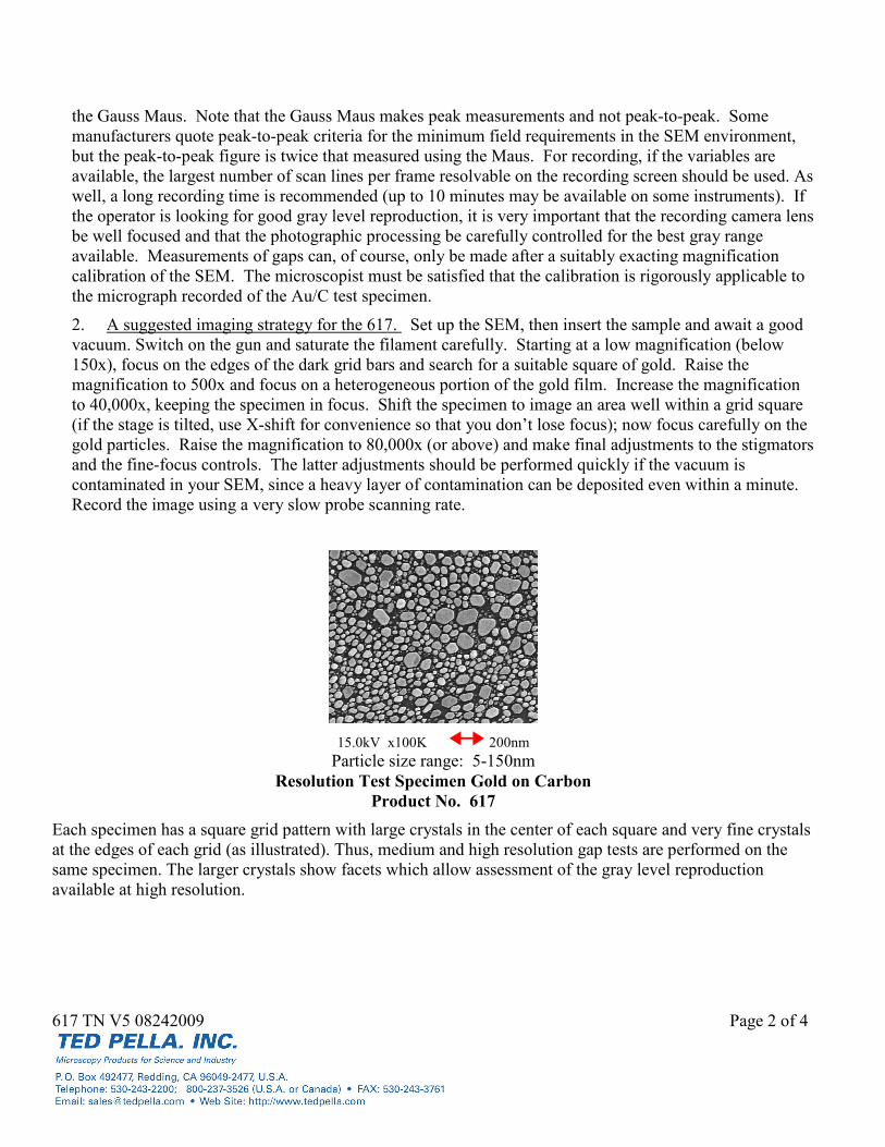
20.0kV x100K 100nm 20.0kV x100K 100nm
Particle size range: 3nm to 50nm Particle size range: 2nm to 30nm
High Resolution Gold on Carbon Ultra High Resolution Gold on Carbon
Product No. 617-2 Product No. 617-3
These gold on carbon calibration specimens have been specifically produced to enable the ultra high
resolution capabilities of the current generation of high performance scanning electron microscopes to be
assessed.
For high resolution performance testing, the #617-2 has a smaller gold island particle size that the #617. The
#617-2 is suitable for testing at instrument magnifications of 50,000x and above.
The ultra high #617-3 is particularly suitable for assessing the image quality of high resolution SEMs such as
those fitted with a field emission electron source. A magnification of at least 80,000x is required to clearly
resolve the gold particles.
Many of the instructions given on this technical note apply also to these ultra high resolution test specimens,
but it should be noted that the instrumental magnification required to resolve the smaller particles will be
higher.
617 TN V5 08242009 Page 4 of 4
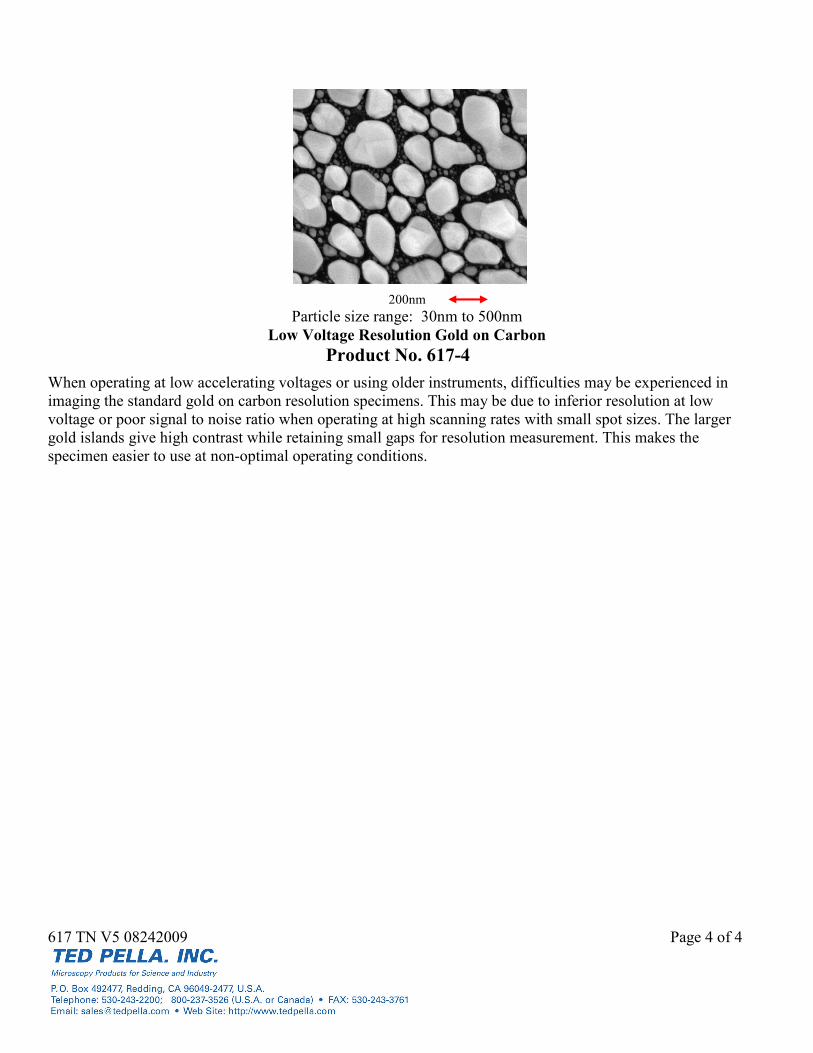
200nm
Particle size range: 30nm to 500nm
Low Voltage Resolution Gold on Carbon
Product No. 617-4
When operating at low accelerating voltages or using older instruments, difficulties may be experienced in
imaging the standard gold on carbon resolution specimens. This may be due to inferior resolution at low
voltage or poor signal to noise ratio when operating at high scanning rates with small spot sizes. The larger
gold islands give high contrast while retaining small gaps for resolution measurement. This makes the
specimen easier to use at non-optimal operating conditions.