gold nanoshell bioconjugates for molecular imaging in living cells
TRANSCRIPT
1012 OPTICS LETTERS / Vol. 30, No. 9 / May 1, 2005
Gold nanoshell bioconjugates for molecularimaging in living cells
Christopher Loo, Leon Hirsch, Min-Ho Lee, Emmanuel Chang, Jennifer West,Naomi Halas, and Rebekah Drezek
Department of Bioengineering, Rice University, MS-142, P.O. Box 1892, Houston, Texas 77251-1892
Received October 4, 2004
Advances in scattering-based optical imaging technologies offer a new approach to noninvasive point-of-caredetection, diagnosis, and monitoring of cancer. Emerging photonics technologies provide a cost-effectivemeans to image tissue in vivo with high resolution in real time. Advancing the clinical potential of theseimaging strategies requires the development of optical contrast agents targeted to specific molecular signa-tures of disease. We describe the use of a novel class of contrast agents based on nanoshell bioconjugates formolecular imaging in living cells. Nanoshells offer significant advantages over conventional imaging probesincluding continuous and broad wavelength tunability, far greater scattering and absorption coefficients, in-creased chemical stability, and improved biocompatibility. We show that nanoshell bioconjugates can be usedto effectively target and image human epidermal growth factor receptor 2 (HER2), a clinically relevant biom-arker, in live human breast carcinoma cells. © 2005 Optical Society of America
OCIS codes: 170.0170, 170.3880, 290.5850.
Optical imaging tools such as reflectance confocal mi-croscopy (RCM) and optical coherence tomography(OCT) offer the potential for noninvasive, high-resolution in vivo imaging at competitive costs rela-tive to current imaging modalities. Scattering-basedoptical technologies rely on inherent changes in indi-ces of refraction for image contrast.1 Strategies thatdepend on only the intrinsic optical contrast withintissue have proved clinically valuable in manyscreening applications including early cancer detec-tion; however, such techniques are not sensitiveenough to resolve an image based solely on the pres-ence of biomarkers of disease. In cases of cancer,when early detection is critical to reducing morbidityand mortality, the use of molecular-specific contrastagents provides the capacity to optically sense andimage abnormalities long before pathologic changesoccur at the anatomic level. In addition, imagingbased on molecular-specific targets allows real-timein vivo monitoring of the treatment course and canprovide fundamental insights into cancer biology.2 Arecent demonstration of scattering-based optical mo-lecular imaging used gold colloid conjugates to anti-bodies to the epidermal growth factor receptor as acontrast agent in imaging cervical cancer cells andbiopsy samples.3 Although gold colloid conjugates arehighly valuable as contrast agents for detecting su-perficial epithelial cancers with visible light, there isparticular need for contrast agents in the near-infrared (NIR) region of the spectrum. This is thespectral region in which tissue is most opticallytransparent,4 allowing imaging of deeper tissuestructures. The NIR region is also the region alreadyexploited by RCM and OCT; thus contrast agentswould provide greatly needed enhancement whereverthese imaging modalities are utilized.
Over the years, the expanding availability of a va-riety of nanostructures with highly controllable opti-cal properties has provided a series of new contrastagents for optical imaging. The use of a variety of na-
nomaterials such as quantum dots, gold nanopar-0146-9592/05/091012-3/$15.00 ©
ticles, and their bioconjugates in biological imaginghas been described in recent literature.5,6 Typically,nanostructures have many properties far superior tomolecular species such as Indocyanine Green. Advan-tages include higher quantum efficiencies, greaterscattering and absorbance cross sections, optical ac-tivities over more biocompatible wavelengths, andsignificantly increased chemical or photochemicalstability. Compared with Indocyanine Green,nanoshells provide a millionfold enhancement in op-tical extinction.7 Nanostructures can also be targetedto specific molecular signatures of interest. Thereforethe systematic control of nanostructure propertiesthat can be obtained by particular size variations isin direct contrast with molecular probes, whose prop-erties vary nonsystematically between molecularspecies.
Nanoshells are a novel class of nanoparticles com-posed of a dielectric silica core surrounded by a thinmetallic shell, which is typically made of gold.Nanoshells have a strong optical resonance whosewavelength can be tuned across much of the visibleand infrared region of the spectrum by varying therelative size of the core and shell layer.8 Varying theabsolute nanoparticle size allows the relative contri-butions of scattering and absorption at a given wave-length of interest to be controlled.9 This extremelyagile tunability of the optical resonance is completelyunique to nanoshells. Gold nanoshells are capable ofscattering light in the NIR and provide appealing op-tical properties for use in conjunction withreflectance-based optical imaging methods. Addition-ally, the gold surface is biologically inert and allowsproteins to be readily conjugated, facilitating in vivouse.10
Nanoshells have demonstrated promise in a vari-ety of biomedical applications ranging from sub-strates for whole-blood immunoassays11 to photother-mal cancer therapy. By use of magnetic resonancethermal guidance, in vitro cancer cells were success-
fully ablated with gold nanoshells tuned to absorb2005 Optical Society of America
May 1, 2005 / Vol. 30, No. 9 / OPTICS LETTERS 1013
NIR light.12 Similar use of nanoshells for photother-mal ablation of tumors in mice further showed com-plete regression of tumors with the mice remaininghealthy compared with controls.13 In contrast withtherapeutic NIR-absorbing nanoshells, we fabricatehighly scattering NIR nanoshells for optical imaging.We then demonstrate the feasibility of using thesetargeted nanoshell bioconjugates as contrast agentsto image human epidermal growth factor receptor 2(HER2) expression in living human breast carcinomacells.
HER2-positive SKBr3 human breast cancer cellswere cultured in McCoy’s 5A modified mediumsupplemented with 10% fetal bovine serum and anti-biotics. HER2-negative MCF7 human breast cancercells were cultured in Eagle’s minimum essential me-dium supplemented with 10% fetal bovine serum,0.01 mg/ml of bovine insulin, and antibiotics. Cellswere maintained at 37°C and 5% CO2.
The synthetic protocol developed for the fabricationof gold nanoshells is based on the principles ofmolecular self-assembly and colloid chemistry inaqueous solution. On the basis of the Stöbermethod,14 we fabricated silica nanoparticle cores byreducing tetraethylorthosilicate in ammonium hy-droxide and ethanol. Particle surfaces were termi-nated with amine groups by reaction with aminopro-pyltriethoxysilane. Small gold colloid was grown withthe method of Duff and Baiker.15 Gold–silicananoshells were then grown by reacting gold saltsHAuCl4d with the silica–colloid particles in the pres-ence of formaldehyde. Nanoshell formation was as-sessed with an UV–Vis spectrophotometer and scan-ning electron microscopy. Nanoshell dimensions weremathematically assessed with Mie scattering theorywith good agreement with scattering electron micros-copy and UV–Vis spectra.
Either anti-HER2 (specific) or anti-immunoglobulin G (anti-IgG) (nonspecific) antibodieswere attached to a polyethyleneglycol (PEG)linker [orthopyridyldisulfide-polyethyleneglycol-N-hydroxysuccinimide (OPSS-PEG-NHS), molecularweight of 2000] through a hydroxysuccinimide group(NHS). The antibody–PEG linker complex was thenattached to the nanoshell surface through a sulfur-containing group located at the distal end of the PEGlinker. By use of NaHCO3 (100 mM, pH of 8.5),OPSS-PEG-NHS was resuspended to a volume equalto that of the antibody. The reaction was allowed toproceed on ice overnight. Excess unbound polymerwas removed by membrane dialysis (molecularweight cutoff of 10,000). PEG-ylated antibodys0.67 mg/mld was added to nanoshells s23109 nanoshells/mld to facilitate targeting. After an-tibody conjugation, nanoshell surfaces were coatedwith PEG-thiol (PEG-SH, molecular weight of 5000,25 mM) to block nonspecific adsorption sites.
HER2-expressing SKBr3 cells were exposed to bio-conjugated nanoshells s8 mg/mLd and observed un-der dark-field microscopy, a form of microscopy sensi-tive only to scattered light. Images were taken with aZeiss Axioskop 2 plus microscope equipped with a
black-and-white CCD camera under the same magni-fication and lighting conditions. Optical contrast wasquantified with the Scion image analysis program.Average intensity values were obtained in each dark-field image. Normality of intensity data was estab-lished through a Shapiro Wilk test before using apaired Student’s t-test (two tailed) to test for signifi-cance.
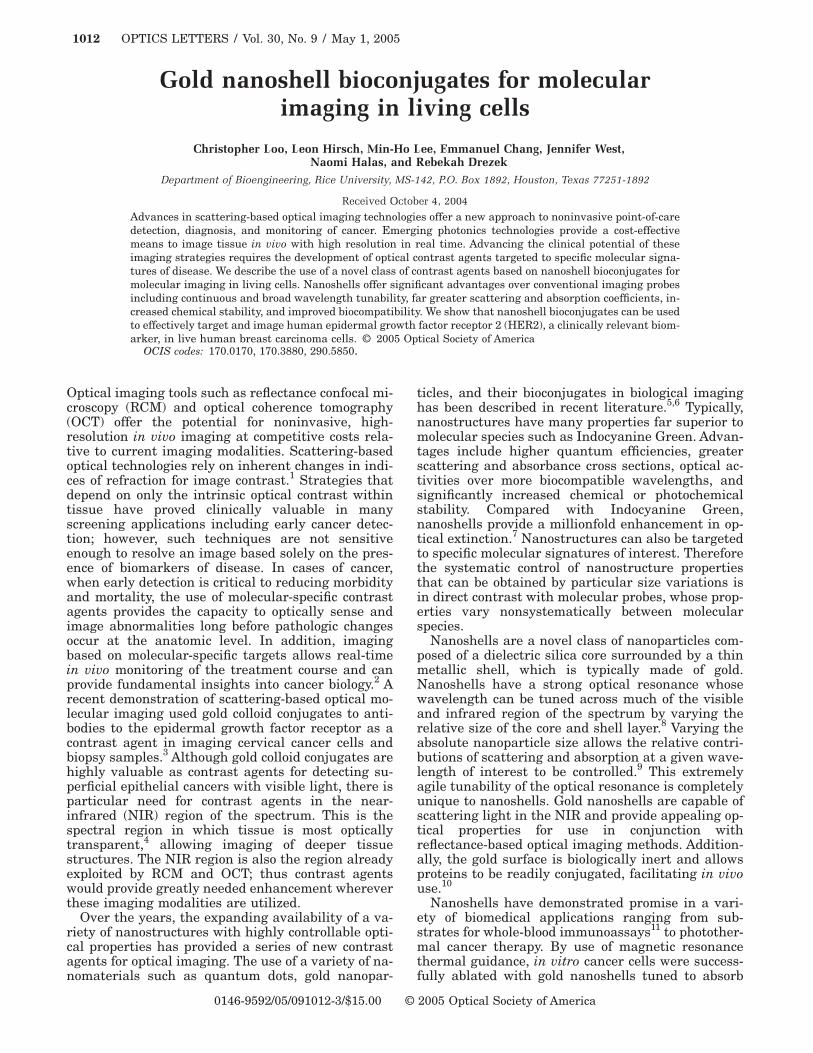
Figure 1 shows the optical properties fornanoshells with a 120-nm silica core radius and 35-nm-thick shell that were used in this study.Nanoshells with these dimensions generate a scatter-ing spectrum beginning at 700 nm and extending farinto the NIR region; thus nanoshells with these spec-tral characteristics are capable of facilitating imag-ing in both the visible and the NIR regions, allowingthe nanoshell conjugates to be used as contrastagents for RCM and OCT.
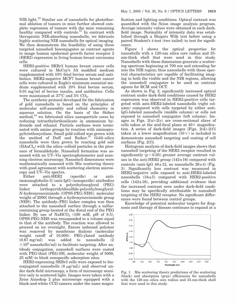
As shown in Fig. 2, significantly increased opticalcontrast under dark-field conditions caused by HER2expression was observed in HER2-positive cells tar-geted with anti-HER2-labeled nanoshells (right col-umn) compared with cells targeted by either anti-IgG-labeled nanoshells (middle column) or cells notexposed to nanoshell conjugates (left column). Im-ages in Figs. 2(a)–2(c) are cross-sectional slices ofcells taken at the mid-focal plane at 403 magnifica-tion. A series of dark-field images [Figs. 2(d)–2(f)]taken at a lower magnification s103 d is included todemonstrate nanoshell targeting and coating of cellsurfaces [Fig. 2(f)].
Histogram analysis of dark-field images shows thatnanoshell targeting of the HER2 receptor resulted insignificantly sp,0.05d greater average contrast val-ues in the anti-HER2 group s142±16d compared withcontrols (anti-IgG 48±12, no nanoshells 26±4) (Fig.3). Significantly less contrast was measured inHER2-negative cells exposed to anti-HER2-labelednanoshells s34±5d compared with HER2-positivecells s142±16d, providing additional evidence thatthe increased contrast seen under dark-field condi-tions may be specifically attributable to nanoshelltargeting of the HER2 receptor. No significant differ-ences were found between control groups.
Knowledge of potential molecular targets for diag-nosis and therapy of disease continues to expand at a
Fig. 1. Mie scattering theory predictions of the scattering(black) and absorption (gray) efficiencies for nanoshellswith the 120-nm silica core radius and 35-nm-thick shell
that were used in this study.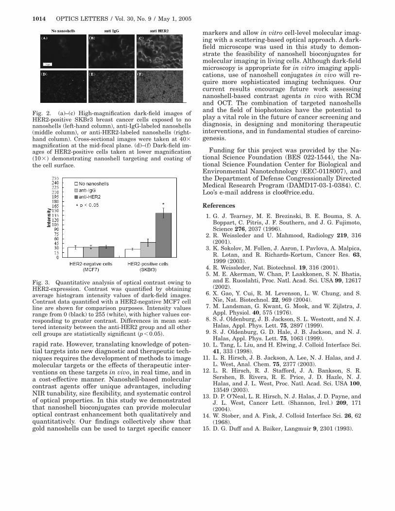
1014 OPTICS LETTERS / Vol. 30, No. 9 / May 1, 2005
rapid rate. However, translating knowledge of poten-tial targets into new diagnostic and therapeutic tech-niques requires the development of methods to imagemolecular targets or the effects of therapeutic inter-ventions on these targets in vivo, in real time, and ina cost-effective manner. Nanoshell-based molecularcontrast agents offer unique advantages, includingNIR tunability, size flexibility, and systematic controlof optical properties. In this study we demonstratedthat nanoshell bioconjugates can provide molecularoptical contrast enhancement both qualitatively andquantitatively. Our findings collectively show that
Fig. 2. (a)–(c) High-magnification dark-field images ofHER2-positive SKBr3 breast cancer cells exposed to nonanoshells (left-hand column), anti-IgG-labeled nanoshells(middle column), or anti-HER2-labeled nanoshells (right-hand column). Cross-sectional images were taken at 403magnification at the mid-focal plane. (d)–(f) Dark-field im-ages of HER2-positive cells taken at lower magnifications103 d demonstrating nanoshell targeting and coating ofthe cell surface.
Fig. 3. Quantitative analysis of optical contrast owing toHER2-expression. Contrast was quantified by obtainingaverage histogram intensity values of dark-field images.Contrast data quantified with a HER2-negative MCF7 cellline are shown for comparison purposes. Intensity valuesrange from 0 (black) to 255 (white), with higher values cor-responding to greater contrast. Differences in mean scat-tered intensity between the anti-HER2 group and all othercell groups are statistically significant sp,0.05d.
gold nanoshells can be used to target specific cancer
markers and allow in vitro cell-level molecular imag-ing with a scattering-based optical approach. A dark-field microscope was used in this study to demon-strate the feasibility of nanoshell bioconjugates formolecular imaging in living cells. Although dark-fieldmicroscopy is appropriate for in vitro imaging appli-cations, use of nanoshell conjugates in vivo will re-quire more sophisticated imaging techniques. Ourcurrent results encourage future work assessingnanoshell-based contrast agents in vivo with RCMand OCT. The combination of targeted nanoshellsand the field of biophotonics have the potential toplay a vital role in the future of cancer screening anddiagnosis, in designing and monitoring therapeuticinterventions, and in fundamental studies of carcino-genesis.
Funding for this project was provided by the Na-tional Science Foundation (BES 022-1544), the Na-tional Science Foundation Center for Biological andEnvironmental Nanotechnology (EEC-0118007), andthe Department of Defense Congressionally DirectedMedical Research Program (DAMD17-03-1-0384). C.Loo’s e-mail address is [email protected].
References
1. G. J. Tearney, M. E. Brezinski, B. E. Bouma, S. A.Boppart, C. Pitris, J. F. Southern, and J. G. Fujimoto,Science 276, 2037 (1996).
2. R. Weissleder and U. Mahmood, Radiology 219, 316(2001).
3. K. Sokolov, M. Follen, J. Aaron, I. Pavlova, A. Malpica,R. Lotan, and R. Richards-Kortum, Cancer Res. 63,1999 (2003).
4. R. Weissleder, Nat. Biotechnol. 19, 316 (2001).5. M. E. Akerman, W. Chan, P. Laakkonen, S. N. Bhatia,
and E. Ruoslahti, Proc. Natl. Acad. Sci. USA 99, 12617(2002).
6. X. Gao, Y. Cui, R. M. Levenson, L. W. Chung, and S.Nie, Nat. Biotechnol. 22, 969 (2004).
7. M. Landsman, G. Kwant, G. Mook, and W. Zijlstra, J.Appl. Physiol. 40, 575 (1976).
8. S. J. Oldenburg, J. B. Jackson, S. L. Westcott, and N. J.Halas, Appl. Phys. Lett. 75, 2897 (1999).
9. S. J. Oldenburg, G. D. Hale, J. B. Jackson, and N. J.Halas, Appl. Phys. Lett. 75, 1063 (1999).
10. L. Tang, L. Liu, and H. Elwing, J. Colloid Interface Sci.41, 333 (1998).
11. L. R. Hirsch, J. B. Jackson, A. Lee, N. J. Halas, and J.L. West, Anal. Chem. 75, 2377 (2003).
12. L. R. Hirsch, R. J. Stafford, J. A. Bankson, S. R.Sershen, B. Rivera, R. E. Price, J. D. Hazle, N. J.Halas, and J. L. West, Proc. Natl. Acad. Sci. USA 100,13549 (2003).
13. D. P. O’Neal, L. R. Hirsch, N. J. Halas, J. D. Payne, andJ. L. West, Cancer Lett. (Shannon, Irel.) 209, 171(2004).
14. W. Stober, and A. Fink, J. Colloid Interface Sci. 26, 62(1968).
15. D. G. Duff and A. Baiker, Langmuir 9, 2301 (1993).