gms bi 793 lecture 8 affinity mass spectrometry ... · affinity mass spectrometry: strategies for...
TRANSCRIPT
1
GMS BI 793 Lecture 8Affinity Mass Spectrometry:
Strategies for Proteome ProfilingJoseph Zaia
March 28, 20171. Discovery versus quantitative proteomics2. Affinity proteomics
• Tandem affinity purification• Isotope coding strategies
3. Affinity MS for identification of intracellular post-translationalmodifications
• Phosphorylation• O-GlcNAc• N- and O-glycosylation
3/28/17

3/28/17 2
Proteomics: using genomic sequences to identify proteins
http://www.isaaa.org/siteimages/pocketkimages/clip_image002_0014.jpg

Choudhary, C. and M. Mann, Nat RevMol Cell Biol, 2010. 11(6): p. 427-439.
Proeomics: wide angle proteinanalysis
3/28/17 3
4
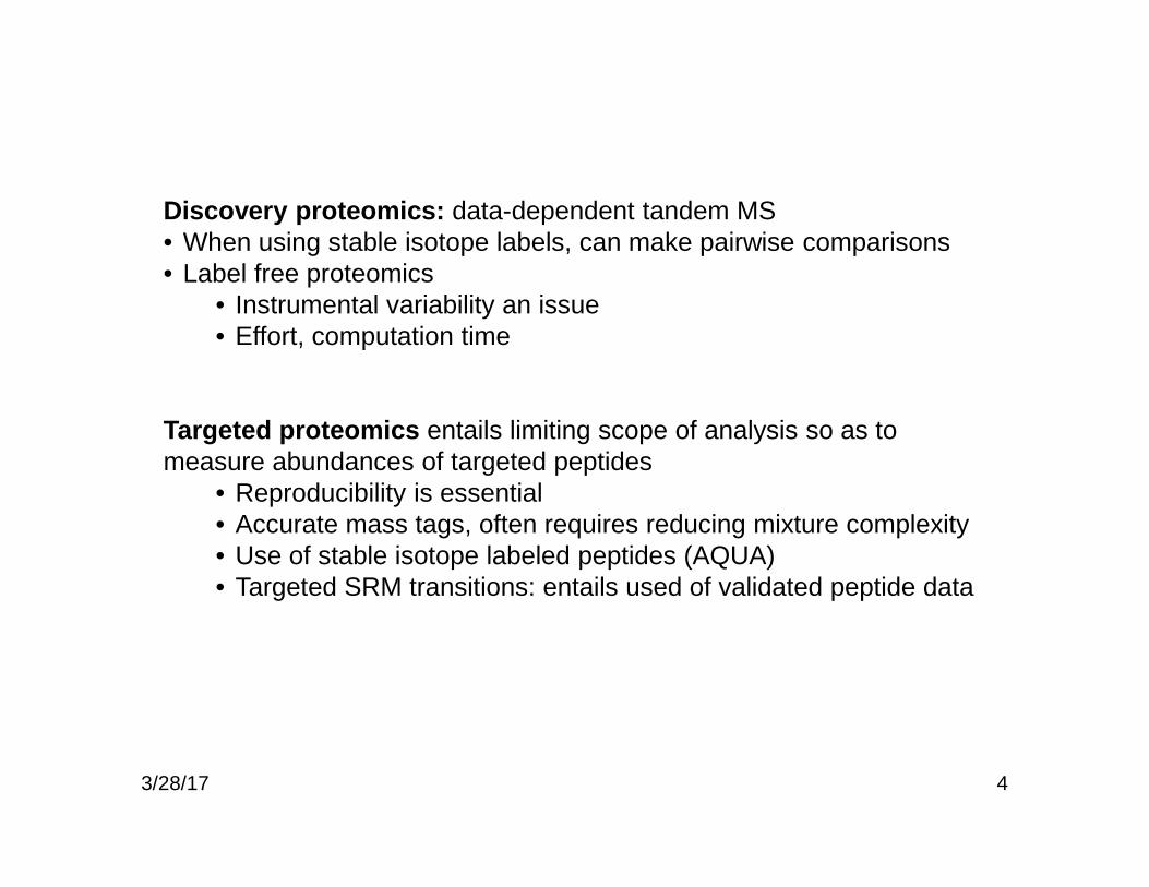
Discovery proteomics: data-dependent tandem MS• When using stable isotope labels, can make pairwise comparisons• Label free proteomics
• Instrumental variability an issue• Effort, computation time
Targeted proteomics entails limiting scope of analysis so as tomeasure abundances of targeted peptides
• Reproducibility is essential• Accurate mass tags, often requires reducing mixture complexity• Use of stable isotope labeled peptides (AQUA)• Targeted SRM transitions: entails used of validated peptide data
3/28/17

3/28/17 5
Proteomics: discovery of proteins
http://www.piercenet.com/media/Proteomics%20Identification%20Workflow-700px.jpg
Proteins
Trypticdigestion
Trypticpeptides RP-HPLC
MS
Peptide massspectrum
Tandem MS sequencing of peptides

3/28/17 6
Proteomics: quantification of proteins
http://www.piercenet.com/media/Quantitative%20Protomics%20Workflows-700px.jpg

3/28/17 7
Selected reaction monitoring
8
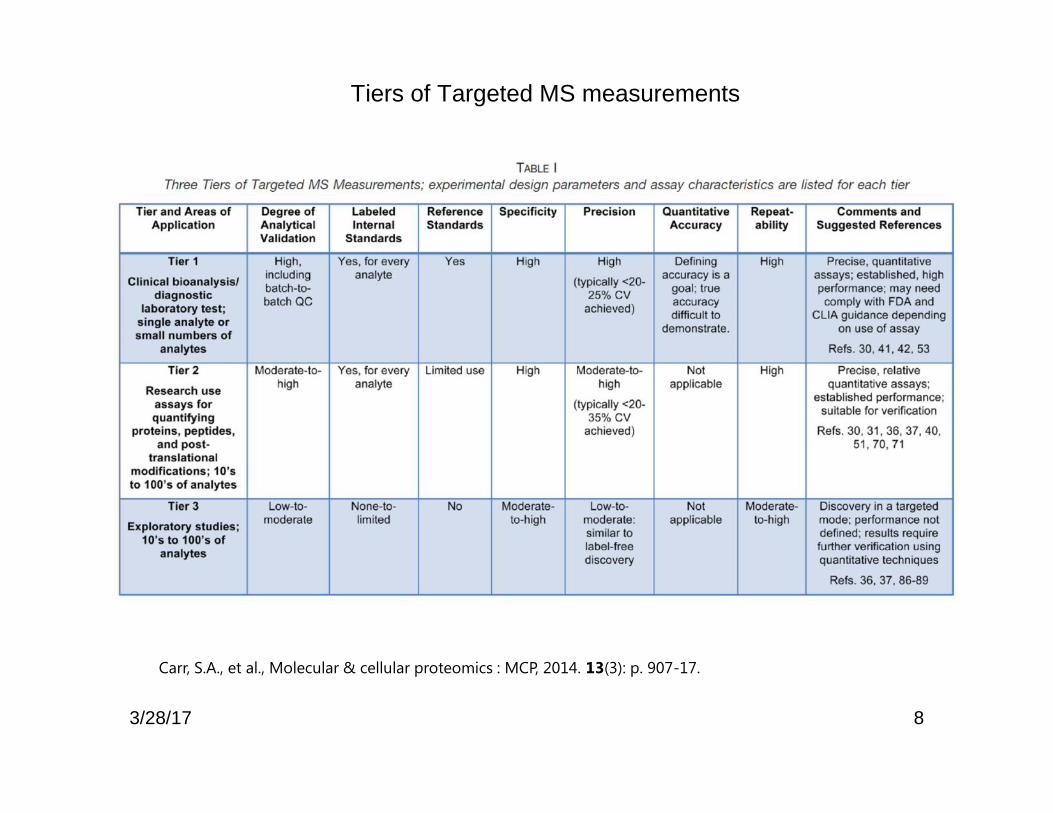
Tiers of Targeted MS measurements
Carr, S.A., et al., Molecular & cellular proteomics : MCP, 2014. 13(3): p. 907-17.
3/28/17

3/28/17 9
Some common post-translational modifications of proteins
Mann, M., and Jensen, O. N. (2003). Nat Biotechnol 21, 255-61.

10
Separation strategies for proteome profiling
Zhang, H.; Yan, W.; Aebersold, R. Curr Opin Chem Biol 2004, 8, 66-75.
(enzyme families)
3/28/17

11
The Isotope Coding Reagents
Affinitygroup(e.g.
biotin)
Linker: isotopelabel, may be
cleavable, mayhave chromophore
Reactive group:thiol reactive,
amino reactive,etc.
Non-isobaric labeling (ICAT, mTRAQ, SILAC) Isobaric labeling (iTRAQ, TMT)
3/28/17

12
ICAT Reagents
VICAT (visible ICAT)
Traditional
Photocleavage rxn
Bottari, P.; Aebersold, R.; Turecek, F.; Gelb, M. H. Bioconjug Chem 2004, 15, 380-388.3/28/17

13
ICAT MS: biotin connected to a thiol-reactive groupthrough an isotope-labeled linker
• Traditional ICAT– Deuterated
– Biotinylated
– Non-cleavable
– No chromophore
– $$$$$$
• Improved ICAT– 13C and 15N
– Reducible or photocleavablelinkers
– Cleavable linker
– Addition of chromophore
– $$$$$$
Bottari, P.; Aebersold, R.; Turecek, F.; Gelb, M. H. Bioconjug Chem 2004, 15, 380-388.3/28/17

14
Isobaric tag for relative and absolute quantitation (iTRAQ, Sciex)
3/28/17

15
iTRAQ
3/28/17
16
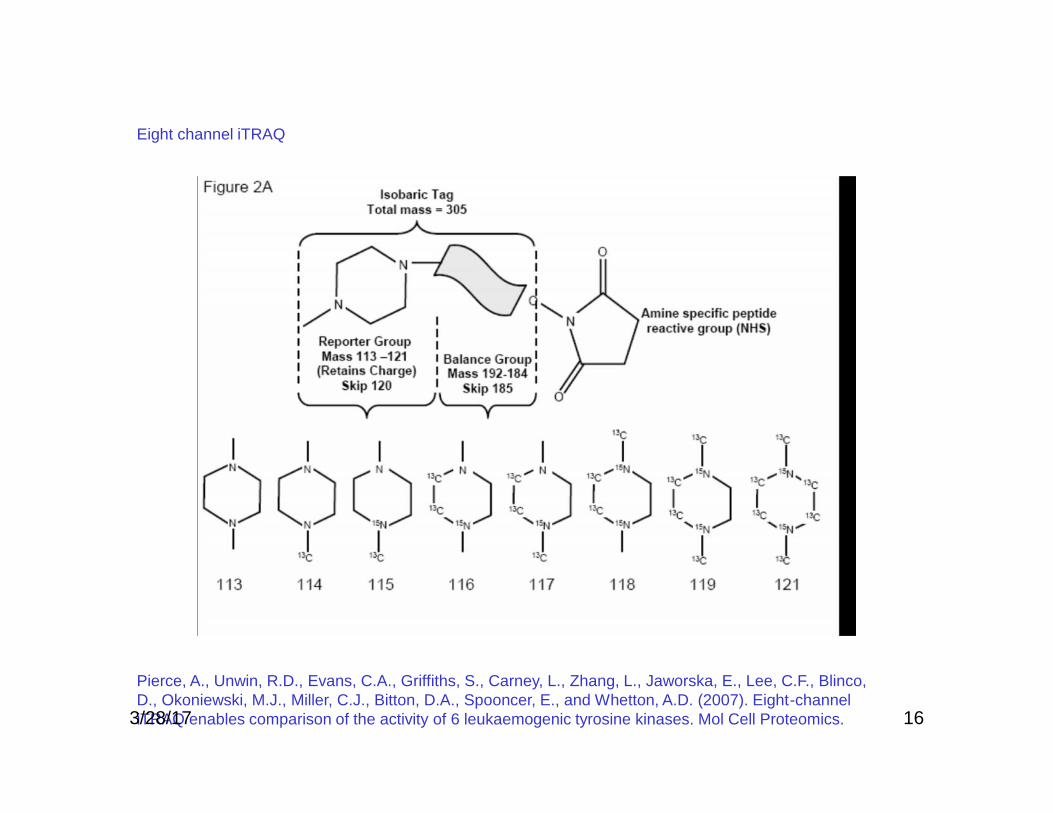
Pierce, A., Unwin, R.D., Evans, C.A., Griffiths, S., Carney, L., Zhang, L., Jaworska, E., Lee, C.F., Blinco,D., Okoniewski, M.J., Miller, C.J., Bitton, D.A., Spooncer, E., and Whetton, A.D. (2007). Eight-channeliTRAQ enables comparison of the activity of 6 leukaemogenic tyrosine kinases. Mol Cell Proteomics.
Eight channel iTRAQ
3/28/17

Tandem mass tags (TMT, Thermo-Fisher)
173/28/17

Iodo TMT reagents
183/28/17

iTRAQ and complex mixtures
19Ow, S.Y., et al., Journal of proteome research, 2009. 8(11): p. 5347-55.
8-plex samples in adilution series(4 protein mix)
4-plex cyanobacterium proteomeused as a complex background
3/28/17

iTRAQ vs mTRAQ
20
Mertins, P., et al., Molecular & cellular proteomics : MCP, 2012.11(6): p. M111 014423.
3/28/17

iTRAQ vs mTRAQ: FIG. 6. iTRAQ quantification is more precise but less accurate than mTRAQ. A,GluC peptide spike-in experiments to test accuracy and variability of quantification.
21
Mertins, P., et al., Molecular & cellular proteomics : MCP, 2012.11(6): p. M111 014423.
3/28/17

22
Systematic identification of protein complexes inSaccharomyces cerevisiae by mass spectrometry
Nature 2002, 415, 180-183.• Ho, Yuen*; Gruhler, Albrecht*; Heilbut, Adrian*; Bader, Gary D.†‡; Moore, Lynda*; Adams, Sally-Lin*; Millar, Anna*; Taylor, Paul*;
Bennett, Keiryn*; Boutilier, Kelly*; Yang, Lingyun*; Wolting, Cheryl*; Donaldson, Ian*; Schandorff, Søren*; Shewnarane, Juanita*;Vo, Mai*†; Taggart, Joanne*†; Goudreault, Marilyn*†; Muskat, Brenda*; Alfarano, Cris*; Dewar, Danielle†; Lin, Zhen†;
Michalickova, Katerina†‡; Willems, Andrew R.†§; Sassi, Holly†; Nielsen, Peter A.*; Rasmussen, Karina J.*; Andersen, Jens R.*;Johansen, Lene E.*; Hansen, Lykke H.*; Jespersen, Hans*; Podtelejnikov, Alexandre*; Nielsen, Eva*; Crawford, Janne*; Poulsen,
Vibeke*; Sørensen, Birgitte D.*; Matthiesen, Jesper*; Hendrickson, Ronald C.*; Gleeson, Frank*; Pawson, Tony†§; Moran,Michael F.*; Durocher, Daniel†§; Mann, Matthias*; Hogue, Christopher W. V.*†‡; Figeys, Daniel*; Tyers, Mike†§
•725 yeast proteins selected and expressed as Flag epitope fusions•100 protein kinases, 36 phosphatases, 86 DNA repair proteins
•One step immunoaffinity purification, eluted with excess FLAG peptide•Complexes separated by SDS-PAGE•15,683 gel slices processed by in-gel tryptic digestion, followed by tandem MS•940,000 tandem mass spectra that matched proteins in the yeast databases•35,000 protein identifications made
•Cited 2551 times as of 3/22/16
3/28/17

23
Systematic identification of protein complexes inSaccharomyces cerevisiae by mass spectrometry
Nature 2002, 415, 180-183.3/28/17

24Nature 2002, 415, 180-183.
Example signaling diagrams determined from theaffinity MS/MS data.
3/28/17

25
TAP: tandem affinity purification, applied to the yeast proteome
Nature 2002, 415, 141-147.3/28/17

26
Connectedcomplexes
Nature 2002, 415, 141-147.
It will take many years tomake sense of these data:bioinformaticstechniques
3/28/17

27
Burckstummer, T.; Bennett, K. L.; Preradovic,A.; Schutze, G.; Hantschel, O.; Superti-Furga,G.; Bauch, A. Nat Methods 2006, 3, 1013-1019.
TAP in mammalianCells: need toimprove yield,sensitivity
3/28/17
A tag based on protein G and thestreptavidin-binding peptide (GS-TAP)resulted in a tenfold increase in protein-complex yield and improved the specificityof the procedure
3/28/17 28
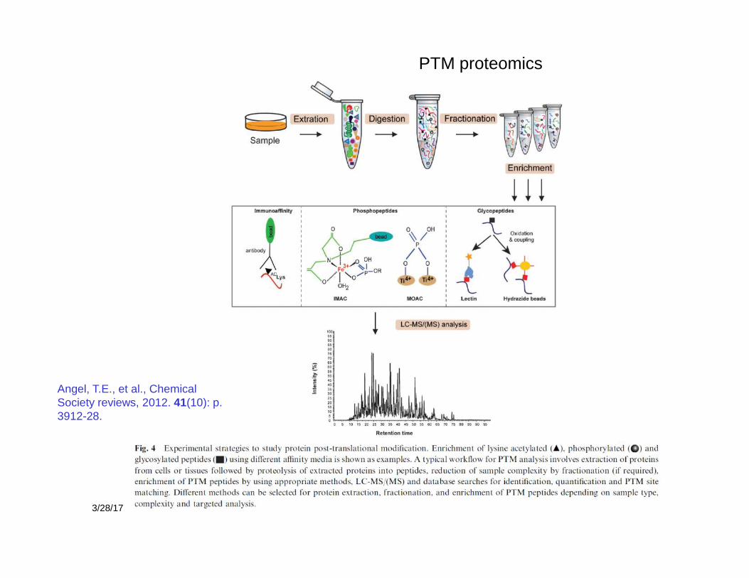
PTM proteomics
Angel, T.E., et al., ChemicalSociety reviews, 2012. 41(10): p.3912-28.
29
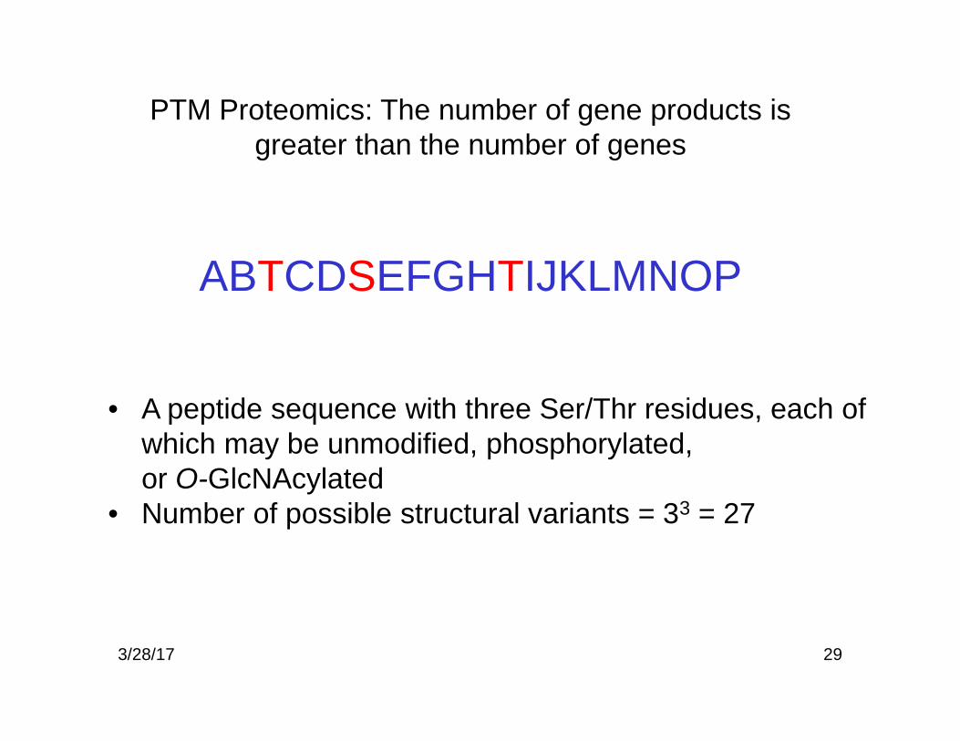
PTM Proteomics: The number of gene products isgreater than the number of genes
ABTCDSEFGHTIJKLMNOP
• A peptide sequence with three Ser/Thr residues, each ofwhich may be unmodified, phosphorylated,or O-GlcNAcylated
• Number of possible structural variants = 33 = 27
3/28/17
30
OHO
HONHAc
O
OH
NH
O
R'
R
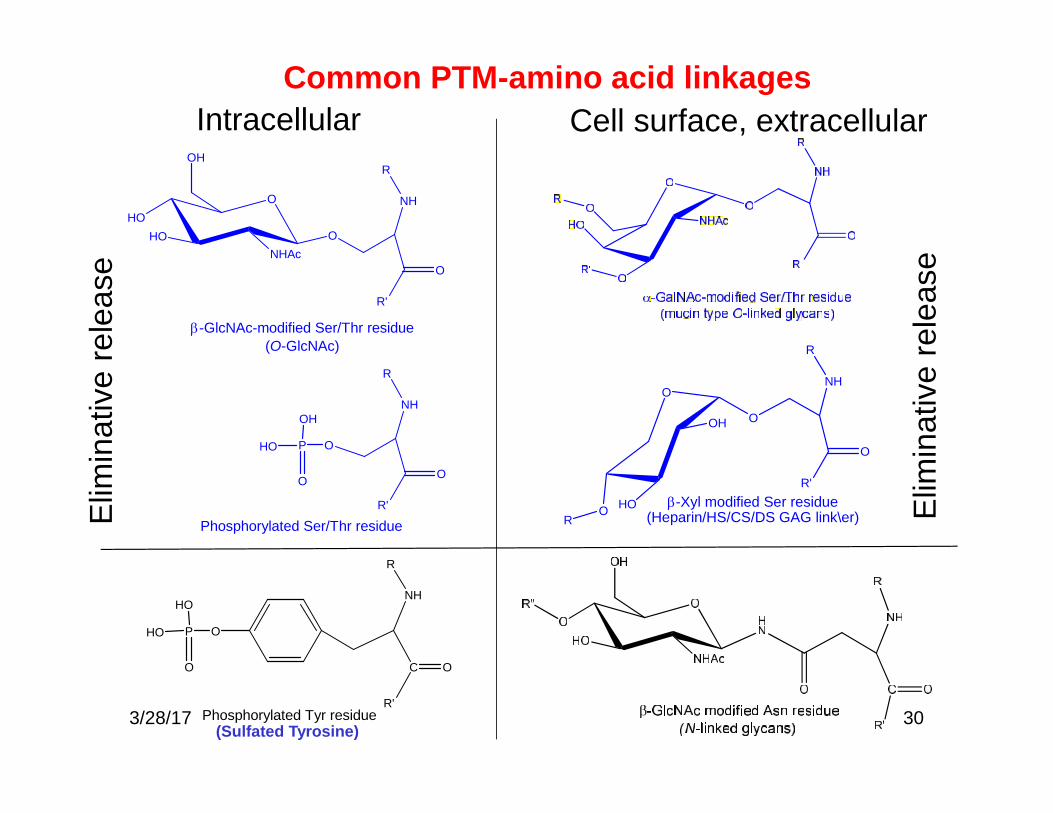
-GlcNAc-modified Ser/Thr residue(O-GlcNAc)
O
NH
O
R'
R
P
OH
HO
O
Phosphorylated Ser/Thr residue
NH
C
R'
O
OP
HO
HO
O
Phosphorylated Tyr residue
R
Common PTM-amino acid linkagesIntracellular Cell surface, extracellular
Elim
inat
ive
rele
ase
Elim
inat
ive
rele
ase
(Sulfated Tyrosine)
NH
O
R'
R
O
O HO
OOH
-Xyl modified Ser residue(Heparin/HS/CS/DS GAG link\er)R
3/28/17
31
HN CH C
CH2
O
O
O
HN CH C
CH2
O
O
P O
OH
HO
Dehydroalanine
Baseor CID
R = phosphate
R
OHO
HONHCOCH3
OH
R = GlcNAc
O
P O
OH
HO
OHO
HONHCOCH3
OH
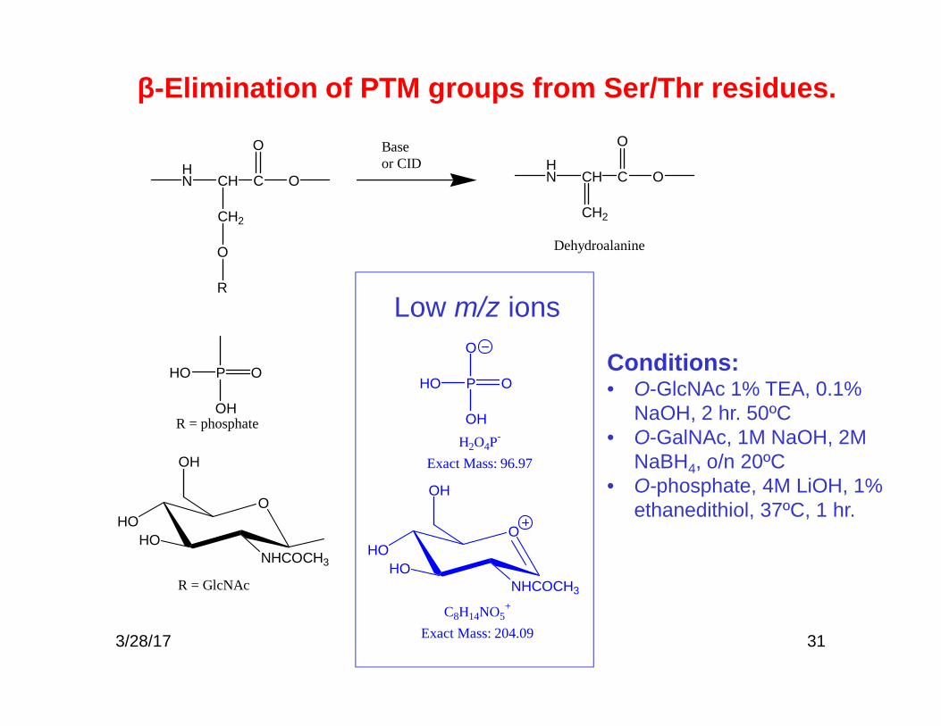
H2O4P-
Exact Mass: 96.97
C8H14NO5+
Exact Mass: 204.09
Low m/z ions
Conditions:• O-GlcNAc 1% TEA, 0.1%
NaOH, 2 hr. 50ºC• O-GalNAc, 1M NaOH, 2M
NaBH4, o/n 20ºC• O-phosphate, 4M LiOH, 1%
ethanedithiol, 37ºC, 1 hr.
β-Elimination of PTM groups from Ser/Thr residues.
3/28/17

32
Precursor ion scans and neutral loss scans
MS1:precursor ion scanning CID MS2:
product ion scanning
Product ion scanFixed m/z fragmentation full scan
Precursor ion scanScanning fragmentation Fixed m/z
Constant neutral loss scanScanning fragmentation Scaning a set
m/z loss3/28/17

33
R
OH
R
O
S
OH
OO
mass shift 79.95736
HSO4-
m/z 96.96010[M-SO3+H]+loss of 79.95736
Neg CID Pos CID
R
OH
R
O
P
OH
OHO
mass shift 79.96688
Post-translationalmodification
Post-translationalmodification
PO2-
m/z 62.96414PO3
-
m/z 78.95905H2PO4
-
m/z 96.96962
[M-H3PO4+H]+loss of 97.97744[M-HPO3+H]+Loss of 79.96688
Neg CID Pos CID
Mass shifts for phosphorylation and sulfation
3/28/17

34
The Phosphoproteome
• ~30% of all proteins are thought to be phosphorylated• Protein kinases are coded by >2000 genes (518 human protein kinases
with a conserved catalytic domain)– Receptor tyrosine kinases (growth factor receptors: EGFR, FGFR,
VEGFR)– Non-receptor tyrosine kinases– Signal transducing serine/threonine kinases (mitogen activated
protein kinases, MAPK)– Cyclin dependent kinases (CDKs, pRb phosphorylation required for
progression through G1 phase)n• Identification of phosphorylation sites is a challenge
– Phosphorylation is dynamic, it is possible that only a few percent ofthe sites of a gene product are phosphorylated at a given time
– 100K potential human phosphoylatioin sites– Phospho groups are somewhat labile during MS/MS, losses of 98
from precursor and product ions often observed
3/28/17

35
Affinity enrichment of phosphopeptides
[1]P.A. Grimsrud, D.L. Swaney, C.D. Wenger, N.A. Beauchene,and J.J. Coon, Phosphoproteomics for the masses. ACS ChemBiol 5 (2010) 105-19.3/28/17

36
IMAC: immobilized metal affinity chromatography
Gioeli, D.; Ficarro, S. B.; Kwiek, J. J.; Aaronson, D.; Hancock, M.; Catling, A. D.; White, F. M.; Christian,R. E.; Settlage, R. E.; Shabanowitz, J.; Hunt, D. F.; Weber, M. J. J Biol Chem 2002, 277, 29304-29314.
3/28/17

37
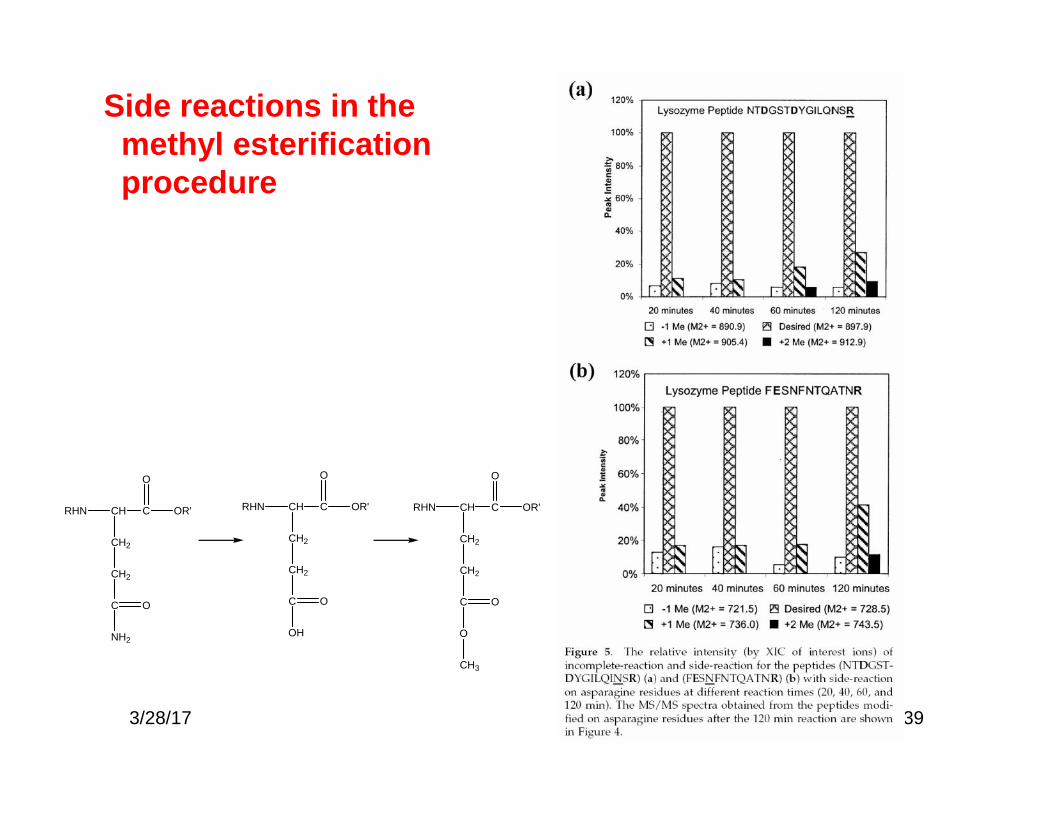
Methyl esterification of acidic amino acidresidues
RHN CH C
CH2
OR'
O
C
OH
O
methanolic HCl
RHN CH C
CH2
OR'
O
C
OCH3
O
3/28/17

38
The effect of methylesterification on IMACphosphoproteomeanalysis
00
IMAC Esterificatrion + IMAC
Ficarro, S. B.; et al. Nat Biotechnol 2002, 20, 301-305.3/28/17
39
Side reactions in themethyl esterificationprocedure
RHN CH C
CH2
OR'
O
CH2
C
OH
O
RHN CH C
CH2
OR'
O
CH2
C
NH2
O
RHN CH C
CH2
OR'
O
CH2
C
O
O
CH3
3/28/17
40
A
B
NH3-Thr-Val-Asp-Ser-Pro-Lys-COOH
NH3COOH+ +
pH=2.7 Net charge
2+
1+NH3-Thr-Val-Asp-Ser-Pro-Lys-COOH
NH3COOH+ +H2PO3
-
01 02 03 04 05 06 07 08 0
% o
f tot
al
Predicted net charge at pH 2.7
C
0 10 20 30 40 50 600.0
0.4
0.8
1.2
OD
214
Time
1+
2+
3+
=4+
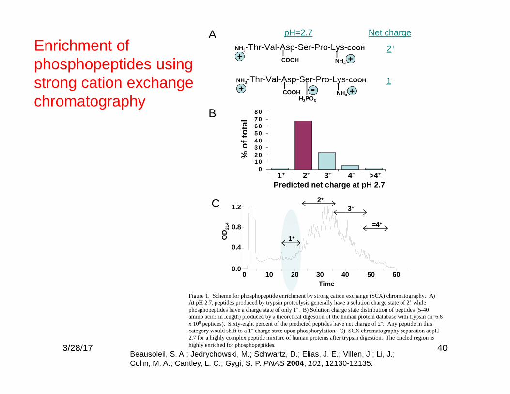
Figure 1. Scheme for phosphopeptide enrichment by strong cation exchange (SCX) chromatography. A)At pH 2.7, peptides produced by trypsin proteolysis generally have a solution charge state of 2+ whilephosphopeptides have a charge state of only 1+. B) Solution charge state distribution of peptides (5-40amino acids in length) produced by a theoretical digestion of the human protein database with trypsin (n=6.8x 10⁸ peptides). Sixty-eight percent of the predicted peptides have net charge of 2+. Any peptide in thiscategory would shift to a 1+ charge state upon phosphorylation. C) SCX chromatography separation at pH2.7 for a highly complex peptide mixture of human proteins after trypsin digestion. The circled region ishighly enriched for phosphopeptides.
1+ 2+ 3+ 4+ >4+
Enrichment ofphosphopeptides usingstrong cation exchangechromatography
Beausoleil, S. A.; Jedrychowski, M.; Schwartz, D.; Elias, J. E.; Villen, J.; Li, J.;Cohn, M. A.; Cantley, L. C.; Gygi, S. P. PNAS 2004, 101, 12130-12135.
3/28/17

41
Absolute quantification (AQUA) of proteins andphosphoproteins from cell lysates by tandem MS
Gerber, S. A.; Rush, J.; Stemman, O.; Kirschner, M. W.; Gygi, S. P. Proc Natl Acad Sci U S A 2003, 100, 6940-6945.3/28/17

423/28/17

433/28/17

3/28/17 44
Influence of phosphorylation on CAD tandem MS
Na, S. and E. Paek, Mass Spectrometry Reviews, 2015. 34(2): p.133-147.

3/28/17 45Humphrey, S.J., S.B. Azimifar, and M. Mann, Nat Biotechnol, 2015. 33(9): p. 990-5.
EasyPhos phosphoproteomics metho
(guanidine HCl)
46
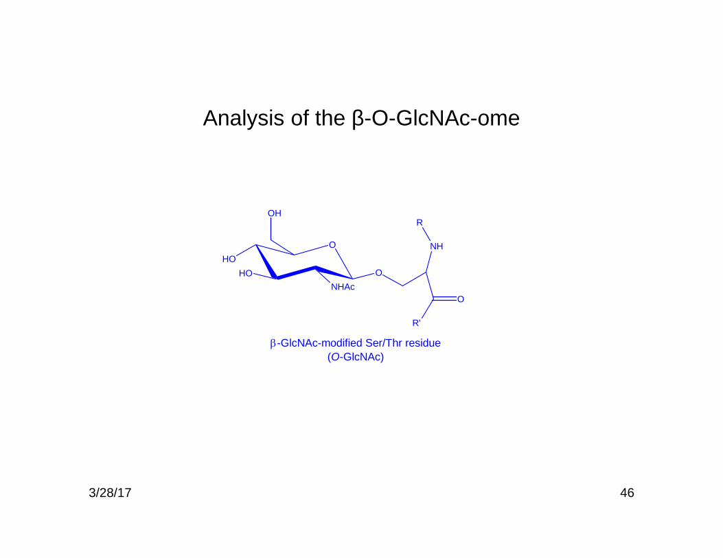
Analysis of the β-O-GlcNAc-ome
OHO
HONHAc
O
OH
NH
O
R'
R
-GlcNAc-modified Ser/Thr residue(O-GlcNAc)
3/28/17

47
• Many nuclear and clytoplasmic proteins are transientlymodified with Ser/Thr-O-GlcNAc.
• All O-GlcNAc modified proteins are potentialphosphoproteins (reciprocal switches)
• Added by UDP-GlcNAc-peptide-β-GlcNAc transferase(OGT) using UDP-GlcNAc
• Removed by N-acetyl-β-D-glucosaminidase (OGlcNAcase)• Anti-O-GlcNAc monoclonal antibodies are now available• Metabolic labeling using Gal transferase and Gal• β-O-GlcNAc is very labile• Need methods to both enrich O-GlcNAc and determine
sites of occupancy
Ser/Thr β-O-GlcNAc Modification
Wells, L.; Vosseller, K.; Cole, R. N.; Cronshaw, J. M.; Matunis, M. J.; Hart, G. W. Mol Cell Proteomics 2002, 1, 791-804.3/28/17

48
Affinitry enrichment of O-GlcNAc using β-elimination andMichael addition (BEMAD)
Wells, L.; Vosseller, K.; Cole, R. N.; Cronshaw, J. M.; Matunis, M. J.; Hart, G. W. Mol Cell Proteomics 2002, 1, 791-804.3/28/17
49
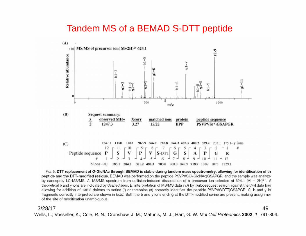
Tandem MS of a BEMAD S-DTT peptide
Wells, L.; Vosseller, K.; Cole, R. N.; Cronshaw, J. M.; Matunis, M. J.; Hart, G. W. Mol Cell Proteomics 2002, 1, 791-804.3/28/17

Organic chemical ligation reactions
50
Staudinger ligation:
Click chemistry:
3/28/17

GalT1 labeling and tagging with photocleavable biotin
51
Wang, Z.; Udeshi, N. D.; O'Malley, M.; Shabanowitz, J.; Hunt, D. F.; Hart, G.W. Mol Cell Proteomics 2010, 9, 153-160.
3/28/17

52Wang, Z.; Udeshi, N. D.; O'Malley, M.; Shabanowitz, J.; Hunt, D. F.; Hart, G. W.Mol Cell Proteomics 2010, 9, 153-160.
CAD of cleaved O-GlcNAc-peptide
3/28/17
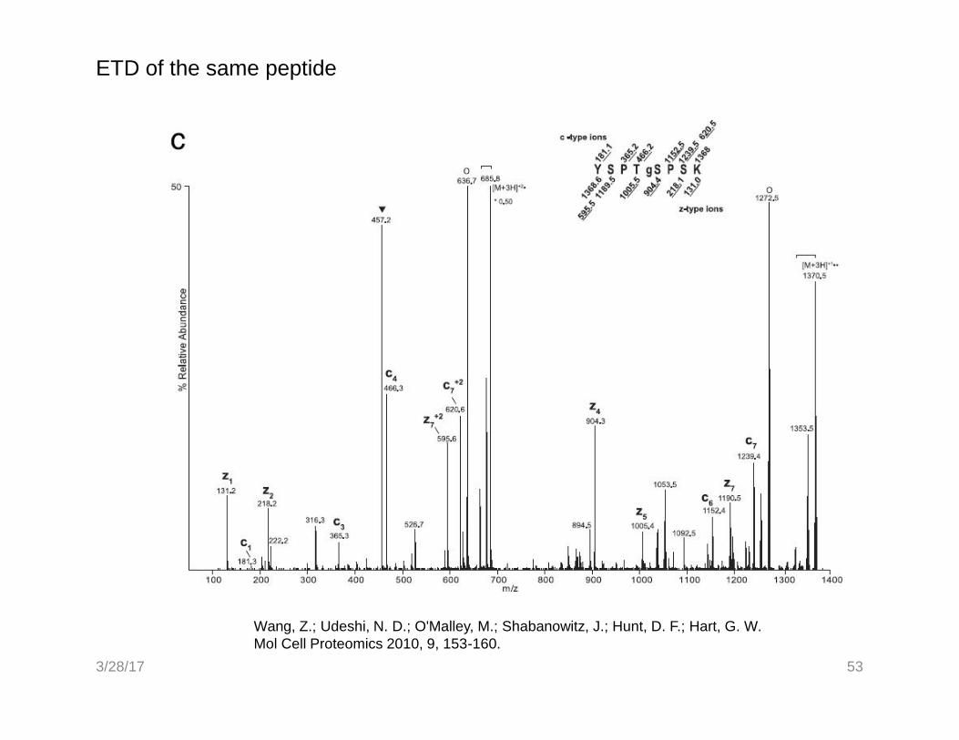
ETD of the same peptide
53
Wang, Z.; Udeshi, N. D.; O'Malley, M.; Shabanowitz, J.; Hunt, D. F.; Hart, G. W.Mol Cell Proteomics 2010, 9, 153-160.
3/28/17

Chapter 8, Figure 4Essentials of Glycobiology
Second Edition
Processing and maturation of an N-glycan
Only Man5 glycans acted upon by Lec1; mostmature glycoproteins contain Man5-9 thatescape Golgi processing and extension
54
N-glycosylation (Essentials of Glycobiology Chapter 8)
543/28/17

55
Glycoproteomics:
A glycoprotein
3/28/17

Peptide N-glycosidase F (PNGase F)
563/28/17

57
Common glycan oxonium ions
O
OH
OH
OH
CH2OH
Hex
C6H11O5+
m/z 163.06
COOH
OH
HO
OCH3COHN
HO
OH
Neu5Ac
C11H18NO8+
m/z 292.10
O
NHCOCH3
OH
OH
CH2OH
HexNAc
C8H14NO5+
m/z: 204.09
• Pos ion CID of glycoconjugates forms oxonium ions,(analogous to peptide immonium ions)
• Precursor ion scans for these ions serve to identifyglycopeptides in a glycoprotein digest
O
OH
OH
OH
O
NHCOCH3
OH
O
HO
OH
HexHexNAc
C14H24NO10+
m/z 366.14
3/28/17
58
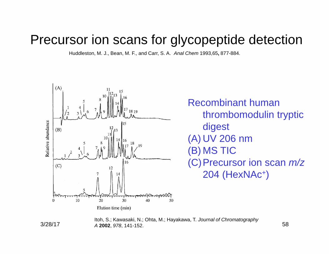
Precursor ion scans for glycopeptide detection
Recombinant humanthrombomodulin trypticdigest
(A) UV 206 nm(B) MS TIC(C)Precursor ion scan m/z
204 (HexNAc+)
Itoh, S.; Kawasaki, N.; Ohta, M.; Hayakawa, T. Journal of ChromatographyA 2002, 978, 141-152.
Huddleston, M. J., Bean, M. F., and Carr, S. A. Anal Chem 1993,65, 877-884.
3/28/17

59
Zhang Nat Biotech 2003 21:660-666
Enrichment ofglycopeptides byperiodate labelingfollowed by amine-affinitychromatography
3/28/17
60
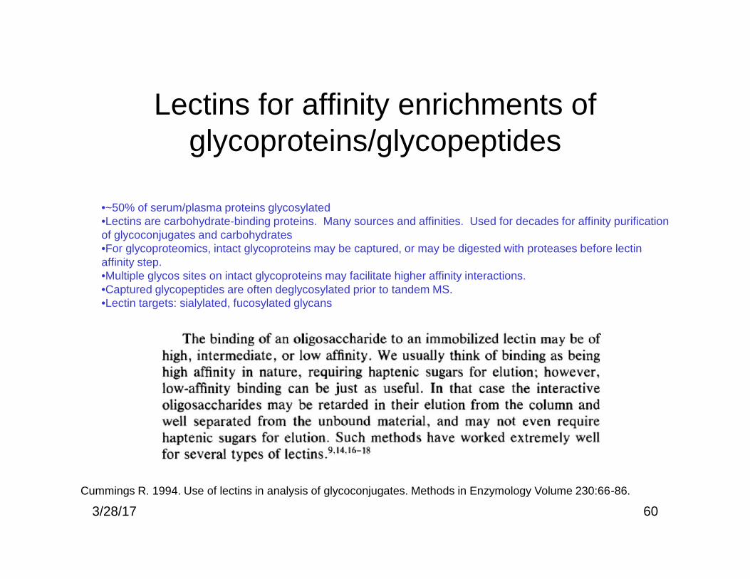
Lectins for affinity enrichments ofglycoproteins/glycopeptides
Cummings R. 1994. Use of lectins in analysis of glycoconjugates. Methods in Enzymology Volume 230:66-86.
•~50% of serum/plasma proteins glycosylated•Lectins are carbohydrate-binding proteins. Many sources and affinities. Used for decades for affinity purificationof glycoconjugates and carbohydrates•For glycoproteomics, intact glycoproteins may be captured, or may be digested with proteases before lectinaffinity step.•Multiple glycos sites on intact glycoproteins may facilitate higher affinity interactions.•Captured glycopeptides are often deglycosylated prior to tandem MS.•Lectin targets: sialylated, fucosylated glycans
3/28/17

61
MLAC: multiple lectin affinity chromatography
• MLAC is a single column with threelectins: ConA; WGA and jacalin
• ConA: specific for glycans with Man andGlc (biantennary NL low affinity, high ManNL, high affinity)
• WGA: GlcNAc, (NeuAc)• Jacalin: Galβ3GlcNAc of O-glycans, αGalof other glycans
• Advantages over single lectin: betteroverall binding coverage
• Initial work showed that high abundanceproteins (albumin, etc) interfere, and betterresults are obtained when they aredepleted (2005. Proteomics 5:3353-3366).
• Poros protein G and Poros anti HSAcartridges used for depletion ($$$)
Plavina T, (2007). Journal of Proteome Research 6:662-671.3/28/17

Filter-aided sample preparation (FASP)
62D.F. Zielinska, F. Gnad, J.R. Wisniewski, and M. Mann, Cell 141 (2010) 897-907.
3/28/17
30 kDaMWCO

Proteomic data on FASP lectin enriched glycopeptides
63D.F. Zielinska, F. Gnad, J.R. Wisniewski, and M. Mann, Cell 141 (2010) 897-907.3/28/17