glycoproteins in - core
TRANSCRIPT
Biochem. J. (1983) 212, 355-363 355Printed in Great Britain
Glycoproteins and glycosaminoglycans synthesized by human keratinocytesin culture
Their role in cell-substratum adhesion
Glyn P. ROBERTS and Lucy JENNERSection ofDermatology, Department ofMedicine, Welsh National School ofMedicine, Heath Park,
CardiffCF4 4XN, Wales, U.K.
(Received 16 November 1982/Accepted 25 January 1983)
Glycoproteins and proteoglycans synthesized by human keratinocytes in mediumcontaining D-[1-14C]glucosamine were extracted and analysed by polyacrylamide-gelelectrophoresis in the presence of sodium dodecyl sulphate. Extraction of the labelledkeratinocytes with 0.5% Triton X-100 removed most of the glycoconjugates and left thecytoskeleton and nuclear residue adherent to the substratum. In addition to thecytoskeletal proteins, there was a relatively simple profile of glycoproteins andglycosaminoglycans associated with this adherent cytoskeleton. These consisted of eightglycoproteins in the mol.wt. range 99000-232000, five proteins in the keratin region(mol.wt. 42000-61000), hyaluronic acid and a sulphated glycosaminoglycan. Surfacelabelling of the keratinocytes with galactose oxidase (with or without neuraminid-ase)/KB3H4 revealed that many of the glycoproteins were exposed on the cell surface.The importance of the glycoproteins and proteoglycans in attaching the keratinocytes tothe substratum was examined by studying their expression after incubation in mediumcontaining tunicamycin and their degradation after digestion with trypsin andhyaluronidase. These studies, together with an examination of the glycoconjugatesreleased by sequential extraction with 0.5% Triton X-100 followed by 0.2% sodiumdodecyl sulphate, revealed that the glycoprotein of mol.wt. 232000 has an importantrole in mediating the attachment of keratinocytes to the substratum.
With the exception of the circulating cells in thevascular system, most eukaryotic cells exist attachedto other cells, to intercellular substances such ascollagen or to a basal lamina. Similarly, attachmentto a substratum is a requirement for the growth ofnormal cells in culture, and this requirement hasbeen termed anchorage dependence (Stoker et al.,1968). In the malignant state cells tend to lose theiranchorage dependence. An understanding of themolecular basis of cellular adhesion and an explana-tion of how this interaction contributes to theformation of tissues is a fundamental goal of manystudies on developmental biology and tumourbiology. There are now several lines of evidenceimplicating cell-surface glycoconjugates in the pro-cess of cell-cell adhesion and cell-substratumadhesion (Hughes & Pena, 1981). Extensive studies
Abbreviations used: SDS, sodium dodecyl sulphate;Hepes, 4-(2-hydroxyethyl)-1-piperazine-ethanesulphonicacid.
Vol. 212
on a major cell-surface glycoprotein called fibro-nectin have shown it to be one of the main agents inmediating the adhesion and spreading of fibroblastson solid supports (Yamada & Olden, 1978). Epi-thelial cells differ from fibroblasts in that fibronectindoes not stimulate their attachment to substratum.However, it has been shown that laminin, aglycoprotein component of basement membranes,will stimulate the attachment of a transformed line ofmouse epidermal cells to plates coated with type IVcollagen (Terranova et al., 1980).
In the mammalian epidermis, mitosis occurs onlyin the innermost cells or basal layer, which are inclose proximity to the basal lamina. These cells thenmove upwards and differentiate to form in turn thespinous, granular and cornified (stratum corneum)layers. Adhesive forces operating in the intercellularspace may be of importance in regulating epidermalstructure, and it is probable that these adhesiveinteractions change with differentiation (Skerrow,
G. P. Roberts and L. Jenner
1978). Furthermore, studies using lectin binding(Holt et al., 1979; Brabec et al., 1980) show thatchanges in glycoconjugate synthesis occur as epi-dermal cells differentiate. When skin slices wereincubated in medium containing radiolabelledsugars, a complex mixture of epidermal glyco-conjugates was synthesized (King et al., 1980;Roberts & Marks, 1982). The investigation of thefunction of epidermal glycoconjugates in cellularadhesion was not easily accomplished with skinslices, and consequently the glycoconjugates syn-thesized by human keratinocytes cultured in vitro asdescribed by Rheinwald & Green (1975) have nowbeen examined.
In the present paper evidence is reported that aglycoprotein of apparent mol.wt. 232000 has animportant role in attaching cultured keratinocytes tothe substratum.
Experimental
MaterialsTunicamycin was a gift from Dr. W. F. J.
Cuthbertson, Glaxo Group Research Ltd., Green-ford, Middx., U.K. Dulbecco's minimum essentialmedium, Ca2+-free Eagle's minimum essentialmedium, foetal-calf serum, calf serum, penicillin-streptomycin, Hepes and 30mm plastic tissueculture dishes were obtained from Flow Labora-tories, Irvine, Scotland, U.K. Cholera toxin and25cm2 tissue-culture flasks were purchased fromBecton Dickinson, Wembley, Middx., U.K. Cor-tisol, bovine testicular hyaluronidase (1.7 units/mg),protease type VI from Streptomyces griseus, trypsintype I from bovine pancreas, galactose oxidase(14.6units/mg), neuraminidase type VI (2.5units/mg) and protein A-Sepharose CL-4B were fromSigma, Poole, Dorset, U.K. Reagents for electro-phoresis and general chemicals were purchased fromBDH Chemicals, Poole, Dorset, U.K. L-[6-3H1-Fucose (23 Ci/mmol), D-[6-3Hlglucosamine hydro-chloride (22.6 Ci/mmol), D-[ 1-14Clglucosaminehydrochloride (61 mCi/mmol), L-[U-14C]leucine(339mCi/mmol), [14C]methylated proteinmolecular-weight markers, 35SO42- and KB3H4(4.3Ci/mmol) were all obtained from The Radio-chemical Centre, Amersham, Bucks., U.K. OsrayM3 X-ray film was from Agfa-Gevaert, Dunstable,Beds., U.K., and X-Omat RP X-ray film wasobtained from Kodak, Western Sales Centre, Bristol,U.K. Sheep antiserum to laminin was obtained fromDr. S. I. Katz, National Institute of Health,Bethesda, MD, U.S.A., and rabbit antiserum tohuman plasma fibronectin was purchased fromBethesda Research Laboratories (U.K.) Ltd., Cam-bridge, U.K. Protein A-bearing Staphylococcalaureus cells were obtained from Calbiochem-Behring Corp., Bishops Stortford, Herts., U.K.
Cell culturesSuspensions of trypsin-treated human epidermal
cells were obtained from foreskins (donor age range2-6 years) and cultured in the presence of irradiatedmouse embryo fibroblasts by the method Rheinwald& Green (1975) as modified by Dykes et al. (1982).Feeder layers of fibroblasts were removed with0.02% EDTA, and keratinocytes were subculturedby using trypsin-EDTA (Rheinwald & Green,1975). In some experiments tunicamycin (1 mg/ml in25 mM-NaOH) was added to the culture medium togive a final concentration of 2,g/ml.Radiolabelling ofkeratinocytes
After the keratinocytes had become 50-100%confluent, the fibroblast feeder layers were removedwith EDTA. Metabolic labelling with L-[6-3H]-fucose (25,uCi/ml), D-[6-3Hlglucosamine (25,uCi/nil), D-[1-_4C]glucosamine (8,uCi/ml), L-[U-'4C]leu-cine (2,uCi/ml) or 35QS42- (66,uCi/ml), either singlyor in various combinations, was performed byaddition of the radioisotope to fresh medium (1 ml)and incubation at 370C for 24h. Externally dis-posed glycoproteins on keratinocytes were labelledby KB3H4 reduction of cells treated with galactoseoxidase or with neuraminidase and galactose oxi-dase by the method of Gahmberg & Hakomori(1973) with minor modifications. The keratinocyteswere first washed five times with phosphate-bufferedsaline (15 mM-sodium phosphate/140mM-NaCI),pH7.0 (4ml), and then incubated with galactoseoxidase (16 units) with or without neuraminidase(0.5 unit) in phosphate-buffered saline, pH 7.0(0.5 ml), at 20°C for 1 h. After removal of thesupernatant the cell layers were washed five timeswith phosphate-buffered saline, pH7.2 (2ml), andthen reduced with 2.5 mCi of KB3H4 in phos-phate-buffered saline, pH7.2 (0.5ml), at 20°C for30min. After both labelling procedures the celllayers were washed six times with phosphate-buffered saline, pH 7.2 (4 ml), and harvested asdescribed below.
Extraction ofkeratinocytesTwo different methods of extraction were used.(a) The labelled keratinocytes were extracted
three times with 0.5% Triton X- 100 in phos-phate-buffered saline, pH 7.4 (0.5 ml), containing2 mM-phenylmethanesulphonyl fluoride, at 200C for20min each extraction. The residue was solubilizedin 0.25 ml of electrophoresis sample buffer [2%(w/v) SDS in 0.05 M-Tris/HCl buffer, pH 6.8, con-taining 10% (v/v) glycerol, 0.001% BromophenolBlue, 0.14M-2-mercaptoethanol and 2mM-phenyl-methanesulphonyl fluoride] at 1000C for 5 min.
(b) The cells were extracted three times with 0.5%Triton X- 100 as in (a) and the residue was thenextracted first with 0.2% SDS (0.5 ml) at 200C for
1983
356

Glycoconjugates and keratinocyte-substratum adhesion
20min and then with 0.25ml of 0.2% SDS con-taining 0.14 M-2-mercaptoethanol at 200C for20min. Before electrophoretic examination the ex-tracts were freeze-dried, extracted with acetone(1 ml) and then solubilized in electrophoresis samplebuffer at 1000C for 5 min.
Polyacrylamide-gel electrophoresisSamples were analysed by SDS/polyacrylamide-
gel electrophoresis in a discontinuous buffer system(Laemmli, 1970). Gel slabs were 1.5mm thick, with alinear gradient of 5-15% (w/v) polyacrylamide inthe separating gel and a 5% (w/v) stacking gel. Afterelectrophoresis for 18 h at constant voltage (80 V)the gels were stained with Coomassie Blue R250.Detection of "4C-labelled compounds was by ex-posure of dried gels to Osray M3 X-ray film.Fluorography (Bonner & Laskey, 1974) with pre-sensitized X-Omat RP films (Laskey & Mills, 1975)was used to detect 3H-labelled compounds. The gelswere calibrated with ["4C]methylated protein mol-ecular-weight markers, namely myosin (200000),phosphorylase b (92500), bovine serum albumin(69000), ovalbumin (46000), carbonic anhydrase(30000) and lysozyme (14 300).
Radioactivity determinationRadioactivity was measured in a Rackbeta
liquid-scintillation counter 12515 (LKB Wallac,Turku, Finland). The scintillation medium usedcontained 3.33% (w/v) 2,5-diphenyloxazole and0.2% 1,4-bis-(5-phenyloxazol-2-yl)benzene in tolu-ene/Triton X- 100 (2: 1, v/v).
Enzyme digestionsKeratinocytes were digested with 0.2% trypsin in
phosphate-buffered saline, pH 7.4, at 370C for15 min. Cell layers were digested with hyaluronidase(0.03 unit) in phosphate-buffered saline, pH 7.0(0.25 ml), at 370C for 1.5h.
Isolation and characterization of glycosaminogly-cans
Extracts of keratinocytes labelled with D-[6-3H1-glucosamine and 35SO42- were freeze-dried, and theresidues were extracted with acetone to removeTriton X-100 or with acetone/triethylamine/aceticacid/water (17:1:1:1, by vol.) to remove SDS(Henderson et al., 1979). The residues were digestedwith 0.25 ml of protease type VI (2 mg/ml) in0.5 M-Tris/HCl buffer, pH 8.0, containing 2mM-CaC12 and a drop of toluene. After incubation at370C for 24h the digest was adjusted to 10% (w/v)trichloroacetic acid and left at 40C for 1 h. Theprecipitate was removed by centrifugation at16 000g for 5min and the supernatant was dialysedagainst water. Unlabelled hyaluronic acid, dermatansulphate and heparin (50,ug of each) were added and
all the glycosaminoglycans were precipitated with3vol. of 5% (w/v) potassium acetate in ethanol at40C overnight. Precipitated glycosaminoglycanswere collected by centrifugation and redissolved in101 of water. The glycosaminoglycans were separ-ated by electrophoresis on cellulose acetate strips in0.05 M-phosphate buffer, pH 7.2 (Breen et al., 1970),and the radioactivity in the individual glycosamino-glycans was determined as described by King(1981).
ImmunoprecipitationsImmunoprecipitations were performed essentially
as described by Kessler (1976). Labelled kera-tinocytes were extracted as described above and the0.2%-SDS extracts were diluted with Nonidet P40 togive a final detergent concentration of 0.1% SDS/0.5% Nonidet P40 in 15 mM-Tris/HCl (pH 7.5)/0.14 M -NaCl / 15 mM- MgC12/2 mM-phenylmethane-sulphonyl fluoride. After incubation with the appro-priate undiluted antisera (10,u1) for 2h at 200C, theantigen-antibody complexes were precipitated byaddition of protein A-bearing Staphylococcal aureuscells [100,ul of a 10% (w/v) suspension] or byaddition of protein A-Sepharose CL-4B 1[501 of a10% (w/v) suspension]. The samples were incu-bated for 1 h at 200C and then centrifuged (16 000gfor 2min). After the precipitates had been washedwith 5 x 0.5 ml of 0.1% SDS/0.5% Nonidet P40in 15 mM-Tris/HCl (pH 7.5)/0.14 M-NaCl/ 15mM-MgCl2/2 mM-phenylmethanesulphonyl fluoride, theimmune complexes were eluted from the pellets withelectrophoresis sample buffer at 1000C for 5 min.The supernatants obtained after centrifugation wereanalysed by SDS/polyacrylamide-gel electro-phoresis and fluorography.
Protein determinationProtein was determined by the method of Lowry
et al. (1951), with bovine serum albumin asstandard.
Results
Extraction of labelled keratinocytes with TritonX-100
Extraction of other cultured cells with non-ionicdetergents such as Triton X-100 has been shown toextract most of the proteins and glycoproteins fromthe cytoplasm and plasma membranes, leaving thecytoskeleton and a nuclear residue adherent to thesubstratum (Osborn & Weber, 1977). A similarresult was obtained by extracting human kera-tinocytes with 0.5% Triton X-100, and in the presentpaper the material left adherent to the substratumhas been called the 'adherent cytoskeleton', since itappears to contain macromolecular components ofthe adhesive substance and the cytoskeleton, i.e.
Vol. 212
357

G. P. Roberts and L. Jenner
(b)(a) (b) (c) (d)
S~~~~~~~~~ 0 3xMol.wt.
i-200
(c)m
10 3X Mol.wt.
-200
-92.5
-92.5-69
-69
-46
-30 -46
-30
-14.3
-14.3
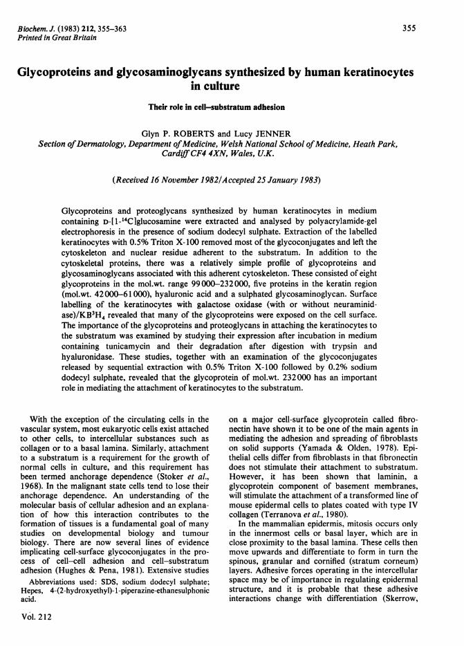
Fig. 1. SDS/polyacrylamide-gel electrophoresis ofkeratinocyte proteins after extraction with 0.5% Triton
X-100Human keratinocytes were incubated in mediumcontaining L-[U-'4Clleucine for 24h and the cellswere then extracted with 0.5% Triton X-100. Theextract and the adherent cytoskeleton were analysedon SDS/polyacrylamide-gradient (5-15% w/v) slabgels as described in the Experimental section. About50,g of protein was loaded on the gels. Lanes (a)and (c) show proteins in the adherent cytoskeleton,and lanes (b) and (d) show proteins in the 0.5%Triton X-100 extract. Lanes (a) and (b) were stainedfor protein with Coomassie Blue and lanes (c) and(d) are autoradiographs.
components of 'stick' and 'grip' (Rees et al., 1977).The proteins extracted into 0.5% Triton X-100 cover
a wide molecular-weight range (<14300 to>200000), whereas the major proteins left adherentto the substratum occur grouped together in themolecular-weight range 42000-61000 after electro-phoresis under reducing conditions (Fig. 1). Thepatterns of proteins labelled by incubation withL-[U-14C]leucine were very similar to those revealedby staining with Coomassie Blue in both the TritonX-100 extract and the adherent cytoskeleton. How-ever, the glycoconjugates metabolically labelled with
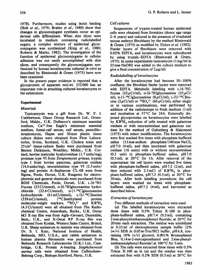
Fig. 2. Effect of tunicamycin and trypsin digestion onglycoconjugates associated with the adherent cytoskele-
tonsKeratinocytes were cultured in the presence ofD-[1-14C]glucosamine for 24h and then extractedwith 0.5% Triton X-100. The adherent cytoskeletonwas dissolved in electrophoresis sample buffer andanalysed on SDS/polyacrylamide-gradient (5-15%,w/v) slab gels. The labelled glycoconjugates were
detected by autoradiography. Lane (a), adherentcytoskeleton of keratinocytes grown in normalmedium; lane (b), keratinocytes grown in mediumcontaining tunicamycin (2,ug/ml); lane (c), keratino-cytes grown in normal medium and then digestedwith 0.5% trypsin before extraction with 0.5%Triton X-100. Approx. 50,ug of protein was appliedto all the gels.
D-[ 1-14Clglucosamine differed considerably from theprotein patterns. In the adherent cytoskeleton therewere eight large glycoproteins, with apparentmol.wts. 232000, 194000, 180000, 165000,148000, 129000, 114000 and 99000, and fivelabelled bands in the keratin region (mol.wts.42000-61 000) (Fig. 2). The molecular-weightvalues were means from seven different experiments
1983
358
Actin a

Glycoconjugates and keratinocyte-substratum adhesion
and, since glycoproteins bind less SDS than doproteins on a weight basis (Pitt-Rivers & Impiom-bato, 1968), these are only approximate values. Ofthe large glycoproteins, those with mol.wts. 232000,180000, 148000 and 129000 were the mostintensely labelled, and the intensities of the otherbands varied in different experiments. One band, ofmol.wt. 54000, in the keratin region was con-sistently well labelled in all experiments, but theintensities of the other bands in this region dependedon the conditions used for their detection. They wererelatively intense when autoradiography was usedfor detection, but weak when fluorography wasemployed.
In addition to the glycoproteins described above,electrophoresis of the adherent cytoskeleton revealedan intensely labelled band close to the origin in thestacking gel. The radioactivity in gel slices from thisarea was rapidly solubilized by digestion withhyaluronidase, but not by digestion with proteasetype VI from Streptomyces griseus. On prolongedincubation (3 days at 370C) with protease type VI asmall amount of radioactivity was solubilized, andthis was shown to be incorporated into proteo-glycans by precipitation with cetylpyridinium chlor-ide followed by salt fractionation as described bySaarni & Tammi (1977). The glycosaminoglycanssynthesized by keratinocytes cultured in the pres-ence of D-[6-3H]glucosamine and 35 042- werecharacterized by electrophoresis on cellulose acet-ate. Hyaluronic acid accounted for 62% of theD-[6-3H]glucosamine in the glycosaminoglycans,and a further 30% was recovered in the areacorresponding to heparan sulphate. A small amount(8%) of the D-[6-3H]glucosamine was incorporatedinto material migrating in the same region aschondroitin 4-sulphate, dermatan sulphate and chon-droitin 6-sulphate. The identity of hyaluronic acidwas confirmed by the fact that it did not contain35S042- and it was degraded by hyaluronidase.A similar profile of glycoconjugates was labelled
with L-[6-3H]fucose and D-[6-3H]glucosamine inboth the Triton X-100 extracts and the adherentcytoskeleton, with the exceptions that the proteo-glycan band was missing and labelling in the keratinregion of the adherent cytoskeleton was decreasedwith L-[6-3H]fucose.
Effect oftunicamycin, trypsin and hyaluronidaseThe importance of the glycoconjugates present in
the adherent cytoskeleton in attaching the kera-tinocytes to the substratum was examined byinvestigating the effect of tunicamycin, trypsin andhyaluronidase on detachment of keratinocytes andalso their effect on the glycoconjugates. Whenkeratinocytes were cultured in the presence oftunicamycin (2,ug/ml) for up to 5 days, thekeratinocytes remained attached to the substratum.
Under these conditions irradiated fibroblasts becamedetached during the first 24h. The electrophoreticpattern of the adherent cytoskeleton from kera-tinocytes labelled with D-[1-_4Clglucosamine in thepresence of tunicamycin for 24 h is shown in lane (b)of Fig. 2. Labelling of the seven glycoproteins withmol.wts. in the range 99000-194000 was greatlydecreased relative to the labelling of the glyco-protein of mol.wt. 232000. The proteoglycans andlabelled bands in the keratin region were not affectedby tunicamycin.
Digestion of D-[ 1-'4C]glucosamine-labelled kera-tinocytes with 0.2% trypsin at 370C for 15minresulted in detachment and dispersal of the cells.Extraction of these cells with 0.5% Triton X-100followed by electrophoretic examination of theresidue left from both the cells and the plasticsurface resulted in the autoradiogram shown in lane(c) of Fig. 2. Whereas the labelled bands in thekeratin regions were not affected by digestion withtrypsin, the amounts of the glycoproteins in themol.wt. range 99000-232000 were greatly de-creased, and most of the proteoglycans werereleased from the cells.
Removal of hyaluronic acid by digestion ofkeratinocytes with hyaluronidase did not result indetachment of the cells. However, a sulphatedglycosaminoglycan associated with the adherentcytoskeleton was resistant to hyaluronidase.
Sequential extraction of labelled keratinocytes with0.5% Triton X-100, 0.2% SDS and 0.2% SDS/0.14 M-2-mercaptoethanol
Keratinocytes labelled with D-[ 1-14C]glucosaminewere extracted sequentially with 0.5% Triton X-100,0.2% SDS and 0.2% SDS/0. 14 M-2-mercapto-ethanol at 200 C. The adherent cytoskeleton left afterextraction with 0.5% Triton X-100 started to detachfrom the substratum in large sheets on incubation in0.2% SDS. The ease with which the adherentcytoskeleton detached varied in different experi-ments and appeared to decrease with increasingculture time. After removal of the supernatant, thesheets and remaining adherent cytoskeleton wereextracted with 0.2% SDS/0.14M-2-mercapto-ethanol. This treatment solubilized the adherentcytoskeleton. An autoradiograph of the electro-phoretic patterns of the three extracts is shown inFig. 3. A complex mixture of glycoconjugates wasextracted with Triton X-100, whereas the 0.2%SDS/0. 14 M-2-mercaptoethanol extract contains asimpler mixture similar to that of the total adherentcytoskeleton shown previously (Fig. 2, lane a). Inthe 0.2%-SDS extract there was considerable en-richment of the glycoprotein of mol.wt. 232000relative to the other glycoproteins. This glyco-Xprotein was not bound by anti-laminin or anti-fibronectin sera in immunoprecipitation experi-
Vol. 212
359

G. P. Roberts and L. Jenner
(a) (b) (c}
_ ~~~10-3 x mol.wt.
.... .....o
30
-14.3
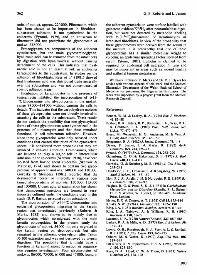
Fig. 3. Sequential extraction Of D-[1-14C]glucosamine-labelled keratinocytes with 0.5% Triton X-100, 0.2%
SDS and 0.2% SDSI0.14M-2-mercaptoethanolKeratinocytes cultured in the presence of D-[ 1-
14C]glucosamine for 24 h were extracted sequen-tially with 0.5% Triton X-100, 0.2% SDS and 0.2%SDS/0.14M-2-mercaptoethanol as described in theExperimental section. The extracts were freeze-driedand analysed by SDS/polyacrylamide-gradient (5-15%, w/v) gel electrophoresis. The labelled glyco-conjugates were detected by autoradiography. Lane(a), 0.5% Triton extract (30Oug of protein); lane (b),0.2% SDS extract (7,ug of protein); lane (c), 0.2%SDS/0.14M-2-mercaptoethanol extract (18pug ofprotein).
ments. Some glycosaminoglycan was also ex-
tracted, and a similar experiment with keratinocyteslabelled with D-[6-3H]glucosamine and 35SO42-revealed that some of the glycosaminoglycan was
sulphated. Electrophoresis of the glycosaminogly-cans on cellulose acetate revealed that hyaluronicacid (90%) was the major glycosaminoglycan, withsmall amounts (10%) of material with the migrationcharacteristics of heparan sulphate.
In addition to the glycoprotein of mol.wt. 232000and glycosaminoglycan, the 0.2%-SDS extractcontained a small amount of a glycoprotein withmol.wt. 47000. However, this material may be anintracellular component since it was not destroyedby trypsin (Fig. 2, lane c).
In order to examine the possibility that in-complete removal of fibroblasts was contributing tothe glycoconjugates found in the adherent cyto-skeletons of keratinocytes, irradiated fibroblastswere metabolically labelled with D-[ 1-14CIglucos-amine for 24h. The labelled fibroblasts were ex-tracted sequentially with 0.5% Triton X-100, 0.2%SDS and 0.2% SDS/0.14M-2-mercaptoethanol, andthe extracts were examined by SDS/polyacryl-amide-gel electrophoresis and autoradiography. The0.5%-Triton-X- 100 extract contained a complexmixture of glycoconjugates, some of which corre-sponded to labelled bands found in the Triton X-100extract from keratinocytes. However, the 0.2%-SDSand 0.2%-SDS/0. 14 M-2-mercaptoethanol extractsfrom fibroblasts differed from the correspondingextracts from keratinocytes. Only a glycoproteinband of mol.wt. 221000, together with a faint bandin the glycosaminoglycan region, was found in the0.29-SDS extract and no bands were detected in the0.296-SDS/0.14M-2-mercaptoethanol extract fromfibroblasts. Furthermore, when a heavy inoculum ofkeratinocytes was subcultured for a short time inthe absence of fibroblasts, the keratinocytes didadhere and spread on to the Petri-dish surface. Thesekeratinocytes were labelled with D-[ 1-14CIglucos-amine and subjected to sequential extraction with0.5% Triton X-100, 0.2% SDS and 0.2% SDS/0.14 M-2-mercaptoethanol. Electrophoretic analysisof these extracts revealed that the glycoproteinssynthesized were similar to those shown in Fig. 3,thus demonstrating that these glycoproteins were notproducts of the fibroblasts.
Surface labelling of keratinocytes with galactoseoxidase/KB3H4
Further characterization of the glycoconjugatesinvolved in cell-substratum adhesion was per-formed by labelling the glycoconjugates exposed onthe surface of keratinocytes with galactose oxi-dase/KB3H4 either with or without neuraminidasedigestion. The labelled keratinocytes were extractedwith 0.5% Triton X-100 and the adherent cyto-skeleton was examined by SDS/polyacrylamide-gelelectrophoresis. Fluorographs of the electrophoreticpatterns are shown in Fig. 4. Neuraminidasedigestion increased the labelling intensity of thebands, but otherwise the electrophoretic patterns ofkeratinocytes labelled with and without neuraminid-ase digestion were similar. Comparison of theelectrophoretic patterns of adherent cytoskeletonsfrom metabolically labelled keratinocytes (Fig. 2,
1983
360
..........

Glycoconjugates and keratinocyte-substratum adhesion
(a) (b)
_l-3 X Mol.wt.
*...*.
-200
-92.5
-69
-46
-30
- 14.3
Fig. 4. Surface labelling of keratinocytes with galactoseoxidase/KB3H4
Keratinocytes were surface labelled by incubationwith galactose oxidase or neuraminidase and galact-ose oxidase, followed by reduction with KB3H4 asdescribed in the Experimental section. The labelledkeratinocytes were extracted with 0.5% TritonX-100 and the adherent cytoskeletons were ana-lysed on SDS/polyacrylamide-gradient (5-15%,w/v) slab-gels. The labelled glycoconjugates weredetected by fluorography. The Figure shows theglycoconjugates of the adherent cytoskeleton afterlabelling wtih (a) galactose oxidase/KB3H4 and (b)neuraminidase/galactose oxidase/KB3H4. About20ug of protein was loaded on the gels.
lane a) with those of surface-labelled keratinocytes(Fig. 4) showed that the glycoproteins with mol.wts.232000, 194000, 148000, 129000 and 99 000 wereboth metabolically labelled and surface labelled. Inthe keratin region three bands with mol.wts. 61 000,51000 and 47000 were intensely surface-labelled,but only one of them, the band with mol.wt. 51 000,was metabolically labelled with D-[ 1-4Clglucos-amine. Two other bands, mol.wts. 75000 and86000, were surface-labelled but not metabolicallylabelled.
DiscussionExtraction of cultured cells with non-ionic deter-
gents has proved useful for detecting the cytoskeletalfilaments which remain firmly attached to thesubstratum (Osborn & Weber, 1977). It is stilluncertain how the cytoskeletons are anchored to thesubstratum, but evidence from intact cells suggeststhat there is a transmembrane linkage of cyto-skeletal structure to the pericellular matrix (Hynes &Destree, 1978; Singer, 1979). Rees et al. (1977) havestudied the relationships between internal andexternal structures in fibroblast adhesion to sub-stratum, and they have introduced the concept of'grip' and 'stick' in the control of cell adhesion.Control of 'sticking' by physical forces at the outersurface is exercised through the 'grip' of thecytoskeleton. In the present study both CoomassieBlue staining and L-[U-_4Clleucine uptake revealed arelatively simple pattern of proteins in the adherentcytoskeleton left after extraction of keratinocyteswith Triton X-100. In addition to a protein with themobility of actin (mol.wt. 42000), there was a seriesof seven proteins in the keratin region, which wereprobably derived from the intermediate filaments.D-[ 1-14C]Glucosamine was incorporated into glycos-aminoglycans (mainly' hyaluronic acid, with smalleramounts of a sulphated glycosaminoglycan with themigration properties of heparin sulphate), into eightlarge glycoproteins (mol.wts. 232000, 194000,180000, 165000, 148000, 129000, 114000 and99000) and into five bands in the keratin region(mol.wts. 42000-61 000). Surface labelling withgalactose oxidase/KB3H4 revealed that the glyco-proteins with mol.wts. 232000, 194000, 148000,129000 and 99000 were located on the exteriorsurface of the keratinocytes. The results of trypsindigestion, tunicamycin treatment and sequentialextraction with Triton X-100 and SDS indicate thatthe glycoprotein of mol.wt. 232000 has an import-ant role in attaching the keratinocytes to thesubstratum. Two non-collagenous glycoproteinswith molecular weights in this region have beendescribed in the basement-membrane zone of theepidermal-dermal junction. These are laminin andbullous-pemphigoid antigen (Briggaman, 1982),both of which are synthesized by keratinocytes inculture (Stanley et al., 1982). Laminin consists ofseveral subunits (mol.wts. 220000 and 440000)linked to each other by disulphide bonds (Timplet al., 1979) and has been shown to stimulate theadhesion of mouse epidermal cells to collagensubstrates (Terranova et al., 1980). However, therewas no evidence of a second subunit of mol.wt.440000 in the present study, and in immuno-precipitation experiments the glycoprotein of mol.wt.232000 was not precipitated by anti-laminin sera.Stanley et al. (1981) have shown that the bullous-pemphigoid antigen contains disulphide-linked sub-
Vol. 212
361
362 G. P. Roberts and L. Jenner
units of mol.wt. approx. 220000. Fibronectin, whichhas been shown to be important in fibroblast-substratum adhesion, is not synthesized in theepidermis (Fyrand, 1979), and an antiserum tofibronectin did not precipitate the glycoprotein ofmol.wt. 232000.
Proteoglycans are components of the adherentcytoskeleton, but the main glycosaminoglycan,hyaluronic acid, can be removed from the intact cellsby digestion with hyaluronidase without causingdetachment of the cells. This indicates that hyal-uronic acid is not an essential link in anchoringkeratinocytes to the substratum. In studies on theadhesion of fibroblasts, Rees et al. (1981) showedthat hyaluronic acid was distributed quite generallyover the substratum and was not concentrated atspecific adhesion areas.
Incubation of keratinocytes in the presence oftunicamycin inhibited the incorporation of D-[ 1-14C]glucosamine into glycoproteins in the mol.wt.range 99000-194000 without causing the cells todetach. This indicates that the carbohydrate moietiesof these glycoproteins were not directly involved inattaching the cells to the substratum. These resultsdo not exclude the possibility that non-glycosylatedforms of these glycoproteins were synthesized in thepresence of tunicamycin and that these remainedfunctional in cell-substratum adhesion. However,since these glycoproteins were only released underconditions that caused disruption of the cytoskeletalsheets, it is considered more probable that they areinvolved in cell-cell adhesion. Desmosomes, whichare believed to play a major role in intercellularadhesion in the epidermis (Skerrow, 1978), have beenisolated from bovine snout epidermis (Skerrow &Matoltsy, 1974) and shown to contain two glyco-proteins of apparent mol.wts. 140000 and 120000.Gorbsky & Steinberg (1981) reported that thedesmosomal 'cores' or intercellular regions con-tained glycoproteins of mol.wts. 150000, 115000and 100000. Ultrastructural examination has shownthat desmosomal junctions are formed in kera-tinocytes cultured under the conditions used in thisstudy (S. P. Barton, personal communication).The incorporation of D-[1-'4C]glucosamine into
epidermal glycoproteins migrating in the keratinregion was investigated previously (Roberts &Marks, 1982) and shown to be mainly due toglycoproteins which co-migrated with the mainkeratin polypeptides. In the present study theglycoprotein of mol.wt. 54000 not only migrated inthe keratin region on electrophoresis but alsoremained in the adherent cytoskeleton after TritonX-100 extraction and was not destroyed by trypsindigestion. The possibility that it might have afunction in keratin-filament formation or organiza-tion requires investigation. Four glycoproteins, ofmol.wts. 86 000, 75 000, 61 000 and 47 000, found in
the adherent cytoskeleton were surface-labelled withgalactose oxidase/KB3H4 after neuraminidase diges-tion, but were not detected by metabolic labellingwith D-[ 1-14C]glucosamine of keratinocytes orirradiated fibroblasts. In view of the probability thatthese glycoproteins were derived from the serum inthe medium, it is noteworthy that one of theseglycoproteins has a similar molecular weight toepibolin, an epidermal spreading factor isolated fromserum (Stenn, 1981). Epibolin is claimed to berequired for epidermal cell migration in vivo andmay be important in areas such as wound healingand epithelial tumour metastases.
We thank Professor R. Marks and Dr. P. J. Dykes foradvice with various aspects of this work and the MedicalIllustration Department of the Welsh National School ofMedicine for preparing the Figures in this paper. Thework was supported by a project grant from the MedicalResearch Council.
References
Bonner, W. M. & Laskey, R. A. (1974) Eur. J. Biochem.46, 83-88
Brabec, R. K., Peters, B. P., Bernstein, I. A., Gray, R. H.& Goldstein, I. J. (1980) Proc. Natl. Acad. Sci.U.S.A. 77,477-479
Breen, M., Weinstein, H. G., Andersen, M. & Veis, A.(1970) Anal. Biochem. 35, 146-159
Briggaman, R. A. (1982) J. Invest. Dermatol. 78, 1-6Dykes, P., Jenner, L. & Marks, R. (1982) Arch.
Dermnatol. Res. 273, 225-231Fyrand, 0. (1979) Br. J. Dermatol. 101, 263-270Gahmberg, C. G. & Hakomori, S. I. (1973) J. Biol.
Chem. 248,4311-4317Gorbsky, G. & Steinberg, M. S. (1981) J. Cell Biol. 90,
243-248Henderson, L. E., Oroszlan, S. & Konigsberg, W. (1979)
Anal. Biochem. 93, 153-157Holt, P. J. A., Anglin, J. H. & Nordquist, R. E. (1979) Br.
J. Dermatol. 100, 237-245Hughes, R. C. & Pena, S. D. J. (1981) in Carbohydrate
Metabolism and its Disorders (Randle, P. J., Steiner,D. F. & Whelan, W. J., eds.), pp. 363-423, AcademicPress, London
Hynes, R. 0. & Destree, A. T. (1978) Cell 15, 875-886Kessler, S. W. (1976) J. Immunol. 117, 1482-1490King, I. A. (1981) Biochim. Biophys. Acta 674, 87-95King, I. A., Tabiowo, A. & Williams, R. H. (1980)
Biochem. J. 190, 65-77Laemmli, U. K. (1970) Nature (London) 227, 680-685Laskey, R. A. & Mills, A. D. (1975) Eur. J. Biochem. 56,
335-341Lowry, 0. H., Rosebrough, N.- J., Farr, A. L. & Randall,
R. J. (1951) J. Biol. Chem. 193, 265-275Osborn, M. & Weber, K. (1977) Exp. Cell Res. 106,
339-349Pitt-Rivers, R. & Impiombato, F. S. A. (1968) Biochem.
J. 109, 825-830Rees, D. A., Lloyd, C. W. & Thom, D. (1977) Nature
(London) 267, 124-128
1983

Glycoconjugates and keratinocyte-substratum adhesion 363
Rees, D. A., Badley, R. A., Bayley, S. A., Couchman,J. R., Smith, C. G. & Woods, A. (1981) in CellularInteractions (Dingle, J. T. & Gordon, J. L., eds.), pp.67-80, Elsevier/North-Holland, Amsterdam
Rheinwald, J. G. & Green, H. (1975) Cell 6, 331-344Roberts, G. P. & Marks, R. (1982) Br. J. Dermatol. 106,
727Saarni, H. & Tammi, M. (1977) Anal. Biochem. 81, 40-46Singer, I. I. (1979) Cell 16, 675-685Skerrow, C. J. (1978) Invest. Cell Pathol. 1, 23-37Skerrow, C. J. & Matoltsy, A. G. (1974) J. Cell Biol. 63,
524-530Stanley, J. R., Hawley-Nelson, P., Yuspa, S. H., Shevach,
E. M. & Katz, S.I. (1981) Cell 24, 897-903
Stanley, J. R., Hawley-Nelson, P., Yaer, M., Martin,G. R. & Katz, S. I. (1982) J. Invest. Dermatol. 82,456-459
Stenn, K. S. (1981) Proc. Natl. Acad. Sci. U.S.A. 78,6907-6911
Stoker, M., O'Neill, C., Berryman, S. & Waxman, V.(1968) Int. J. Cancer 3, 683-693
Terranova, V. P., Rohrbach, D. H. & Martin, G. R.(1980) Cell 22, 719-726
Timpl, R., Rohde, H., Robey, P. G., Rennard, S. I.,Foidart, J. M. & Martin, G. R. (1979) J. Biol. Chem.254, 9933-9937
Yamada, K. M. & Olden, K. (1978) Nature (London)275, 179-184
Vol. 212