glycogen in normal human iris pigment epithelium
TRANSCRIPT
GLYCOGEN IN NORMAL HUMAN IRIS PIGMENT EPITHELIUM
JOSEPH W. BERKOW, M.D., AND BEN S. F INE, M.D.
Washington, D.C.
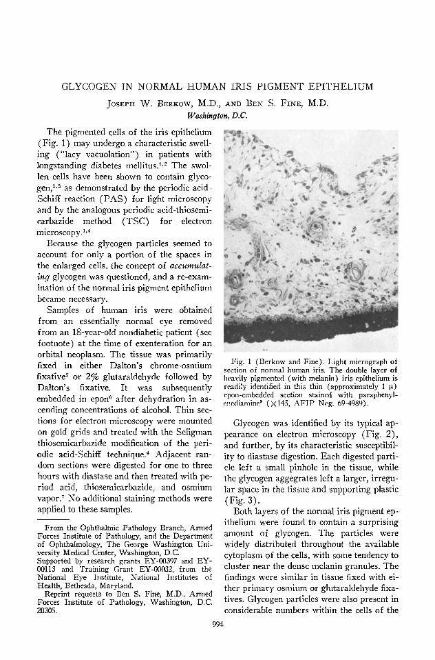
The pigmented cells of the iris epithelium ( Fig. 1 ) may undergo a characteristic swelling ("lacy vacuolation") in patients with longstanding diabetes mellitus.1'2 The swollen cells have been shown to contain glycogen,1·3 as demonstrated by the periodic acid-Schiff reaction (PAS) for light microscopy and by the analogous periodic acid-thiosemi-carbazide method (TSC) for electron microscopy.1'4
Because the glycogen particles seemed to account for only a portion of the spaces in the enlarged cells, the concept of accumulating glycogen was questioned, and a re-examination of the normal iris pigment epithelium became necessary.
Samples of human iris were obtained from an essentially normal eye removed from an 18-year-old nondiabetic patient (see footnote) at the time of exenteration for an orbital neoplasm. The tissue was primarily fixed in either Dalton's chrome-osmium fixative5 or 2% glutaraldehyde followed by Dalton's fixative. It was subsequently embedded in epone after dehydration in ascending concentrations of alcohol. Thin sections for electron microscopy were mounted on gold grids and treated with the Seligman thiosemicarbazide modification of the periodic acid-Schiff technique.4 Adjacent random sections were digested for one to three hours with diastase and then treated with period acid, thiosemicarbazide, and osmium vapor.7 No additional staining methods were applied to these samples.
From the Ophthalmic Pathology Branch, Armed Forces Institute of Pathology, and the Department of Ophthalmology, The George Washington University Medical Center, Washington, D.C. Supported by research grants EY-00397 and EY-00113 and Training Grant EY-00032, from the National Eye Institute, National Institutes of Health, Bethesda, Maryland.
Reprint requests to Ben S. Fine, M.D., Armed Forces Institute of Pathology, Washington, D.C. 2030S.
Fig. 1 (Berkow and Fine). Light micrograph of section of normal human iris. The double layer of heavily pigmented (with melanin) iris epithelium is readily identified in this thin (approximately 1 μ) epon-embedded section stained with paraphenyl-enediamine* (Xl45, AFIP Neg. 69-4989).
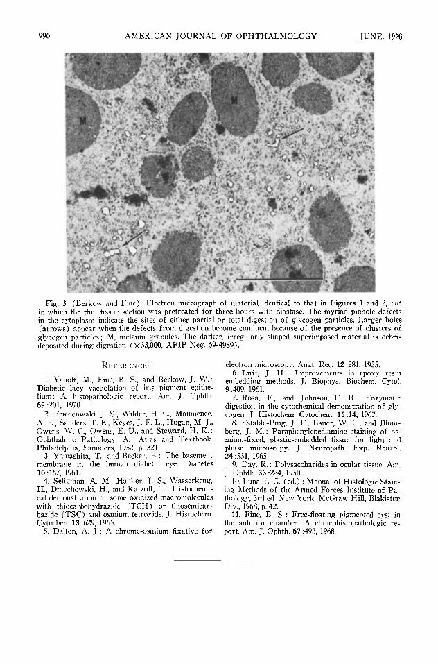
Glycogen was identified by its typical appearance on electron microscopy (Fig. 2) , and further, by its characteristic susceptibility to diastase digestion. Each digested particle left a small pinhole in the tissue, while the glycogen aggegrates left a larger, irregular space in the tissue and supporting plastic (Fig. 3) .
Both layers of the normal iris pigment epithelium were found to contain a surprising amount of glycogen. The particles were widely distributed throughout the available cytoplasm of the cells, with some tendency to cluster near the dense melanin granules. The findings were similar in tissue fixed with either primary osmium or glutaraldehyde fixatives. Glycogen particles were also present in considerable numbers within the cells of the
994

VOL. 69, NO. 6 NORMAL IRIS PIGMENT EPITHELIUM 995
Fig. 2 (Berkow and Fine). Electron micrograph of iris pigment epithelium fixed in glutaraldehyde and osmium and treated with periodic acid-thiosemicarbazide. The clusters of small dense particles (arrows) represent the glycogen particles; M, melanin granules (X33.000, AFIP Neg. 69-4989).
dilator muscle, a neuroectodermal smooth muscle that is derived from, and remains in continuity with, the anterior layer of pigment epithelium.*
The presence of glycogen in the iris pigment epithelium had not been previously detected by light microscopy,9 presumably because the PAS-positive material is effectively masked by the presence of the melanin granules. Methods of bleaching the melanin granules would also destroy the glycogen.10
By electron microscopy, glycogen particles have been reported to be present within the pigmented epithelial cells of the iris that have separated to become a free-floating pigmented cyst in the anterior chamber.11 These
* Similar TSC-reactive particles were also found in the pigment epithelium of the iris from a five-month-old child, a 10-month-old child, and 11-month-old child, a 50-year-old woman, and a normal Rhesus monkey, all of whom had anterior segments that appeared morphologically normal.
cells were considered to be essentially normal. Separation of the melanin granules by
materials accumulating within the pigmented epithelial cells of the iris might serve to unmask the glycogen in the diabetic iris and thus create the illusion of accumulating glycogen.2 No quantitation has been made here of the glycogen present in either the normal or the abnormal diabetic iris.
S U M M A R Y
Glycogen, characteristically observed in the iris pigment epithelium in some cases of diabetes mellitus, was demonstrated to be present in considerable amount in the normal human iris pigment epithelium. By electron microscopy, the particulate intracellular material was demonstrated by a technique analogous to the P A S reaction commonly used in light microscopy. This material was also identified as glycogen by its susceptibility to digestion by diastase.
996 A M E R I C A N J O U R N A L O F O P H T H A L M O L O G Y JUNE, 1970
"tm
Fig. 3. (Berkow and Fine). Electron micrograph of material identical to that in Figures 1 and 2, but in which the thin tissue section was pretreated for three hours with diastase. The myriad pinhole defects in the cytoplasm indicate the sites of either partial or total digestion of glycogen particles. Larger holes (arrows) appear when the defects from digestion become confluent because of the presence of clusters of glycogen particles ; M, melanin granules. The darker, irregularly shaped superimposed material is debris deposited during digestion (χ33,000, A F I P Neg. 69-4989).
R E F E R E N C E S
1. Yanoff, M., Fine, B. S., and Berkow, J. W.: Diabetic lacy vacuolation of iris pigment epithelium: A histopathologic report. Am. J. Ophth. 69:201, 1970.
2. Friedenwald, J. S., Wilder, H. C , Maumcnee, A. E., Sanders, T. E., Keyes, J. E. L., Hogan, M. J., Owens, W. C, Owens, E. U., and Steward, H. K. : Ophthalmic Pathology. An Atlas and Textbook. Philadelphia, Saunders, 1952, p. 321.
3. Yamashita, T., and Becker, B. : The basement membrane in the human diabetic eye. Diabetes 10:167, 1961.
4. Seligman, A. M., Hanker, J. S., Wasserkrug, H., Dmochowski, H., and Katzoff, L. : Histochemi-cal demonstration of some oxidized macromolecules with thiocarbohydrazide ( T C H ) or thiosemicar-bazide ( T S C ) and osmium tetroxide. J. Histochem. Cytochem.13 :629, 1965.
5. Dalton, A. J. : A chrome-osmium fixative for
electron microscopy. Anat. Rec. 12 :281, 1955. 6. Luft, J. H. : Improvements in epoxy resin
embedding methods. J. Biophys. Biochem. Cvtol. 9 :409, 1961.
7. Rosa, F., and Johnson, F. B. : Enzymatic digestion in the cytochemical demonstration of glycogen. J. Histochem. Cytochem. 15:14, 1967.
8. Estable-Puig, J. F., Bauer, W. C , and Blum-berg, J. M. : Paraphenylenediamine staining of osmium-fixed, plastic-embedded tissue for light and phase microscopy. J. Neuropath. Exp. Neurol. 24:531, 1965.
9. Day, R. : Polysaccharides in ocular tissue. Am. J. Ophth. 33 :224, 1950.
10. Luna, L. G. (cd.) : Manual of Histologie Staining Methods of the Armed Forces Institute of Pa-thologv, 3rd ed. New York, McGraw-Hill, Blakiston Div., 1968, p. 42.
11. Fine, B. S. : Free-floating pigmented cyst in the anterior chamber. A clinicohistopathologic report. Am. J. Ophth. 67 :493, 1968.