glycobiology and extracellular matrices: o-glcnac … · jürgen roth and jin won cho sung-min kim,...
TRANSCRIPT

Jürgen Roth and Jin Won ChoSung-Min Kim, Jong In Yook, Yong-Il Park,Insook Jang, Sujin Park, Hyun Sil Kim, Jeong Gu Kang, Sang Yoon Park, Suena Ji, Deprivation through Glycogen DegradationCells Increases in Response to Glucose
-GlcNAc Protein Modification in CancerOGlycobiology and Extracellular Matrices:
doi: 10.1074/jbc.M109.026351 originally published online October 15, 20092009, 284:34777-34784.J. Biol. Chem.
10.1074/jbc.M109.026351Access the most updated version of this article at doi:
.JBC Affinity SitesFind articles, minireviews, Reflections and Classics on similar topics on the
Alerts:
When a correction for this article is posted•
When this article is cited•
to choose from all of JBC's e-mail alertsClick here
Supplemental material:
http://www.jbc.org/content/suppl/2009/10/15/M109.026351.DC1.html
http://www.jbc.org/content/284/50/34777.full.html#ref-list-1
This article cites 40 references, 20 of which can be accessed free at
at YO
NSE
I UN
IVE
RSIT
Y on January 21, 2014
http://ww
w.jbc.org/
Dow
nloaded from
at YO
NSE
I UN
IVE
RSIT
Y on January 21, 2014
http://ww
w.jbc.org/
Dow
nloaded from

O-GlcNAc Protein Modification in Cancer CellsIncreases in Response to Glucose Deprivation throughGlycogen Degradation*□S
Received for publication, May 28, 2009, and in revised form, September 24, 2009 Published, JBC Papers in Press, October 15, 2009, DOI 10.1074/jbc.M109.026351
Jeong Gu Kang‡1, Sang Yoon Park‡1, Suena Ji‡1, Insook Jang‡1, Sujin Park‡1, Hyun Sil Kim§, Sung-Min Kim¶,Jong In Yook§, Yong-Il Park¶, Jurgen Roth�, and Jin Won Cho‡�2
From the Departments of ‡Biology and §Oral Pathology, Oral Cancer Research Institute, and �World Class University IntegratedOMICS for Biomedical Science, Yonsei University, 134 Shinchon-dong, Seodaemun-gu, Seoul 120-749, Korea, and the¶Department of Biotechnology and Biomaterial Engineering Research Center, Catholic University of Korea, Bucheon 420-743, Korea
When cellular glucose concentrations fall below normal lev-els, in general the extent of protein O-GlcNAc modification(O-GlcNAcylation) decreases. However, recent reports de-monstrated increased O-GlcNAcylation by glucose deprivationin HepG2 and Neuro-2a cells. Here, we report increasedO-GlcNAcylation in non-small cell lung carcinoma A549 cellsand various other cells in response to glucose deprivation.Although the level ofO-GlcNAc transferase was unchanged, theenzyme contained lessO-GlcNAc, and its activitywas increased.Moreover,O-GlcNAcase activity was reduced. The studied cellscontain glycogen, and we show that its degradation in responseto glucose deprivation provides a source for UDP-GlcNAcrequired for increased O-GlcNAcylation under this condition.This required active glycogen phosphorylase and resulted inincreased glutamine:fructose-6-phosphate amidotransferase,the first and rate-limiting enzyme in the hexosamine biosyn-thetic pathway. Interestingly, glucose deprivation reduced theamount of phosphofructokinase 1, a regulatory glycolytic en-zyme, and blocked ATP synthesis. These findings suggest thatglycogen is the source for increased O-GlcNAcylation but notfor generating ATP in response to glucose deprivation and thatthis may be useful for cancer cells to survive.
Extracellular glucose is transferred into cells by glucosetransporters (1, 2), where it is rapidly converted into glucose6-phosphate by hexokinase. Glucose 6-phosphate is either usedfor glycogen synthesis or enters the glycolytic pathway afterconversion to fructose 6-phosphate. Most of the transferredglucose is used for ATP synthesis through the glycolytic path-way and the tricarboxylic acid cycle. Only about 2–5% of thetransferred glucose enters the hexosamine biosynthetic path-
way (HBP)3 and is converted into uridine 5�-diphospho-N-acetylglucosamine (UDP-GlcNAc) (3). UDP-GlcNAc is trans-ferred by O-GlcNAc transferase (OGT) to serine or threonineresidues to yield the single O-linked �-N-acetylglucosamine(O-GlcNAc)modification (4). UDP-GlcNAc represents a nutri-ent sensor being the product of various metabolic pathwayssuch as the glucose, fatty acid, amino acid, nucleotide, andenergy metabolism (5, 6). Because protein O-GlcNAc modifi-cation (O-GlcNAcylation for brevity) by OGT is highly depen-dent onUDP-GlcNAc concentration,O-GlcNAcmetabolism isthought to have a critical role in nutrient homeostasis (5, 6).Since its first description as a novel posttranslational protein
modification by Torres and Hart (7), �600 proteins as diverseas transcription factors, enzymes, cytoskeletal proteins, andribosomal proteins have been identified to carry O-linkedGlcNAc (4–6). Thus, protein O-GlcNAcylation appears to beinvolved in many different cellular activities, which include theregulation of transcription and translation (8–12), of proteindegradation (12–16), and localization (17), as well as of protein-protein interactions (13, 18). Of particular interest are reportsthatO-GlcNAcylation of nucleocytoplasmic proteins can occurunder various forms of cellular stress such as heat shock, UVlight, and high salt concentration (19, 20). Under such condi-tions, the O-GlcNAcylation may have a function for cell sur-vival, but the mechanism is not clear.Recently, it was reported that glucose deprivation
increases protein O-GlcNAcylation in HepG2 (21) andNeuro-2a cells (22). Further studies have revealed thedynamics of O-GlcNAcylation in HepG2 cells during glucosedeprivation and have shown that up-regulation of OGT ismediated by decreasedHBP flux (23). In light of the abovemen-tioned importance of glucose as a source for UDP-GlcNAc syn-thesis and thus protein O-GlcNAcylation, increased O-Glc-NAcylation under glucose deprivation appears paradoxical.However, it may provide a clue for the importance of theO-GlcNAcylation as a nutrient sensor, in glucose homeostasis,
* This work was supported by the Korea Science and Engineering Foundation(KOSEF) funded by Ministry of Education, Science, and Technology GrantR0A-2007-000-20011-0, Korea Research Foundation Grant KRF-2006-005-J04502, and World Class University Project R31-2008-000-10086-0. Thiswork was made possible through the use of research facilities in the YonseiCenter for Biotechnology.
□S The on-line version of this article (available at http://www.jbc.org) con-tains supplemental “Experimental Procedures” and Figs. 1 and 2.
1 Fellowship awardees of the Brain Korea 21 program.2 To whom correspondence should be addressed: Shinchon-dong, Seodae-
mun-gu, Seoul 120-749, Korea. Fax: 82-2-312-5657; E-mail: [email protected].
3 The abbreviations used are: HBP, hexosamine biosynthetic pathway; AMPK,AMP-activated protein kinase; DON, 6-diazo-5-oxo-L-norleucine; GFAT,glutamine:fructose-6-phosphate amidotransferase; GP, glycogen phos-phorylase; O-GlcNAc, O-linked �-N-acetylglucosamine; OGT, O-GlcNActransferase; PAS, periodic acid-Schiff; PBS, phosphate-buffered saline;sWGA, succinylated wheat germ agglutinin; UDP-GlcNAc, uridine5�-diphospho-N-acetylglucosamine.
THE JOURNAL OF BIOLOGICAL CHEMISTRY VOL. 284, NO. 50, pp. 34777–34784, December 11, 2009© 2009 by The American Society for Biochemistry and Molecular Biology, Inc. Printed in the U.S.A.
DECEMBER 11, 2009 • VOLUME 284 • NUMBER 50 JOURNAL OF BIOLOGICAL CHEMISTRY 34777
at YO
NSE
I UN
IVE
RSIT
Y on January 21, 2014
http://ww
w.jbc.org/
Dow
nloaded from

and for cell survival. We observed increased protein O-Glc-NAcylation in glucose-deprived A549 lung cancer cells andother cell lines, similar to that reported for HepG2 andNeuro-2a cells (21, 22). However, we observe that increasedO-GlcNAcylation in response to glucose deprivation was notdue to up-regulated OGT (21, 22) and not dependent on theactivation of AMP-activated protein kinase (AMPK) (22).It was reported that inhibition of glycogen phosphorylase
(GP), which catalyzes glycogen degradation, suppressed thegrowth of A549 cells (24), but the role of glycogen in cancerbiology is not well understood. We demonstrate here that gly-cogen is a source for protein O-GlcNAcylation in glucose-de-prived A549 cells. Thus, protein O-GlcNAcylation in tumorsmay have a function in overcoming the effects of hypoxia andhypoglycemia. Our findings have the interesting implicationthatO-GlcNAcylationmay act as an intermediary between glu-cose and glycogen metabolism in cancer cells.
EXPERIMENTAL PROCEDURES
Cell Culture, Glucose Deprivation, and Inhibitor Treatments—Human non-small cell lung carcinoma cell lines A549, H1299,and H460, human lung L132 fibroblasts, human breast cancerMCF7 cells, and human embryonic kidney HEK293 cells weregrown in high glucose (25 mM) Dulbecco’s modified Eagle’smedium (Invitrogen) containing 10% (v/v) fetal bovine serum(Lonza) and 1% (v/v) antibiotics (Invitrogen) at 37 °C in 5%CO2. Cells were rinsed with PBS and then cultured in glucose-free Dulbecco’s modified Eagle’s medium (WelGENE, Inc.)containing 10% (v/v) fetal bovine serumand 1% (v/v) antibioticsfor up to 36 h. For the inhibitor treatment, cells were cultured inmedium containing either 1 �g/ml cycloheximide (Sigma), 20�M 6-Diazo-5-oxo-L-norleucine (DON, Sigma), 20 �M Com-pound C (Calbiochem), or 20 �M glycogen phosphorylaseinhibitor (Calbiochem) for 1 h. Afterward, cells were washedwith PBS and incubated in the glucose-freemedium containinginhibitor.Transient Overexpression of OGT—Human full-length OGT
was cloned into the p3XFLAG-CMV-7.1 expression vector(Sigma). The vector encoding FLAG-tagged OGT or emptyvector was transfected using Tranfectin (Bio-Rad).Cell Lysis, Immunoprecipitation, and WGA Precipitation—
Cells were washed twice in ice-cold PBS and solubilized in lysisbuffer (50 mM Tris-HCl, pH 7.4, 150 mM NaCl, 1% NonidetP-40, 1 mM EDTA, and protease inhibitors). The lysates werecentrifuged at 18,000� g for 20min at 4 °C. Protein concentra-tions were determined by Bradford assay (Bio-Rad). To immu-noprecipitate FLAG-tagged OGT, 1 mg of a given lysate wasmixed with EZviewTM Red anti-FLAGM2 Affinity Gel (Sigma)overnight at 4 °C under gentle agitation. Beads were washedfour times with lysis buffer. Pellet was resuspended in 2� SDSsample buffer. Samples were boiled for 5 min and subjectedto Western blotting. For succinylated wheat germ agglutinin(sWGA) precipitation, 1mg of cell lysate was incubatedwith 25�l of agarose-sWGA (Vector Laboratories) overnight at 4 °C.The precipitates were washed with lysis buffer and then elutedin 2� SDS sample buffer.Western Blotting—Soluble protein samples or precipitates
were subjected to SDS-PAGE and transferred to nitrocellulose
membranes (GEHealthcare) using a PROTEIN II xi cell system(Bio-Rad) and mini-TransBlot cell system (Bio-Rad). Themembranes were blocked in 5% skimmedmilk in TBST (20mM
Tris, pH 7.4, 150mM sodium chloride, and 1%Tween 20) for 1 hat ambient temperature and then incubated overnight at 4 °C in1% skimmed milk containing diluted primary antibodies. Thefollowing antibodies were used: anti-O-GlcNAc (CTD110.6,Covance), anti-OGT (DM17, Sigma), anti-actin, anti-glyco-gen phosphorylase (Santa Cruz Biotechnology), anti-FLAG(Sigma), anti-AMPK, anti-phospho-AMPK and anti-glycogensynthase (Cell Signaling). Rabbit polyclonal O-GlcNAcaseantibodies were raised by immunizing two rabbits with humanfull-length O-GlcNAcase (AbFrontier). Rabbit polyclonalglutamine:fructose-6-phosphate amidotransferase (GFAT)antibodies were kindly provided by Dr. Pann-Ghill Suh(POSTECH, Pohang, Korea). Afterward, membranes werewashed several times and incubated with horseradish peroxi-dase-conjugated appropriate secondary antibodies (Santa CruzBiotechnology) for 1 h. Following several washes, the mem-branes were developed using ECL reagent (GE Healthcare) andthen exposed to Medical x-ray Film Blue (Agfa) or AmershamBiosciences HyperfilmTM ECL (GE Healthcare). Densitometrymeasurements were performed using aMulti Gauge version 2.3program (Fuji Photo Film).OGT and O-GlcNAcase Assay—OGT activity was measured
in whole cell lysates, which were incubated with reaction buffercontaining recombinant glutathione S-transferase-tagged p62and 100 �M UDP-GlcNAc for 2 h (25). Recombinant glutathi-one S-transferase-tagged p62 fragment used as substrate forOGT was extracted from Escherichia coli and purified on glu-tathione-Sepharose (GEHealthcare). The reactionwas stoppedby the addition of 2 mM glutathione, and supernatants weremixed with SDS-PAGE sample buffer, subjected to SDS-PAGE,and immunoblotted using anti-O-GlcNAc antibodies.O-GlcNAcase assayswere performed as described previously
with minor modifications (10). The whole cell lysates wereincubated with assay buffer (50 mM sodium cacodylate, pH 6.5,50 mM N-acetylgalactosamine, 2 mM p-nitrophenyl-N-acetyl-D-glucosaminide) for 1 h at 37 °C. The reaction was stopped byadding 0.5 M sodium carbonate. Hydrolyzed p-nitrophenol wasmeasured spectrophotometrically at 400 nm.GFAT Assay—The GFAT assay was performed as described
previously with minor modifications (26). Cells were washedwith ice-cold PBS andmechanically removed in 500�l ofGFATbuffer (50 mM Tris-HCl, 5 mM EDTA, 5 mM glucose 6-phos-phate and glutathione, 5mMglucose 6-phophateNa2, and 5mM
KCl, pH7.5). The harvested cells were sonicated andpelleted bycentrifugation (18,000 � g, 20 min). Protein concentration wasdetermined with Bio-Rad protein assay reagent. Samples wereadjusted to the same protein concentration in 500 �l of GFATbuffer. Then 500 �l of reaction buffer (0.8 mM D-fructose6-phosphate, 6.0 mM glutamine, 0.3 mM acetylpyridine adeninedinucleotide, 50 mM KCl, 0.1 mM KH2PO4, 6 units of glutamicacid dehydrogenase, pH 7.8) was added to the respective sam-ples. After incubation for 2 h at 37 °C, changes of absorbance at370 nm due to the reduction of acetylpyridine adenine dinucle-otide were measured.
O-GlcNAc Modification Increases in Glucose-deprived A549
34778 JOURNAL OF BIOLOGICAL CHEMISTRY VOLUME 284 • NUMBER 50 • DECEMBER 11, 2009
at YO
NSE
I UN
IVE
RSIT
Y on January 21, 2014
http://ww
w.jbc.org/
Dow
nloaded from
Periodic Acid-Schiff (PAS) Staining and ATP ContentDetermination—The cell plateswere treatedwith 0.5%periodicacid (Janssen, Geel, Belgium) solution for 10 min. The plateswere rinsed with PBS. Then the cell plates were incubated withSchiff solution (Muto PureChemicals) overnight, washed in tapwater, and counterstained with Harris hematoxylin. ATP con-centrations were determined using the ATP Bioluminescentassay kit (Sigma).Statistical Analysis—Statistical significance of enzyme activ-
ities or ATP level was determined by unpaired Student’s t testusing Microsoft Excel-based application. Results are expressedas mean � S.E.
RESULTS
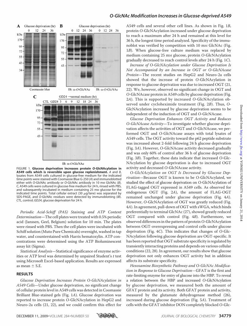
Glucose Deprivation Increases Protein O-GlcNAcylation inA549 Cells—Under glucose deprivation, no significant changeof cellular protein level inA549 cells was detected inCoomassieBrilliant Blue-stained gels (Fig. 1A). Glucose deprivation wasreported to increase protein O-GlcNAcylation in HepG2 andNeuro-2a cells (21, 22), and we could confirm this effect for
A549 cells and several other cell lines. As shown in Fig. 1B,protein O-GlcNAcylation increased under glucose deprivationto reach a maximum after 24 h and remained at this level for36 h, the longest time period analyzed. Specificity of the immu-noblot was verified by competition with 10 mM GlcNAc (Fig.1B). When glucose-free culture medium was replaced bymedium containing 25 mM glucose, protein O-GlcNAcylationgradually decreased to reach control levels after 24 h (Fig. 1C).Increase of O-GlcNAcylation under Glucose Deprivation Is
Not Accompanied by an Increase in OGT or O-GlcNAcaseProtein—The recent studies on HepG2 and Neuro-2a cellsshowed that the increase of protein O-GlcNAcylation inresponse to glucose deprivation was due to increased OGT (21,22). We, however, observed no significant change in OGT andO-GlcNAcase protein in A549 cells by glucose deprivation (Fig.2A). This is supported by increased O-GlcNAcylation ob-served under cycloheximide treatment (Fig. 2B). Thus, O-GlcNAcylation increased by glucose deprivation seems to beindependent of the induction of OGT and O-GlcNAcase.Glucose Deprivation Enhances OGT Activity and Reduces
O-GlcNAcase Activity—To investigate whether glucose depri-vation affects the activities of OGT and O-GlcNAcase, we per-formed OGT and O-GlcNAcase assays with total lysates ofA549 cells. The OGT activity toward the p62 peptide substratewas increased about 2-fold following 24 h glucose deprivation(Fig. 3A). However, O-GlcNAcase activity decreased graduallyand was only 60% of control after 36 h of glucose deprivation(Fig. 3B). Together, these data indicate that increased O-Glc-NAcylation by glucose deprivation is due to increased OGTactivity and decreased O-GlcNAcase activity.O-GlcNAcylation on OGT Is Decreased by Glucose Dep-
rivation—Because OGT is known to be O-GlcNAcylated, westudied the effect of glucose deprivation on O-GlcNAcylation ofFLAG-tagged OGT expressed in A549 cells. As observed forendogenous OGT (Fig. 2A), the amount of FLAG-OGTremained unchanged under glucose deprivation (Fig. 4A).However, O-GlcNAcylation of OGT was greatly reduced (Fig.4A). In agreement, pull-down ofOGTwith sWGA,which bindspreferentially to terminal GlcNAc (27), showed greatly reducedOGT compared with control (Fig. 4B). Furthermore, weobserved differences in the pattern of proteinO-GlcNAcylationbetween OGT-overexpressing and control cells under glucosedeprivation (Fig. 4C). This indicates that changes of O-Glc-NAcylation following glucose deprivation are OGT-specific. Ithas been reported thatOGT substrate specificity is regulated bytransiently interacting proteins and depends on various cellularconditions (22, 28). In agreement, our results show that glucosedeprivation not only enhances OGT activity but in additionaffects its substrate specificity.Hexosamine Biosynthetic Pathway and O-GlcNAc Modifica-
tion in Response to Glucose Deprivation—GFAT is the first andrate-limiting enzyme for entry of glucose into the HBP. To reveala relation between the HBP and increased O-GlcNAcylationby glucose deprivation, we measured both the amount ofGFAT protein and its activity. Both GFAT protein and activity,measured by the glutamate dehydrogenase method (26),increased during glucose deprivation (Fig. 5A). Treatment ofcells with the GFAT inhibitor DON completely blockedO-Glc-
FIGURE 1. Glucose deprivation increases protein O-GlcNAcylation inA549 cells which is reversible upon glucose replenishment. A and B,lysates from A549 cells cultured in glucose-free medium for the indicatedtime points were stained with Coomassie Blue G-250 (A) and immunoblottedeither with O-GlcNAc antibody or O-GlcNAc antibody in 10 mM GlcNAc (B).C, A549 cells were cultured in glucose-free medium for 24 h, rinsed with PBS,and subsequently incubated in medium containing 25 mM glucose for theindicated time points. Total cellular extract (30 �g/lane) was separated bySDS-PAGE, and O-GlcNAc residues were detected by immunoblotting (IB).CTL, control; GD24, glucose deprivation for 24 h.
O-GlcNAc Modification Increases in Glucose-deprived A549
DECEMBER 11, 2009 • VOLUME 284 • NUMBER 50 JOURNAL OF BIOLOGICAL CHEMISTRY 34779
at YO
NSE
I UN
IVE
RSIT
Y on January 21, 2014
http://ww
w.jbc.org/
Dow
nloaded from

NAcylation under glucose deprivation (Fig. 5B). These dataindicate a relation between the HBP and increased O-Glc-NAcylation under conditions of glucose deprivation. However,glucose deprivation reduced the amount of phosphofructoki-nase 1 (Fig. 5C) that converts fructose 6-phosphate into fruc-tose 1,6-bisphosphate under consumption of ATP. Under ourexperimental conditions, we also observed that generation of
ATPwas blocked (Fig. 5D). Because ATP is not only a substratebut also al allosteric inhibitor of phosphofructokinase 1 (forreview, see ref. 29), a possible relationship between the reduc-tion of phosphofructokinase 1 and the exhaustion of ATPremains to be clarified. Despite, we conclude that glucose dep-rivation abolishes the glycolytic pathway in A549 cells.Increase of O-GlcNAcylation in Response to Glucose Depriva-
tion Occurs through Glycogen Degradation—Because A549 andH1299 cells contain glycogen (24), we considered glycogen as apossible source for fructose 6-phosphate in glucose-deprivedcells. By PAS staining, both A549 and H1299 cells containedglycogen (Fig. 6A) and became PAS-negative after 24 h of glu-cose deprivation (Fig. 6A). Subsequently, we analyzed GP,which catalyzes the phosphorylation of glycogen to glucose1-phosphate. Inhibition of GP prevented the effect of glucosedeprivation onO-GlcNAcylation (Fig. 6B). GP protein in A549cells remained unchanged during glucose deprivation (Fig. 6C).When cells were grown in medium containing 25 mM glucose,inhibition of GP had no effect on O-GlcNAcylation (data notshown). Together, these data indicate a relation betweenincreased O-GlcNAcylation by glucose deprivation and glyco-gen degradation, the latter providing a source for UDP-Glc-NAc. To analyze the relationship between UDP-GlcNAc levels
FIGURE 2. Increase of O-GlcNAcylation by glucose deprivation is notaccompanied by OGT induction but by O-GlcNAcase reduction and isindependent of de novo protein synthesis. A, lysates from A549 cells cul-tured in glucose-free medium for the indicated times were resolved by SDS-PAGE, and membranes were immunoblotted (IB) for OGT and O-GlcNAcase.B, cells were cultured in the presence of cycloheximide (CHX; 1 �g/ml) for 1 h,washed with PBS, and subsequently incubated in the glucose-free mediumcontaining cycloheximide (1 �g/ml) for the indicated times. Total cellularextract (30 �g/lane) was resolved SDS-PAGE, and O-GlcNAc was determinedby immunoblotting. Densitometry measurements were performed using aMulti Gauge version 2.3 program (Fuji Photo Film).
FIGURE 3. Glucose deprivation increases OGT activity and reducesO-GlcNAcase activity. A, whole cell lysates from glucose-deprived A549cells were assayed for OGT activity at the indicated time points asdescribed under “Experimental Procedures.” The O-GlcNAcylation level ofp62 was taken as measure for OGT activity and normalized to the amountof p62. IB, immunoblot. B, whole cell lysates from glucose-deprived A549cells were assayed for O-GlcNAcase activity as described under “Experi-mental Procedures” for the indicated times. Hydrolyzed p-nitrophenolwas measured spectrophotometrically at 400 nm. **, p � 0.01 comparedwith control; ***, p � 0.001 compared with control. CTL, control; NAG,1,2-dideoxy-2�-methyl-�-D-glucopyranoso-[2,1-d]-�2�-thiazoline.
O-GlcNAc Modification Increases in Glucose-deprived A549
34780 JOURNAL OF BIOLOGICAL CHEMISTRY VOLUME 284 • NUMBER 50 • DECEMBER 11, 2009
at YO
NSE
I UN
IVE
RSIT
Y on January 21, 2014
http://ww
w.jbc.org/
Dow
nloaded from

and glycogen breakdown, we measured the intracellular UDP-GlcNAc levels of glucose-deprived cells under GP inhibitortreatment. Although UDP-GlcNAc could be detected in con-trol A549 cells, we were unable to measure UDP-GlcNAc fol-lowing 24 h of glucose deprivation whether or not GP wasinhibited (supplementary Fig. 1). Most probably, all UDP-Glc-NAc was rapidly used for O-GlcNAcylation.Increased O-GlcNAcylation in Response to Glucose Depriva-
tion Is Independent of Activation of AMPK—Next, we investi-gated the correlation between AMPK and increased O-Glc-NAcylation by glucose deprivation because AMPK is known to
be a nutrient sensor and activated by glucose depletion (30–32). Previously, Cheung et al. (22) demonstrated that glucosedeprivation inducesO-GlcNAcylation by OGT induction in anAMPK-dependent manner in Neuro-2a cells. However, weobtained no evidence for AMPK dependence of increasedO-GlcNAcylation by glucose deprivation in A549 cells. AMPKwas already phosphorylated in control conditions, and its phos-phorylation rather decreased than increased following glucosedeprivation (Fig. 7A). In line with this, treatment with theAMPK inhibitor Compound C did not significantly affect thepatterns of O-GlcNAcylation following glucose deprivation,albeit the increase ofO-GlcNAcylationwas less prominent (Fig.7B, compare left two lanes with left two lanes) which may be
FIGURE 4. O-GlcNAcylation on OGT is decreased by glucose deprivation.A, whole cell lysates from A549 cells transfected with either FLAG vector orFLAG-tagged OGT were immunoprecipitated (IP) with FLAG antibodies andimmunoblotted (IB) for FLAG and O-GlcNAc. B, whole cell lysates from controland glucose-deprived A549 cells were precipitated with sWGA-agarose. Theprecipitates were separated by SDS-PAGE and immunoblotted for OGT.C, whole cell lysates from A549 cells transfected with FLAG vector or FLAG-OGT immunoblotted for O-GlcNAc, FLAG, and �-actin. CTL, control; GD24,glucose deprivation for 24 h.
FIGURE 5. HBP is activated in response to glucose deprivation. A, wholecell lysates from glucose-deprived A549 cells were assayed for GFAT activityas described under “Experimental Procedures” at indicated time points. IB,immunoblot. *, p � 0.05 compared with control. B, cells were treated with 20�M DON, washed with PBS, and then incubated with glucose-free mediumcontaining 20 �M DON for the indicated time points. Total cellular extract (30�g/lane) was resolved by SDS-PAGE, and O-GlcNAc was detected by immu-noblotting (IB). C, whole lysates from glucose-deprived A549 cells wereimmunoblotted for phosphofructokinase 1 (PFK1) at the indicated timepoints. D, ATP content of glucose-deprived A549 cells was determined at theindicated time points as described under “Experimental Procedures.” ***, p �0.001 compared with control.
O-GlcNAc Modification Increases in Glucose-deprived A549
DECEMBER 11, 2009 • VOLUME 284 • NUMBER 50 JOURNAL OF BIOLOGICAL CHEMISTRY 34781
at YO
NSE
I UN
IVE
RSIT
Y on January 21, 2014
http://ww
w.jbc.org/
Dow
nloaded from

related to reduced OGT protein expression under CompoundC treatment (Fig. 7A). Recently, Taylor et al. (23) reported noincrease in AMPK pathway activation in glucose-deprivedHepG2 cells. Furthermore, we observed that Compound Creduced the amount of glycogen synthase but not of GP (Fig.7A). From this, we speculate that the glycogen metabolism inA549 cells may be influenced by AMPK through its effect onglycogen synthase.Glucose Deprivation Increases O-GlcNAcylation in Various
Cell Lines—To verify that increased O-GlcNAcylation by glu-cose deprivation is not a particular feature of A549 cells, othercell lines were studied. As observed for A549 cells, O-Glc-NAcylation was increased in glucose-deprived H460 andH1299 cells as well as in HEK293 andMCF7 cells (Fig. 8). BothH460 cells, another non-small cell lung carcinoma line, andMCF7 cells, human breast cancer cells, contained glycogen and
became PAS-negative after 24 h of glucose deprivation (supple-mentary Fig. 2). In contrast,O-GlcNAcylationwas decreased inglucose-deprived L132 lung fibroblasts (Fig. 8A, right panel).Thus, glucose deprivation resulted in increased O-Glc-NAcylation in various carcinoma cell lines and in kidney-derivedembryonic epithelia, but not in lung fibroblasts. Other glycogen-containing carcinoma cell lines also showed increased O-Glc-NAcylation following glucose deprivation (data not shown).
DISCUSSION
An increase of O-GlcNAcylation by glucose deprivation wasreported by Taylor et al. (21) for HepG2 cells and by Cheung etal. (22) forNeuro-2a cells and could be confirmed in the presentstudy for several other carcinoma cell lines and embryonic kid-ney epithelia.We havemade the following new observations: (i)the increased O-GlcNAcylation in A549 cells was due to an
FIGURE 6. Increase of O-GlcNAcylation in response to glucose deprivationoccurs through glycogen degradation. A, control and glucose-deprivedA549 and H1299 cells were stained by PAS reagent as described under “Exper-imental Procedures.” PAS-stained cells appear in pink. B, GP inhibitor (20 �M)treatment. Total cell lysates were separated by SDS-PAGE and immuno-blotted (IB) for O-GlcNAc or stained with Coomassie Blue G-250. C, whole celllysates from glucose-deprived A549 cells were immunoblotted for GP.
FIGURE 7. Increased O-GlcNAcylation in response to glucose deprivationis not wholly dependent on activation of AMPK. Cells were cultured inpresence of 20 �M Compound C for 1 h, washed with PBS, and incubated inglucose-free medium containing 20 �M Compound C for 24 h. Total cellularextract (30 �g/lane) was resolved by SDS-PAGE and immunoblotted (IB) forAMPK, pAMPK, OGT, GP, and glycogen synthase (GS) (A) and O-GlcNAc (B).
O-GlcNAc Modification Increases in Glucose-deprived A549
34782 JOURNAL OF BIOLOGICAL CHEMISTRY VOLUME 284 • NUMBER 50 • DECEMBER 11, 2009
at YO
NSE
I UN
IVE
RSIT
Y on January 21, 2014
http://ww
w.jbc.org/
Dow
nloaded from

increase of OGT activity rather than OGT protein and a con-current decrease ofO-GlcNAcase activity, and (ii) the increasedO-GlcNAcylation under conditions of glucose deprivation inthe studied cell lines is apparently due to glycogen degradation.In previous studies by others (21, 22), O-GlcNAcylation
increased by glucose deprivation was found to be paralleled byinduction of OGT. Our analysis of A549 lung carcinoma cellsshowed no change in the amount of OGT protein during glu-cose deprivation but a significant increase in OGT activity aswell as decrease of O-GlcNAcase protein and activity. This2-fold effect may explain the observed increase of O-Glc-NAcylation. Interestingly, and in contrast to the increase inO-GlcNAcylation of cellular proteins, we detected a decrease ofO-GlcNAcylation of OGT itself. This was accompanied notonly by an increase of its activity but also a change in substratespecificity. Based on these observations, we propose that thereduced O-GlcNAcylation of OGT has an effect on both itscatalytic activity and substrate specificity. It is known that thesubstrate specificity of OGT is regulated by transiently inter-acting proteins and depends on various cellular conditions (28).It can be speculated that the O-GlcNAcylation state of OGTmay influence its interaction with such regulatory proteins,
which potentially suggests a regulatory role of O-Glc-NAcylation for OGT. This proposal should be tested in futureexperiments.Also, we showed that O-GlcNAcase activity decreased
gradually under glucose deprivation without simultaneousdecreases of O-GlcNAcase proteins. We speculate that post-translational regulation of O-GlcNAcase probably influencesO-GlcNAcase activity under glucose deprivation.Although certain details of the mechanism leading to in-
creased O-GlcNAcylation upon glucose deprivation are nowestablished (21, 23, 28), the origin of theUDP-GlcNAc requiredfor increased O-GlcNAcylation is unknown. Our results indi-cate that glycogen provides an important source for the neededglucose because the inhibition of GP, the rate-limiting enzymefor glycogenolysis, prevented increased O-GlcNAcylationunder glucose deprivation. Furthermore, we demonstrate theinvolvement of the HBP by showing an increased amount ofGFAT protein under glucose deprivation and that GFAT inhi-bition blocked the increased O-GlcNAcylation. Glycogen is apolymeric form of glucose, which functions as the short termenergy storage in animal cells. It ismost abundant in liver hepa-tocytes and skeletal muscle fibers, but most other cell typessynthesize some glycogen as well. Muscle glycogen is used togenerate ATP by rapid anaerobic glycolysis, and liver glycogenis used as a source of glucose that is released into the bloodduring fasting (for review, see ref. 33). However the role of gly-cogen metabolism in other cell types, especially cancer cells, isnotwell understood, even though the carbohydratemetabolismhas long been associated with cancer cells (34). Because of thelimited supply with nutrients and oxygen in rapidly growingcarcinomas, glycogenmay be used by carcinoma cells as sourcefor energy. As our present study show, cancer cells in cultureuse glycogen for O-GlcNAcylation in response to glucose dep-rivation and not for generating ATP. How can this unexpectedphenomenonbe explained?Under glucose deprivation, energy-consuming gene transcription and protein synthesis might bereduced, and protein stability might be increased until glucosestress is overcome.We hypothesize that the general increase ofO-GlcNAcylation might induce the transient block of genetranscription and protein synthesis. This is based on the findingthat the highlyO-GlcNAcylatedCTDdomain of RNApolymer-ase II may inhibit transcriptional elongation induced by phos-phorylation of CTD (9, 35). Also, O-GlcNAc modificationprobably protects proteins from proteasomal degradation viaassociation byHSP70 (13–15, 19).HSP70 has a lectin-like activ-ity toward O-GlcNAc (13), and several heat shock proteinsincluding HSP70 are modified by O-GlcNAc, too (36–38). Adirect link between increased O-GlcNAcylation and the regu-lation of protein degradation has emerged from studies of pro-teosomes because it was demonstrated that O-GlcNAcylationis inhibitory for the 19 S cap of the proteosome (16, 39). Takentogether, we suggest that O-GlcNAcylation may protect pro-teins from damage under glucose deprivation and therefore,may help cells to survive as proposed by others (19, 20, 40).The recent reports (21, 23, 28) and the present study about
increased O-GlcNAcylation under glucose deprivation areinteresting in a more general sense because they demonstrate ahigh degree of diversity. The mechanism leading to increased
FIGURE 8. Glucose deprivation increases protein O-GlcNAcylation ofother cell lines except L132 lung fibroblasts. Whole lysates from glucose-deprived H460, L132, H1299, HEK293T, and MCF7 cells were immunoblotted(IB) for O-GlcNAc following glucose deprivation at the indicated time points.
O-GlcNAc Modification Increases in Glucose-deprived A549
DECEMBER 11, 2009 • VOLUME 284 • NUMBER 50 JOURNAL OF BIOLOGICAL CHEMISTRY 34783
at YO
NSE
I UN
IVE
RSIT
Y on January 21, 2014
http://ww
w.jbc.org/
Dow
nloaded from

O-GlcNAcylation under glucose deprivation not only seems tovary in a cell type but also in a metabolism-related manner,which suggests that further studies on the regulation O-Glc-NAcylation arewarranted because of its importance for cellularfunction.
Acknowledgments—We thank Dr. Pann-Ghill Suh (POSTECH,Pohang, Korea) for providing the glutamine:fructose-6-phosphateamidotransferase antibody.
REFERENCES1. Hruz, P. W., and Mueckler, M. M. (2001)Mol. Membr. Biol. 18, 183–1932. Mueckler, M. (1994) Eur. J. Biochem. 219, 713–7253. McClain, D. A., and Crook, E. D. (1996) Diabetes 45, 1003–10094. Wells, L., Vosseller, K., and Hart, G. W. (2001) Science 291, 2376–23785. Love, D. C., and Hanover, J. A. (2005) Sci. STKE 2005, re136. Zachara,N. E., andHart, G.W. (2004)Biochim. Biophys. Acta1673, 13–287. Torres, C. R., and Hart, G. W. (1984) J. Biol. Chem. 259, 3308–33178. Gao, Y., Miyazaki, J., and Hart, G.W. (2003) Arch. Biochem. Biophys. 415,
155–1639. Comer, F. I., and Hart, G. W. (2001) Biochemistry 40, 7845–785210. Kang,H.T., Ju, J.W., Cho, J.W., andHwang, E. S. (2003) J. Biol. Chem.278,
51223–5123111. Kuo, M., Zilberfarb, V., Gangneux, N., Christeff, N., and Issad, T. (2008)
Biochimie 90, 679–68512. Yang, W. H., Kim, J. E., Nam, H. W., Ju, J. W., Kim, H. S., Kim, Y. S., and
Cho, J. W. (2006) Nat. Cell Biol. 8, 1074–108313. Guinez, C., Lemoine, J., Michalski, J. C., and Lefebvre, T. (2004) Biochem.
Biophys. Res. Commun. 319, 21–2614. Guinez, C., Losfeld, M. E., Cacan, R., Michalski, J. C., and Lefebvre, T.
(2006) Glycobiology 16, 22–2815. Zhang, F., Su, K., Yang, X., Bowe, D. B., Paterson, A. J., and Kudlow, J. E.
(2003) Cell 115, 715–72516. Zachara, N. E., and Hart, G. W. (2004) Trends Cell Biol. 14, 218–22117. Andrali, S. S., Qian, Q., and Ozcan, S. (2007) J. Biol. Chem. 282,
15589–1559618. Yang, W. H., Park, S. Y., Nam, H. W., Kim do, H., Kang, J. G., Kang, E. S.,
Kim, Y. S., Lee,H.C., Kim,K. S., andCho, J.W. (2008)Proc. Natl. Acad. Sci.U.S.A. 105, 17345–17350
19. Zachara, N. E., and Hart, G. W. (2006) Biochim. Biophys. Acta 1761,599–617
20. Zachara, N. E., O’Donnell, N., Cheung, W. D., Mercer, J. J., Marth, J. D.,and Hart, G. W. (2004) J. Biol. Chem. 279, 30133–30142
21. Taylor, R. P., Parker, G. J., Hazel, M. W., Soesanto, Y., Fuller, W., Yazzie,M. J., and McClain, D. A. (2008) J. Biol. Chem. 283, 6050–6057
22. Cheung, W. D., and Hart, G. W. (2008) J. Biol. Chem. 283, 13009–1302023. Taylor, R. P., Geisler, T. S., Chambers, J. H., and McClain, D. A. (2009)
J. Biol. Chem. 284, 3425–343224. Schnier, J. B., Nishi, K.,Monks, A., Gorin, F. A., and Bradbury, E.M. (2003)
Biochem. Biophys. Res. Commun. 309, 126–13425. Konrad, R. J., Zhang, F., Hale, J. E., Knierman, M. D., Becker, G. W., and
Kudlow, J. E. (2002) Biochem. Biophys. Res. Commun. 293, 207–21226. Ye, F., Maegawa, H., Morino, K., Kashiwagi, A., Kikkawa, R., Xie, M., and
Shen, Z. (2004) J. Biochem. Biophys. Methods 59, 201–20827. Monsigny, M., Sene, C., Obrenovitch, A., Roche, A. C., Delmotte, F., and
Boschetti, E. (1979) Eur. J. Biochem. 98, 39–4528. Cheung, W. D., Sakabe, K., Housley, M. P., Dias, W. B., and Hart, G. W.
(2008) J. Biol. Chem. 283, 33935–3394129. Rodicio, R., Strauss, A., and Heinisch, J. J. (2000) J. Biol. Chem. 275,
40952–4096030. Culmsee, C., Monnig, J., Kemp, B. E., and Mattson, M. P. (2001) J. Mol.
Neurosci. 17, 45–5831. McCullough, L. D., Zeng, Z., Li, H., Landree, L. E., McFadden, J., and
Ronnett, G. V. (2005) J. Biol. Chem. 280, 20493–2050232. Carling, D. (2009) Cell Metab. 9, 7–833. McBride, A., and Hardie, D. G. (2009) Acta Physiol. 196, 99–11334. Mathupala, S. P., Rempel, A., and Pedersen, P. L. (1995) J. Biol. Chem. 270,
16918–1692535. Kelly, W. G., Dahmus, M. E., and Hart, G. W. (1993) J. Biol. Chem. 268,
10416–1042436. Roquemore, E. P., Chevrier, M. R., Cotter, R. J., and Hart, G. W. (1996)
Biochemistry 35, 3578–358637. Walgren, J. L., Vincent, T. S., Schey, K. L., and Buse, M. G. (2003) Am. J.
Physiol. Endocrinol. Metab. 284, E424–E43438. Wells, L., Vosseller, K., Cole, R. N., Cronshaw, J. M., Matunis, M. J., and
Hart, G. W. (2002)Mol. Cell. Proteomics 1, 791–80439. Liu, K., Paterson, A. J., Zhang, F., McAndrew, J., Fukuchi, K., Wyss, J. M.,
Peng, L., Hu, Y., and Kudlow, J. E. (2004) J. Neurochem. 89, 1044–105540. Ngoh, G. A., Facundo, H. T., Hamid, T., Dillmann,W., Zachara, N. E., and
Jones, S. P. (2009) Circ. Res. 104, 41–49
O-GlcNAc Modification Increases in Glucose-deprived A549
34784 JOURNAL OF BIOLOGICAL CHEMISTRY VOLUME 284 • NUMBER 50 • DECEMBER 11, 2009
at YO
NSE
I UN
IVE
RSIT
Y on January 21, 2014
http://ww
w.jbc.org/
Dow
nloaded from