gliding bacteria substrata with different energies · cytophagaceae, flexibacter columnaris ek28,...
TRANSCRIPT

Vol. 56, No. 8APPLIED AND ENVIRONMENTAL MICROBIOLOGY, Aug. 1990, p. 2529-25340099-2240/90/082529-06$02.00/0Copyright C) 1990, American Society for Microbiology
Adhesion and Motility of Gliding Bacteria on Substrata withDifferent Surface Free Energies
ROBERT P. BURCHARD,1* DAN RITTSCHOF,2 AND JOSEPH BONAVENTURA2
Department of Biological Sciences, University of Maryland Baltimore County, Baltimore, Maryland 21228,1 andMarine Biomedical Center, Duke University Marine Laboratory, Beaufort, North Carolina 285162
Received 12 March 1990/Accepted 5 June 1990
The adhesion and motility of several aquatic and terrestrial gliding bacteria on slides differing in their criticalsurface energies have been examined. In general, adhesion was tenacious on low-critical surface energy
(hydrophobic) surfaces and tenuous on hydrophilic surfaces. Gliding was inhibited on very hydrophobicsubstrata and skittish on very hydrophilic surfaces.
Motility of the gliding bacteria requires association with aninterface, typically a solid or semisolid substratum. Neitherthe mechanism(s) of adhesion nor those of motility of thistaxonomically heterogeneous assemblage of bacteria areunderstood (reviewed in references 9, 13, 25, 31, and 39).Several reports indicate that motility is affected by substra-tum characteristics. Reichenbach (37) reported that Arch-angium violaceum glides "shakily" in wet mounts andsmoothly on agar. Certain motility mutants of Cytophagajohnsonae are unable to glide on glass but able to glide onagar (14). Additionally, some gliding bacteria require thepresence of polyanions for motility on gel substrata (3).
Gliding bacteria in wet mounts prepared on standardmicroscope slides make contact with and adhere to bothslide and cover slip surfaces; they begin to glide shortlythereafter. In some species, translocation is interrupted byflipping, spinning, or pendulumlike movements (10, 21, 28,32, 38, 41). However, in our recent studies of Flexibactermaritimus, the motility of cells from comparable cultureswas erratic when examined microscopically, leading to thehypothesis that differences in the surface characteristics ofslides might influence the adhesion and motility of thebacteria. This report describes the adhesion and motility ofF. maritimus and several other gliders on slides derivatizedwith a variety of silanes so as to modify their critical surfaceenergies (CSEs) while maintaining identical substratum tex-ture.
MATERIALS AND METHODSBacterial strains. The bacteria examined in this study and
their origins and descriptions are presented in Table 1.Culture conditions. Myxococcus xanthus was initially cul-
tured without agitation in CT liquid medium (18) at 30°C,after which it was incubated overnight at 25°C on a rotaryshaker (100 rpm). The other strains were incubated inshallow layers of culture media (Table 1) without agitation at250C.
Slide silanization. Borosilicate glass slides (Fisherfinestpremium microscope slides, Fisher Inc.) were treated withsilanizing reagents to covalently modify the surfaces. Beforetreatments were applied, all slides were baked (i.e., muffled)at 500°C for a minimum of 4 h. Muffled glass slides were usedas one of the seven surface treatments. The other six surfacetreatments were aminopropyltrimethoxysilane (APS), 3-chloropropyltrimethoxysilane (ClPrS), diphenyldichlorosi-
* Corresponding author.
lane (DPS), n-octadecyldimethyl[3-(trimethoxysilyl)-propyl]ammonium chloride (ODAPQ), N-trimethoxysilylpropyl-N,N,N-trimethylammonium chloride (QAP), and (tridecaflu-oro-1,1,2,2-tetrahydrooctyl)-1-trichlorosilane (TDF). Si-lanization reagents were purchased from Petrarch ChemicalCo.
Either 1 or 2% solutions of the silanization reagents weremade in one of four solvents (Table 2). Solvent selection wasdependent upon the specific reagent. All solvents werehigh-performance liquid chromatography grade. Once a si-lane reagent solution was made, it was used immediately.The slides were completely covered and incubated accordingto the procedure for the specific reagent. After the rinse step,the slides were dried at 60°C for 30 min. Before use, theslides were rinsed three times with high-performance liquidchromatography-grade methylene chloride, cured in a 105°Coven for 30 min, and stored overnight in a desiccator undervacuum. In some experiments, the muffled glass slides werenot rinsed with methylene chloride.APS-COOH was generated by treating APS-derivatized
glass with excess succinic anhydride in acetone containing0.25% triethylamine (46a).
Wettability measurements. The wettability coefficient(WC) of the slides was determined by measuring the spreadof 25-ixl drops of a series of solutions of water and methanolaccording to the method of D. Rittschof and J. D. Costlow(in J. Ros, ed., Proceedings of the 22nd European MarineBiology Symposium, in press). Solutions used were 100, 80,60, 40, 30, 20, 10, and 0% distilled water in high-performanceliquid chromatography-grade methanol. Drops were appliedto the surface, and their longest horizontal dimensions weremeasured. When a drop spread .20 mm, all higher methanolconcentrations were assigned a value of 20. Drop spreadmeasurements were reduced to a single number and scaledfrom 0 to 100 according to the following equation:
8WC = R1/W100 + 1/W80 + 1/W60 + 1/W40 + 1VW30 + 1/W20 + 1/W10 + 1/WO
- 4)/16] x 100
where W100 is the millimeters over which a drop of 100%water spreads, W80 is the millimeters over which a drop of80% water in methanol spreads, W60 is the millimeters overwhich a drop of 60% water in methanol spreads, etc., 8 is thenumber of solvent concentrations used, 4 is the minimumpossible drop measurement in millimeters, and 16 is themeasurable range in millimeters. The scaling results invalues for surfaces with known wettabilities that are roughly
2529
on May 5, 2019 by guest
http://aem.asm
.org/D
ownloaded from

2530 BURCHARD ET AL.
TABLE 1. Bacteria examined in this study
Strain Growth medium Source (originalStrain (reference) description)
Cytophaga sp. strain U67 C62 (23) J. Henrichsen (23)Cytophaga sp. strain Adh3 C62 a This laboratory (11)Cytophaga sp.-like RB1058 CAS This laboratoryFlexibacter columnaris EK28 Cytophaga (33) J. F. Bernardet (7)Flexibacter maritimus lyl-1 CAS D. Baxa (6)Flexibacter sp. strain FS-1 YE/2 (16) D. White (45)Microscilla sp. strain RB-1 CAS This laboratoryMyxococcus xanthus DK1622 CT (18) D. Kaiser (26)
a Composed of 0.2% Casamino Acids (Difco Laboratories) in artificialseawater made up like formula 1 of Smibert and Krieg (46) with the exceptionthat we employed 0.1 mg of FeSO4 per liter. The final pH was 7.5.
comparable to the critical surface tensions obtained with acontact angle goniometer (4; Rittschof and Costlow, inpress).
Observations. Droplets (10 ,ul) of actively motile cells inculture medium were applied to the derivatized slide surfaceand covered with a 12- by 12-mm cover slip (Assistent) thathad been washed with 0.1 N HCl, rinsed with distilled water,and dried at 150°C. This provided a 70 ,uM column of cellsuspension. Cells in the plane of focus of the slide wereobserved with a Wild phase-contrast microscope at a mag-nification of x400. Microphotographs were taken with aZeiss Universal phase-contrast microscope equipped with anOlympus OM-2 camera with Tmax 125 film.
Cells that no longer demonstrated Brownian motion wereconsidered to be firmly adherent to the substratum. Weaklyadherent cells demonstrated some Brownian motion butwere not dislodged by streaming of the suspending medium.Gliding motility was assessed by observation of adherentbacteria moving in a direction parallel to the cell long axis.Flexing, flipping, and pivoting movements were also noted.Adhesion quantitation. Slides were submerged for 1 min in
a suspension of exponential-phase bacteria (A540 of 0.1) ingrowth medium. Loosely adherent bacteria were removedby two successive 5-s submersions in sterile culture medium.For those strains growing in marine salts-containing me-dium, the second rinse was in C62 medium (23). The slideswere then carefully blotted dry with bibulous paper. Thebacteria in five randomly selected 0.094-mm2 fields werecounted either on the dry slide or after rehydrating the fixedbacteria by preparing a wet mount with sterile culturemedium.
TABLE 2. Proceduresa for derivitization of microscopeslides with silanes
Derivatized % Coupling Reaction Rinse stepsurface Silane solventb 1min) solvent(s)b(min)
APS 2 95% ETOH 15-30 ETOH (twice),MEOH (once)
APS-COOH _a 15-30 Acetone (twice),H20 (once)
ClPrS 1 95% ETOH + 30 ETOH (twice),0.2% HOAC MEOH (once)
DPS 1 MECL 60 MECL (twice)ODAPQ 2 H20 30 H20 (twice)QAP 2 95% ETOH 15-30 ETOH (twice)TDF 1 MECL 60 MECL (twice)
a See Materials and Methods for procedural details.b ETOH, Ethanol; HOAC, acetic acid; MEOH, methanol; MECL, meth-
ylene chloride.
TABLE 3. Adhesion and motility of bacteriaon derivatized slides
Results with:
Cytophaga sp. F. maritimus
Derivatized WCa
strain U67 lyl-1surface No. of No. of
Adh/ adherent adherentMotb bacteria/ Adh bacteria/
fieldc field
Expt 1TDF 8 +/- 28 ± 3 + 107 85ODAPQ 14 +/- 68 ± 47 + 87 54APS 26 +/+ 73 ± 38 + 66 31DPS 38 +/± 52 ± 18 + 31 5ClPrS 38 +/+ 48 ± 18 + 42 10Muffled glass 92 +/+ 24 ± 19 ± 22 ± 5
Expt 2TDF 10 +/- 22 ± 5 + 220 118ODAPQ 15 +/+ 85 ± 32 + 116 ± 49APS 15 +/+ 11 ± 4 + 154 ± 107CIPrS 21 +/± 37 ± 5 + 77 ± 43DPS 26 +/+ 25 ± 14 + 54 ± 23QAP 34 +/+ 13 ± 6 + 20 ± 7Muffled glass 94 +/± 28 ± 12 + 5 ± 3
a Scaled from 0 to 100.b Adhesion/motility as determined by observations of viable bacteria. +
and -, Positive and negative for adhesion or motility; +, tenuous adhesion orlimited motility.
C Mean ± standard deviation as described in Adhesion quantitation (seeMaterials and Methods).
RESULTS
Cytophaga sp. strain U67, a model bacterium in ourstudies, demonstrated distinct adhesion and motility behav-iors across the range of derivatized slides of different CSEstested (Tables 3 and 4). On TDF-derivatized glass, whichhad the lowest CSE of the series, cells initially made contactwith the slide by one pole. This was immediately followed bya pivot, at the end of which the entire cell length was incontact with the slide. In the absence of detectable Brownianmotion and gliding movement, we concluded that adhesionto the substratum was firm. On slides derivatized withODAPQ and DPS, silanes conferring higher CSEs on theslides, initial contact and subsequent adhesion appearedsimilar to that on TDF. However, in some experiments,
TABLE 4. Adhesion and motility of bacteria onderivatized slidesa
Adhesion/motility of:
Derivatized WC Cytophaga sp. Flexibacter sp. Cytophaga sp.-surface strain: strain: like strain
U67 Adh3b EK28 FS-1 RB1058
TDF 8 +1- + +/- +1+ +1-ODAPQ 18 +/- + +I+ +I± +l-DPS 37 +l- + +I+ +1+ +I+ClPrS 38 +I+ - +I+ +I+ +I+APS 44 +I+ - +I± +I± +I+APS-COOH 51 +/+ - +I+ +I± +I+QAP 57 +/+ - +I+ +I+ +I+Muffled glass 64 l/++ - +/++ +/++ ±1+
a For scales, see footnotes a and b of Table 3. + +, Very strong motility.b Results for strain Adh3 are for adhesion only.
APPL. ENVIRON. MICROBIOL.
on May 5, 2019 by guest
http://aem.asm
.org/D
ownloaded from
ADHESION AND MOTILITY OF GLIDING BACTERIA 2531
limited gliding motility was observed. On slides of interme-diate CSE (APS, ClPrS, QAP), motility was observed inalmost all cases. It was difficult to assess the tenacity ofadhesion since the bacteria, many of which had a fixedlongitudinal curvature, rotated around their long axes duringgliding (21). This gave an erratic or "bumpy" appearance totheir translocation. On the very high CSE muffled glass,adhesion appeared to be tenuous; Brownian motion of thebacteria occurred in place. The bacteria did, however, resistbeing dislodged by streaming. For some muffled-glass wetmounts, it was difficult to assay motility because of weakadhesion and Brownian motion (Table 3). In others, glidingwas very active; the bacteria appeared to skitter on thesubstratum (Table 4). Like the other bacteria in this study,Cytophaga sp. strain U67 adhered to and glided on theuntreated cover slips (the WC was 18.7 in one set ofmeasurements).We attempted to quantitate adhesion by counting the
Cytophaga sp. strain U67 cells remaining adherent to thesilane-derivatized slides after submersion in bacterial sus-pension. The numbers of adherent bacteria had no correla-tion to the results from direct observations of motile cells(Table 3).The behavior of Cytophaga sp. strain Adh3, an adhesion-
defective mutant of Cytophaga sp. strain U67 (11), wasdistinct from that of its parent (Table 4). It adhered weaklyor not at all to substrata of both low and high CSEs.Adhesion was observed consistently only on slides deriva-tized with DPS (WC range of 23 to 38). In one experiment,several observers detected gliding of Adh3 on DPS.Adhesion of another freshwater member of the family
Cytophagaceae, Flexibacter columnaris EK28, appeared tobe comparable over the entire range of derivatized slides,distinguishing its behavior from that of Cytophaga sp. strainU67 (Table 4). However, the two strains were similar in thatthere was little or no motility on low-CSE substrata. F.columnaris cells were occasionally observed to pivot or spincontinuously on one pole attached to the derivatized slides,including those on which they were unable to translocate.A terrestrial isolate, Flexibacter sp. strain FS-1, demon-
strated a pattern of adhesion similar to that of Cytophaga sp.strain U67. However, this strain was distinguished by itsability to glide and flex on all substrata examined (Table 4).We have also observed the adhesion and motility behav-
iors of three marine strains of the family Cytophagaceae. F.maritimus, the cultures of which become detectably viscousbecause of an accumulation of extracellular polymers duringstatic incubation (R. P. Burchard, unpublished results),demonstrated firm adhesion on all substrata examined ex-cept muffled glass (highest CSE), to which the cells appearedto attach tenuously. Gliding on any one derivatized surfacewas inconsistent. However, motility was observed on all ofthe substrata tested. Quantitative adhesion results for thisstrain are presented in Table 3. In contrast to the resultsfrom Cytophaga sp. strain U67, there was an orderedprogression in the data; the density of adherent bacteriadecreased with increasing CSE.Adhesion and motility of Cytophaga sp.-like strain
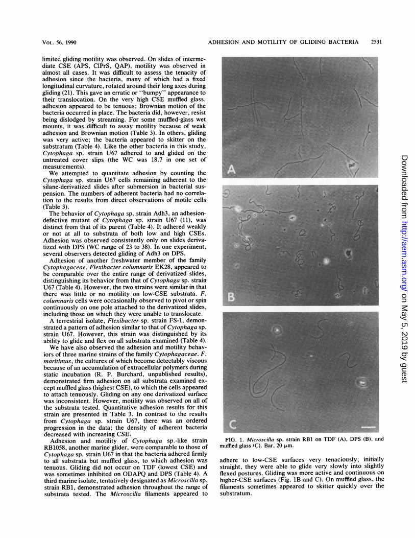
RB1058, another marine glider, were comparable to those ofCytophaga sp. strain U67 in that the bacteria adhered firmlyto all substrata but muffled glass, to which adhesion wastenuous. Gliding did not occur on TDF (lowest CSE) andwas sometimes inhibited on ODAPQ and DPS (Table 4). Athird marine isolate, tentatively designated as Microscilla sp.strain RB1, demonstrated adhesion throughout the range ofsubstrata tested. The Microscilla filaments appeared to
FIG. 1. Microscilla sp. strain RB1 on TDF (A), DPS (B), andmuffled glass (C). Bar, 20 p.m.
adhere to low-CSE surfaces very tenaciously; initiallystraight, they were able to glide very slowly into slightlyflexed postures. Gliding was more active and continuous on
higher-CSE surfaces (Fig. 1B and C). On muffled glass, thefilaments sometimes appeared to skitter quickly over thesubstratum.
VOL. 56, 1990
on May 5, 2019 by guest
http://aem.asm
.org/D
ownloaded from

2532 BURCHARD ET AL.
FIG. 2. M. xanthus DK1622 rosettes on muffled glass at t = 0(A), muffled glass at t = 50 min (B), and ODAPQ at t = 50 min (C).Bar, 20 Lm.
M. xanthus, a gliding bacterium that is unrelated to thefamily Cytophagaceae (39), demonstrated tenuous adhesionover the entire range of CSE substrata tested. These bacteriaglided with velocities 1 to 2 orders of magnitude slower thanthose of members of the family Cytophagaceae, making it
difficult to determine whether single cells were activelytranslocating on the derivatized slides. As an alternativeassessment of motility, the displacement of bacteria fromrosettes that had settled on the slides was determined (Fig.2A). Rosettes are tight clusters of radially arrayed cells thatform in rotary shake cultures of some M. xanthus strains (8,40). After incubation periods of approximately 25 min,bacteria from the settled rosettes appeared to have movedout from the rosette on all substrata but muffled glass (Fig.2B). The displacement of bacteria was likely to have beendue to straight-line gliding, since most of them were stillarrayed radially, as they had been in the intact rosettes.
DISCUSSION
Our observations demonstrate that the adhesion and mo-tility of diverse gliding bacteria are affected by the surfaceenergies of their substrata. These bacteria adhere moretenaciously to surfaces of low CSE than to hydrophilic ones,suggesting that one or more of the bacterial surface compo-nents that make contact with substrata are relatively hydro-phobic. Some of these components appear to be cell enve-lope proteins (11; unpublished results). They may belocalized in specific surface domains, since initial contactwith hydrophobic substrata by Cytophaga sp. strain U67 andsome of the other strains was typically made via the bacterialpole. This accords with an early observation of polar adhe-sion by a Flexibacter sp., suggesting that its poles are morehydrophobic than are the lateral surfaces (29).Adhesion may be mediated, at least in part, by a water
exclusion mechanism (49). On very hydrophobic substrata,water may be excluded so well that the bacteria stick and areprevented from gliding. In contrast, on very hydrophilicsubstrata (high CSE), water may be poorly excluded, pre-venting close bacterial contact and accounting for limited orskittering motility. As has been suggested for other bacteria(20, 34, 44), electrostatic interactions may account for thelimited adhesion of gliding bacteria to hydrophilic substrata.The silanes that we employed to achieve intermediate-rangeCSEs were both charged and uncharged. We were unable todistinguish reproducible differences in adhesion and motilityon slides derivatized with these silanes. Since adhesion wasmost tenacious on low-CSE surfaces, our results suggest amajor role of van der Waals forces rather than charge-chargeinteractions. Once a gliding bacterium has attached to asubstratum, whether tenaciously or weakly, it is not readilydislodged in a low-shear environment. It is apparent that theadhesion of gliding bacteria is biophysically complex, as isthe case for other bacteria (e.g., references 1, 5, 27, and 43).Cytophaga sp. strain U67, with its relatively hydrophobic
cell surface (M. Sorongon and R. P. Burchard, unpublisheddata), typifies the behavior of other gliders in its progres-sively more tenacious adhesion to more-hydrophobic sub-strata. Its adhesion-defective mutant Adh3, selected by theinability to adhere to hexadecane in a bacterial adherence tohydrocarbon enrichment protocol (11, 42), had a relativelyhydrophilic surface compared with that of its parent strain.Charge repulsion may account for the inability of the strainto adhere to more-hydrophilic (high-CSE) substrata. Poorexclusion of water may explain the loss of adhesion tolow-CSE surfaces. Only on DPS-derivatized glass, interme-diate in CSE, did Cytophaga sp. strain Adh3 adhere repro-ducibly. In one set of experiments, Adh3 was observed toglide on DPS, indicating that the motility apparatus isfunctional in this mutant. Since DPS-derivatized slides var-ied in CSE, the lack of reproducible motility suggests that
APPL. ENVIRON. MICROBIOL.
on May 5, 2019 by guest
http://aem.asm
.org/D
ownloaded from

ADHESION AND MOTILITY OF GLIDING BACTERIA 2533
the mutant cells will glide only on substrata with veryspecific surface properties.Other gliding bacteria examined in this study, including
those that produce substantial amounts of extracellularpolymers ("slime"), also have cell surface properties thatpermit them to adhere more firmly to substrata of low CSEthan to those that are hydrophilic. The extracellular poly-mers do not appear to interfere with the cell envelopemaking direct contact with the substratum (11; unpublisheddata).
In a recent quantitative adhesion study of another Flexi-bacter sp., similar numbers of bacteria attached to bothhydrophilic and hydrophobic polystyrene substrata (30). Incontrast, the number of adherent F. maritimus cells in-creased with increasing hydrophobicity of the substratum.These data correlated with the adhesion and motility behav-ior of the bacteria in wet mounts. A variety of species ofnongliding bacteria have also been reported to adhere ingreater numbers to hydrophobic surfaces than to hydrophilicsurfaces (e.g., references 2, 20, 44, and 47). However, thisheld true only when the surface tension of the test bacteriumwas less than that of the suspending medium (2) or only forrelatively low concentrations of bacteria in the suspensionsin which the substrata were submerged (47). Native popula-tions of aquatic bacteria have been reported to demonstratean adhesion minimum on substrata in the middle range ofCSE (critical surface tension of -2.0 x 10⁻⁴ to 2.5 x 10⁻⁴N/cm). For marine bacteria, adhesion maxima were foundfor both higher and lower CSEs (17). Freshwater bacterialpopulations demonstrated an adhesion maximum and asecond minimum on successively more hydrophilic substrata(36).
Quantitation of adhesion of Cytophaga sp. strain U67, afreshwater strain, demonstrated no correlation with obser-vations of its apparent tenacity of adhesion and motility(Table 3). A similar lack of correlation between the numbersof adherent bacteria and hydrophobicity has been reportedby others (27). These data exemplify the conclusion ofBusscher et al. (12) and van Pelt et al. (47) that enumerationof bacteria adherent to a surface after a fixed period ofsubmersion in cell suspension may not be an appropriatemeasure of binding strength. We are currently developingassays of the strength of adhesion of gliding bacteria.The bacterial-adhesion literature illustrates that the pres-
ence of organic molecules, ions, and free radicals; ionicstrength; and bacterial physiology all affect adhesion tosurfaces (e.g., references 1, 19, 24, 30, 35, and 49). Weminimized substratum effects due to the adsorption of or-ganics and ions since most of our observations were madeimmediately after preparing the wet mount. However, pre-liminary observations suggest that physiology and culturemedium affect the adhesion and motility of the glidingbacteria. For example, Microscilla sp. strain RB1 grown inHSM (0.2% tryptone, 0.05% yeast extract, 0.3% gelatin, 3%marine salts [Forty Fathoms Marine Enterprises, Towson,Md.]), a richer and much less defined medium prepared withan aquarium marine salts mixture, demonstrated more ten-uous adhesion on both high- and low-CSE substrata than didcomparable cells grown in CAS, a Casamino Acids (DifcoLaboratories)-artificial seawater medium. Furthermore, F.maritimus grown in shake culture behaved differently onmuffled glass than when grown without agitation. We areexploring some of these phenomena.
Tenacity of adhesion affects the expression of the machin-ery of gliding. The bacteria that we examined adhered firmlyto but glided little if at all on low-CSE substrata. On highly
hydrophilic (high-CSE) substrata, adhesion appeared to betenuous and gliding, if it occurred, often appeared to berelatively fast; cells sometimes seemed to skitter across themuffled glass slides. The differences in cell contact and inmotility were particularly striking with Microscilla sp. strainRB1 (Fig. 1). In contrast, the gliding diatom Nitzschialinearis adheres to glass (high CSE) more strongly than tolow-CSE polystyrene (22). Another diatom demonstrated an
adhesion minimum on glass derivatized with dichlorodime-thylsilane (surface energy of 2.5 x 1i-' N/cm; 15). Thesestudies suggest different mechanisms of adhesion and motil-ity between the structurally distinct gliding bacteria anddiatoms.
ACKNOWLEDGMENTS
This research was supported by Office of Naval Research con-
tracts N00014-88-K-0158 to R.P.B. and N00014-86-K-0261 to J.B.,D.R., and J. D. Costlow and by Public Health Service grantES01908 to J.B. from the National Institutes of Health.We gratefully acknowledge the expert technical assistance of
Cindy-Lou Dull, Tracy Mathiessen, Maria Sorongon, Alva R.Schmidt, Gail Cannon, Dierdre Roberts, and Kelly Eisenman.
LITERATURE CITED1. Abbott, A., P. R. Rutter, and R. C. W. Berkeley. 1983. The
influence of ionic strength, pH and a protein layer on theinteraction between Streptococcus mutans and glass surfaces.J. Gen. Microbiol. 129:439-445.
2. Absolom, D. R., F. V. Lamberti, Z. Policova, W. Zingg, C. J. vanOss, and A. W. Neumann. 1983. Surface thermodynamics ofbacterial adhesion. Appl. Environ. Microbiol. 46:90-97.
3. Arlauskas, J., and R. P. Burchard. 1981. Substratum require-ments for bacterial gliding motility. Arch. Microbiol. 133:137-141.
4. Baier, R. E. 1973. Influence of the initial surface condition ofmaterials on bioadhesion, p. 633-639. In R. F. Acker (ed.),Proceedings of the 3rd International Congress on Marine Cor-rosion and Fouling. Northwestern University Press, Evanston,Ill.
5. Baier, R. E. 1980. Substrata influences on adhesion of microor-ganisms and their resultant new surface properties, p. 59-104. InG. Bitton and K. C. Marshall (ed.), Adsorption of microorgan-isms to surfaces. John Wiley & Sons, Inc., New York.
6. Baxa, D. V., K. Kawai, and R. Kusuda. 1986. Characteristics ofgliding bacteria isolated from diseased cultured flounder, Par-alichthys olivaceous. Fish Pathol. 21:251-258.
7. Bernardet, J. F. 1989. 'Flexibacter columnaris': first descriptionin France and comparison with bacterial strains from otherorigins. Dis. Aquat. Org. 6:37-44.
8. Burchard, R. P. 1974. Studies on gliding motility in Myxococcusxanthus. Arch. Microbiol. 99:271-280.
9. Burchard, R. P. 1981. Gliding motility of prokaryotes: ultra-structure, physiology and genetics. Annu. Rev. Microbiol.35:497-529.
10. Burchard, R. P. 1984. Inhibition of Cytophaga sp. strain U67gliding motility by inhibitors of polypeptide synthesis. Arch.Microbiol. 139:248-254.
11. Burchard, R. P., and R. A. Bloodgood. 1990. Surface proteins ofthe gliding bacterium Cytophaga sp. strain U67 and its mutantsdefective in adhesion and motility. J. Bacteriol. 172:3379-3387.
12. Busscher, H. J., A. H. Weerkamp, H. C. van der Mei, A. W. J.van Pelt, J. P. de Jong, and J. Arends. 1984. Measurement of thesurface free energy of bacterial cell surfaces and its relevancefor adhesion. Appl. Environ. Microbiol. 48:980-983.
13. Castenholz, R. W. 1982. Movements, p. 320-339. In N. G. Carrand B. A. Whitton (ed.), The biology of cyanobacteria. Univer-sity of California Press, Berkeley.
14. Chang, L.-Y. E., J. L. Pate, and R. J. Betzig. 1984. Isolation andcharacterization of nonspreading mutants of the gliding bacte-rium Cytophaga johnsonae. J. Bacteriol. 159:26-35.
15. Characklis, W. G., and K. E. Cooksey. 1983. Biofilms and
VOL. 56, 1990
on May 5, 2019 by guest
http://aem.asm
.org/D
ownloaded from

2534 BURCHARD ET AL.
microbial fouling. Adv. Appl. Microbiol. 29:93-138.16. Dayrell-Hart, B., and R. P. Burchard. 1979. Association of
flexing and gliding in Flexibacter. J. Bacteriol. 137:1417-1420.17. Dexter, S. C. 1979. Influence of substratum critical surface
tension on bacterial adhesion-in situ studies. J. Colloid Inter-face Sci. 70:346-354.
18. Dworkin, M. 1962. Nutritional requirements for vegetativegrowth of Myxococcus xanthus. J. Bacteriol. 84:250-257.
19. Fletcher, M. 1988. Attachment of Pseudomonas fluorescens toglass and influence of electrolytes on bacterium-substratumseparation distance. J. Bacteriol. 170:2027-2030.
20. Fletcher, M., and G. I. Loeb. 1979. Influences of substratumcharacteristics on the attachment of a marine pseudomonad tosolid surfaces. Appl. Environ. Microbiol. 37:67-72.
21. Godwin, S. L., M. Fletcher, and R. P. Burchard. 1989. Interfer-ence reflection microscopic study of sites of association be-tween gliding bacteria and glass substrata. J. Bacteriol. 171:4589-4594.
22. Harper, M. A., and J. F. Harper. 1967. Measurements of diatomadhesion and their relationship with movement. Br. Phycol.Bull. 3:195-207.
23. Henrichsen, J. 1972. Bacterial surface translocation: a surveyand a classification. Bacteriol Rev. 36:478-503.
24. Hsieh, Y.-L., and D. A. Timm. 1988. Relationship of substratumwettability measurements and initial Staphylococcus aureusadhesion to films and fabrics. J. Colloid Interface Sci. 123:275-286.
25. Humphrey, B. A., M. R. Dickson, and K. C. Marshall. 1979.Physicochemical and in situ observations on the adhesion ofgliding bacteria to surfaces. Arch. Microbiol. 120:231-238.
26. Kaiser, D. 1979. Social gliding is correlated with the presence ofpili in Myxococcus xanthus. Proc. Natl. Acad. Sci. USA 76:5952-5956.
27. Kjelleberg, S. 1984. Adhesion to inanimate surface, p. 51-71. InK. C. Marshall (ed.), Microbial adhesion and aggregation.Springer-Verlag, New York.
28. Lapidus, I. R., and H. C. Berg. 1982. Gliding motility ofCytophaga sp. strain U67. J. Bacteriol. 151:384-398.
29. Marshall, K. C., and R. H. Cruikshank. 1973. Cell surfacehydrophobicity and the orientation of certain bacteria at inter-faces. Arch. Mikrobiol. 91:29-40.
30. McEldowney, S., and M. Fletcher. 1988. Effect of pH, temper-ature and growth conditions on the adhesion of a glidingbacterium and three nongliding bacteria to polystyrene. Microb.Ecol. 16:183-195.
31. Pate, J. L. 1988. Gliding motility in procaryotic cells. Can. J.Microbiol. 34:459-465.
32. Pate, J. L., and L.-Y. E. Chang. 1979. Evidence that glidingmotility in prokaryotic cells is driven by rotary assemblies in thecell envelope. Curr. Microbiol. 2:59-64.
33. Pate, J. L., and E. J. Ordal. 1967. Fine structure of Chondro-coccus columnaris. I. Structure and function of mesosomes. J.Cell Biol. 35:1-13.
34. Paul, J. H., and W. H. Jeffrey. 1985. Evidence for separate
adhesion mechanisms for hydrophilic and hydrophobic surfacesin Vibrio proteolytica. Appl. Environ. Microbiol. 50:431-437.
35. Pratt-Terpstra, I. H., A. H. Weerkamp, and H. J. Busscher.1989. Microbial factors in a thermodynamic approach of oralstreptococcal adhesion to solid substrata. J. Colloid InterfaceSci. 129:568-574.
36. Pringle, J. H., and M. Fletcher. 1983. Influence of substratumwettability on attachment of freshwater bacteria to solid sur-faces. Appl. Environ. Microbiol. 45:811-817.
37. Reichenbach, H. 1965. Untersuchungen an Archangium viola-ceum. Arch. Mikrobiol. 52:376-403.
38. Reichenbach, H. 1966. Myxococcus spp. (Myxobacteriales):Schwarmentwicklung und Bildung von Protocysten. In G. Wolf(ed.), Encyclopaedia cinematographica. Institut fur den Wissen-schaftlichen Film, Gottingen, Federal Republic of Germany.
39. Reichenbach, H. 1981. Taxonomy of the gliding bacteria. Annu.Rev. Microbiol. 35:339-364.
40. Reichenbach, H., and M. Dworkin. 1981. Introduction to thegliding bacteria, p. 315-327. In M. P. Starr (ed.), The prokary-otes, vol. 1. Springer-Verlag, New York.
41. Ridgway, H. F., and R. A. Lewin. 1988. Characterization ofgliding motility in Flexibacter polymorphus. Cell Motil. Cy-toskeleton 11:46-63.
42. Rosenberg, M. 1984. Bacterial adherence to hydrocarbons: auseful technique for studying cell surface hydrophobicity.FEMS Microbiol. Lett. 22:289-295.
43. Rutter, P. R., and B. Vincent. 1980. The adhesion of micro-organisms to surfaces: physico-chemical aspects, p. 79-92. InR. C. W. Berkeley, J. M. Lynch, J. Melling, P. R. Rutter, andB. Vincent (ed.), Microbial adhesion to surfaces. Ellis HorwoodLtd., Chichester, England.
44. Satou, N., J. Satou, H. Shintani, and K. Okuda. 1988. Adher-ence of streptococci to surface-modified glass. J. Gen. Micro-biol. 134:1299-1305.
45. Simon, G. D., and D. White. 1971. Growth and morphologicalcharacteristics of a species of Flexibacter. Arch. Mikrobiol.78:1-16.
46. Smibert, R. M., and N. R. Krieg. 1981. General characteriza-tion, p. 409-443. In P. Gerhardt, R. G. E. Murray, R. N.Costilow, E. W. Nester, W. A. Wood, N. R. Krieg, and G. B.Phillips (ed.), Manual of methods for general bacteriology.American Society for Microbiology, Washington, D.C.
46a.Swaisgood, H. E., and H. R. Horton. 1987. Sulfhydryl oxidasefrom milk. Methods Enzymol. 143:504-510.
47. van Pelt, A. W. J., A. H. Weerkamp, M. H. W. J. C. Uyen, H. J.Busscher, H. P. de Jong, and J. Arends. 1985. Adhesion ofStreptococcus sanguis CH3 to polymers with different surfacefree energies. Appl. Environ. Microbiol. 49:1270-1275.
48. Waite, H. 1983. Adhesion in byssally attached bivalves. Biol.Rev. 58:209-231.
49. Wrangstadh, M., P. L. Conway, and S. Kjelleberg. 1986. Theproduction and release of an extracellular polysaccharide duringstarvation of a marine Pseudomonas sp. and the effect thereofon adhesion. Arch. Microbiol. 145:220-227.
APPL. ENVIRON. MICROBIOL.
on May 5, 2019 by guest
http://aem.asm
.org/D
ownloaded from