glaucoma - government medical college and …gmch.gov.in/e-study/e...
TRANSCRIPT

GLAUCOMA
Dr Suresh KumarAssociate Professor
Department of OphthalmologyGMCH-32
What is Glaucoma?
• Glaucoma is a progressive optic neuropathy with characteristic appearance of the optic disc and specific pattern of visual field defects , irrespective of IOP level .
Physiology of aqueous productionCiliaryBody: Ant Pars plicata (2mm wide)
Post Pars plana (4mm wide)
Aqueous humor is actively secreated by non pigmented epithelium of ciliary processes
Trabecular Meshwork: Uveal meshworkCorneoscleral meshwork
Endothelial(juxtacanalicular)meshwok

Pathogenesis of Sec. Glaucoma

Outflow of aqueous
• Posterior chamber – via pupil – Anterior chamber – exits eye by 2 routes :
– Trabecular (conventional) route : 90%
– Uveoscleral (unconventional) route : 10%

Factors determining IOP
• Rate of aqueous secretion
• Rate of aqueous outflow – difference between the IOP and episcleral venous pressure
• Normal range of IOP 11-21mm Hg

Investigations• Visual acuity and refractive state• Slit-lamp biomicroscopy (optic disc; 90D)• Goldmann Applanation tonometry• Gonioscopy• Perimetry• OCT
Tonometry
• Indentation tonometry : Schiotz• Applanation tonometry :
– Goldmann – Perkin– Mackey Marg – Tonopen
• Non contact tonometry : AirpuffPulsair 2000 Keeler

• Applaanation tonometry based on Imbert-Fick principle : For an ideal, dry, thin walled sphere, pressure inside sphere (P) equals force needed to flatten its surface(F) divided by area of flattening (A) - 3.06mm Goldmanntonometry
Angle of anterior chamber
• Formed by root of iris , anterior most part of ciliary body , scleral spur , trabecular meshwork and schwalbe line.
• Aqueous humor fills (0.25ml) of anterior chamber and (0.06ml) of posterior chamber

Gonioscopy• Biomicroscopic visualisation of angle of
anterior chamber using goniolensTypes of Goniolens• Direct : Koeppe goniolens
Swan -Jacob• Indirect : Goldmann three mirror
Zeiss goniolensPosner and Sussman

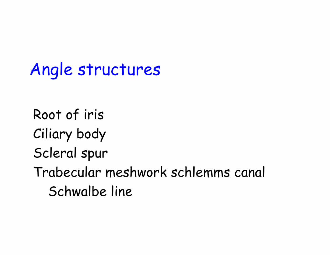
Angle structures
Root of irisCiliary bodyScleral spurTrabecular meshwork schlemms canal
Schwalbe line
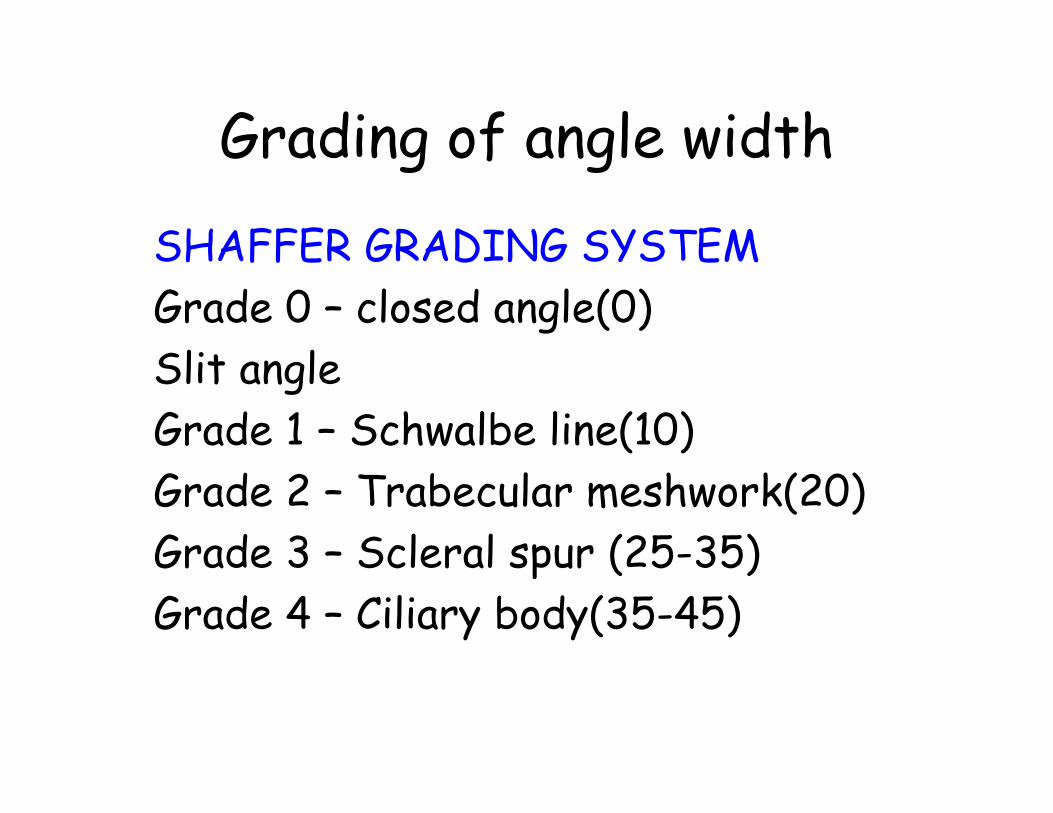
Grading of angle width
SHAFFER GRADING SYSTEMGrade 0 – closed angle(0)Slit angleGrade 1 – Schwalbe line(10)Grade 2 – Trabecular meshwork(20)Grade 3 – Scleral spur (25-35)Grade 4 – Ciliary body(35-45)

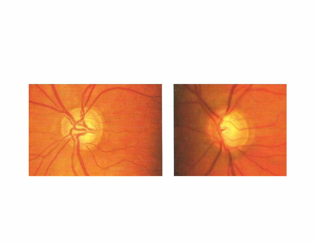

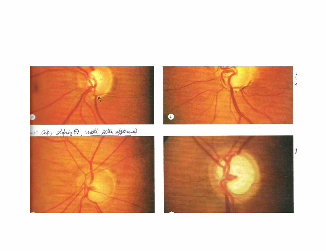
Optic disc signs

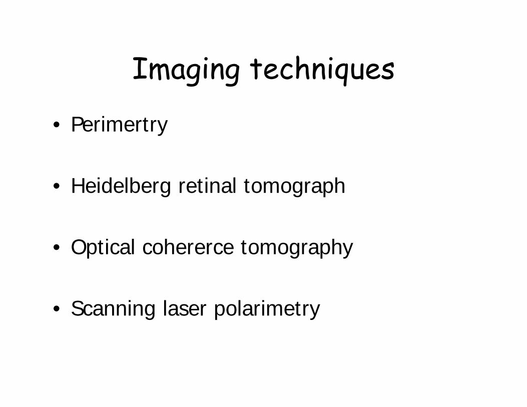
Imaging techniques
• Perimertry
• Heidelberg retinal tomograph
• Optical cohererce tomography
• Scanning laser polarimetry


PRIMARY OPEN ANGLE GLAUCOMA
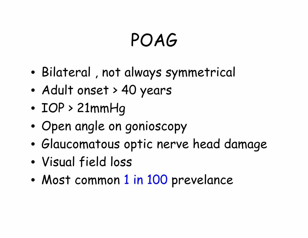
POAG• Bilateral , not always symmetrical• Adult onset > 40 years• IOP > 21mmHg • Open angle on gonioscopy• Glaucomatous optic nerve head damage• Visual field loss• Most common 1 in 100 prevelance

Risk factors and associations• Age : older patients >40 years• Race : black people more than white• Family history and inheritance :
Multifactoral inheritanceIst degree relatives and siblings(10%)
Myopia and DiabetesRetinal diseases : CRVO ,Rheg RD, RP

Steroid responsiveness• Normal population divided into 3 Groups
IOP response to 6wk course of topical betamethasone
High responders(>30mmHg) 5%Moderate responders(22-30mmHg)
35%Non responders(no change) 60%
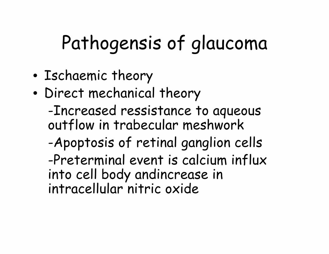
Pathogensis of glaucoma• Ischaemic theory• Direct mechanical theory
-Increased ressistance to aqueous outflow in trabecular meshwork-Apoptosis of retinal ganglion cells-Preterminal event is calcium influx into cell body andincrease in intracellular nitric oxide
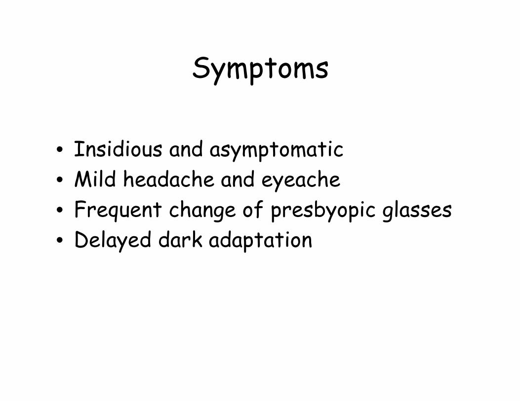
Symptoms
• Insidious and asymptomatic• Mild headache and eyeache• Frequent change of presbyopic glasses• Delayed dark adaptation
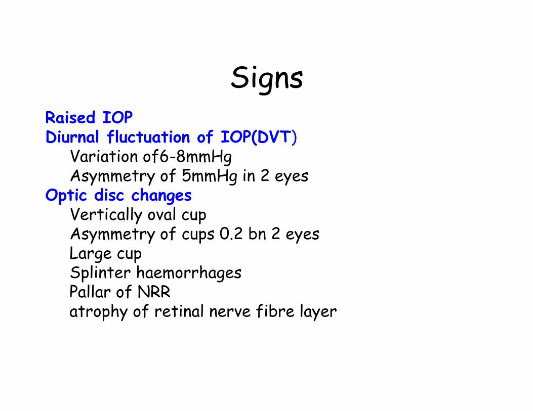
SignsRaised IOPDiurnal fluctuation of IOP(DVT)
Variation of6-8mmHgAsymmetry of 5mmHg in 2 eyes
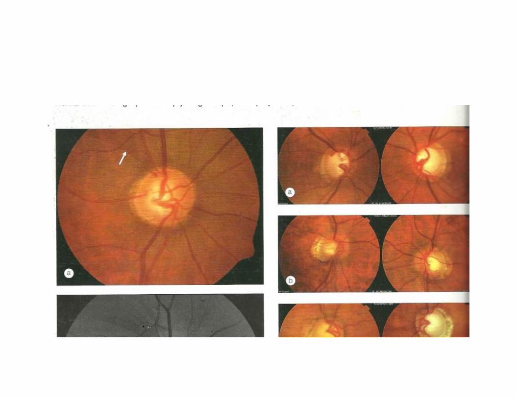
Optic disc changesVertically oval cupAsymmetry of cups 0.2 bn 2 eyesLarge cupSplinter haemorrhagesPallar of NRRatrophy of retinal nerve fibre layer

• Advanced changesMarked cupping (0.7-0.9)Thinning of NRRBayonetting signLamellar dot signPulsations of retinal arterioles
Glaucomatous optic atrophy
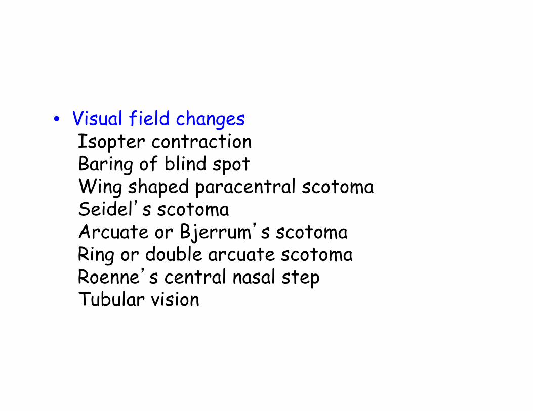
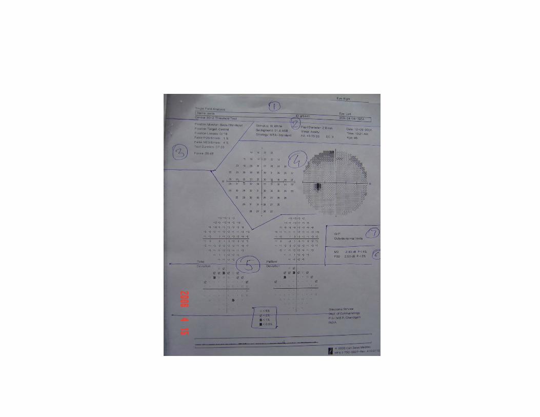
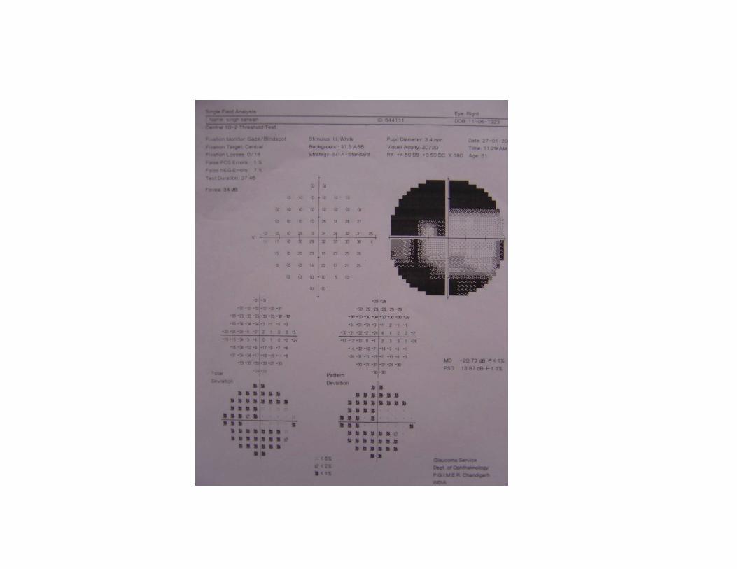
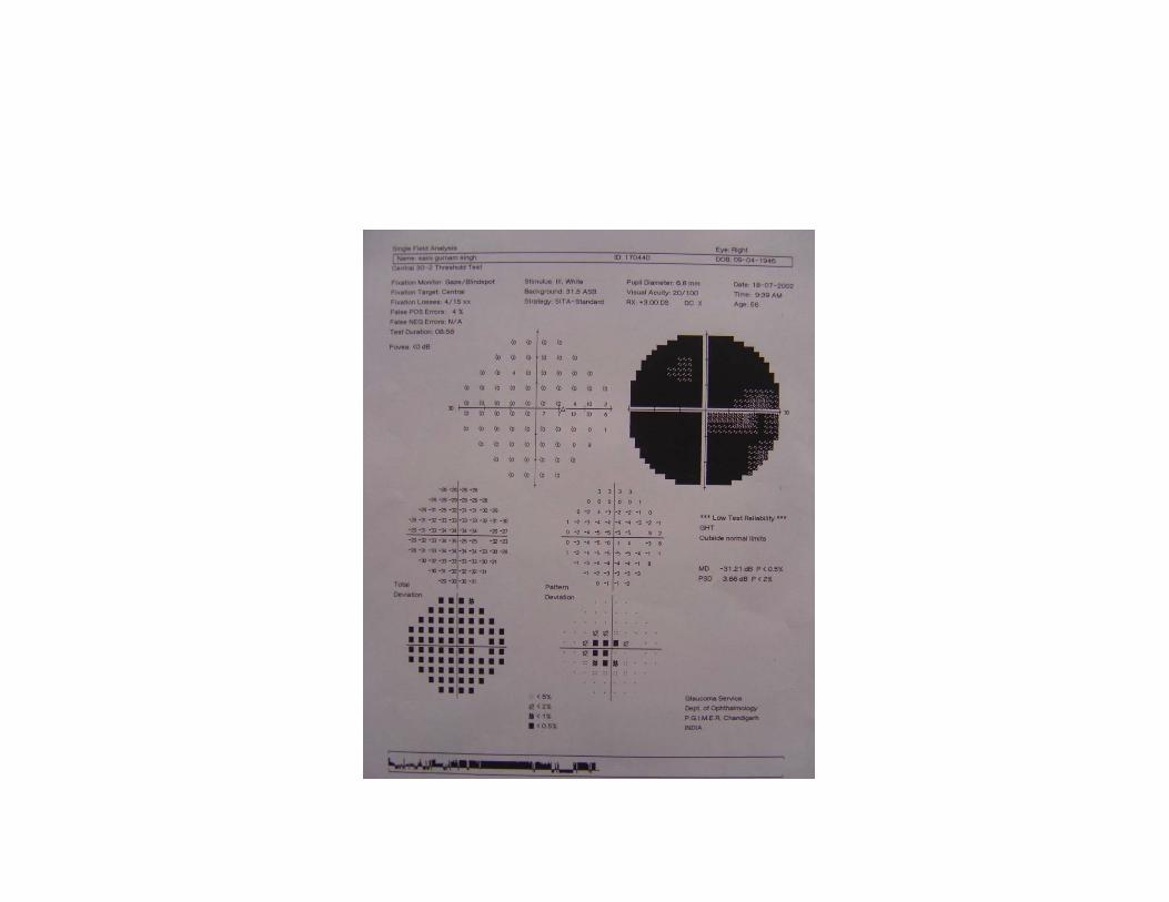
• Visual field changesIsopter contractionBaring of blind spotWing shaped paracentral scotomaSeidel’s scotomaArcuate or Bjerrum’s scotomaRing or double arcuate scotomaRoenne’s central nasal stepTubular vision

• GonioscopyNormal open angle

Grading of glaucomatous damageMild damage : early visual field defects
MD <-6dB mild cuppingModerate damage : definite arcuate
scotoma MD <-12dB moderate thinning of NRR
Severe damage : extensive VF lossMD >-12dBMarked cupping
End-stage disease : small residual fieldMinimal residual NRR

Treatment
• Baseline evaluation• Achive target pressure• Monitoring optic nerve and visual field
THERAPUTIC CHOICEMedical therapyArgon or diode laser trabeculoplastyFilteration surgery

Single drug therapy
• Prostaglandin analoguesLatanoprost (0.005%)Bimatoprost (0.03%)Travoprost (0.004%)
Topical beta blockersTimolol maleate (0.25,0.5%) Betaxolol (0.5%) Carteolol (1%,2%)
Levobunolol (0.5%) Metipranolol ( 0.1%)
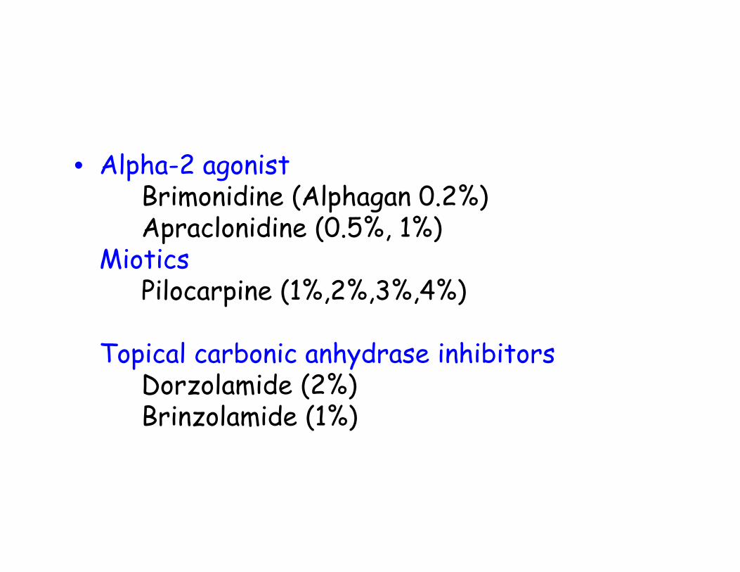
• Alpha-2 agonistBrimonidine (Alphagan 0.2%)Apraclonidine (0.5%, 1%)
MioticsPilocarpine (1%,2%,3%,4%)
Topical carbonic anhydrase inhibitorsDorzolamide (2%)Brinzolamide (1%)
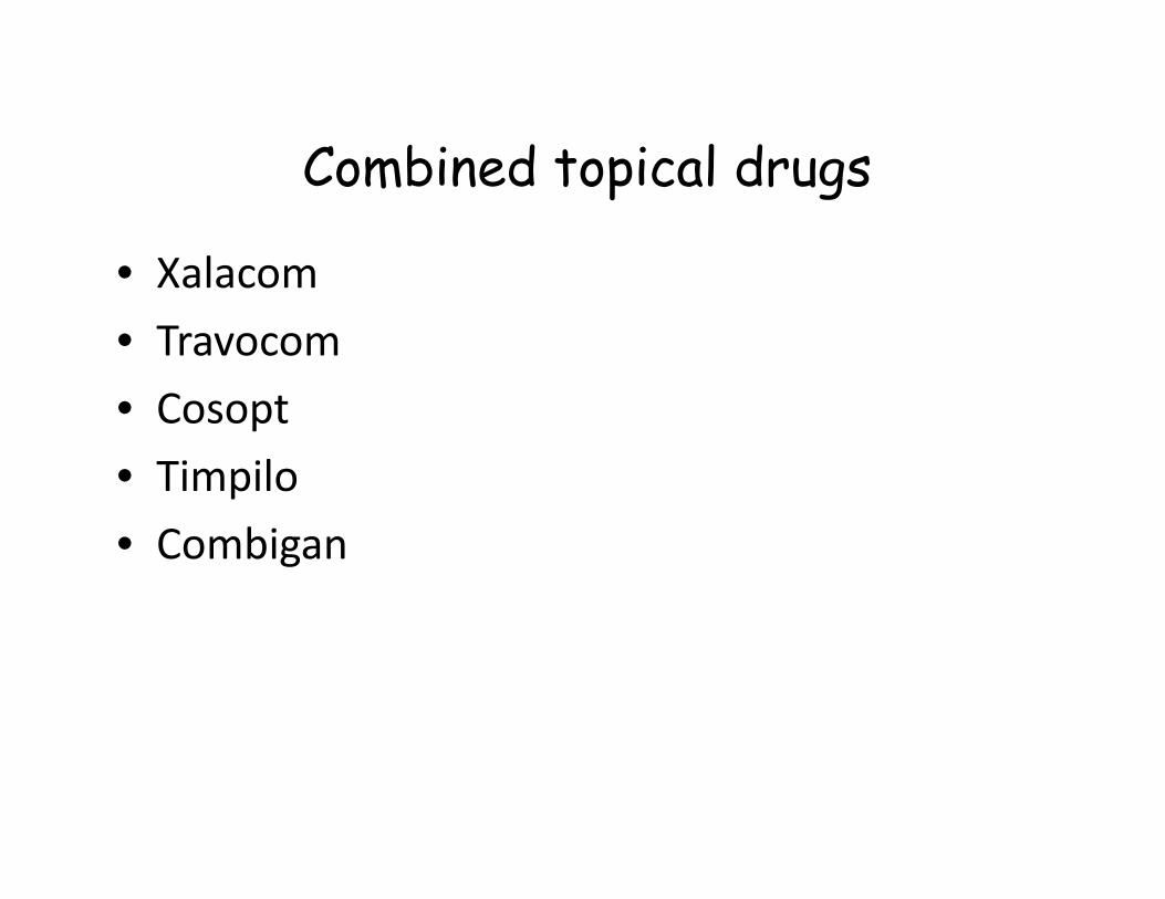
Combined topical drugs
• Xalacom• Travocom• Cosopt• Timpilo• Combigan
Systemic carbonic anhydrase inhibiters
• Acetazolamide 250-1000mg• Methazolamide 50-100mg
• Hyperosmotic agentsGlycerol (50%)Mannitol (20%)Isosorbide

Argon or diode laser trabeculoplasty
• IndicationsAvoidance of polypharmacyAvoidance of surgeryNon compliant patient

Trabeculectomy
• IndicationsFailed medical therapyFailed laser trabeculoplastyAdvanced diseaseUnsuitability for laser therapy

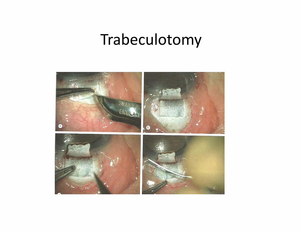
Trabeculotomy
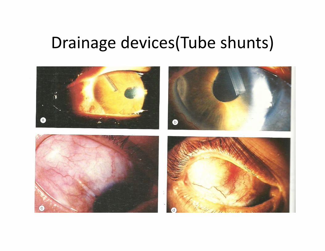
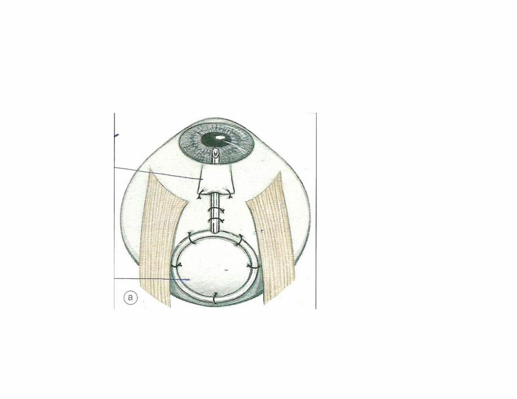
Drainage devices(Tube shunts)
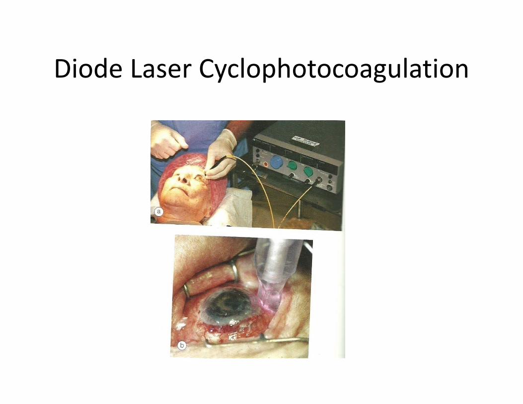
Diode Laser Cyclophotocoagulation
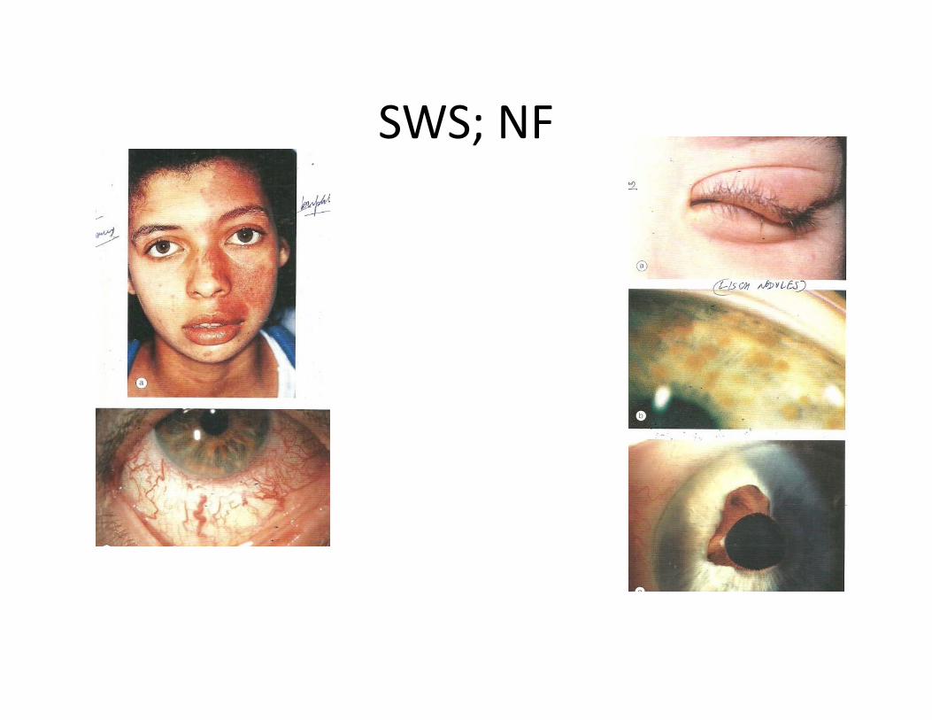
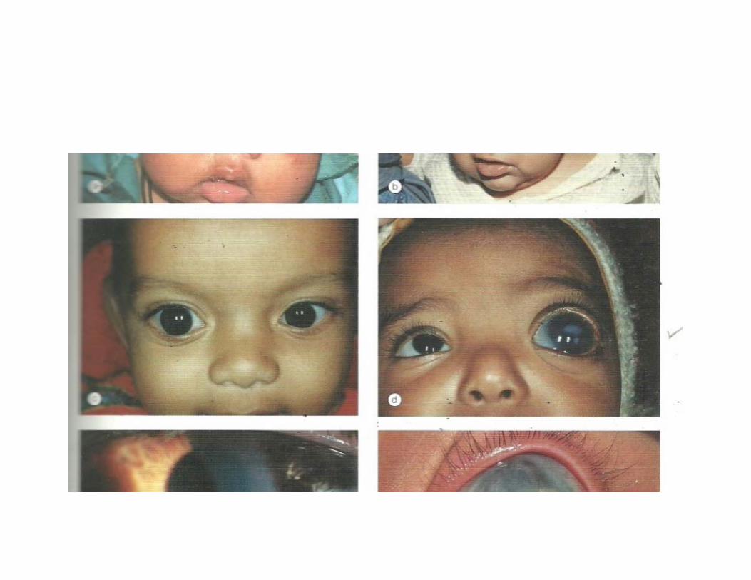
SWS; NF
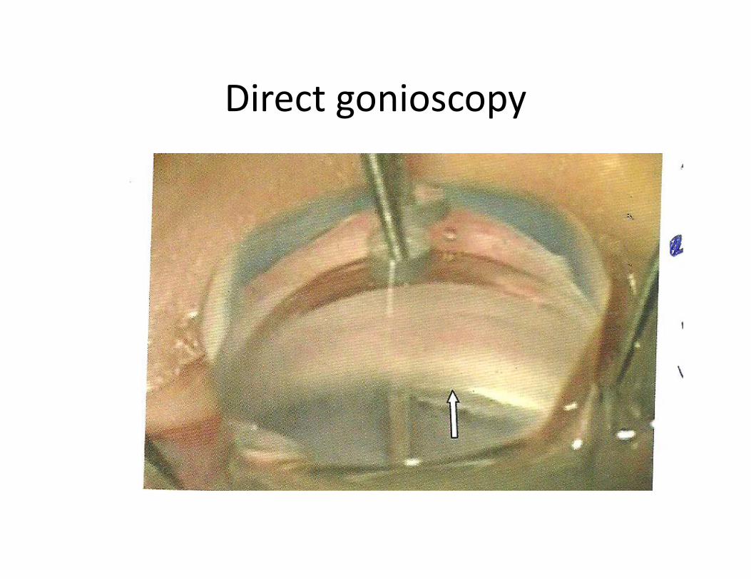
Direct gonioscopy

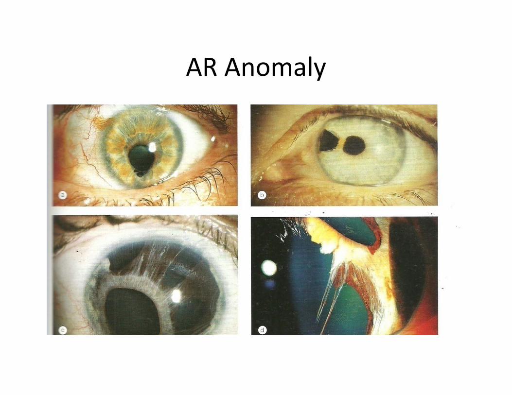
AR Anomaly

Traumatic Glaucoma: Hyphaema
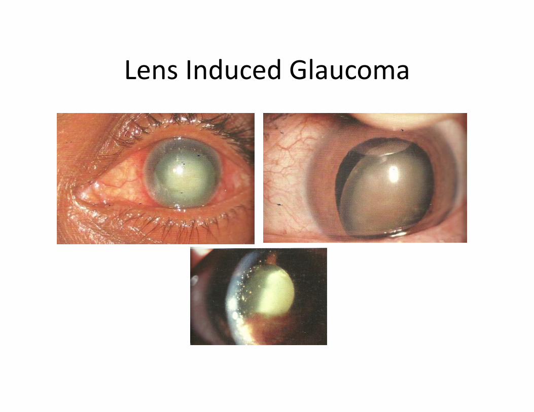
Lens Induced Glaucoma
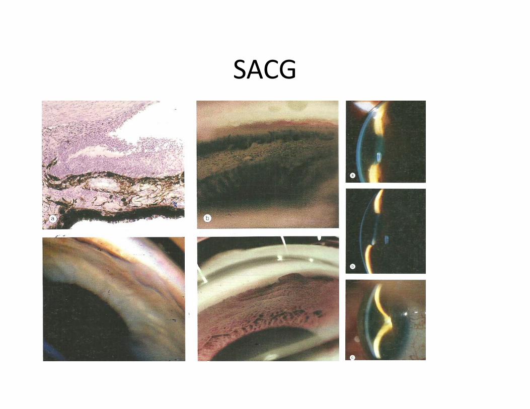
SACG
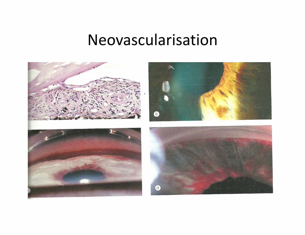
Neovascularisation

Pigment Dispersion

Pseudoexfoliation
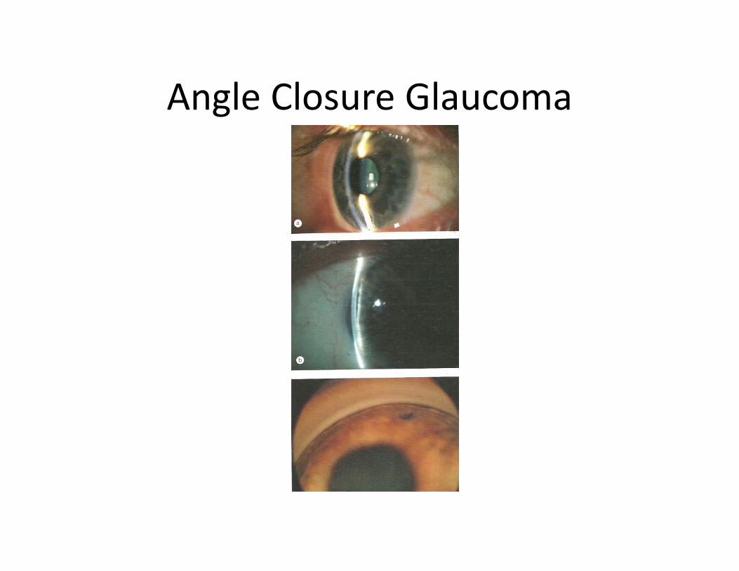
Angle Closure Glaucoma