geographical distribution of peronosclerospora spp., the ... · darussalam, north sumatera,...
TRANSCRIPT
AAB Bioflux, 2016, Volume 8, Issue 3. http://www.aab.bioflux.com.ro
143
Geographical distribution of Peronosclerospora spp., the causal organism of maize downy mildew, in Indonesia Amran Muis, Nurnina Nonci, Marcia B. Pabendon Indonesian Cereals Research Institute, Maros, Indonesia. Corresponding author: A. Muis,
Abstract. Downy mildew is a major disease in maize caused by a number of species of the genus Peronosclerospora and Sclerospora. This study aimed to determine the geographical distribution of dominant species of downy mildew that infects maize in corn production areas in Indonesia. The research was conducted applying survey by visiting the centers of maize production in Indonesia. Samples of leaves affected by disease were taken to the laboratory of plant diseases of Indonesia Cereals Research Institute (ICERI) to observe its morphology under microscope evaluation. The results show that Peronosclerospora spp. has spread in almost all of the island or a large area in Indonesia. In one area it can be found in more than one species, whereas one species was found in some endemic areas of downy mildew. The P. philippinensis only found in Sulawesi Island, while P. maydis and P. sorghi found in all islands. Key Words: corn downy mildew, map, genetic diversity, morphology.
Introduction. One of the major problem in increasing corn production in Indonesia is an attack of a plant disease, especially downy mildew which attacks the plants, especially at young stage toward susceptible varieties, and it can cause yield loss up to 100% (Sudjono 1988; Wakman 2004). Downy mildew is caused by fungi Peronosclerospora spp. that infects maize plant through spores carried by the wind in the morning. So far, it has been reported that there were 10 species from three genera which cause downy mildew on maize, namely P. maydis, P. phillipinensis, P. sacchari, P. Sorgi, P. spontanea, P. miscanthi, P. heteropogani, Sclerospora macrospora, S. philippinensis, S. rayssiae, and S. graminicola (Shurtleff 1980; Rathore et al 2002; Wakman & Djatmiko 2002; Yen et al 2004; Kutama et al 2010; Nagabhushan et al 2014). Telle et al (2011) has discovered one downy mildew pathogen species, i.e. P. eriochloae which has not been reported as pathogen in maize.
In Indonesia, downy mildew at first only occurs in some maize production areas, but along with the spread of maize, downy mildew has spread in several provinces. During epidemics incidence, in an endemic area, spacious incidence can be in the tens of hectares. In endemic areas such as East Java, Lampung, and South Sulawesi, it was found downy mildew infected spacious and cause significant losses at the farm level (Pakki et al 2006). Downy mildew caused a yield loss of approximately 90%, especially if infection occurs in early vegetative growth stage (Burhanuddin & Pakki 1999; Jabbar & Talanca 1999). Nowadays, there are three species of Peronosclerospora that have been found spreading on different islands, namely P. maydis, P. philippinensis, and P. sorghi (Mikoshiba et al 1977; Wakman & Hasanuddin 2003; Lukman et al 2013; Muis et al 2013; Rustiani et al 2015). However the area of distribution of each of these species has not been widely reported.
This study aimed to determine the geographical distribution of dominant species of downy mildew that infects maize in some endemic areas in Indonesia. Material and Method. This study was conducted from 2012 to 2015 usng survey method, by visiting maize production areas in several provinces such as: Nangro Aceh Darussalam, North Sumatera, Lampung, West Java, Central Java, Daerah Istimewa
AAB Bioflux, 2016, Volume 8, Issue 3. http://www.aab.bioflux.com.ro
144
Yogyakarta, East Java, West Kalimantan, South Kalimantan, North Sulawesi, Gorontalo, Central Sulawesi, and South Sulawesi. Morphological characterization of Peronosclerospora spp. Sampling was done by placing masking tape on the under surface of the infected leaves then the tape affixed to glass slides and labeled to include the location and date of sampling. Sampling was done several times at each location. The collected samples were placed in the box and then taken to the Plant Pathology Laboratory of Indonesian Cereals Research Institute (ICERI) to observe the form of conidia under the microscope evaluation.
Downy mildew pathogen species identification is based on morphological characteristics proposed by Quimio & Hanlin (1999) and CIMMYT (2012) as shown in Table 1.
Table 1
Morphological characteristics of various downy mildew pathogens of maize (Source: CIMMYT 2012)
Morphological characteristics Pathogen
(Name of Disease) Conidiophores/ Sporangiophores Conidia/Sporangia Oospores
Peronosclerospora sorghi (Sorghum downy mildew)
Erect, dichotomously
branched, 180 to 300µm in length.
Emerge singly or in groups from stomata
Oval (14.4-27.3 × 15-28.9µm), borne
on sterigmata (about 13µm long
Spherical (36µm in diameter on
average), light yellow or brown in
color
P. maydis (Java downy mildew)
Clustered conidiophores (150
to 550 µm in length) emerge from stomata. Dichotomously
branched two to four times
Spherical to subspherical in shape (17-23 µm x 27-39
µm)
Not reported
P. philippinensis (Philippine downy
mildew)
Erect and dichotomously
branched two to four times. 150 to 400 µm in length and emerge from
stomata
Ovoid to cyclindrincal (17-21 µm x 27-38
µm), slightly rounded at apex
Rare, spherical (25 to 27 µm in
diameter and smooth walled
P. sacchari (Sugarcane downy
mildew)
160 to 170 µm in length erect and arise singly or in
pairs from stomata
Elliptical, oblong(15-23 µm x 25-41 µm)
with round apex
40 to 50 µm in diameter, globular,
yellow
Sclerospora graminicola
(Graminicola downy mildew or green ear)
Average length of 268 µm
Borne on short sterigmata, elliptical (12-21 x 14-31 µm)
with distinctive papillate operculum
at apex
Pale brown and 22 to 35 µm in diameter
Sclerophthora macrospora (crazy
top)
Very short (14 µm on average)
Lemon shaped (30-65 x 60-100 µm),
operculate
Pale yellow, circular (45-75 µm)
Scleropthora rayssiae var. zeae
(Brown stripe downy mildew)
- Oval to cyclindrical (18-26 x 29-67 µm)
Spherical (29-37 µm in diameter), brown in color.

AAB Bioflux, 2016, Volume 8, Issue 3. http://www.aab.bioflux.com.ro
145
Genetic diversity of downy mildew based on SSR markers. For genetic diversity testing purposes, downy mildew infected leaf samples were collected from Kediri of East Java (15 samples), Landak and Bengkayang of West Kalimantan (6 samples), Pidie and Aceh Besar of Nangro Aceh Darussalam (5 samples), Langkat of North Sumatra (3 samples), 1 sample of Bogor (West Java), and a number of samples from South Sulawesi (2 samples of Maros, 4 samples of Barru, 7 samples of Sidrap, 6 samples of Tana Toraja, and 9 samples of Bone).
DNA collection was done by selecting a downy mildew infected plants as much as 10 DNA collections that spread on 10 different points for each location. Determining the location of a collection based on clear information about the downy mildew endemic areas. Two mL tube for each DNA collection was prepared and each filled with 800 µL of CTAB (Cetyl Trymetyil Ammonium Bromide) buffer. Every collected DNA was cut with a hole punch tool as many as 180 cuts which was equal to 0.4 g per DNA collection tube as much as 2 per one point then labeled. Beside that, five pieces of diseased leaves from every site of DNA collection were prepared, cleaned or dried if were wet by using tissue paper, packed plastic bag flops, labeled the same as the label on the tube for each DNA collection, then stored in ice containing box. After arriving at the laboratory, the leaves was immediately stored in a freezer at -300C. The DNA collection of leaves in plastic bags was kept as stock if there is a failure at the time of DNA extraction. The number of primers used were 24 namely: DM1, DM3, DM4, DM6, DM7, DM9, DM10, DM13, DM14, DM16, DM18, DM19, DM24, DM29, DM31, DM33, DM36, DM39, DM43, DM46, DM47, DM51, DM52 and DM54.
DNA extraction procedure following the protocol recommended by CIMMYT used by George et al (2004), but modified that replace liquid nitrogen with CTAB buffer (Khan et al 2004). PCR stages, the process of staining and visualization of DNA banding pattern also followed the protocol of George et al (2004). Taq polymerase used is GoTag®Green Master Mix obtained from Biorad Company. Scoring DNA banding pattern done in a way: 0 if there is no band and 1 if there is a band, and if the band is very dubious appearance written 9 (missing data). Genotypic data analysis uses NTSYS-pc, 2.1 (Rohlf 2000). Data were analyzed: (1). Level of Polymorphism (PIC = Polymorphisms Information Content). PIC level of primers used were calculated for each SSR markers (Smith et al 1997), using formula:
n
ifPIC1
21 i = 1, 2, 3,………n,
Where: 2if is the frequency of allele to i.
(2). Estimates of genetic distance and cluster analysis of the level of genetic similarity (GS = genetic similarity) was estimated using Jaccard coefficient (Rohlf 2000) with the formula:
unmS
Where: m = number of DNA bands (alleles) that have the same position, n = total DNA bands (alleles), and u = number of bands (alleles) that DNA is not the same position. Genetic similarity was analyzed by using the computer program NTSYS-PC ver. 2.1 (Rohlf 2000). Analysis of genetic distance matrix was obtained from the analysis of genetic similarity (Lee 1998), using formula:
S = 1 – GS
Where: S = jarak genetic, GS = genetic similarity. Result and Discussion Morphological characterization of Peronosclerospora spp. Collection of DM conidia was successfully obtained from districts of Kediri and Malang (East Java), Maros, Barru,
AAB Bioflux, 2016, Volume 8, Issue 3. http://www.aab.bioflux.com.ro
146
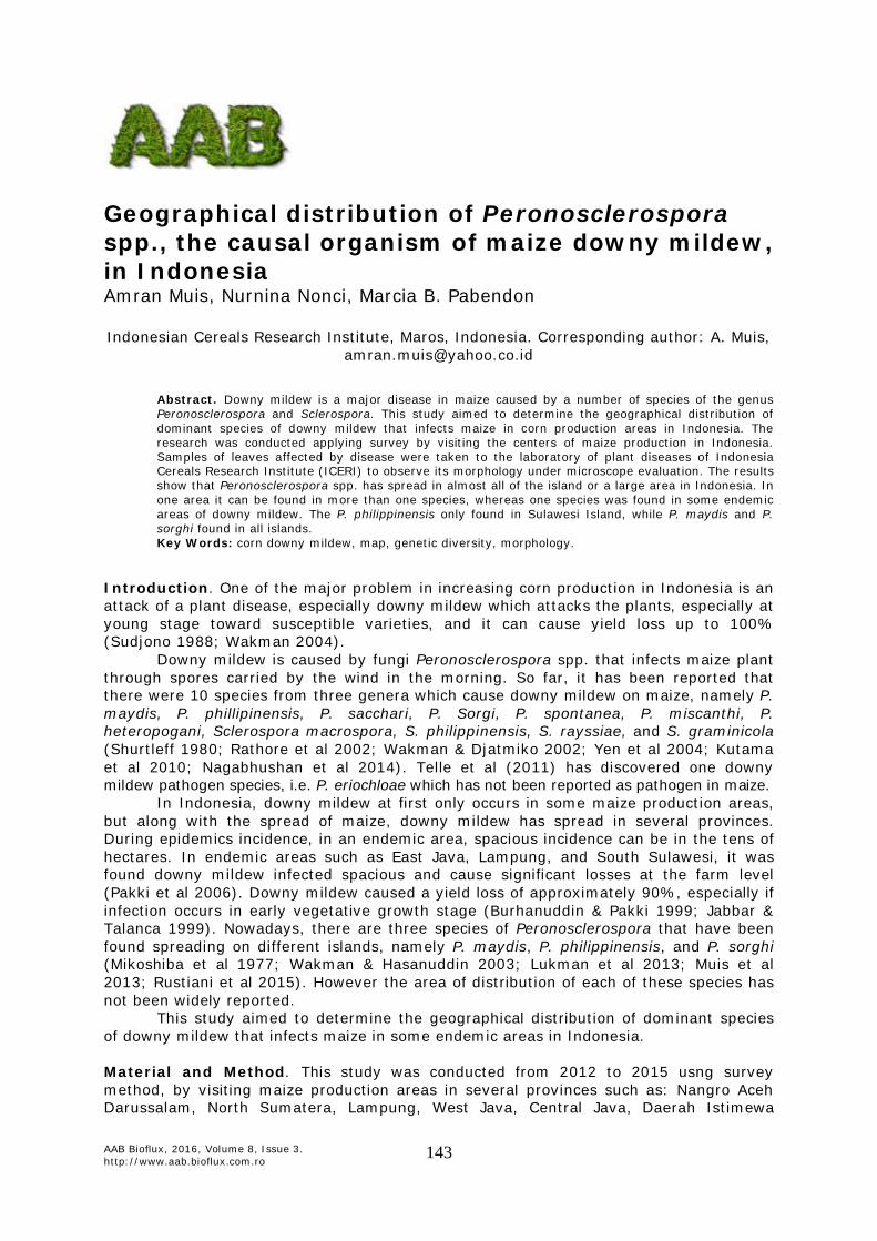
Sidrap, Enrekang, Tana Toraja, Bone, Soppeng, Wajo, Gowa, and Jeneponto (South Sulawesi), Tomohon (North Sulawesi), Palu, Donggala, Sigi, and Parigi Moutong (Central Sulawesi), Gorontalo (Gorontalo), Landak and Bengkayang (South Kalimantan), Langkat (North Sumatera), Central Lampung (Lampung), Bogor (West Java), Sleman and Gunung Kidul (Yogyakarta), Pati, Klaten, and Grobogan (Central Java). While samples from Aceh did not successfully obtained of DM conidia. The observation under microscope showed that conidia form downy mildew pathogens in all three provinces are different from each other. Conidia derived from Maros, Barru, Sidrap, Enrekang, Tana Toraja, Bone, Soppeng, Wajo, Gowa, Jeneponto (South Sulawesi), Gorontalo (Gorontalo), and Tomohon (North Sulawesi) was ovoid indicating that DM species was P. philippinensis, conidia derived from Kediri (East Java), Landak, Bengkayang (West Kalimantan), Palu, Donggala, Sigi, Parigi Moutong (Central Sulawesi), Sleman (Yogyakarta), Klaten, Pati, and Grobogan (Central Java) was spherical indicating that DM species was P. maydis, conidia derived from Langkat (North Sumatra), Central Lampung (Lampung), Bogor (West Java), Gunung Kidul (Yogyakarta), and Malang (East Java) was oval indicating that DM species was P. sorghi (Figure 1).
P. maydis P. sorghi P. philippinensis
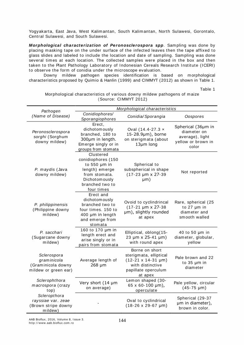
Figure 1. Conidia form of three species of Peronosclerospora found from different provinces in Indonesia (original). The results of the field observations in the downy mildew infected areas showed that the symptoms shown by the three species were similar (Figure 2).
Symptom of P. maydis Symptom of P. sorghi Symptom of P. philippinensis
Figure 2. Symptoms of downy mildew infected plants with three different species found in Indonesia (original). Common symptoms of downy mildew were characterized by chlorotic striping or partial symptoms in leaves and leaf sheaths, along with dwarfing. It was stated by CIMMYT
AAB Bioflux, 2016, Volume 8, Issue 3. http://www.aab.bioflux.com.ro
147
(2004) that downy mildew symptoms become clearer with the appearance of downy growth under the leaf surfaces due to conida formation produced early in the morning.
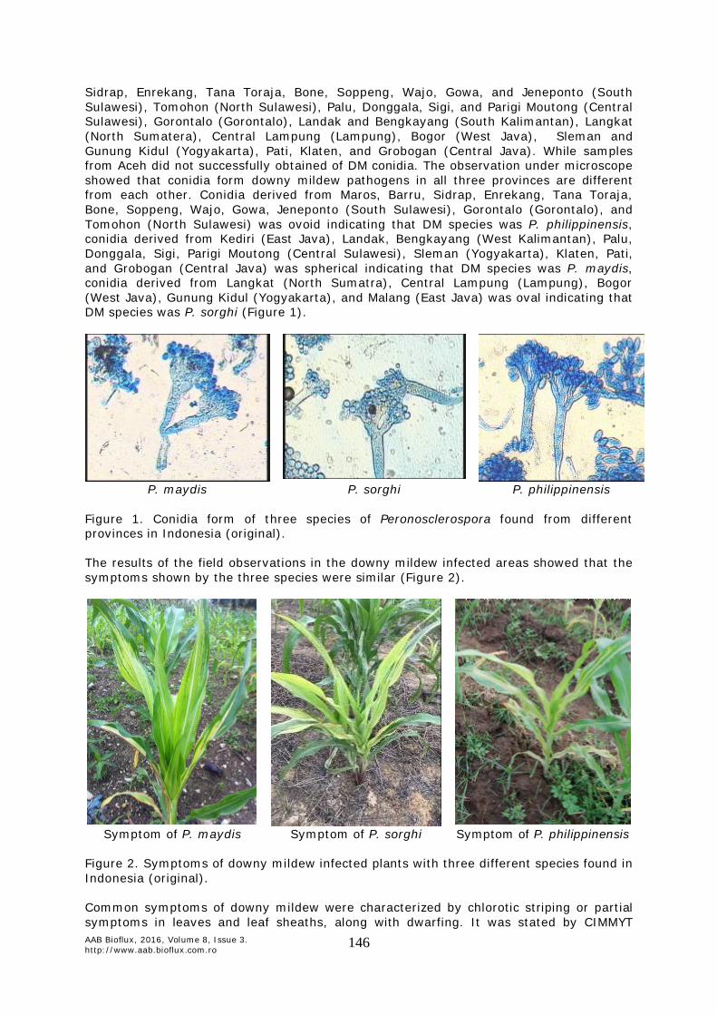
Based on the data mentioned above, it is also known that P. maydis and P. philippinensis commonly found in low lands, while P. sorghi mostly found in the highlands (Figure 3).
Figure 3. Geographical distribution of Peronosclerospora spp. in maize production areas of Indonesia. The map above is a summary of data from previous studies of Muis et al (2013). In Figure 3 shows that P. maydis is found in West Kalimantan, South Kalimantan, Central Java, D.I. Yogyakarta, East Java, Central Sulawesi and a part of South Sulawesi. P. philippinensis is found in North Sulawesi, Gorontalo, and most of South Sulawesi. While P. sorghi is found in Aceh, North Sumatra, Lampung, West Java, East Java, D.I. Yogyakarta, and Southeast Sulawesi. For some provinces such as Bali, West Nusa Tenggara, East Nusa Tenggara, East Kalimantan, Central Kalimantan, Maluku, Papua and West Papua we had no samples of downy mildew. However, the map in Figure 3, shows that there were three species causes downy mildew of maize in Indonesia and P. philippinensis was found only in Sulawesi Island. These results reinforce earlier study that Java DM (P. maydis) was found widespread in Java especially in area with temperature range of 25-300C, relative humidity 80-100%, and 1000-3000 mm annual rainfall (Rustiani et al 2015). High levels of diversity of DM in Java could be due to two causes, due to genetic variation within P. maydis, or due to presence of further DM species besides P. maydis (Lukman et al 2013).
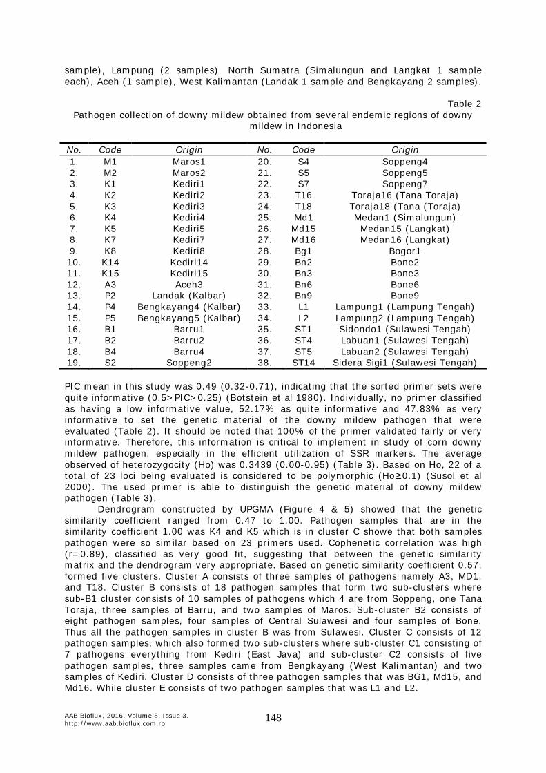
Genetic diversity of downy mildew based on SSR markers. Pathogen collection of downy mildew obtained from several endemic regions of downy mildew in Indonesia is shown in Table 2, i.e. South Sulawesi (Maros 2 samples, Bone 5 samples, Barru 4 samples, Soppeng 4 samples, and Toraja 2 samples), Central Sulawesi (Sidondo 1 sample and Labuan 2 samples), East Java (Kediri 5 samples), West Java (Bogor 1
P. maydis
P. sorghi
P. philippinensis
AAB Bioflux, 2016, Volume 8, Issue 3. http://www.aab.bioflux.com.ro
148
sample), Lampung (2 samples), North Sumatra (Simalungun and Langkat 1 sample each), Aceh (1 sample), West Kalimantan (Landak 1 sample and Bengkayang 2 samples).
Table 2
Pathogen collection of downy mildew obtained from several endemic regions of downy mildew in Indonesia
No. Code Origin No. Code Origin 1. M1 Maros1 20. S4 Soppeng4 2. M2 Maros2 21. S5 Soppeng5 3. K1 Kediri1 22. S7 Soppeng7 4. K2 Kediri2 23. T16 Toraja16 (Tana Toraja) 5. K3 Kediri3 24. T18 Toraja18 (Tana (Toraja) 6. K4 Kediri4 25. Md1 Medan1 (Simalungun) 7. K5 Kediri5 26. Md15 Medan15 (Langkat) 8. K7 Kediri7 27. Md16 Medan16 (Langkat) 9. K8 Kediri8 28. Bg1 Bogor1 10. K14 Kediri14 29. Bn2 Bone2 11. K15 Kediri15 30. Bn3 Bone3 12. A3 Aceh3 31. Bn6 Bone6 13. P2 Landak (Kalbar) 32. Bn9 Bone9 14. P4 Bengkayang4 (Kalbar) 33. L1 Lampung1 (Lampung Tengah) 15. P5 Bengkayang5 (Kalbar) 34. L2 Lampung2 (Lampung Tengah) 16. B1 Barru1 35. ST1 Sidondo1 (Sulawesi Tengah) 17. B2 Barru2 36. ST4 Labuan1 (Sulawesi Tengah) 18. B4 Barru4 37. ST5 Labuan2 (Sulawesi Tengah) 19. S2 Soppeng2 38. ST14 Sidera Sigi1 (Sulawesi Tengah) PIC mean in this study was 0.49 (0.32-0.71), indicating that the sorted primer sets were quite informative (0.5>PIC>0.25) (Botstein et al 1980). Individually, no primer classified as having a low informative value, 52.17% as quite informative and 47.83% as very informative to set the genetic material of the downy mildew pathogen that were evaluated (Table 2). It should be noted that 100% of the primer validated fairly or very informative. Therefore, this information is critical to implement in study of corn downy mildew pathogen, especially in the efficient utilization of SSR markers. The average observed of heterozygocity (Ho) was 0.3439 (0.00-0.95) (Table 3). Based on Ho, 22 of a total of 23 loci being evaluated is considered to be polymorphic (Ho≥0.1) (Susol et al 2000). The used primer is able to distinguish the genetic material of downy mildew pathogen (Table 3).
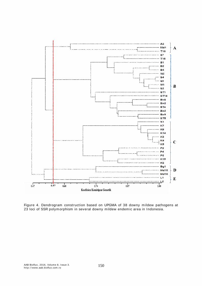
Dendrogram constructed by UPGMA (Figure 4 & 5) showed that the genetic similarity coefficient ranged from 0.47 to 1.00. Pathogen samples that are in the similarity coefficient 1.00 was K4 and K5 which is in cluster C showe that both samples pathogen were so similar based on 23 primers used. Cophenetic correlation was high (r=0.89), classified as very good fit, suggesting that between the genetic similarity matrix and the dendrogram very appropriate. Based on genetic similarity coefficient 0.57, formed five clusters. Cluster A consists of three samples of pathogens namely A3, MD1, and T18. Cluster B consists of 18 pathogen samples that form two sub-clusters where sub-B1 cluster consists of 10 samples of pathogens which 4 are from Soppeng, one Tana Toraja, three samples of Barru, and two samples of Maros. Sub-cluster B2 consists of eight pathogen samples, four samples of Central Sulawesi and four samples of Bone. Thus all the pathogen samples in cluster B was from Sulawesi. Cluster C consists of 12 pathogen samples, which also formed two sub-clusters where sub-cluster C1 consisting of 7 pathogens everything from Kediri (East Java) and sub-cluster C2 consists of five pathogen samples, three samples came from Bengkayang (West Kalimantan) and two samples of Kediri. Cluster D consists of three pathogen samples that was BG1, Md15, and Md16. While cluster E consists of two pathogen samples that was L1 and L2.

AAB Bioflux, 2016, Volume 8, Issue 3. http://www.aab.bioflux.com.ro
149
Table 3 Data profile of 23 SSR markers to 38 corn downy mildew samples
No. Marker Frequency Allele Number of allele Diversity of genes Heterozygocity PIC
1. DM1 0.49 4.00 0.56 0.92 0.46 2. DM3 0.57 2.00 0.49 0.87 0.37 3. DM4 0.33 5.00 0.75 0.89 0.71 4. DM6 0.47 6.00 0.69 0.97 0.65 5. DM7 0.62 4.00 0.57 0.50 0.53 6. DM9 0.58 3.00 0.57 0.46 0.51 7. DM10 0.59 2.00 0.48 0.82 0.37 8. DM13 0.73 2.00 0.39 0.54 0.32 9. DM14 0.69 4.00 0.48 0.54 0.44 10. DM16 0.59 4.00 0.59 0.61 0.55 11. DM18 0.50 2.00 0.50 0.68 0.38 12. DM19 0.43 4.00 0.66 0.56 0.59 13. DM24 0.41 4.00 0.70 0.80 0.64 14. DM29 0.59 3.00 0.50 0.82 0.40 15. DM33 0.56 3.00 0.59 0.50 0.52 16. DM36 0.47 3.00 0.60 0.65 0.51 17. DM39 0.66 3.00 0.50 0.35 0.45 18. DM43 0.51 2.00 0.50 0.97 0.37 19. DM46 0.50 6.00 0.64 0.39 0.58 20. DM47 0.49 5.00 0.66 0.95 0.61 21. DM51 0.71 2.00 0.41 0.58 0.33 22. DM52 0.55 2.00 0.50 0.00 0.37 23. DM54 0.49 7.00 0.71 0.64 0.68
Total 12.53 82.00 13.05 15.00 11.34 Average 0.54 3.57 0.57 0.65 0.49
Based on the morphology of the spores on previous study, downy mildew pathogen collected from Sulawesi belonging to P. philippinensis (Muis et al 2013) that are in cluster B, pathogen collected from Kediri classified as P. maydis (Muis et al 2013) that in cluster C, while the downy mildew pathogen collected from Bogor (West Java), and Langkat (North Sumatra) that are in the cluster D pertained P. sorghi (Wakman et al 2003; Muis et al 2013).
Based on the lowest level of genetic similarity in this set is 0.56, then the cluster A joined with cluster B and C, respectively as sub-cluster while cluster D and E into a separate cluster of clusters A, B, and C. It showed that was more like P. philippinensis but changes due to environmental influences. The same was expected to occur in the collection of Lampung (cluster E), at the level of similarity 0.56 into one cluster to cluster D that was P. sorghi (Muis et al 2013), but formed a separate sub-clusters because agroecological differences. The obtained data showed that in one region there were more than one species Peronosclerospora spp. It was very reasonable because of the influence of environmental factors such as wind, water, soil, and the transfer of seeds from one area to another. For example, in North Sumatra, there were two species evolved, namely P. sorghi developed in Langkat while P. philippinensis developed in Simalungun (Md1). However, the possibility of P. philippinensis in Simalungun has undergone genetic changes due to environmental factors and formed a separate cluster that was the cluster A. Conversely, there were also species grown in more than one region as P. sorghi dominant in Langkat (North Sumatra) also found in Bogor (West Java) and in Central Lampung (Lampung). P. philippinensis besides it was found in Sulawesi, we also found in Aceh. P. maydis were predominantly found in Kediri (East Java) wass also found in Landak and Bengkayang (West Kalimantan).
AAB Bioflux, 2016, Volume 8, Issue 3. http://www.aab.bioflux.com.ro
150
Figure 4. Dendrogram construction based on UPGMA of 38 downy mildew pathogens at 23 loci of SSR polymorphism in several downy mildew endemic area in Indonesia.
AAB Bioflux, 2016, Volume 8, Issue 3. http://www.aab.bioflux.com.ro
151
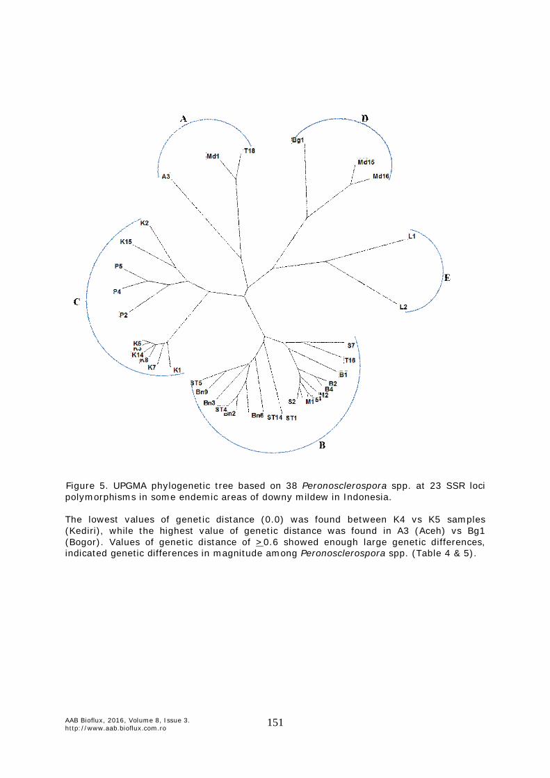
Figure 5. UPGMA phylogenetic tree based on 38 Peronosclerospora spp. at 23 SSR loci polymorphisms in some endemic areas of downy mildew in Indonesia. The lowest values of genetic distance (0.0) was found between K4 vs K5 samples (Kediri), while the highest value of genetic distance was found in A3 (Aceh) vs Bg1 (Bogor). Values of genetic distance of >0.6 showed enough large genetic differences, indicated genetic differences in magnitude among Peronosclerospora spp. (Table 4 & 5).
AAB Bioflux, 2016, Volume 8, Issue 3. http://www.aab.bioflux.com.ro
152
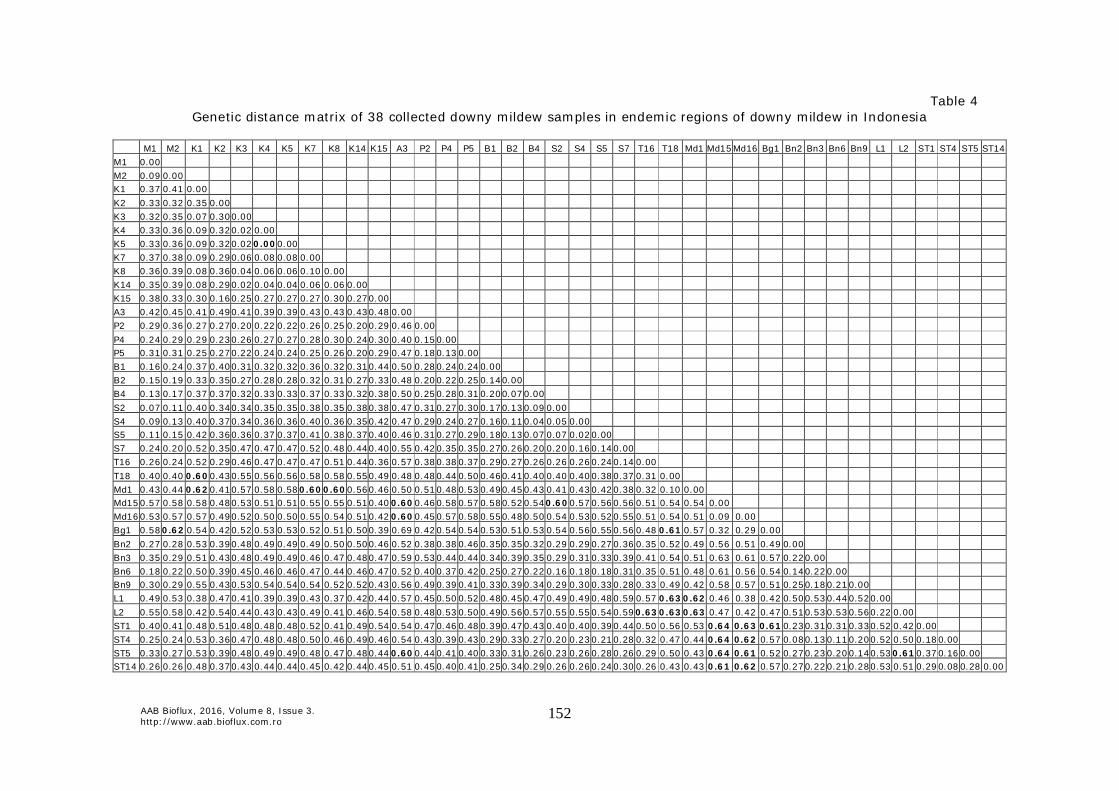
Table 4 Genetic distance matrix of 38 collected downy mildew samples in endemic regions of downy mildew in Indonesia
M1 M2 K1 K2 K3 K4 K5 K7 K8 K14 K15 A3 P2 P4 P5 B1 B2 B4 S2 S4 S5 S7 T16 T18 Md1 Md15 Md16 Bg1 Bn2 Bn3 Bn6 Bn9 L1 L2 ST1 ST4 ST5 ST14 M1 0.00 M2 0.09 0.00 K1 0.37 0.41 0.00 K2 0.33 0.32 0.35 0.00 K3 0.32 0.35 0.07 0.30 0.00 K4 0.33 0.36 0.09 0.32 0.02 0.00 K5 0.33 0.36 0.09 0.32 0.02 0.00 0.00 K7 0.37 0.38 0.09 0.29 0.06 0.08 0.08 0.00 K8 0.36 0.39 0.08 0.36 0.04 0.06 0.06 0.10 0.00 K14 0.35 0.39 0.08 0.29 0.02 0.04 0.04 0.06 0.06 0.00 K15 0.38 0.33 0.30 0.16 0.25 0.27 0.27 0.27 0.30 0.27 0.00 A3 0.42 0.45 0.41 0.49 0.41 0.39 0.39 0.43 0.43 0.43 0.48 0.00 P2 0.29 0.36 0.27 0.27 0.20 0.22 0.22 0.26 0.25 0.20 0.29 0.46 0.00 P4 0.24 0.29 0.29 0.23 0.26 0.27 0.27 0.28 0.30 0.24 0.30 0.40 0.15 0.00 P5 0.31 0.31 0.25 0.27 0.22 0.24 0.24 0.25 0.26 0.20 0.29 0.47 0.18 0.13 0.00 B1 0.16 0.24 0.37 0.40 0.31 0.32 0.32 0.36 0.32 0.31 0.44 0.50 0.28 0.24 0.24 0.00 B2 0.15 0.19 0.33 0.35 0.27 0.28 0.28 0.32 0.31 0.27 0.33 0.48 0.20 0.22 0.25 0.14 0.00 B4 0.13 0.17 0.37 0.37 0.32 0.33 0.33 0.37 0.33 0.32 0.38 0.50 0.25 0.28 0.31 0.20 0.07 0.00 S2 0.07 0.11 0.40 0.34 0.34 0.35 0.35 0.38 0.35 0.38 0.38 0.47 0.31 0.27 0.30 0.17 0.13 0.09 0.00 S4 0.09 0.13 0.40 0.37 0.34 0.36 0.36 0.40 0.36 0.35 0.42 0.47 0.29 0.24 0.27 0.16 0.11 0.04 0.05 0.00 S5 0.11 0.15 0.42 0.36 0.36 0.37 0.37 0.41 0.38 0.37 0.40 0.46 0.31 0.27 0.29 0.18 0.13 0.07 0.07 0.02 0.00 S7 0.24 0.20 0.52 0.35 0.47 0.47 0.47 0.52 0.48 0.44 0.40 0.55 0.42 0.35 0.35 0.27 0.26 0.20 0.20 0.16 0.14 0.00 T16 0.26 0.24 0.52 0.29 0.46 0.47 0.47 0.47 0.51 0.44 0.36 0.57 0.38 0.38 0.37 0.29 0.27 0.26 0.26 0.26 0.24 0.14 0.00 T18 0.40 0.40 0.60 0.43 0.55 0.56 0.56 0.58 0.58 0.55 0.49 0.48 0.48 0.44 0.50 0.46 0.41 0.40 0.40 0.40 0.38 0.37 0.31 0.00 Md1 0.43 0.44 0.62 0.41 0.57 0.58 0.58 0.60 0.60 0.56 0.46 0.50 0.51 0.48 0.53 0.49 0.45 0.43 0.41 0.43 0.42 0.38 0.32 0.10 0.00 Md15 0.57 0.58 0.58 0.48 0.53 0.51 0.51 0.55 0.55 0.51 0.40 0.60 0.46 0.58 0.57 0.58 0.52 0.54 0.60 0.57 0.56 0.56 0.51 0.54 0.54 0.00 Md16 0.53 0.57 0.57 0.49 0.52 0.50 0.50 0.55 0.54 0.51 0.42 0.60 0.45 0.57 0.58 0.55 0.48 0.50 0.54 0.53 0.52 0.55 0.51 0.54 0.51 0.09 0.00 Bg1 0.58 0.62 0.54 0.42 0.52 0.53 0.53 0.52 0.51 0.50 0.39 0.69 0.42 0.54 0.54 0.53 0.51 0.53 0.54 0.56 0.55 0.56 0.48 0.61 0.57 0.32 0.29 0.00 Bn2 0.27 0.28 0.53 0.39 0.48 0.49 0.49 0.49 0.50 0.50 0.46 0.52 0.38 0.38 0.46 0.35 0.35 0.32 0.29 0.29 0.27 0.36 0.35 0.52 0.49 0.56 0.51 0.49 0.00 Bn3 0.35 0.29 0.51 0.43 0.48 0.49 0.49 0.46 0.47 0.48 0.47 0.59 0.53 0.44 0.44 0.34 0.39 0.35 0.29 0.31 0.33 0.39 0.41 0.54 0.51 0.63 0.61 0.57 0.22 0.00 Bn6 0.18 0.22 0.50 0.39 0.45 0.46 0.46 0.47 0.44 0.46 0.47 0.52 0.40 0.37 0.42 0.25 0.27 0.22 0.16 0.18 0.18 0.31 0.35 0.51 0.48 0.61 0.56 0.54 0.14 0.22 0.00 Bn9 0.30 0.29 0.55 0.43 0.53 0.54 0.54 0.54 0.52 0.52 0.43 0.56 0.49 0.39 0.41 0.33 0.39 0.34 0.29 0.30 0.33 0.28 0.33 0.49 0.42 0.58 0.57 0.51 0.25 0.18 0.21 0.00 L1 0.49 0.53 0.38 0.47 0.41 0.39 0.39 0.43 0.37 0.42 0.44 0.57 0.45 0.50 0.52 0.48 0.45 0.47 0.49 0.49 0.48 0.59 0.57 0.63 0.62 0.46 0.38 0.42 0.50 0.53 0.44 0.52 0.00 L2 0.55 0.58 0.42 0.54 0.44 0.43 0.43 0.49 0.41 0.46 0.54 0.58 0.48 0.53 0.50 0.49 0.56 0.57 0.55 0.55 0.54 0.59 0.63 0.63 0.63 0.47 0.42 0.47 0.51 0.53 0.53 0.56 0.22 0.00 ST1 0.40 0.41 0.48 0.51 0.48 0.48 0.48 0.52 0.41 0.49 0.54 0.54 0.47 0.46 0.48 0.39 0.47 0.43 0.40 0.40 0.39 0.44 0.50 0.56 0.53 0.64 0.63 0.61 0.23 0.31 0.31 0.33 0.52 0.42 0.00 ST4 0.25 0.24 0.53 0.36 0.47 0.48 0.48 0.50 0.46 0.49 0.46 0.54 0.43 0.39 0.43 0.29 0.33 0.27 0.20 0.23 0.21 0.28 0.32 0.47 0.44 0.64 0.62 0.57 0.08 0.13 0.11 0.20 0.52 0.50 0.18 0.00 ST5 0.33 0.27 0.53 0.39 0.48 0.49 0.49 0.48 0.47 0.48 0.44 0.60 0.44 0.41 0.40 0.33 0.31 0.26 0.23 0.26 0.28 0.26 0.29 0.50 0.43 0.64 0.61 0.52 0.27 0.23 0.20 0.14 0.53 0.61 0.37 0.16 0.00 ST14 0.26 0.26 0.48 0.37 0.43 0.44 0.44 0.45 0.42 0.44 0.45 0.51 0.45 0.40 0.41 0.25 0.34 0.29 0.26 0.26 0.24 0.30 0.26 0.43 0.43 0.61 0.62 0.57 0.27 0.22 0.21 0.28 0.53 0.51 0.29 0.08 0.28 0.00
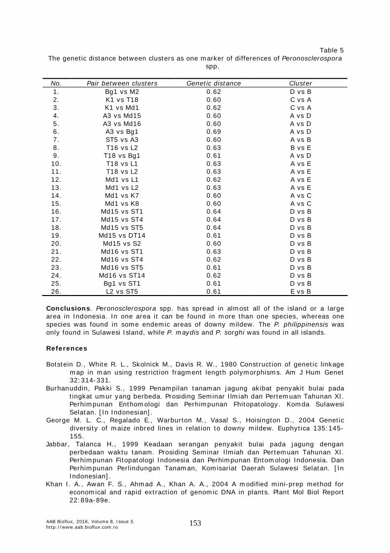
AAB Bioflux, 2016, Volume 8, Issue 3. http://www.aab.bioflux.com.ro 153
Table 5 The genetic distance between clusters as one marker of differences of Peronosclerospora
spp.
No. Pair between clusters Genetic distance Cluster 1. Bg1 vs M2 0.62 D vs B 2. K1 vs T18 0.60 C vs A 3. K1 vs Md1 0.62 C vs A 4. A3 vs Md15 0.60 A vs D 5. A3 vs Md16 0.60 A vs D 6. A3 vs Bg1 0.69 A vs D 7. ST5 vs A3 0.60 A vs B 8. T16 vs L2 0.63 B vs E 9. T18 vs Bg1 0.61 A vs D 10. T18 vs L1 0.63 A vs E 11. T18 vs L2 0.63 A vs E 12. Md1 vs L1 0.62 A vs E 13. Md1 vs L2 0.63 A vs E 14. Md1 vs K7 0.60 A vs C 15. Md1 vs K8 0.60 A vs C 16. Md15 vs ST1 0.64 D vs B 17. Md15 vs ST4 0.64 D vs B 18. Md15 vs ST5 0.64 D vs B 19. Md15 vs DT14 0.61 D vs B 20. Md15 vs S2 0.60 D vs B 21. Md16 vs ST1 0.63 D vs B 22. Md16 vs ST4 0.62 D vs B 23. Md16 vs ST5 0.61 D vs B 24. Md16 vs ST14 0.62 D vs B 25. Bg1 vs ST1 0.61 D vs B 26. L2 vs ST5 0.61 E vs B
Conclusions. Peronosclerospora spp. has spread in almost all of the island or a large area in Indonesia. In one area it can be found in more than one species, whereas one species was found in some endemic areas of downy mildew. The P. philippinensis was only found in Sulawesi Island, while P. maydis and P. sorghi was found in all islands.
References Botstein D., White R. L., Skolnick M., Davis R. W., 1980 Construction of genetic linkage
map in man using restriction fragment length polymorphisms. Am J Hum Genet 32:314-331.
Burhanuddin, Pakki S., 1999 Penampilan tanaman jagung akibat penyakit bulai pada tingkat umur yang berbeda. Prosiding Seminar Ilmiah dan Pertemuan Tahunan XI. Perhimpunan Enthomologi dan Perhimpunan Fhitopatology. Komda Sulawesi Selatan. [In Indonesian].
George M. L. C., Regalado E., Warburton M., Vasal S., Hoisington D., 2004 Genetic diversity of maize inbred lines in relation to downy mildew. Euphytica 135:145-155.
Jabbar, Talanca H., 1999 Keadaan serangan penyakit bulai pada jagung dengan perbedaan waktu tanam. Prosiding Seminar Ilmiah dan Pertemuan Tahunan XI. Perhimpunan Fitopatologi Indonesia dan Perhimpunan Entomologi Indonesia. Dan Perhimpunan Perlindungan Tanaman, Komisariat Daerah Sulawesi Selatan. [In Indonesian].
Khan I. A., Awan F. S., Ahmad A., Khan A. A., 2004 A modified mini-prep method for economical and rapid extraction of genomic DNA in plants. Plant Mol Biol Report 22:89a-89e.

AAB Bioflux, 2016, Volume 8, Issue 3. http://www.aab.bioflux.com.ro 154
Kutama A. S., Aliyu B. S., Emechebe A. M., 2010 State of sorghum downy mildew in maize in the Sudan and Sahel Savanna agro-ecological zones of Nigeria. Bayero Journal of Pure and Applied Science 3(1):233-237.
Lee M., 1998 DNA markers for detecting genetic relationship among germplasm rvealed for establishing heterotic groups. Presented at the maize Training Course, CIMMYT, Texcoco, Mexico, 25 August 1998.
Lukman R., Afifuddin A., Lubberstedt T., 2013 Unraveling the genetic diversity of maize downy mildew in Indonesia. J Plant Pathol Microb 4:162 doi:10.4172/2157-7471.1000162.
Mikoshiba F., Sudjadi M., Soediarto A., 1977 Dispersion of conidia of Sclerospora maydis in outbreaks of maize downy mildew disease in Indonesia. Tropical Agriculture Research Center, Japan, pp. 186-189.
Muis A., Pabendon M. B., Nonci N., Waskito W. P. S., 2013 Keragaman genetik Peronosclerospora maydis penyebab penyakit bulai pada jagung berdasarkan analisis marka SSR. Jurnal Penelitian Pertanian Tanaman Pangan 32(3):139-147. [In Indonesian].
Nagabhushan, Lohithaswa H. C., Sreemarasetty T. A., Puttaramanaik, Hittalmani S., 2014 Identification of stable source of resistance to sorghum downy mildew in maize (Zea mays L.). Journal of Agroecology and Natural Resource Management 1(3):176-178.
Pakki S., Talanca H., Gusnawaty, 2006 Sebaran penyakit bulai (Peronosclerospora sp) pada beberapa sentra pertanaman jagung di Sulawesi Selatan. Prosiding dan Loka Karya Nasional. Balisereal. [In Indonesian].
Quimio T. H., Hanlin R. T., 1999 Illustrated genera and species of plant pathogenic fungi in the tropics. College of Agriculture, University of the Philippines Los Banos, College, Laguna, Philippines, 259 pp.
Rathore R. S., Trivedi A., Mathur K., 2002 Rajasthan downy mildew: The problem and management perspectives. Proceedings of the Eight Asian Regional Maize Workshop: New Technologies for New Millenium. Bangkok, Thailand, 5-8 August 2002, pp. 366-379.
Rohlf F. J., 2000 NTSYS-PC numerical taxonomyand Multivariate Analysis System Version 2.1. Applied Biostatistics Inc.
Rustiani U. S., Sinaga M. S., Hidayat S. H., Wiyono S., 2015 Ecological characteristic of Peronosclerospora maydis in Java, Indonesia. International Journal of Sciences: Basic and Applied Research (IJSBAR) 19(1):159-167.
Shurtleff M. C., 1980 Compendium of corn diseases. Second edition, The American Phytopathological Society, 105 pp.
Smith J. S. C., Chin E. C .L., Shu H., Smith O. S., Wall S. J., Senior M. L., 1997 An evaluation of the utility of SSR loci as molecular markers in maize (Zea mays L.) – Comparisons with data from RFLPs and pedigree. Theor Appl Genet 95:163-173.
Sudjono M. S. 1988 Penyakit jagung dan pengendaliannya. In: Subandi, M. Syam dan A. Widjono. Jagung. Puslitbantan Tan. Pangan. Bogor: pp. 205-217. [In Indonesian].
Susol E., Eyre S., John S., 2000 High-throughput genotyping of microsatellite markers, In: SNP and microsatellite genotyping. Markers for genetic analysis. Worthington J., John S. (eds), pp. 49-66, Eaton Publishing.
Telle S., Shivas R. G., Ryley M. J., Thines M., 2011 Molecular phylogenetic analysis of Peronosclerospora (Oomycetes) reveals cryptic species and genetically distinct species parasitic to maize. Eur J Plant Pathol 130:521-528.
Wakman W., Djatmiko H. A., 2002 Sepuluh spesies cendawan penyebab penyakit bulai pada tanaman jagung. Makalah disajikan pada Seminar PFI di Universitas Negeri Jenderal Sudirman Purwokerto. 7 September 2002. [In Indonesian].
Wakman W., Hasanuddin, 2003 Penyakit bulai (Peronosclerospora sorghi) pada jagung di dataran tinggi Karo Sumatera Utara. Makalah disajikan pada Seminar Nasional PFI di Bandung. [In Indonesian].
AAB Bioflux, 2016, Volume 8, Issue 3. http://www.aab.bioflux.com.ro 155
Wakman W., 2004 Penyakit bulai pada tanaman jagung di Indonesia: masalah, penelitian dan cara mengatasinya. Prosiding Seminar Tahunan PFI Komda Sulsel. [In Indonesian].
Yen T. T. O., Prasanna B. M., Setty T. A. S., Rathore R. S., 2004 Genetic variability for resistance to sorghum downy mildew (Peronosclerospora sorghi) and Rajasthan downy mildew (P. heteropogoni) in the tropical/sub-tropical Asian maize germplasm. Euphytica 138:23-31.
*** CIMMYT, 2012 Maize Doctor. http://maizedoctor.cimmyt.org/index.php [1 May 2012].
Received: 10 November 2016. Accepted: 17 December 2016. Published online: 20 December 2016. Authors: Amran Muis, Indonesian Cereals Research Institute, Indonesia, Jl. Dr. Ratulangi No. 274 Maros 90514, e-mail: [email protected] Nurnina Nonci, Indonesian Cereals Research Institute, Indonesia, Jl. Dr. Ratulangi No. 274 Maros 90514, e-mail: [email protected] Marcia Bunga Pabendon, Indonesian Cereals Research Institute, Indonesia, Jl. Dr. Ratulangi No. 274 Maros 90514, e-mail: [email protected] This is an open-access article distributed under the terms of the Creative Commons Attribution License, which permits unrestricted use, distribution and reproduction in any medium, provided the original author and source are credited. How to cite this article: Muis A., Nonci N., Pabendon M. B., 2016 Geographical distribution of Peronosclerospora spp., the causal organism of maize downy mildew, in Indonesia. AAB Bioflux 8(3):143-155.