genotoxicity of three mouthwash products, cepacol®, periogard®, and plax®, in the drosophila...
TRANSCRIPT

Research Article
Genotoxicity of ThreeMouthwash Products, Cepacol1,Periogard1, and Plax1, in the Drosophila
Wing-Spot Test
Fa¤ bio Rodrigues, Maur|¤ cio Lehmann, Viviane Souza do Amaral,Maria Lu|¤ za Reguly, and Helo|¤ sa Helena Rodrigues de Andrade*
Laboratorio da Toxicidade Genetica – TOXIGEN, Universidade Luterana doBrasil – ULBRA/Canoas, Canoas, RS, Brazil
Antiseptic mouthwashes used in biofilm control arewidely available in the marketplace, despite incon-sistent data concerning their genetic and cellular tox-icity. In the present study, we investigated the geno-toxic potential of three antiseptics currently used forodontologic treatment, Cepacol1 (containing cetyl-pyridinium chloride), Periogard1 (chlorhexidinedigluconate), and Plax1 (triclosan). Genotoxicitywas evaluated using the Somatic Mutation andRecombination Test (SMART) in Drosophila mela-nogaster, employing flies having normal bioactiva-tion (the standard cross) and flies with increased cyto-
chrome P450-dependent biotransformation capacity(the high bioactivation cross). Periogard and Plaxproduced negative responses in both types of flies;however, Cepacol (75 and 100%) produced posi-tive responses in both the standard and high bioacti-vation assays, with the genotoxic responses mainlydue to the induction of mitotic recombination. Assaysperformed with ethanol and cetylpirydinium chlo-ride, two major ingredients of Cepacol, indicatedthat the genotoxity of the mouthwash is likely to bedue to ethanol. Environ. Mol. Mutagen. 48:644–649, 2007. VVC 2007Wiley-Liss, Inc.
Key words: SMART; cetylpyridinium chloride (CPC); chlorhexidine digluconate (CXD); triclosan (TCS)
INTRODUCTION
Antiseptic mouthwashes for personal oral hygiene are
widely used for their ability to inhibit dental plaque, a
complex biofilm formed in a series of discrete steps. Pla-
que begins with the accumulation of gram-positive strep-
tococci, followed by increasing deposits, which involve
gram-negative anaerobic bacteria [Xie et al., 2000; Lewis,
2001].
Three of the most extensively used mouthwash antisep-
tics are cetylpyridinium chloride (CPC), chlorhexidine
digluconate (CXD), and triclosan (TCS). Although these
agents have demonstrated the ability to inhibit the forma-
tion of biofilm, there is little publicly available informa-
tion on their genetic and cellular toxicity. A survey of the
effects of CPC and CPX in various experimental systems
indicates that CPC is cytotoxic [Burgalassi et al., 2001;
Chetoni et al., 2003] and that CXD can induce DNA dam-
age [Sakagami et al., 1988a,b; Eren et al., 2002; Ribeiro
et al., 2004]. Additionally, TCS was shown to induce so-
matic mutations in mice [Russel and Montgomery, 1980],
although a more recent report [Bargava and Leonard,
1996] failed to confirm this finding.
The present investigation was designed to evaluate
the genotoxic potential of three antiseptic mouthwash
products containing CPC, CXD, and TCS: Cepacol1 (0.05%
CPC), Periogard1 (0.12% CPX), and Plax1 (0.03% TCS).
The study employed the in vivo wing Somatic Mutation and
Recombination Test (SMART) in Drosophila melanogaster,which quantitatively measures both mutation and homolo-
gous recombination. There exists an extraordinary conserva-
tion of not only individual domains and proteins but also of
entire complexes and multi-step pathways between fly and
man [St. John and Xu, 1997]. This suggests that Drosophila
is a reasonable model for evaluating the potential of environ-
mental agents for inducing genetic damage in humans
[Bishop and Schiestl, 2003].
*Correspondence to: Heloısa H. R. de Andrade, Laboratorio da Toxicidade
Genetica – ULBRA, Predio 22, 48 andar, Sala 25, Av. Farroupilha, 8001,
92450-900, Canoas, RS, Brazil. E-mail: [email protected]
Grant sponsors: Conselho Nacional de Desenvolvimento Cientıfico e
Tecnologico (CNPq), Financiadora de Estudos e Projetos (FINEP), Fun-
dacao e Coordenacao de Aperfeicoamento de Pessoal de Nıvel Superior
(CAPES).
Received 4 August 2006; provisionally accepted 17 May 2007; and in
final form 1 June 2007
DOI 10.1002/em.20332
Published online 18 September 2007 in Wiley InterScience (www.interscience.
wiley.com).
VVC 2007Wiley-Liss, Inc.
Environmental andMolecular Mutagenesis 48:644^649 (2007)
MATERIALS ANDMETHODS
Chemicals
Cepacol [0.05% CPC (CAS no. 123-03-5)] was obtained from Aventis
Pharma (Sao Paulo, Brazil), while Periogard [0.12% CXD (CAS no. 55-
56-1)] and Plax [0.03% TCS (CAS no. 3380-34-5)] were purchased from
Colgate-Palmolive Company (Sao Paulo, Brazil). CPC was obtained
from Labsynth (Sao Paulo, Brazil). The molecular structures of the three
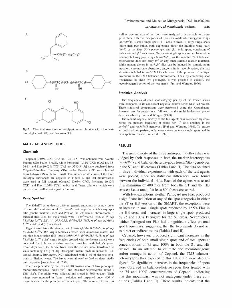
antiseptic substances are depicted in Figure 1. The test mouthwashes
were used at full strength (Cepacol [0.05% CPC], Periogard [0.12%
CXD] and Plax [0.03% TCS]) and/or in different dilutions, which were
prepared in distilled water just before use.
Wing Spot Test
The SMART assay detects different genetic endpoints by using crosses
of three different strains of Drosophila melanogaster which carry spe-
cific genetic markers (mwh and flr3) on the left arm of chromosome 3.
Parental flies used for the crosses were (i) flr3/In(3LR)TM3, ri pp sep
l(3)89Aa bx34ee Bds, (ii) ORR/ORR, flr3/In(3LR)TM3, ri pp sep l(3)89Aabx34e e Bds, and (iii) mwh/mwh.
Eggs derived from the standard (ST) cross (flr3/In(3LR)TM3, ri pp sep
l(3)89Aa bx34ee Bds virgin females crossed with mhw/mwh males) and
the high bioactivation (HB) cross (ORR/ORR, flr3/In(3LR)TM3, ri pp sepl(3)89Aa bx34e e Bds virgin females crossed with mwh/mwh males) were
collected for 8 hr on standard medium enriched with baker’s yeast.
Three days later, the larvae from both the crosses were transferred to
vials containing 1.5 g of dry Drosophila instant medium (Carolina Bio-
logical Supply, Burlington, NC) rehydrated with 5 ml of the test solu-
tions or distilled water. The larvae were allowed to feed on these media
until pupation [Andrade et al., 2004].
The flies generated by the ST and HB crosses were of two genotypes:
marker-heterozygous (mwhþ/flr3) and balancer-heterozygous (mwhþ/
TM3, Bds). The adults were collected and stored in 70% ethanol. Their
wings were mounted in Faure’s solution and inspected under 400 3magnification for the presence of mutant spots. The number of spots, as
well as type and size of the spots were analyzed. It is possible to distin-
guish three different categories of spots on marker-heterozygous wings
(mwh/flr3): (i) small single spots (1–2 cells in size), (ii) large single spots
(more than two cells), both expressing either the multiple wing hairs
(mwh) or the flare (flr3) phenotype, and (iii) twin spots, consisting of
both mwh and flr3 subclones. Only mwh single spots can be observed on
balancer heterozygous wings (mwh/TM3), as the inverted TM3 balancer
chromosome does not carry flr3 or any other suitable marker mutation.
While mutant clones in mwh/flr3 flies can be induced by somatic point
mutation, chromosome aberration, and/or mitotic recombination, this last
alteration is lethal in mwh/TM3 flies because of the presence of multiple
inversions in the TM3 balancer chromosome. Thus, by comparing spot
frequencies in these two genotypes, it was possible to quantify the
recombinagenic action of the test agents [Frei and Wurgler, 1996].
Statistical Analysis
The frequencies of each spot category per fly of the treated series
were compared to its concurrent negative control series (distilled water).
These statistical comparisons were performed using the Kastenbaum-
Bowman test for proportions, followed by the multiple-decision proce-
dure described by Frei and Wurgler (1988).
The recombinagenic activity of the test agents was calculated by com-
paring the standard frequency of clones per 105 cells obtained in the
mwh/flr3 and mwh/TM3 genotypes [Frei and Wurgler, 1996]. To ensure
an unbiased comparison, only mwh clones in mwh single spots and in
twin spots were used [Frei et al., 1992].
RESULTS
The genotoxicity of the three antiseptic mouthwashes was
judged by their responses in both the marker-heterozygous
(mwh/flr3) and balancer-heterozygous (mwh/TM3) genotypesin the ST and HB crosses (Tables I and II). The data obtained
in three individual experiments with each of the test agents
were pooled, since no statistical differences were found
between the individual trials. Each of the agents was tested
in a minimum of 400 flies from both the ST and the HB
crosses; i.e., a total of at least 800 flies were scored.
With few exceptions, neither Periogard nor Plax produced
a significant induction of any of the spot categories in either
the ST or HB version of the SMART; the exceptions were
an increase in small single spots produced by 12.5% Plax in
the HB cross and increases in large single spots produced
by 25 and 100% Periogard for the ST cross. Nevertheless,
neither Periogard nor Plax had a significant effect on total
spot frequencies, suggesting that the two agents do not act
as direct or indirect toxins (Tables I and II).
Cepacol, however, produced significant increases in the
frequencies of both small single spots and of total spots at
concentrations of 75 and 100% in both the ST and HB
crosses. In an attempt to estimate the recombinagenic
and/or mutagenic action of Cepacol, the TM3-balancer-
heterozygous flies exposed to this antiseptic were also an-
alyzed. No significant increases in the frequencies of spots
were observed in balancer-heterozygous flies treated with
the 75 and 100% concentrations of Cepacol, indicating
that this mouthwash was not mutagenic under these con-
ditions (Tables I and II). These results indicate that the
Fig. 1. Chemical structures of cetylpyridinium chloride (A), chlorhexi-
dine digluconate (B), and triclosan (C).
Environmental and Molecular Mutagenesis. DOI 10.1002/em
Genotoxicity of Mouthwash Products 645

genotoxicity of Cepacol in the SMART assay is restricted
to the agent’s ability to induce mitotic recombination.
Periogard and Plax contain, respectively, 9 and 6% of
ethanol, whereas Cepacol contains 16.8% of ethanol
(Eth). In attempting to determine whether ethanol or CPC
(0.05%) should be regarded as representing the genotoxic
hazard of Cepacol both drugs were tested individually at
the same concentrations present in the test concentrations
of the mouthwash. Treatment with CPC did not produce a
significant increase in any spot category in either ST or
HB cross flies (Table III). In contrast, 12.5 and 16.8%
Eth, which corresponds to the 75 and 100% doses of
Cepacol, produced significant increases in total spot fre-
quencies in ST cross flies, and 8.4, 12.5, and 16.8% Eth,
corresponding to the 50, 75, and 100% concentrations of
Cepacol, produced significant increases in total spots in
assays run with the HB cross (Table III).
DISCUSSION
The present study used the Drosophila SMART assay
to evaluate the genotoxic potential of three commercial
antiseptic mouthwashes: Cepacol (containing CPC), Perio-
gard (containing CXD), and Plax (containing TCS). The
results indicate that neither Periogard nor Plax is geno-
toxic in the assay (Tables I and II).
Although TCS was reported to be positive for somatic
mutation in the mouse spot test [Russel and Montgomery,
1980], acute, subacute/subchronic, and chronic treatments
with TCS were negative for mutagenicity, carcinogenicity,
and teratogenicity in male and female Sprague-Dawley rats
[Russel and Montgomery, 1980; Bhargava and Leonard,
1996]. Treatment with CDX induced primary DNA damage
both in rat peripheral blood and in oral mucosa cells as
assessed by the Comet assay, but did not induce micronuclei
[Ribeiro et al., 2004]. CDX also induced DNA damage in
human buccal epithelial cells and peripheral lymphocytes,
as detected by the Comet assay [Sakagami et al., 1988a,b;
Eren et al., 2002]. It is possible that the DNA damage
detected by Comet assay in these studies was an early event
that was repaired quickly, resulting in negative responses in
the micronucleus assay and our SMART assays.
In contrast to the negative results with Periogard and
Plax, Cepacol was positive in both the ST and HB crosses
TABLE I. Genotoxicity of Cepacol1, Plax1, and Periogard1 in the D. melanogaster Wing SpotTest using the Standard (ST) Cross
Genotypes
Dilutions
(%)
Number
of flies (N)
Spots per fly (number of spots)/statistical diagnosisa
Total mwh
clonesc (n)
Small single spots
(1–2 cells)b (m ¼ 2)
Large single spots
(>2 cells)b (m ¼ 5)
Twin spots
(m ¼ 5)
Total spots
(m ¼ 2)
CEPACOL1
mwh/flr3 0 40 0.88 (35) 0.15 (6) 0.03 (1) 1.05 (42) 38
12.5 40 0.83 (33)� 0.18 (7)i 0.05 (2)i 1.05 (42)� 40
25 40 1.08 (43)� 0.03 (1)� 0.03 (1)i 1.13 (45)� 45
50 40 0.88 (35)� 0.20 (8)i 0.08 (3)i 1.15 (46)� 46
75 40 1.68 (67)þ 0.15 (6)i 0.00 (0)i 1.83 (73)þ 73
100 40 1.60 (64)þ 0.18 (7)i 0.03 (1)i 1.80 (72)þ 72
mwh/TW3 0 40 0.90 (36) 0.03 (1) d 0.93 (37) 37
75 40 0.88 (35)� 0.00 (0)i 0.88 (35)� 35
100 40 0.85 (34)� 0.08 (3)i 0.93 (37)� 37
PLAX1
mwh/flr3 0 40 0.85 (34) 0.15 (6) 0.03 (1) 1.03 (41) 41
6.25 40 0.93 (37)� 0.20 (8)i 0.03 (1)i 1.15 (46)� 46
12.50 40 0.73 (29)� 0.13 (5)i 0.03 (1)i 0.88 (35)� 35
25 40 0.95 (38)� 0.08 (3)� 0.05 (2)i 1.08 (43)� 43
50 40 1.08 (43)� 0.10 (4)i 0.03 (1)i 1.20 (48)� 47
75 40 0.90 (36)� 0.35 (14)i 0.15 (6)i 1.40 (56)� 54
PERIOGARD1
mwh/flr3 0 40 0.93 (37) 0.05 (2) 0.05 (2) 1.03 (41) 41
12.5 40 0.93 (37)� 0.20 (8)i 0.00 (0)i 1.13 (45)� 44
25 40 0.78 (31)� 0.28 (11)þ 0.05 (2)i 1.10 (44)� 44
50 40 0.75 (30)� 0.08 (3)i 0.10 (4)i 0.93 (37)� 37
75 40 0.73 (29)� 0.08 (3)i 0.10 (4)i 0.90 (36)� 36
100 40 0.65 (26)� 0.33 (13)þ 0.10 (4)i 1.08 (43)� 43
ENU 0.05 mM 10 5.50 (55)þ 6.50 (65)þ 2.50 (25)þ 14.50 (145)þaStatistical diagnosis according to Frei and Wurgler (1988): þ, positive; �, negative; i, inconclusive; m, multiplication factor. Probability levels a ¼b ¼ 0.05.bIncluding rare flr3 spots.cConsidering mwh clones from mwh single spots and from twin spots.dOnly mwh single spots can be observed in mwh/TM3 heterozygotes as the balancer chromosome TM3 does not carry flr3 mutation.
Environmental and Molecular Mutagenesis. DOI 10.1002/em
646 Rodrigues et al.

in the SMART assay, with the positive responses being
mainly due to recombination (Tables I and II). Since
Cepacol contains 16.8% Eth and 0.05% CPC, both drugs
were tested individually at the same concentrations that
were present in the mouthwash (Table III). CPC was neg-
ative, and Eth positive in these assays. Consequently, the
induction of recombination that we observed for Cepacol
was probably due to the Eth contained in this mouthwash.
Published information relevant to the assessment of the
possible genotoxic potential of ethanol gave clear evi-
dence that it is not a bacterial or mammalian cell muta-
gen, although the in vitro assays carried out have gener-
ally not included exogenous metabolic activation. The
reported in vivo tests for chromosome aberration are all
negative and only few micronucleus tests have given posi-
tive results. The majority of SCE assays in cultured cells
have shown negative responses; however, these studies
did not use an exogenous metabolic activation system.
SCE can be induced in lymphocytes in vitro by acetalde-
hyde or ethanol treatment in the presence of alcohol dehy-
drogenase enzyme [Obe et al., 1986], which suggests that
acetaldehyde may be the agent responsible for SCE induc-
tion by ethanol in vitro and in animals.
Drosophila is well equipped to tolerate and utilize high
levels of ethanol encountered in its rotting-fruit niche,
which means that this metabolite is formed in significant
amounts in different tissues in vivo. Therefore, the genetic
effects observed in the present study—mitotic recombina-
tion increments in somatic cells of Drosophila mela-nogaster—might be due to an ethanol metabolite, presum-
ably acetaldehyde [Phillips and Jenkinson, 2001]. Acetal-
dehyde has been found to induce SCE and chromosome
aberrations in cultured mammalian cells. Although there
have been very few studies in intact mammals, the avail-
able evidence suggests that acetaldehyde produces similar
cytogenetic effects in vivo, which may be related to its
ability to form DNA–DNA and/or DNA–protein cross-
links. All in all, this might explain our data, as well as
the significant increases induced by ethanol in D. mela-nogaster X-chromosome nondisjunction, and sex-linked
recessive lethals [Rey et al., 1992; Phillips and Jenkinson,
2001; Montooth et al., 2006].
TABLE II. Genotoxicity of Cepacol1, Plax1, and Periogard1 in the D. melanogaster Wing SpotTest using the High Bioactivation (HB) Cross
Genotypes
Dilutions
(%)
Number
of flies (N)
Spots per fly (number of spots)/statistical diagnosisa
Total mwh
clonesc (n)
Small single spots
(1–2 cells)b (m ¼ 5 )
Large single spots
(>2 cells)b (m ¼ 5)
Twin spots
(m ¼ 5)
Total spots
(m ¼ 2)
CEPACOL1
mwh/flr3 0 40 0.90 (36) 0.20 (8) 0.08 (3) 1.18 (47) 47
12.5 40 1.03 (41)� 0.08 (3)� 0.08 (3)i 1.18 (47)� 47
25 40 1.38 (55)þ 0.18 (7)i 0.00 (0)� 1.55 (62)� 62
50 40 1.00 (40)� 0.10 (4)� 0.13 (5)i 1.23 (49)� 47
75 40 1.60 (64)þ 0.18 (7)i 0.03 (1)i 1.80 (72)þ 72
100 40 1.65 (66)þ 0.25 (10)i 0.03 (1)i 1.93 (77)þ 77
mwh/TM3 0 40 1.00 (40) 0.03 (1) d 1.03 (41) 41
75 40 0.93 (37)� 0.15 (6)i 1.08 (43)� 43
100 40 1.33 (53)� 0.03 (1)i 1.35 (54)� 54
PLAX1
mwh/flr3 0 40 0.90 (36) 0.20 (8) 0.08 (3) 1.18 (47) 47
6.25 40 1.03 (41) 0.25 (10)i 0.10 (4)i 1.38 (55)� 54
12.5 40 1.38 (55)þ 0.03 (1)� 0.03 (1)i 1.43 (57)� 57
25 40 1.05 (42)� 0.33 (13)i 0.03 (1)i 1.40 (56)� 56
50 40 1.00 (40)� 0.28 (11)i 0.03 (1)i 1.30 (52)� 50
75 40 1.25 (50)i 0.20 (8)i 0.03 (1)i 1.48 (59)� 59
PERIOGRAD1
mwh/flr3 0 40 1.08 (43) 0.30 (12) 0.08 (3) 1.45 (58) 57
12.5 40 0.83 (33)� 0.15 (6)� 0.05 (2)� 1.03 (41)� 41
25 40 0.85 (34)� 0.25 (10)� 0.10 (4)� 1.20 (48)� 47
50 40 0.98 (39)� 0.20 (8)� 0.15 (6)� 1.33 (53)� 53
75 40 0.95 (38)� 0.30 (12)i 0.05 (2)i 1.30 (52)� 52
100 40 1.05 (42)� 0.38 (15)i 0.08 (3)i 1.50 (60)� 60
URE 20 mM 10 19.10 (191)þ 9.80 (98)þ 2.60 (26)þ 31.50 (315)þaStatistical diagnosis according to Frei and Wurgler (1998): þ, positive; �, negative; i, inconclusive; m, multiplication factor. Probability levels a ¼b ¼ 0.05.bIncluding rare flr3 spots.cConsidering mwh clones from mwh single spots and from twin spots.dOnly mwh single spots can be observed in mwh/TM3 heterozygotes as the balancer chromosome TM3 does not carry flr3 mutation.
Environmental and Molecular Mutagenesis. DOI 10.1002/em
Genotoxicity of Mouthwash Products 647

In conclusion, the results of this study indicate that the
higher ethanol concentration present in Cepacol induces
mitotic recombination between homologous chromosomes
in the Drosophila SMART assay. Homologous mitotic
recombination can result in loss of heterozygosity or
genetic rearrangements, and these events are involved in
the genesis of numerous diseases, including cancer
[Bishop and Schiestl, 2003]. The evidence indicating that
the major effect of Cepacol is an increased frequency of
HR suggests that there may be a risk associated with the
use of this mouthwash, due to the high concentration of
ethanol present in its formulation. These results suggest
that it may be prudent to reevaluate currently available
odontology products by means of assays that are able to
detect a wide range of genetic lesions. In this context, the
SMART assay is one of the methodologies capable of
evaluating the induction of a range of genetic events
induced by complex mixtures like the mouthwashes ana-
lyzed in this study [Andrade et al., 2004].
REFERENCES
Andrade HHR, Reguly ML, Lehmann M. 2004. Wing Somatic Mutation
and Recombination Test (SMART). In: Henderson DS, editor. Dro-
sophila Cytogenetics Protocols. Totowa: Human Press. pp 389–413.
Bhargava HN, Leonard PA. 1996. Triclosan: Applications and safety.
Am J Infect Control 24:209–218.
Bishop AJR, Schiestl RH. 2003. Role of homologous recombination in
carcinogenesis. Exp Mol Pathol 74:94–105.
Burgalassi S, Chetoni P, Monti D, Saetonne MF. 2001. Cytotoxicity of
potential ocular permeation enhancers evaluated on rabbit and
human corneal epithelial cell lines. Toxicol Lett 122:1–8.
Chetoni P, Burgalassi S, Monti D, Saetonne MF. 2003. Ocular toxicity
of some corneal penetration enhancers evaluated by electrophysi-
ology measurements on isolated rabbit corneas. Toxicol In Vitro
17:497–504.
Eren K, Ozmric N, Sardas S. 2002. Monitoring of buccal epithelial cells
by alkaline comet assay (single cell gel electrophoresis technique)
in cytogenetic evaluation of chlorhexidine. Clin Oral Investig
6:150–154.
Frei H, Wurgler FE. 1988. Statistical methods to decide whether mutage-
nicity test data from Drosophila assays indicate positive, negative
or inconclusive result. Mutat Res 203:297–308.
TABLE III. Genotoxicity of Ethanol and Cetylpyridinium Chloride in the D. melanogaster Wing Spot Test using both theStandard (ST) and High Bioactivation (HB) Crosses
Genotypes
Dilutions
(%)
Number
of flies (N)
Spots per fly (number of spots)/statistical diagnosisa
Total mwhc
clones (n)
Small single spots
(1–2 cellsb) (m ¼ 2)
Large single spots
(>2 cells)b (m ¼ 5)
Twin spots
(m ¼ 5)
Total spots
(m ¼ 2)
Standard cross
Ethanol
mwh/flr3 0 40 0.55 (22) 0.35 (14) 0.05 (2) 0.95 (38) 38
2.1 40 0.95 (38)þ 0.15 (6)� 0.00 (0)i 1.10 (44)� 44
4.2 40 1.00 (40)þ 0.15 (6)� 0.00 (0)i 1.15 (46)� 46
8.4 40 0.60 (24)� 0.25 (10)� 0.20 (8)i 1.05 (42)� 42
12.5 40 1.55 (62)þ 0.20 (8)� 0.25 (10)þ 2.00 (80)þ 76
16.8 40 0.90 (36)þ 0.50 (20)i 0.05 (2)i 1.45 (58)þ 58
Cetylpyridinium chloride
mwh/flr3 0 40 0.85 (34) 0.10 (4) 0.10 (4) 1.05 (42) 42
12.5 40 1.05 (42)� 0.15 (6)i 0.05 (2)i 1.25 (50)� 50
25 40 0.85 (34)� 0.20 (8)i 0.00 (0)� 1.05 (42)� 42
50 40 0.95 (38)� 0.15 (6)i 0.10 (4)i 1.20 (48)� 48
75 40 1.10 (44)� 0.05 (2)i 0.00 (0)� 1.15 (46)� 46
100 40 1.15 (46)i 0.10 (4)i 0.03 (1)� 1.28 (51)� 51
High bioactivation cross
Ethanol
mwh/flr3 0 40 0.90 (36) 0.10 (4) 0.10 (4) 1.10 (44) 44
2.1 40 1.25 (50)i 0.00 (0)� 0.00 (0)� 1.25 (50)� 50
4.2 40 1.05 (42)� 0.15 (6)i 0.10 (4)i 1.30 (52)� 52
8.4 40 1.65 (66)þ 0.35 (14)þ 0.40 (16)þ 2.40 (96)þ 96
12.5 40 1.45 (58)þ 0.40 (16)þ 0.20 (8)i 2.05 (82)þ 82
16.8 40 1.75 (70)þ 0.30 (12)þ 0.15 (6)i 2.20 (88)þ 88
Cetylpyridinium chloride
mwh/flr3 0 40 0.95 (38) 0.10 (4) 0.10 (4) 1.15 (46) 46
12.5 40 1.10 (44)� 0.15 (6)i 0.05 (2)i 1.30 (52)� 52
25 40 0.90 (36)� 0.10 (4)i 0.10 (4)i 1.10 (44)� 44
50 40 1.25 (50)� 0.20 (8)i 0.00 (0)� 1.45 (58)� 58
75 40 1.28 (51)� 0.18 (7)i 0.00 (0)� 1.45 (58)� 58
100 40 1.15 (46)� 0.10 (4)i 0.03 (1)� 1.28 (51)� 51
aStatistical diagnosis according to Frei and Wurgler (1988): þ, positive; �, negative; i, inconclusive; m, multiplication factor. Probability levels a ¼b ¼ 0.05.bIncluding rare flr3 spots.cConsidering mwh clones from mwh single spots and from twin spots.
Environmental and Molecular Mutagenesis. DOI 10.1002/em
648 Rodrigues et al.

Frei H, Wurgler FE. 1996. Induction of somatic mutation and recombination
by four inhibitors of eukaryotic topoisomerases assayed in the wing
spot test ofDrosophila melanogaster. Mutagenesis 11:315–332.
Frei H, Clements J, Howe D, Wurgler FE. 1992. The genotoxicity of
anticancer drug mitoxantrone in somatic and germ cells of Dro-
sophila melanogaster. Mutat Res 279:21–33.
Lewis K. 2001. Riddle of biofilm resistance—Minireview. Antimicrob
Agents Chemother 45:999–1007.
Montooth KL, Siebenthal KT, Clark AG. 2006. Membrane lipid physiol-
ogy and toxin catabolism underlie ethanol and acetic acid toler-
ance in Drosophila melanogaster. Exp Biol 209:3837–3850.
Obe, G, Jonas R, Schmidt S. 1986. Metabolism of ethanol in vitro pro-
duces a compound which induces sister-chromatid exchanges in
human peripheral lymphocytes in vitro: Acetaldehyde not ethanol
is mutagenic. Mutat Res 174:47–51.
Phillips BJ, Jenkinson P. 2001. Is ethanol genotoxic? A review of the
published data. Mutagenesis 16:91–101.
Rey M, Palermo AM, Munoz ER. 1992. Nondisjunction induced by etha-
nol in Drosophila melanogaster females. Mutat Res 268:95–104.
Ribeiro DA, Bazo AP, Silva FCA, Marques MEA, Salvadori DMF.
2004. Chlorhexidine induces DNA damage in rat peripheral leu-
kocytes and oral mucosal cells. J Periodontal Res 39:358–361.
Russel LB, Montgomery CS. 1980. Use of the mouse spot test to investi-
gate the mutagenic potential of triclosan (Irgasan1 DP300).
Mutat Res 79:7–12.
Sakagami Y, Yamasaky H, Yokoyama H, Ose Y, Sato T. 1988a. DNA repair
test of disinfectants by liquid rec-assay. Mutat Res 193:21–30.
Sakagami Y, Yamazaki H, Ogasavara N, Yokoyama H, Ose Y, Sato T.
1988b. The evaluation of genotoxic activities of disinfectants and
their metabolites by umu test. Mutat Res 209:155–160.
St. John MAR, Xu T. 1997. Insights from model systems. Understanding
human cancer in a fly? Am J Hum Genet 61:1006–1010.
Xie H, Cook GS, Costerton JW, Bruce G, Rose T, Lamont R. 2000.
Intergeneric communication in dental plaque biofilms. J Bacteriol
182:7067–7069.
Accepted by—R. Snyder
Environmental and Molecular Mutagenesis. DOI 10.1002/em
Genotoxicity of Mouthwash Products 649