genetic toxicology and cancer prevention - icsbs.cz · 9:45 – 10:15 andrea rossnerova, tereza...
TRANSCRIPT
Czech and Slovak
Environmental Mutagen Society
Bilateral Czech and Slovak Meeting
Genetic Toxicology and Cancer Prevention
Smolenice, June 12-15, 2017
Organizers

Book of Abstracts
Bilateral Czech and Slovak Workshop
Genetic Toxicology and Cancer Prevention
Edited by
Alena Gábelová, Ph.D. Cancer Research Institute, Biomedical Research Center SAS, Bratislava
and
Monika Šramková, PhD. Cancer Research Institute, Biomedical Research Center SAS, Bratislava
Rewievers
Lucia Mentelová, PhD. Department of Genetics, Faculty of Natural Sciences, Comenius University in Bratislava,
Jana Tulinská, MD, PhD. Department of Immunology and Immunotoxicology, Slovak Medical University in Bratislava
Printed by TYPOSET s.r.o., Tomášikova 26, 821 01 Bratislava, www.typoset.sk Published by
Cancer Research Institute, Biomedical Research Center of the Slovak Academy of Sciences, Bratislava, Slovak Republic
ISBN 978-80-972247-2-1

Congress Centre Smolenice
Slovak Academy of Sciences
Genetic Toxicology and Cancer Prevention
Congress Centre Smolenice Castle, Slovakia, June 12 – 15, 2017
Table of contents
Programme i Key Note Lectures 1 Lectures 7 Posters 57 List of Authors 69 Sponsors & Exhibitors 73
Scientific and Organizing Committees
Scientific Committee Alena Gábelová, PhD., Cancer Research Institute, Biomedical Research Center SAS, Bratislava, Slovakia Július Brtko, DSc., Institute of Experimnetal Endocrinology, Biomedical Research Center SAS,Bratislava, Slovakia Mária Dušinská, PhD., Norwegian Institute for Air Research, Kjeller, Norway Hana Lehocká, MD, PhD., Institute of Health, Ostrava, Czech Republic Assoc. Prof. Andrea Ševčovičová, PhD., Comenius university, Bratislava, Slovakia Jan Topinka, DSc., Institute of Experimental Medicine, AS CR, Prague, Czech Republic
Local Organizing committee Katarína Kozics, PhD.
Andrea Bábelová, PhD. Eva Horváthová, PhD. Annamária Srančíková, PhD. Michal Šelc, MSc. Monika Šramková, PhD.
Genetic Toxicology and Cancer Prevention, June 12 – 15, 2017
i
Programme
Monday, 12th June 2017
12:30 – 15:00 REGISTRATION
12:30 – 18:00 DISPLAYING THE POSTERS
15:20 – 15:50 Coffee/tea
15:50 – 16:00 Opening ceremony
Chairpersons: Hana Lehocká, MD, PhD. & Miroslav Machala, PhD. 16:00 – 16:30 Miroslav Machala, Lenka Pálková, Simona Strapáčová, Kateřina
Pěnčíková, Jiří Neča, Jana Schmuczerová, Jan Topinka, Zdeněk Dvořák, Jan Vondráček Polycyclic aromatic hydrocarbons with molecular weight 302 (L1)
16:30 – 16:50 Beáta Holečková, Katarína Šiviková, Viera Schwarzbacherová,
Martina Galdíková, Ján Dianovský, Monika Drážovská, Simona Kováčová Assessment of genotoxic potential of selected pesticides (L2)
16:50 – 17:10 Július Brtko, Lucia Toporová, Dana Macejová, Ľuba Hunáková, Ladislav Novotný, Janette Bobálová, Zdeněk Dvořák Nuclear retinoid X receptors and adverse role of their triorganotin-based agonists in organism (L3)
17:10 – 17:30 Stanislav Kyzek, Karolína Ondriašová, Filip Uhrin, Andrea Ševčovičová, Veronika Medvecká, Anna Zahoranová, Eliška Gálová Potential genotoxic effect of low temperature plasma in pea seeds (L4)
17:30 – 17:50 Katarína Kozics, Monika Mesérošová Cyto/genotoxic activity of some essential oils in human lung cells (L5)
17:50 – 18:10 Dominika Mániková, Patrícia Lukáčová, Jana Rendeková,
Danuša Vlasáková, Zuzana Šestáková, Pavel Arsenyan, Miroslav Chovanec Synthetic phenylbenzo[b]selenophenes: first glimpse on the mode of their action through yeast cells (L6)
Genetic Toxicology and Cancer Prevention, June 12 – 15, 2017
ii
18:10 – 18:40 Eva Horváthová, Eliška Gálová, Andrea Ševčovičová, Martina Klapáková, Andrej Boháč, Vladimír Mastihuba, Elena Potocká, Mária Mastihubová
Structure - activity relationship of tyrosol glycosides studied by cell-free assays and in human cells cultured in vitro (L7)
19:00 Welcome drink
19:20 Dinner
Genetic Toxicology and Cancer Prevention, June 12 – 15, 2017
iii
Tuesday, 13th June 2017
Chairpersons: Mária Dušinská, PhD. & Július Brtko, DSc.
9:00 – 9:45 Zubair Ahmed, Richard Tuxworth and Boris Kysela
Nanoparticles and Brain Tumours: from Nanotoxicity to Neuroregeration (KNL1)
9:45 – 10:05 Petra Mazancová, Filip Rázga, Veronika Némethová, Igor Lacík Straightforward but non-trivial concept of chitosan-TPP particles (L8)
10:05 – 10:25 Veronika Némethová, Filip Rázga, Petra Mazancová, Barbora
Svitková, Andrea Bábelová, Michal Šelc, Alena Gábelová Nanoparticle characterization is critical for correct interpretation of biological data (L9)
10:25 – 11:00 Coffee/tea
11:00 – 11:30 Filip Rázga, Veronika Némethová Therapy resistance in CML: The complexity that stands behind (L10)
11:30 – 12:00 Soňa Marvanová, Pavlína Šimečková, Pavel Kulich, Josef Mašek, František Hubatka, Miroslav Ciganek, Radim Skoupý, Jan Hovorka, Miroslav Machala Methods of study the behavior and fate of nanoparticles in cell cultures (L11)
12:00 – 12:20 Monika Šramková, Katarína Kozics, Vlasta Závišová, Martina Koneracká, Alena Gábelová Biological effect of betulinic acid coupled to magnetite nanoparticles in colorectal cancer cell lines (L12)
12:20 – 12:50 Michal Šelc, Naďa Gregušová, Alena Gábelová, Andrea
Bábelová The mechanisms of action of gold and iron oxide nanoparticles for the selected types of kidney cells (L13)
12:50 – 14:30 Lunch
Genetic Toxicology and Cancer Prevention, June 12 – 15, 2017
iv
Chairpersons: Andrea Bábelová, PhD. & Jan Topinka, DSc.
14:30 – 15:00 Jan Topinka, Jiří Horák, František Hopan, Alena Milcová,
Antonín Ambrož, Vlasta Švecová, Pavel Rossner, Kamil Krpec, Petr Kubesa
Genotoxic potential of particulate emissions from residential solid fuel boilers: the effect of technology, fuel, and operation output (L14)
15:00 – 15:30 Pavel Rossner, Jr., Helena Líbalová, Tereza Červená, Andrea
Rossnerová, Alena Milcová, Kristýna Vrbová, Antonín Ambrož, Fatima Elzeinová Mechanisms of lipid peroxidation induced by polycyclic aromatic hydrocarbons and extractable organic matter from particulate matter <2.5 µm (L15)
15:30 – 15:45 Jitka Sikorová, Táňa Brzicová, Alena Milcová, Kristýna Vrbová, Jiří Kléma, Petr Pikal, Jan Topinka, Pavel Rossner, Jr. Nanoparticle stability and size as important factors in nano-TiO2 toxicity in macrophage-like cells (L16)
15:45 – 16:00 Táňa Brzicová, Jitka Sikorova, Kristyna Vrbova, Jan Topinka
Comparison of dynamic light scattering instruments in size analysis of nanoparticles (L17)
16:00 – 16:30 Coffee/tea
16:30 – 17:00 Karol Mičieta, Gustáv Murín, Eva Záhradníková, Andrea
Pogányová, Jozef Dušička Retrospective monitoring ecogenotoxicity of environment of the Bratislava center by native of local flora (L18)
17:00 – 17:25 Karol Mičieta, Jozef Dušička, Gustáv Murín
Native flora in Bratislava: monitoring of ecogenotoxicity at selected industrial sites (L19)
17:25 – 17:40 Alexandra Rejhová, Alena Opattová, Daniel Slíva, Pavel
Vodička Cannabidiol enhance antitumor effect of 5-FU treatment in colorectal cancer cell lines (L20)
Genetic Toxicology and Cancer Prevention, June 12 – 15, 2017
v
17:40 – 17:55 Monika Buríková, Alexandra Poturnayová, Jozef Bizík, Tibor
Hianik Atomic force microscopy: a powerful tool for high-resolution imaging of breast cancer cells (L21)
18:00 - Dinner
20:00 Slovak wine degustation
Genetic Toxicology and Cancer Prevention, June 12 – 15, 2017
vi
Wednesday, 14th June 2017
Chairpersons: Monika Šramková, PhD. & Boris Kysela, PhD. 8:30 – 9:15 Mária Dušinská, Alena Gábelová, Naouale El Yamani, Elisabeth
Elje, Elise Rundén-Pran Small, Smart and Safe (3S) Nanoparticles: How to address their Risk Assessment with in vitro (3R) Approaches (KNL1)
9:15 – 9:45 Božena Smolková, Alena Gábelová, Monika Šramková, Katarína Kozics, Annamária Srančíková, Mária Dušinská Epigenetic changes induced by nanomaterials and possible impact on health (L22)
9:45 – 10:05 Alena Gábelová, Katarína Kozics, Monika Šramková,
Annamária Srančíková, Božena Smolková A multimodular high throughput screening platform for nano-safety assessment (L23)
10:05 – 10:25 Annamária Srančíková, Monika Šramková, Katarína Kozics, Božena Smolková, Alena Gábelová
Searching for appropriate in vitro kidney model to measure drug-induced toxicity (L24)
10:25 – 11:00 Coffee/tea 11:00 – 11:30 Miroslava Šarlinová, Martina Krutáková, Anton Dzian, Tatiana
Matáková, Ľudovít Mušák, Marián Grendár, Erika Halašová Use of microRNA in Lung Carcinoma Diagnosis (L25)
11:30 – 12:00 Alexandra Poturnayová, Monika Buríková, Jozef Bizík, Andreas
Ebner, Michael Leitner, Tibor Hianik Molecular recognition of PTK7 receptors on leukemic T-cells using DNA aptamers (L26)
12:00 – 12:20 Andrea Bábelová, Martina Baliová, Wasiliki Tsalastra, Liliana Schaefer Decorin-mediated regulation of fibrillin-1 is necessary for renal tissue preservation during obstruction-induced injury (L27)
12:20 Lunch

Genetic Toxicology and Cancer Prevention, June 12 – 15, 2017
vii
13:30 SOCIAL PROGRAMME
1. The limestone cave “DRINY”
Around 1 h walking tour through the forest. The Driny cave is the greatest attraction of the Smolenice karst and is the only karst formation in the Low Carpathian Mountains. Moreover, it is the only cave in Slovakia that originated as a result of cleavage. As opposed to the caves with oval-shaped galleries and underground water flows, this cave has the galleries shaped by fissures in the rocky massif. Individual passageways are perpendicular to each other and display a rich stalactites and stalagmites of different heights and colors. Over 550 meters of artificially illuminated corridors are open to visitors practically all year round. The visit to this cave takes 35 minutes. Walking distance from the cave to the Smolenice Castle is about 1 hour. Those who wish to visit the Driny cave bring a sweater, pullover or light jacket and tracking shoe. The middle temperature in the cave is 8 °C, the air humidity is about 97%.
Admissions: 15:00 or/and 16:00
Admission fee: 6,00 Eur
2. Smolenice Castle – Havrania skala – Havranica – Zaruby (highest peak in
Low Carpathian) - Diablov žlab – Občasný vodopád – dolina Hlboča – Smolenice Castle.
This is half day guided tour. Those who wish to joint us in hiking trip bring with you a tracking shoes, some light jacket (or raincoat) and a backpack to take with you some mineral water, food, fruits, etc. Please, do not underestimate Low Carpathians Mountains. This hiking tour is not for participants with a weak fitness. Part of the route is not marked out.
Departure at 13:30
Individual walking tours
3. Dolina Hlboče
4. Havrania skala
5. Molpír hradisko
6. Záruby

Genetic Toxicology and Cancer Prevention, June 12 – 15, 2017
viii
17:00 – 19:00 Poster discussion
Coffee/tea
20:00 Conference Dinner – Barbecue
Genetic Toxicology and Cancer Prevention, June 12 – 15, 2017
ix
Thursday, 15th June 2017
Chairpersons: Eva Horváthová, PhD. & Pavel Rössner Jr., PhD. 9:00 – 9:45 Andrew Collins
The comet assay in human biomonitoring: COST Action hCOMET (KNL3)
9:45 – 10:15 Andrea Rossnerova, Tereza Cervena, Pavel Rossner Jr.
Challenges for micronucleus assay in genetic toxicology (L28) 10:15 – 10:45 Ľudovít Mušák, Martin Petráš, Miroslava Šarlinová, Jela
Valachová, Oto Osina, Erika Halašová Level of heavy toxic metals, selected biochemical parameters and chromosomal aberrations in welders (L29)
10:45 – 11:15 Coffee Break
11:15 – 11:45 Hana Lehocká, Ivona Závacká, Jana Vavrošová, Vladimír
Janout The results of interconnection of the evidence of professional exposure to genotoxic factors (regex)and cancer registry in the Czech Republic (L30)
11:45 – 12:05 Antonín Ambrož, Veronika Vlková, Pavel Rössner, Jr, Andrea
Rössnerová, Vlasta Švecová, Miloš Velemínský, Jr., Radim J. Šrám Impact of air pollution to oxidative DNA damage and lipid peroxidation of mothers and newborns (L31)
12:05 – 12:25 Kateřina Honková, Andrea Rössnerová, Jitka Pavlíková, Hans Gmuender, Vlasta Švecová, Jana Pulkrabová, Jana Hajslová, Miloš Veleminský, Radim J. Šrám Analysis of gene expression profile of newborns from districts with different level of air pollution (L32)
12:25 – 12:30 Closing ceremony
12:30 – Lunch
14:15 – Bus departure


KEYNOTE LECTURES


Genetic Toxicology and Cancer Prevention, June 12 – 15, 2017
1
KL1 Nanoparticles and Brain Tumours: from Nanotoxicity to Neuroregeration Zubair Ahmed, Richard Tuxworth and Boris Kysela
University of Birmingham, College of Medical and Dental Sciences Edgbaston, Birmingham, B15 2TT, United Kingdom
In the UK, glioblastoma (GBM) represents 1.5% of cancer diagnosis, and 2.5% of cancer deaths, and is associated with the largest years of life lost of any cancer. Tumours are highly infiltrating, have a diffuse border region which is very difficult to treat with either surgery or conventional radiotherapy. At present, patients with glioblastoma survive on average for ≤15 months, and virtually no patients with glioblastoma multiforme survive 5 years after treatment. As a consequence of treatment, patients also experience progressive structural brain deterioration. Damage to normal brain cells often affects mental sharpness and the ability to think and perform complex tasks. It is therefore of vital importance that new treatments are developed to manage this lethal disease whilst minimising toxicity and protecting normal brain function. The past decades have seen outstanding progress in our understanding of fundamental cancer biology of DNA damage responses but this development was not accompanied by comparable advances in the clinic. For example radiation therapy in humans benefited much more from technical progress and computerization rather than from knowledge-based manipulation of DNA repair in cancer cells to therapeutic radiation. To address this gap, we have designed, synthesized and tested in vitro and in vivo the new, stable, non-toxic and biocompatible targeted gold and platinum nanoparticles (NPs) for experimental cancer therapy. The NPs are functionalized with a universal designer peptide targeting system for the autonomous translocation of the nanoparticles to the nucleus of targeted cells combined with the in situ inhibition of DNA damage responses. A pilot animal toxicity study revealed no toxicity effects of newly developed nanovectors in the target organ for therapy (brain). The pilot studies of biological efficiency in combination with radiotherapy in the in vivo animal model (F98 glioblastoma in rats) indicated improved survival outcomes for nanovectors treated animals. These preclinical studies have also revealed unexpected positive effects on regenerating potential of injured neurons.

Genetic Toxicology and Cancer Prevention, June 12 – 15, 2017
2
Genetic Toxicology and Cancer Prevention, June 12 – 15, 2017
3
KL2 Small, Smart and Safe (3S) Nanoparticles: How to address their Risk Assessment with in vitro (3R) Approaches Maria Dusinska1, Alena Gabelova2, Naouale El Yamani1, Elisabeth Elje1, Elise Rundén-Pran1 1Health Effects Group, Department of Environmental Chemistry, NILU- Norwegian Institute for Air Research, Kjeller, Norway 2Cancer Research Institute, Biomedical Research Center SAS, Dubravska cesta 9, 845 05 Bratislava, Slovakia Innovative nanotechnology research aims to develop nanoparticles (NPs) that are small, smart and safe (3S) thus can improve our everyday life without affecting negatively our health. In general, the safety evaluation of NPs is based on principles of risk assessment applied to bulk chemical substances. However, more information is needed especially on physicochemical properties of NPs, their behaviour in different environments, and interactions with biological system. NPs can be taken up by cells, can pass through biological membranes and be transported to different organs and tissues. These properties are advantageous for nanomedicine but bring safety concerns for the NPs developed in other nanotechnology industries. To understand which physicochemical properties of NPs are coupled with adverse effects is thus critical for designing 3S NPs. With the increasing production of NPs and their accelerating use it is not possible to investigate the mechanism of action in vivo with all NPs and thus the emphasis for supporting 3S is on developing alternative in vitro tests and high throughput methods following the 3R principle (reduce, refine, and replace animal testing). There are many methodological challenges with 3R approaches as existing models may not be sufficient to fully identify and characterise all the hazards associated with NPs. New models mimicking the in vivo situation more closely are being developed. Endpoints such as cytotoxicity, oxidative stress, inflammation, immunotoxicity, genotoxicity, and carcinogenicity are appropriate for understanding modes of action of NPs that can affect our health. However, it is questionable whether they can fully identify and characterise all the hazards that may be associated with NP exposure. Different exposure schedules may be needed, with emphasis on lower chronic and repeated exposures. For in vitro genotoxicity assessment, DNA damage, gene mutations, chromosome breakage and/or rearrangements (clastogenicity), and numerical chromosome aberrations (aneuploidy) should be evaluated. The ability of NPs to penetrate cellular and nuclear membranes has added a new dimension to particle toxicology, and should always be taken into account in in vitro genotoxicity studies to understand modes of action. Additional in vitro approaches such as cell transformation assays, toxicogenomic or epigenetic toxicity may also be useful for identification of mechanisms leading to adverse effects. Examples of integrated genotoxicity testing with several metal NPs will be presented.

Genetic Toxicology and Cancer Prevention, June 12 – 15, 2017
4
Acknowledgement Supported by EC FP7 NANoREG (NMP4-LA-2013-310584), NRC NorNANoREG (239199/O70), NANoREG2 (H2020-NMP-2014-2015- 646221), HISENTS (H2020-NMP-2015-685817), and ERA-NET EuroNanoMed II GEMNS and INNOCENT projects.
Genetic Toxicology and Cancer Prevention, June 12 – 15, 2017
5
KL2 The comet assay in human biomonitoring: COST Action hCOMET Andrew Collins University of Oslo, Department of Nutrition, PB 1046 Blindern, 0316 Oslo, Norway Members of the COST Action hCOMET, from 23 countries, have a common interest in using the comet assay to measure DNA damage and repair in humans. The main aim of the Action is to collect as much human comet assay data as possible into a single database, and then to carry out a pooled analysis, so that we can make definitive statements about the influence of factors such as sex, age, smoking, nutrition, lifestyle etc. on DNA (in)stability. In the first year we have succeeded in creating the database, with DNA damage estimates for around 20,000 human samples; the challenging task of statistical analysis is now commencing. In addition, we are seeking to improve the inter-laboratory reproducibility of the assay. Though the assay is already 30 years old, there are still some unanswered questions concerning technical factors that affect assay performance, and working groups are actively engaged in resolving these issues. Improvements in scoring are a high priority. The lack of a generally accepted protocol is a problem; individual labs have their own preferred version of the assay, which makes inter-laboratory comparisons difficult. We will test standardised methods in a ring study, and incorporate the findings of this into standard operating procedures that, we hope, will be adopted as best practice in future biomonitoring studies. Another working group is studying the applicability of the comet assay to different cell types. The most commonly used cells in biomonitoring are mononuclear cells isolated from fresh blood, but recently frozen whole blood – or white cells isolated from frozen blood – have been shown to be suitable for the assay. In clinical studies, cells from normal and tumour tissue may be available. Other possible sources include buccal epithelial cells, and cells from the surface of the eye, or from tears, representing target cells for environmental/occupational exposure to genotoxins. Our COST Action supports training schools and short-term scientific missions, for which anyone from a member country can apply. An important aim of hCOMET is to develop a well-trained cohort of young researchers. Acknowledgment COST (European Cooperation in Science and Technology) Action 15132

Genetic Toxicology and Cancer Prevention, June 12 – 15, 2017
6

LECTURES

Genetic Toxicology and Cancer Prevention, June 12 – 15, 2017
7
L1 Polycyclic aromatic hydrocarbons with molecular weight 302 Miroslav Machala1, Lenka Pálková1, Simona Strapáčová1, Kateřina Pěnčíková1, Jiří Neča1, Jana Schmuczerová2, Jan Topinka2, Zdeněk Dvořák3, Jan Vondráček4 1Department of Chemistry and Toxicology, Veterinary Research Institute, Brno, Czech Republic 2Department of Genetic Toxicology and Nanotoxicology, Institute of Experimental Medicine AS CR, Prague, Czech Republic 3Department of Cell Biology and Genetics, Faculty of Science, Palacky University, Olomouc, Czech Republic 4Department of Cytokinetics, Institute of Biophysics AS CR, Brno, Czech Republic Polycyclic aromatic hydrocarbons (PAHs) with molecular weight 302 have been identified in various environmental compartments, such as river sediments and airborne particles. Dibenzo[a,l]pyrene (DB[a,l]P), the best known PAH with molecular weight 302, is one of the most mutagenic and carcinogenic PAHs and several other dibenzopyrenes have been recently classified as probable human carcinogens. Generally, relatively little is known about the occurence and toxicity of this diverse group of six-ring PAHs. In our study we determined PAHs with molecular weight 302 in selected abiotic environmental samples, including extracts from airborne and diesel exhaust particles. Additionally, we evaluated their genotoxic and nongenotoxic effects in liver and lung epithelial cells with focus on the processes associated with aryl hydrocarbon receptor (AhR) activation. Cytotoxicity and the potencies to induce AhR-dependent gene expression were determined in luciferase reporter gene assays using stable transfected rat hepatoma H4IIE.Luc cells (DR-CALUX assay) and human hepatoma AZ-AhR cell line. High AhR-inducing potencies were found after exposure to naphtho[1,2-k]fluoranthene (N[1,2-k]F) and several other PAHs. Further, significant AhR-dependent induction of CYP1A1 mRNA was also found in human lung adenocarcinoma epithelial cell line A549. Surprisingly, with exception of DB[a,l]P, PAHs with molecular weight 302 did not produce significantly stable DNA adducts, cell cycle modulations and apoptotic events. On the other hand, we confirmed high AhR-mediated expression of both adaptation genes, such as TIPARP, and the genes associated with tumor promotion, such as Axin2 and BMP6, in A549 cells exposed to N[1,2-k]F. We can conclude that studied individual PAHs with molecular weight 302 elicit high AhR activation leading to nongenotoxic effects and low or negligible cytotoxicity and genotoxicity. Acknowledgement Supported by the Czech Science Foundation, grant no. P503-12-G147.
Genetic Toxicology and Cancer Prevention, June 12 – 15, 2017
8
L2 Assessment of genotoxic potential of selected pesticides Beáta Holečková, Katarína Šiviková, Viera Schwarzbacherová, Martina Galdíková, Ján Dianovský, Monika Drážovská1, Simona Kováčová Institute of Genetics, Department of Biology and Genetics, University of Veterinary Medicine and Pharmacy in Košice, Komenského 73, 041 81 Košice 1Institute of Epizootiology and Preventive Veterinary Medicine, Department of Epizootiology and Parasitology, University of Veterinary Medicine and Pharmacy in Košice, Komenského 73, 041 81 Košice Pesticides ensure higher crop yields in agricultural production. The modes of action for pesticides are not strictly species-specific; for this reason concerns have been raised about environmental risks associated with their exposure through various routes. Tango®Super is a commercially available pesticide formulation containing epoxiconazole (84 g.l-1) and fenpropimorph (250 g.l-1). This agrochemical is widely used two-compound fungicide against leaf spot and spike grain diseases. In this study, both the formulation of Tango®Super and pure epoxiconazole were tested in vitro to assess their potential genotoxic/cytotoxic effects. Single cell gel electrophoresis (SCGE) and cytogenetic assays such as chromosomal aberrations, sister chromatid exchanges, micronuclei and fluorescence in situ hybridisation in cultured bovine lymphocytes were used for investigation. DNA fragmentation assay was used to test apoptosis. No statistically significant elevations of DNA damage were detected in cells exposed to Tango®Super. Similarly, no increases in cytogenetic endpoints were seen. However, a statistically significant decrease in mitotic (MI) and proliferation (PI) indices were recorded after exposure of bovine lymphocytes to the fungicide for 24 and 48 h at concentrations ranging from 3.0 to 15,0 µg.ml-1 (p<0.05; p<0.01; p<0.001). An inhibition in the cytokinesis block proliferation index (CBPI) was observed in each donor from 1.5 to 15.0 µg.ml-1 (p<0.01; p<0.001) after 24 h exposure. DNA laddering typical for apoptosis was obtained at all concentrations and times tested. After exposure to pure epoxiconazole, no direct genotoxic effect in the induction of DNA damage and/or clastogenic/aneugenic effects were recorded. However, epoxiconazole has the ability to significantly affect cell cycle kinetics: decreased proliferation in the cytokinesis block proliferation index CBPI and identically in the PI were observed with a dose-dependent pattern. Using SCGE assay, slightly increased DNA damage was found in bovine lymphocytes at the two highest concentrations for 2 h. The cytostatic/cytotoxic effects of epoxiconazole were recorded: prolonged exposure time at the highest concentration caused inhibition of replication. DNA ladder assay confirmed potential of epoxiconazole to induce the ladder-like patterns of DNA fragments which are the hallmark of apoptosis. Acknowledgement This study was supported by VEGA projects 1/0043/15 and 1/0176/16.
Genetic Toxicology and Cancer Prevention, June 12 – 15, 2017
9
L3 Nuclear retinoid X receptors and adverse role of their triorganotin-based agonists in organism Július Brtko1, Lucia Toporová1, Dana Macejová1, Ľuba Hunáková2, Ladislav Novotný3, Janette Bobálová4, Zdeněk Dvořák5
1Institute of Experimental Endocrinology, BMC, Slovak Academy of Sciences, Bratislava, Slovakia 2Cancer Research Institute, BMC, Slovak Academy of Sciences, Bratislava, Slovakia 3Department of Pharmaceutical Chemistry, Faculty of Pharmacy, Kuwait University, Kuwait City, Kuwait 4Institute of Analytical Chemistry of the CAS, v. v. i., Brno, Czech Republic 5Department of Cell Biology and Genetics, Faculty of Science, Palacky University, Olomouc, Czech Republic Retinoids acting through all-trans retinoic acid nuclear receptors (RARs) are known to inhibit carcinogenesis, suppress premalignant epithelial lesions, tumour growth and invasion in a variety of tissues. Nuclear retinoid X receptors (RXRs) act predominantly as heterodimers with other nuclear receptors as permissive heterodimers with peroxisome proliferator-activated receptors, liver X receptors, farnesoid X receptor, pregnane X receptor and constitutive androstan receptor or as non-permissive heterodimer with vitamin D receptor, and as conditional heterodimers with retinoid receptors (RAR), and thyroid hormone receptors. RXR – “partner” receptor heterodimers are considered to be ligand-activated, DNA-binding, trans-acting, transcription-modulating proteins involved in a general molecular mechanism responsible for transcriptional responses in target genes. Trialkyltin and triaryltin compounds, a class of organometallic compounds are known to act as nuclear retinoid X receptors (RXR) agonists. Tributyltins, a highly stable compound in sea water, at even pico- or nanomolar concentrations may cause the superimposition of male genitalia on females in several aquatic organisms, since they are DNA-targeted, mitotic, and their actions are occurring through target gene(s)-mediated pathways (1). Triorganotins may cause molecular interactions with reproductive system in mammals, and as potent environmental obesogens, they promote adipocyte differentiation (2). Retinoids acting through RAR are known to inhibit carcinogenesis, suppress premalignant epithelial lesions, tumour growth and invasion in a variety of tissues. Several natural or synthetic retinoids and rexinoids have therapeutical effects due to their antiproliferative and apoptosis-inducing effects. In the current presentation, we summarize our in vitro data on the biological effects of selected trialkyltin and triaryltin derivatives, cognate RXR ligands, in human breast cancer cell lines. Acknowledgement Supported by the APVV-15-0372, APVV-0160-11, VEGA 2/0171/17, SAV-15-01 and SAV-AVCR-15-01 grants and by the institutional support RVO:68081715 of the Institute of Analytical Chemistry of the CAS, v. v. i.

Genetic Toxicology and Cancer Prevention, June 12 – 15, 2017
10
References [1] Brtko J., Dvorak Z.: Triorganotin compounds - ligands for "rexinoid" inducible
transcription factors: Biological effects. Toxicol Lett., 234: 50-58, 2015. [2] Macejova D., Toporova L., Brtko J.: The role of retinoic acid receptors and their cognate
ligands in reproduction in a context of triorganotin based endocrine disrupting chemicals. Endocr. Regul., 50: 154–164, 2016.
Genetic Toxicology and Cancer Prevention, June 12 – 15, 2017
11
L4 Potential genotoxic effect of low temperature plasma in pea seeds Stanislav Kyzek1, Karolína Ondriašová1, Filip Uhrin1, Andrea Ševčovičová1, Veronika Medvecká2, Anna Zahoranová2, Eliška Gálová1 1Department of Genetics, Faculty of Natural Sciences, Comenius University, Ilkovičova 6, Mlynská dolina, Bratislava 842 15, Slovakia, [email protected] 2Department of Experimental Physics, Faculty of Mathematics, Physics and Informatics, Comenius University, Mlynskádolina, Bratislava 842 48, Slovakia The low temperature plasma (LTP) has become a subject of the significant research effort in recent years. In addition to common areas of plasma applications, such as surface finishing, more scientific teams are focused on the study of plasma interactions with cells, microorganisms, for example to sterilization or disinfection [1]. The results of many works suggest that plasma treatment has also a positive impact on the germination and the surface sterilization of the seeds. Current results demonstrate the possibility of plasma application in medicine, pharmaceutical and food industry, but its potential genotoxic effect is not still fully clarified. The plasma generated in the air is able to induce an adaptive response. The term “adaptive response “usually means that a relatively small radiation dose or chemical substance concentration induces increased resistance when the cells are irradiated or treated with higher doses or concentrations several hours later [2]. The effect of the low temperature plasma generated in other gases, like oxygen or nitrogen, on the adaptive response is still unknown. Our work is focused on the potential genotoxic effect of the low temperature plasma treated pea seeds using the comet assay method and the constant field gel electrophoresis. The comet assay is method used for a primary damage detection of DNA in eukaryotic cells. The constant field gel electrophoresis is a method used for double strand breaks detection [3, 4]. The plasma treatment of pea seeds was performed by a planar source of the low temperature plasma based on the Diffuse Coplanar Surface Barrier Discharge (DCSBD) working at atmospheric pressure in ambient air, oxygen or nitrogen [5, 6]. Acknowledgement This work was financially supported by VEGA 1/0904/14 and VEGA 1/0053/14.
References [1] M. Laroussi, Plasma Process. Polymers, 2, 391–400 (2005) [2] J. Hillova, V. Drasil, Int. J. Radiat. Biol. Relat. Stud. Phys. Chem. Med., 12, 201 (1967) [3] T. Gichner, Z. Patková, J. Száková, et al., Environ. Exp. Bot., 62, 113 (2008) [4] S. Chanková, P.E. Bryant, Radiat. Biol. Radioecol., 42 (6), 600 (2002) [5] M. Černák, Ľ. Černáková, I. Hudec, et al., Eur. Phys. J. Appl. Phys., 47, 22806 (2009) [6] A. Zahoranová, M. Henselová, D. Hudecová, et al., Plasma Chem. Plasma Process., 36, 397 (2015)
Genetic Toxicology and Cancer Prevention, June 12 – 15, 2017
12
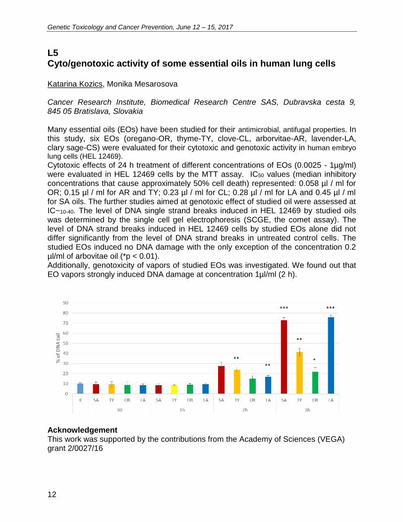
L5 Cyto/genotoxic activity of some essential oils in human lung cells Katarina Kozics, Monika Mesarosova Cancer Research Institute, Biomedical Research Centre SAS, Dubravska cesta 9, 845 05 Bratislava, Slovakia Many essential oils (EOs) have been studied for their antimicrobial, antifugal properties. In this study, six EOs (oregano-OR, thyme-TY, clove-CL, arborvitae-AR, lavender-LA, clary sage-CS) were evaluated for their cytotoxic and genotoxic activity in human embryo lung cells (HEL 12469).
Cytotoxic effects of 24 h treatment of different concentrations of EOs (0.0025 - 1µg/ml) were evaluated in HEL 12469 cells by the MTT assay. IC50 values (median inhibitory concentrations that cause approximately 50% cell death) represented: 0.058 µl / ml for OR; 0.15 µl / ml for AR and TY; 0.23 µl / ml for CL; 0.28 µl / ml for LA and 0.45 µl / ml for SA oils. The further studies aimed at genotoxic effect of studied oil were assessed at IC~10-40. The level of DNA single strand breaks induced in HEL 12469 by studied oils was determined by the single cell gel electrophoresis (SCGE, the comet assay). The level of DNA strand breaks induced in HEL 12469 cells by studied EOs alone did not differ significantly from the level of DNA strand breaks in untreated control cells. The studied EOs induced no DNA damage with the only exception of the concentration 0.2 µl/ml of arbovitae oil (*p < 0.01). Additionally, genotoxicity of vapors of studied EOs was investigated. We found out that EO vapors strongly induced DNA damage at concentration 1µl/ml (2 h).
Acknowledgement This work was supported by the contributions from the Academy of Sciences (VEGA) grant 2/0027/16
Genetic Toxicology and Cancer Prevention, June 12 – 15, 2017
13
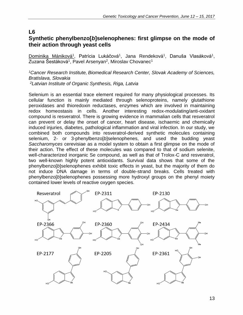
L6 Synthetic phenylbenzo[b]selenophenes: first glimpse on the mode of their action through yeast cells Dominika Mániková1, Patrícia Lukáčová1, Jana Rendeková1, Danuša Vlasáková1, Zuzana Šestáková1, Pavel Arsenyan2, Miroslav Chovanec1 1Cancer Research Institute, Biomedical Research Center, Slovak Academy of Sciences, Bratislava, Slovakia 2Latvian Institute of Organic Synthesis, Riga, Latvia Selenium is an essential trace element required for many physiological processes. Its cellular function is mainly mediated through selenoproteins, namely glutathione peroxidases and thioredoxin reductases, enzymes which are involved in maintaining redox homeostasis in cells. Another interesting redox-modulating/anti-oxidant compound is resveratrol. There is growing evidence in mammalian cells that resveratrol can prevent or delay the onset of cancer, heart disease, ischaemic and chemically induced injuries, diabetes, pathological inflammation and viral infection. In our study, we combined both compounds into resveratrol-derived synthetic molecules containing selenium, 2- or 3-phenylbenzo[b]selenophenes, and used the budding yeast Saccharomyces cerevisiae as a model system to obtain a first glimpse on the mode of their action. The effect of these molecules was compared to that of sodium selenite, well-characterized inorganic Se compound, as well as that of Trolox-C and resveratrol, two well-known highly potent antioxidants. Survival data shows that some of the phenylbenzo[b]selenophenes exhibit toxic effects in yeast, but the majority of them do not induce DNA damage in terms of double-strand breaks. Cells treated with phenylbenzo[b]selenophenes possessing more hydroxyl groups on the phenyl moiety contained lower levels of reactive oxygen species.

Genetic Toxicology and Cancer Prevention, June 12 – 15, 2017
14
Acknowledgement This work was financially supported by VEGA 2/0056/14 and APVV-14-0783 funding.

Genetic Toxicology and Cancer Prevention, June 12 – 15, 2017
15
L7 Structure-activity relationship of tyrosol glycosides studied by cell-free assays and in human cells cultured in vitro Eva Horváthová1, Eliška Gálová2, Andrea Ševčovičová2, Martina Klapáková2, Andrej Boháč3, Vladimír Mastihuba4, Elena Potocká4, Mária Mastihubová4
1Department of Genetics, Cancer Research Institute BMC, Slovak Academy of Sciences, Dúbravská cesta 9, 845 05 Bratislava, Slovakia, e-mail: [email protected] 2Department of Genetics, Faculty of Natural Sciences, Comenius University, Mlynská dolina, 842 15 Bratislava, Slovakia 3Department of Organic Chemistry, Faculty of Natural Sciences, Comenius University, Mlynská dolina, 842 15 Bratislava, Slovakia 4Institute of Chemistry, Slovak Academy of Sciences, Dúbravská cesta 9, 845 38 Bratislava, Slovakia DNA damage associated with different changes at the genetic level of the cell is generally considered as the most important stimulus for the initiation of the multistage process of carcinogenesis. The study of chemoprotective potential of selected natural compounds and their analogues, which can be potentially used in the prevention and health protection, might be therefore of great importance. Plants are a rich source of phytochemicals possessing such properties. Salidroside as the main tyrosol glycoside present in plants of the genus Rhodiola is characterized by many beneficial pharmacological effects. The structure of the compound, a wide range of biological activities and limited availability of the most productive species has inspired scientists to synthesize salidroside, its analogues, tyrosol β-galactoside (TYBGAL), tyrosol α-galactoside (TYAGAL), tyrosol β-fructofuranoside (TYBFRU), and hydroxysalidroside (HOSALI) in preparative scale. The aimes of our study were (i) to prepare tyrosol glycosides by chemical or less conventional enzymatic approaches; (ii) to determine their antioxidant, chelating, reducing and DNA-protective capacity using cell-free methods; (iii) in experimental system utilizing human hepatoma HepG2 cells to evaluate their cytotoxicity (MTT test) and chemoprotective potential against DNA damage induced by hydrogen peroxide (single cell gel electrophoresis, comet assay). Differences in the effectiveness of the synthesized tyrosol glycosides found in this study revealed that their structures can be related to the different activity detected in various test systems. Acknowledgement Supported by the grants APVV-0846-12, VEGA 2/0084/16, 2/0157/16 and the project implementations: TRANSMED, ITMS: 26240120008 and ITMS: 26240220071 supported by the Research & Development Operational Programme funded by the ERDF.
Genetic Toxicology and Cancer Prevention, June 12 – 15, 2017
16
L8 Straightforward but non-trivial concept of chitosan-TPP particles
Petra Mazancová, Filip Rázga, Veronika Némethová, Igor Lacík
Polymer Institute SAS, Dúbravská cesta 9, 845 41 Bratislava, Slovakia
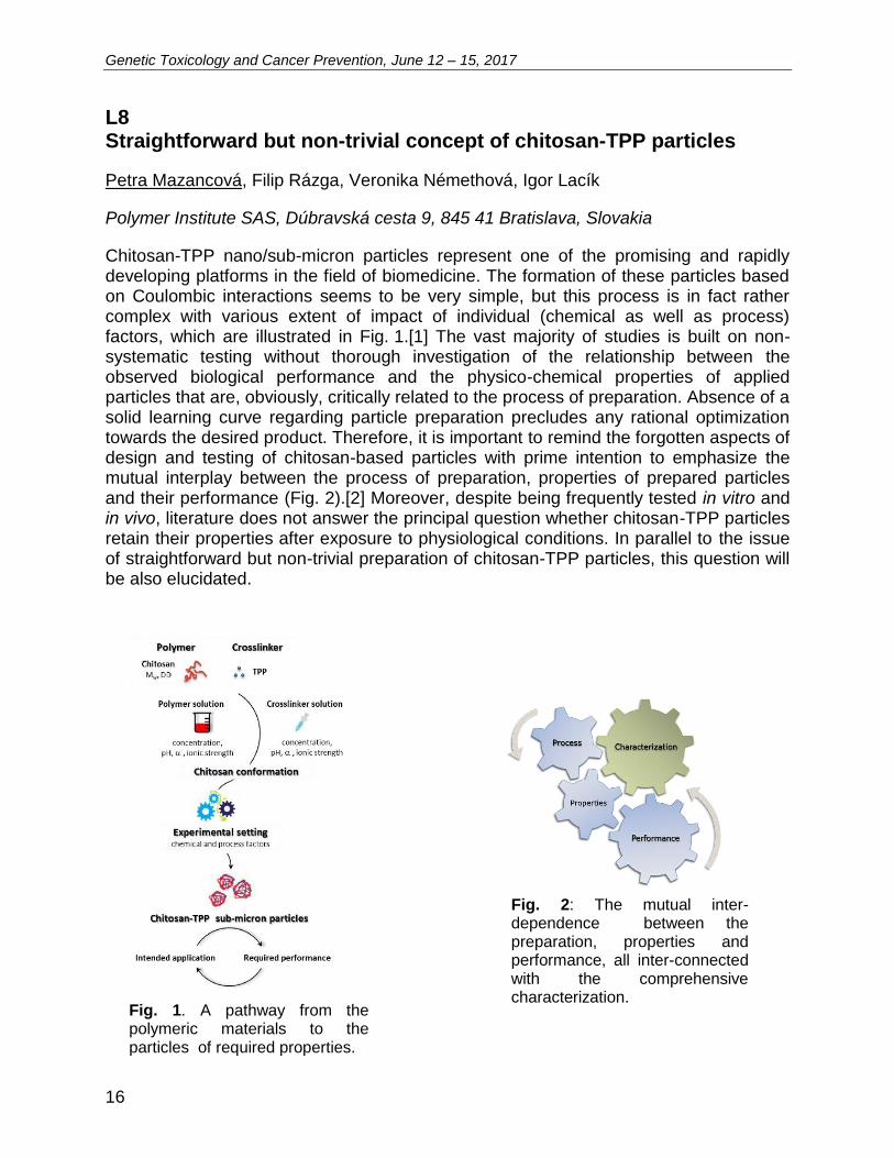
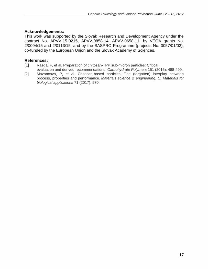
Chitosan-TPP nano/sub-micron particles represent one of the promising and rapidly developing platforms in the field of biomedicine. The formation of these particles based on Coulombic interactions seems to be very simple, but this process is in fact rather complex with various extent of impact of individual (chemical as well as process) factors, which are illustrated in Fig. 1.[1] The vast majority of studies is built on non-systematic testing without thorough investigation of the relationship between the observed biological performance and the physico-chemical properties of applied particles that are, obviously, critically related to the process of preparation. Absence of a solid learning curve regarding particle preparation precludes any rational optimization towards the desired product. Therefore, it is important to remind the forgotten aspects of design and testing of chitosan-based particles with prime intention to emphasize the mutual interplay between the process of preparation, properties of prepared particles and their performance (Fig. 2).[2] Moreover, despite being frequently tested in vitro and in vivo, literature does not answer the principal question whether chitosan-TPP particles retain their properties after exposure to physiological conditions. In parallel to the issue of straightforward but non-trivial preparation of chitosan-TPP particles, this question will be also elucidated.
Fig. 1. A pathway from the polymeric materials to the particles of required properties.
Fig. 2: The mutual inter-dependence between the preparation, properties and performance, all inter-connected with the comprehensive characterization.
Genetic Toxicology and Cancer Prevention, June 12 – 15, 2017
17
Acknowledgements: This work was supported by the Slovak Research and Development Agency under the contract No. APVV-15-0215, APVV-0858-14, APVV-0658-11, by VEGA grants No. 2/0094/15 and 2/0113/15, and by the SASPRO Programme (projects No. 0057/01/02), co-funded by the European Union and the Slovak Academy of Sciences.
References: [1] Rázga, F, et al. Preparation of chitosan-TPP sub-micron particles: Critical
evaluation and derived recommendations. Carbohydrate Polymers 151 (2016): 488-499. [2] Mazancová, P, et al. Chitosan-based particles: The (forgotten) interplay between
process, properties and performance. Materials science & engineering. C, Materials for biological applications 71 (2017): 570.

Genetic Toxicology and Cancer Prevention, June 12 – 15, 2017
18
L9 Nanoparticle characterization is critical for correct interpretation of biological data Veronika Némethová1, Filip Rázga1, Petra Mazancová1, Barbora Svitková2, Andrea Bábelová2, Michal Šelc2, Alena Gábelová2 1Polymer Institute SAS, Dúbravská cesta 9, 845 41 Bratislava, Slovakia 2Cancer Research Institute BMC SAS, Dúbravská cesta 9, 845 05 Bratislava, Slovakia Magnetite nanoparticles (MNPs) with tunable properties and controlled preparation have long been of scientific and biomedical interest. Coating of MNPs has shown to substantially alter their size and surface charge density with a direct impact not only on their colloidal stability, but more importantly, on their cellular uptake efficacy and biodistribution. The presented results serve as evidence that without proper controls and comprehensive investigations, the interpretation of internalization experiments and induced biological effects can be hard to elucidate. On prime examples we demonstrate the promotion of errors resulting from incorrect interpretation of data from particle characterization into the results obtained from model biological experiments, leading to false conclusions [1]. In addition, cellular uptake depending on the number of treated cells is shown and the definition of cellular uptake efficacy reflecting the size distribution of particles beside their absolute internalization is postulated. The derived conclusions are important for experiments concerned with cellular uptake of MNPs for accurate data collection, proper evaluation of experimental results and especially for reliable confrontation of data across independent studies.
Acknowledgement This work was supported by the Slovak Research and Development Agency under the contract No. APVV-15-0215, by VEGA grants No. 2/0094/15 and 2/0113/15, by the grant through the EEA FM and the NFM (project SK0020), and by the SASPRO
Genetic Toxicology and Cancer Prevention, June 12 – 15, 2017
19
Programme (projects No. 0057/01/02 and 0084/01/02), co-funded by the European Union and the Slovak Academy of Sciences. References: [1] Némethová et al. (2017) Intracellular uptake of magnetite nanoparticles: A focus on
physico-chemical characterization and interpretation of in vitro data. Mater Sci Eng C 70, 161-168.
Genetic Toxicology and Cancer Prevention, June 12 – 15, 2017
20
L10 Therapy resistance in CML: The complexity that stands behind Filip Rázga, Veronika Némethová Polymer Institute SAS, Dúbravská cesta 9, 845 41 Bratislava, Slovakia Imatinib mesylate (IMA), an inhibitor of BCR-ABL1 tyrosine kinase activity, has demonstrated significant clinical efficacy in the treatment of chronic myelogenous leukemia (CML) allowing durable response and prolonged survival, and has become the standard treatment for patients with CML. Unfortunately, there is still a significant portion of patients who fail IMA therapy, or do not achieve an optimal treatment response. Therefore, studies dealing with clinical IMA resistance are of extreme importance. It is well known that beside BCR-ABL1 dependent resistance caused by the presence of mutations in the BCR-ABL1 kinase domain, also BCR-ABL1 independent resistance may lead to overall failure of IMA treatment. In this contribution we point out the complexity and non-trivial interpretation of BCR-ABL1 independent clinical resistance, the prospective prediction of which is based on independent monitoring of, in fact, mutually inter-related pharmacokinetic variables such as therapy compliance, IMA trough plasma level, expression/activity of IMA influx and efflux transporters, or intracellular IMA concentration. In this context, the principal hurdles that lag smooth translation of these potential biomarkers into routine clinic are discussed. Acknowledgement This work was supported by the Slovak Research and Developmental Agency under Contract No. APVV-15-0215 and by VEGA Grant No. 2/0094/15. Rázga F. is receiving support within the SASPRO Programme (Project No. 0057/01/02) co-funded by the European Union and the Slovak Academy of Sciences.
Genetic Toxicology and Cancer Prevention, June 12 – 15, 2017
21
L11 Methods of study the behavior and fate of nanoparticles in cell cultures Soňa Marvanová1, Pavlína Šimečková1, Pavel Kulich1, Josef Mašek1, František Hubatka1, Miroslav Ciganek1, Radim Skoupý2, Jan Hovorka3, Miroslav Machala1 1Department of Chemistry and Toxicology, Veterinary Research Institute, Hudcova 70, Brno, 62100, Czech Republic 2Institute of Scientific Instruments of the CAS, Královopolská 147, Brno, 612 64, Czech Republic 3Institute for Environmental Studies, Faculty of Science, Charles University, Benátská 2, Prague 2, 12801, Czech Republic Our research interest in the field of nanotoxicology is focused both on aerosol particulate matter (PM) and engineered nanoparticles (NP) with the aim to elucidate their intracellular fate and effects. Size-segregated particulate matter is frequently used in chemical and toxicological studies. Nevertheless, toxicological in vitro studies working with the whole particles often lack a proper evaluation of PM real size distribution and characterization of agglomeration under the experimental conditions. In our study, coarse (aerodynamic diameter dae 1 – 10 µm), upper accumulation (dae 0.5 – 1 µm), lower accumulation (dae 0.17 – 0.5 µm), and ultrafine (dae < 0.17 µm) PM fractions, collected by high volume cascade impactor in Prague city center, were examined using electron microscopy and the elemental composition of single particles was determined by energy dispersive X-ray spectroscopy. Dynamic light scattering was used to measure particle size distribution in water suspension and in cell culture medium. Both lower accumulation and ultrafine fractions were highly agglomerated in cell culture medium without fetal bovine serum, while the presence of fetal bovine serum prevented the agglomeration as measured during 24 h. In order to study the intracellular fate of nanoparticles, we aim to implement a system of methods for detection of nanoparticles in cells and some of their effects. We use fluorescent nanodiamonds (Adamas, 100 nm, fluorescent, carboxylated) as model nanoparticles to study the type of NP internalization and deposition (using immunofluorescent markers of clathrin, caveolin, early endosomes and lysosomes). As nanoparticles are able to affect the autophagy, we aim to use the detection of LC3B protein, a marker of autophagy, by confocal microscopy and western blotting. Moreover, fluorescent probe LysoTracker can be used as a marker of increased total lysosomal content (increased size and/or number of lysosomes) by flow cytometry. Flow cytometric detection of LysoTracker probe was employed and slight, but significant increase of total lysosomal content was found after 24 h exposure of A549 cells to PM fraction (dae 0.17 – 0.5 µm), similarly to nanodiamonds. As immunofluorescent detection by confocal microscopy is not applicable for airborne particulate matter, we have tested the possibility to detect the nanoparticles also by reflectance independently on their fluorescence. This was achieved in case of 100 nm nanodiamonds. Concerning the PM fraction, part of internalized airborne particles was detected by reflectance, but this sample was not suitable for such measurement due to high heterogeneity of particles.

Genetic Toxicology and Cancer Prevention, June 12 – 15, 2017
22
We aim to use other types of non-fluorescent nanoparticles (possibly metal oxides, at least 100 nm size) to prove the possibility of their detection using reflectance and then to test their colocalization with immunofluorescent markers of endocytosis, autophagy, and lysosomes. Acknowledgement Supported by the Czech Science Foundation, project No. P503-12-G147.
Genetic Toxicology and Cancer Prevention, June 12 – 15, 2017
23
L12 Biological effect of betulinic acid coupled to magnetite nanoparticles in colorectal cancer cell lines Monika Sramkova1, Katarina Kozics1, Vlasta Zavisova2, Martina Koneracka2, Alena Gabelova1
1Biomedical Research Center of Slovak Academy of Sciences, Bratislava, Slovak Republic 2Institute of Experimental Physics, Slovak Academy of Sciences, Kosice, Slovak Republic The incidence of colorectal cancer has still alarming trend worldwide with the frequent occurrence of resistance towards chemotherapy, especially in metastasis. Therefore, the development of drugs that can bypass the chemoresistance and/or augment the cytotoxicity of conventional chemotherapeutics can be the strategy. Betulinic acid (BA) as a natural compound represents one of promising anti-tumor agent. To overcome the main impediment to clinical use of BA which is its poor solubility in aqueous media such as blood serum, different approaches are used, e.g. synthesis of various derivatives of BA or coupling to other carriers. With the increased interest in nanoparticle research, coupling of BA to widely used magnetite nanoparticles (MNP-BA) represents a novel approach from the field of nanotherapy. Despite the number of studies describing the biological effect of BA, characterization of MNP-BA still remains to be addressed. Our aim is to perform comprehensive assessment of the biological effect of MNP-BA with specific regard to its cytotoxic, cytostatic and genotoxic effects in colorectal cell lines (LS180, HCT116, HT29). Pilot experiments showed different sensitivity of cell lines towards the treatment with BA and MNP-BA but overall, the cell survival was decreased after 2hr as well as 24hr treatment. Also the genotoxic potential of MNP-BA was tested in these cell lines using comet assay. Further experiments (generation of ROS, activity of antioxidant enzymes upon MNP-BA treatment) will contribute to gaining an insight about the mode of betulinic acid action in targeted cells. Acknowledgement This work was financially supported by VEGA grant 2/0056/17.
Genetic Toxicology and Cancer Prevention, June 12 – 15, 2017
24
L13 The mechanisms of action of gold and iron oxide nanoparticles for the selected types of kidney cells. Michal Šelc1, Naďa Gregušová2, Alena Gábelová1, Andrea Bábelová1
1Department of Genetics, Cancer Research Institute BMC SAS, Bratislava, Slovakia 2Department of Genetics, Faculty of Natural Sciences, Comenius University, Bratislava, Slovakia Introduction Cancer theranostics combines cancer diagnosis and cancer therapy, aiming for early diagnosis, accurate molecular imaging, and precise treatment at the right time and proper dose, followed by real-time monitoring of treatment efficacy. Therapeutic strategies such as drug delivery, chemotherapy, hyperthermia, photodynamic, and radiation therapy are combined with one or more imaging functionalities for both in vitro and in vivo studies [1]. In terms of theranostics, there is a potentially large role for nanoparticles (NPs). Material, surface charge, shape, and size determine therapeutic effects of NPs [2]. In this study, we worked with two types of NPs: iron oxide and gold nanoparticles. Iron oxide nanoparticles have a number of interesting applications, especially in the biomedical field, that make them one of the most fascinating nanomaterials. They are used as contrast agents for magnetic resonance imaging, in targeted drug delivery, and for induced hyperthermia cancer treatments [3]. Gold nanopartlicles are particularly attractive for use in biological applications for several reasons. First, gold is a noble metal with inert chemical properties, resistant to corrosion, and has low toxicity based on past clinical experience. Gold nanoparticles are also easy to synthesize. Lastly, the gold surface can be easily funcionalized with biological molecules, such as antibodies and nucleic acids [4]. Some of the NPs are cleared through kidney. Ideal disease targeting of renal clearable NPs in clinical practices: the NPs specifically target the diseases and untargeted ones are rapidly cleared out of the body through the urinary system [5]. Blood is filtered in the glomerulus, which consists of glomerular endothelial cells, podocytes, and mesangial cells, which cooperate with each other for proper glomerular filtration [6]. For this reason, we are interested in whether iron oxide and gold nanoparticles can damage the specialized kidney cells that play an important role in blood filtration. Material and methods Three types of NPs were used in this study: a) iron oxide nanoparticles coated with sodium oleate and polyethylene glycol Mw=1000 (PEG-SO-Fe3O4), b) iron oxide nanoparticles coated with sodium oleate and bovine serum albumin (BSA-SO-Fe3O4) and c) gold nanoparticles coated with polyethylene glycol Mw 1000 (PEG-Au). The basic characteristics of used NPs are shown in Table 1.

Genetic Toxicology and Cancer Prevention, June 12 – 15, 2017
25
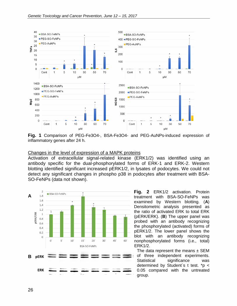
Table 1 Type of nanoparticles Two types of cells (mesangial cells and podocytes) were used in our experiments. Primary culture of both type of cells were isolated from mouse kidney. The cells were magnetically separated using magnetic beads Dynabeads (Invitrogen). These beads are accumulated in glomeruli after transcardial perfusion. Glomeruli are then incubated in Petri dishes with a specific cell media. Cellular signaling was monitored at two levels: RNA and protein expression. To monitor RNA expression, total RNA was isolated from cells, mRNA was transcripted into cDNA and detected by real-time PCR. Changes in protein expression were analyzed by western blotting. Cell growth was monitored using the IncuCyte system. Next, various types of microscopy (light, fluorescent, confocal, electron, etc.), and other molecular biology techniques were used to effectively investigate nanoparticle-induced-effects. Results The expression level of inflammatory genes is dependent on the type of nanoparticles. BSA-SO-FeNPs has no significant effect to change expression level of inflammatory genes such as tumor necrosis factor alpha (TNFa), interleukin-6 (IL6), macrophage inflammatory protein 2 (Mip2), nitric oxide synthase 2, inducible (NOS2i) (Fig. 1). On the other side, PEG-SO-FeNPs significantly increased expression level those genes. Data from PEG-Au were preliminary. They appear increased level Mip2 and NOS2i only. The data iron oxide nanoparticles represent the means ± SEM of three independent experiments. Statistical significance was determined by Student´s t test. *p < 0.05 compared with the untreated group. PEG-AuNPs data represent preliminary data from one experiment. TNFa - tumor necrosis factor alpha, IL6 – interleukin 6, MIP2 - macrophage inflammatory protein 2, NOS2i - nitric oxide synthase 2, inducible.
Coat-type Size of
core Shape
PEG-SO-Fe3O4 10 nm spheric
BSA-SO-Fe3O4 10 nm spheric
PEG-Au 10 nm spheric
Genetic Toxicology and Cancer Prevention, June 12 – 15, 2017
26
pERK
ERK
Fig. 1 Comparison of PEG-Fe3O4-, BSA-Fe3O4- and PEG-AuNPs-induced expression of inflammatory genes after 24 h.
Changes in the level of expression of a MAPK proteins Activation of extracellular signal-related kinase (ERK1/2) was identified using an antibody specific for the dual-phosphorylated forms of ERK-1 and ERK-2. Western blotting identified significant increased pERK1/2, in lysates of podocytes. We could not detect any significant changes in phospho p38 in podocytes after treatment with BSA-SO-FeNPs (data not shown).
Fig. 2 ERK1/2 activation. Protein treatment with BSA-SO-FeNPs was examined by Western blotting. (A) Densitometric analysis presented as the ratio of activated ERK to total ERK (pERK/ERK). (B) The upper panel was probed with an antibody recognizing the phosphorylated (activated) forms of pERK1/2. The lower panel shows the blot with an antibody recognizing nonphosphorylated forms (i.e., total) ERK1/2.
The data represent the means ± SEM of three independent experiments. Statistical significance was determined by Student´s t test. *p < 0.05 compared with the untreated group.
A
B
Genetic Toxicology and Cancer Prevention, June 12 – 15, 2017
27
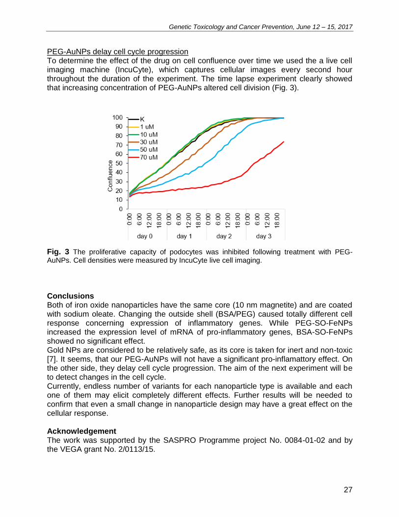
PEG-AuNPs delay cell cycle progression To determine the effect of the drug on cell confluence over time we used the a live cell imaging machine (IncuCyte), which captures cellular images every second hour throughout the duration of the experiment. The time lapse experiment clearly showed that increasing concentration of PEG-AuNPs altered cell division (Fig. 3).
Fig. 3 The proliferative capacity of podocytes was inhibited following treatment with PEG-AuNPs. Cell densities were measured by IncuCyte live cell imaging. Conclusions Both of iron oxide nanoparticles have the same core (10 nm magnetite) and are coated with sodium oleate. Changing the outside shell (BSA/PEG) caused totally different cell response concerning expression of inflammatory genes. While PEG-SO-FeNPs increased the expression level of mRNA of pro-inflammatory genes, BSA-SO-FeNPs showed no significant effect. Gold NPs are considered to be relatively safe, as its core is taken for inert and non-toxic [7]. It seems, that our PEG-AuNPs will not have a significant pro-inflamattory effect. On the other side, they delay cell cycle progression. The aim of the next experiment will be to detect changes in the cell cycle. Currently, endless number of variants for each nanoparticle type is available and each one of them may elicit completely different effects. Further results will be needed to confirm that even a small change in nanoparticle design may have a great effect on the cellular response. Acknowledgement The work was supported by the SASPRO Programme project No. 0084-01-02 and by the VEGA grant No. 2/0113/15.
Genetic Toxicology and Cancer Prevention, June 12 – 15, 2017
28
References
[1] Kelkar, S. S. & Reineke, T. M. (2011). Theranostics: combining imaging and therapy. Bioconjugate chemistry, 22(10), 1879-1903.
[2] Sun, T., Zhang, Y. S., Pang, B., Hyun, D. C., Yang, M., & Xia, Y. (2014). Engineered nanoparticles for drug delivery in cancer therapy. Angewandte Chemie International Edition, 53(46), 12320-12364.
[3] Valdiglesias, V., Fernández-Bertólez, N., Kiliç, G., Costa, C., Costa, S., Fraga, S., Bessa, M. J., Pásaro, E., Teixeira, J. P. & Laffon, B. (2016). Are iron oxide nanoparticles safe? Current knowledge and future perspectives. Journal of Trace Elements in Medicine and Biology, 38, 53-63.
[4] Webster, T. J. Safety of Nanoparticles: From Manufacturing to medical applications. 2009. Providence, RI, USA.
[5] Liu, J., Yu, M., Zhou, C., & Zheng, J. (2013). Renal clearable inorganic nanoparticles: a new frontier of bionanotechnology. Materials Today, 16(12), 477-486.
[6] Kurihara, H., & Sakai, T. (2016). Cell biology of mesangial cells: the third cell that maintains the glomerular capillary. Anatomical science international, 1-14.
[7] Bahadar, H., Maqbool, F., Niaz, K., & Abdollahi, M. (2016). Toxicity of nanoparticles and an overview of current experimental models. Iranian biomedical journal, 20(1),
1.
Genetic Toxicology and Cancer Prevention, June 12 – 15, 2017
29
L14 Genotoxic potential of particulate emissions from residential solid fuel boilers: the effect of technology, fuel, and operation output Jan Topinka1, Jiří Horák2, František Hopan2, Alena Milcová1, Antonín Ambrož1, Vlasta Švecová1, Pavel Rossner1, Kamil Krpec2, Petr Kubesa2 1Institute of Experimental Medicine CAS, Prague, Czech Republic 2VSB – Technical University of Ostrava, Ostrava, Czech Republic Introduction & Background Combustion of various solid fuels in different types of small boilers is a widely used form of heating family houses. However, these types of combustion processes emit large quantities of harmful gaseous and PM emissions. Epidemiological studies show that PM created in small heating appliances contains carcinogens and mutagens and thus may have undesirable and harmful impacts on health. Previous studies have noted that the quality of combustion is affected by the combustion technology, user operation, and fuel used, all of which affect the formation of emissions.
Methodology Various solid fuels (hard coal, lignite, dry wood, wet wood, lignite briquettes, wood pellets) were burnt in four different types of boilers representing both old structural designs (over-fire and under-fire boilers) and also up-to-date combustion devices (gasification and automatic boilers). Two different performance outputs (i.e., nominal and reduced) of boilers were tested to compare the concentration of organic PM components, their toxicity and biological response. For this purpose, the organic components of collected total particulate matter (formed by 93-100% by PM2.5) were extracted, 16 priority PAHs were quantified in extracts by GC-MS, and the analysis of the genotoxic potential of extracts using acellular assay of DNA adducts in calf thymus DNA (relatively simple method to identify genotoxic potential of complex mixtures) was employed.
Results & Conclusions We found that depending on the boiler’s technology, fuel quality and output (reduced or nominal) the mass of emitted PM2.5 varied from 0.2 to 84 kg/ton of fuel. The concentrations of the representative carcinogenic PAH – benzo[a]pyrene varied from 5 to 18,000 mg/ton of fuel. Such huge differences in PAH content are reflected by results of measurement of genotoxic potential in acellular assay: DNA adducts in calf thymus DNA after metabolic activation of PAHs (by S9 microsomal fraction) varied from 6 to 140 adducts/108 nucleotides. Differences in adduct levels are even higher after normalization of results per kg of fuel. The results of the study suggest that: (1) Mass of particulate emissions from boilers highly correlate with PM2.5 and PAH content; (2) For all fuels the highest genotoxicity was observed for over-fire and down-draft boilers compared to boilers gasification and automatic boilers; (3) Reduced output exhibited more emissions and higher toxicity than nominal output; (4) In over-fire boiler are emissions from coal substantially higher and more genotoxic worse than from biomass;
Genetic Toxicology and Cancer Prevention, June 12 – 15, 2017
30
(5) Modern boilers (gasification and automatic) produced lower emissions and exhibited lower genotoxicity. In summary, the results of the study suggest huge differences in mass, composition and genotoxic potential of complex mixtures of organic compounds forming PM emissions from various small boilers. These results reveal the need of further study in target human cells aiming to identify mechanisms of the action, targeted biological processes and human health risk. Acknowledgement Supported by Czech Science Foundation (grant No. P-503-12-G147). The authors acknowledge the assistance provided by the Research Infrastructure NanoEnviCz, supported by the Ministry of Education, Youth and Sports of the Czech Republic under Project No. LM2015073.
Genetic Toxicology and Cancer Prevention, June 12 – 15, 2017
31
L15 Mechanisms of lipid peroxidation induced by polycyclic aromatic hydrocarbons and extractable organic matter from particulate matter <2.5 µm
Pavel Rossner, Jr., Helena Líbalová, Tereza Červená, Andrea Rossnerová, Alena Milcová, Kristýna Vrbová, Antonín Ambrož, Fatima Elzeinová
Institute of Experimental Medicine AS CR, Videnska 1083, 14220, Prague, Czech Republic
Particulate matter of aerodynamic diameter <2.5 µm (PM2.5) and polycyclic aromatic hydrocarbons (PAHs) bound to it pose a significant risk to human health. After metabolic activation, PAHs may either bind to DNA forming bulky DNA adducts, or contribute to generation of reactive oxygen species (ROS) by formation of PAH o-quinones. ROS are highly reactive and cause damage to cellular macromolecules including lipids. In the present study, we investigated processes associated with lipid peroxidation following treatment of two model cell lines (human embryonic lung fibroblasts, HEL cells; human alveolar basal epithelial cells, A549) with extractable organic matter (EOM) from PM2.5 and selected PAHs (benzo[a]pyrene, BaP; 3-nitrobenzanthrone, 3-NBA). We analyzed ROS production, total antioxidant capacity (TAC), levels of lipid peroxidation products (15-F2t-isoprostane, 15-F2t-IsoP; malondialdehyde, MDA) and expression of selected genes and/or proteins associated with metabolism of xenobiotics and/or antioxidant response (CYP1A1, AKR1C2, HMOX1, PTGS2, ALDH3A1, TXNRD1). The treatment with the tested compounds was mostly accompanied by a decrease in the levels of ROS suggesting activation of antioxidant mechanisms. The decrease was time- and dose-dependent and more pronounced for BaP and 3-NBA treatment. An increase of ROS levels was found upon the short-time (30 min) exposure to EOMs and the highest concentration of 3-NBA. Individual PAHs had relatively weak effects on total antioxidant capacity; a dose-response increase of TAC was observed after 4h BaP treatment. The effects induced by EOMs were stronger, particularly for 4h exposure. The exposure to PAHs and EOMs had minimal effect on MDA levels, either after 4h or 24h treatment period. For 15-F2t-IsoP levels and mRNA expression, we observed marked differences between both cell lines. The levels of 15-F2t-IsoP were elevated in A549 cells following 4h exposure to BaP and 3-NBA; in HEL cells, a decrease was found for this time period and increase was detected after 24h exposure. EOMs induced the levels of 15-F2t-IsoP in HEL cells, but the effects in A549 cells, mostly associated with a decrease of 15-F2t-IsoP concentration, were weak. In HEL cells, no expression of CYP1A1 and ALDH3A1 mRNA was found. For other genes, the changes of expression were weak and non-consistent, most of them observed for PTGS2. In contrast, the tested compounds affected mRNA expression levels in A549 cells, notably CYP1A1, ALDH3A1 and TXNRD1. In summary, our data suggest the ability of individual PAHs and EOMs to impact ROS generation, induce antioxidant mechanisms and affect lipid peroxidation levels. However, the extent of these effects depends on the cell line used for the tests.
Acknowledgement Supported by Czech Science Foundation (16-14631S).
Genetic Toxicology and Cancer Prevention, June 12 – 15, 2017
32
L16 Nanoparticle stability and size as important factors in nano-TiO2 toxicity in macrophage-like cells Jitka Sikorová1, Táňa Brzicová1, Alena Milcová1, Kristýna Vrbová1, Jiří Kléma2, Petr Pikal3, Jan Topinka1 and Pavel Rossner, Jr.1 1Department of Genetic Toxicology and Nanotoxicology, Institute of Experimental Medicine CAS , Prague, 142 20, Czech Republic 2Department of Computer Science, Czech Technical University, Prague, 121 35, Czech Republic 3Precheza, Prerov, 751 52, Czech Republic
Toxicity of TiO2 NMs relies on characteristics of NMs such as shape, size, crystal structure, zeta potential, aggregation and agglomeration tendency, surface characteristics and coatings. However, their influence to toxicity remains unclear due to ambiguous results from different studies. In vivo studies revealed target organs (spleen and liver) and macrophages seem to be target cells as they have to cope with engulfed TiO2 NMs load. The presented study measured the cytotoxic effect without photoactivation of fourteen divers TiO2 NMs on human monocytic cell lines THP-1 differentiated into macrophage-like cells. A set of NM consists of 5 variants of anatase and 5 variants of rutile nanoparticles differing in their diameter (from 3 to 165 nm), 3 variants of anatase high aspect ratio nanomaterials. TiO2 samples were characterized in the powder form using following methods: x-ray diffraction, thermogravimetric analysis and Brunauer Emmet Teller measurements. Following dispersion, the size distribution in water and cell culture medium and zeta potential in cell culture medium were measured by dynamic light scattering.Three cytotoxicity assays were used: MTS, WTS-1, and LDH. For all nanomaterials, three independent repetitions were carried out. Over all, cytotoxicity of all NMs was low even at the highest concentration of 256 µg/ml. The viability did not decrease below 60% for WTS-1 and MST assays and 80% for the LDH assay. Despite low toxicity, polydispersity index, besides concentration, was identified as the important cytotoxic factor. Crystal size seemed to have also an influence. There is visible a nonlinear shape for crystalline size and cytotoxicity relationship with the highest toxicity between 20-60 nm. Nonlinear relationship was also shown in Chang review (2013) who concluded that the highest toxicity occurred within particles with diameter between 10-40 nm. Increase cytotoxicity in given diameter size range would give an answer to inconsistent findings at size and cytotoxicity relationship. Acknowledgement The authors acknowledge the assistance provided by the Research Infrastructure NanoEnviCz, supported by the Ministry of Education, Youth and Sports (MEYS) of the Czech Republic under Project No. LM2015073, and CENATOX (GACR P503/12/G147) and NANOGEN (MEYS LO1508) projects. References [1] Chang, X., Zhang, Y., Tang, M. and Wang, B. (2013) Nanoscale Res. Lett., 8:51.
Genetic Toxicology and Cancer Prevention, June 12 – 15, 2017
33
L17 Comparison of dynamic light scattering instruments in size analysis of nanoparticles Tana Brzicova1, 2, Jitka Sikorova1, Kristyna Vrbova1, Jan Topinka1
1Institute of Experimental Medicine CAS, Prague, Czech Republic 2Faculty of Safety Engineering, VSB - Technical University of Ostrava, Ostrava, Czech Republic 3Institute of Environmental Studies, Charles University, Prague, Czech Republic Dynamic light scattering (DLS) is a technique for nanoparticle (NP) size characterization in a liquid environment (e.g. cell culture medium). It provides crucial information for toxicological assessment as it gives insight into dynamic changes, aggregation and agglomeration vs. dissolution, of NPs and their evolution along with time. Besides electron microscopy DLS is a powerful tool for understanding NP behavior in physiologically relevant media within toxicity testing as well as efficiency of a dispersion procedure. Two state-of-art DLS instruments were compared in this study – a NanoBrook Omni (Brookhaven Instruments Corporation, USA) and a Zetasizer Nano ZS (Malvern Instruments Ltd., UK). The integrated EU project NANoREG – “A common European approach to the regulatory testing of Manufactured Nanomaterials” – provides diverse well characterized NPs in order to compare toxicity results among EU laboratories. Comparative tests were performed on standard NPs (NANOSPHERE™ Size Standards, Thermo Scientific) and batch dispersions (2.56 mg/ml) of the NANoREG project NPs under identical experimental conditions. The standard was measured undiluted and gradually diluted. NANoREG NP dispersions were prepared using probe sonication according to the generic NANOGENOTOX dispersion protocol. The standard and NANoREG NPs were measured by back-scattering to prevent issues arising from sample opacity. Standard size was correctly measured in undiluted form, approx. 10 mg/ml, by the Zetasizer Nano ZS; the NanoBrook Omni was able to measure the size correctly at a concentration of 0.4 mg/ml or lower. Not only that it is uncomfortable for the operator, the dilution itself also affects the extent of aggregation and agglomeration of the suspended NPs. Differences among sizes of the NANoREG NPs measured by the two instruments increased with rising sample opacity. Intensity distributions of the NANoREG samples were more stable with the Zetasizer Nano ZS. We concluded that under identical experimental conditions, the Zetasizer Nano ZS is suitable for quick and reliable DLS measurements of NP suspensions, while the NanoBrook Omni requires dilution of more concentrated suspensions, and thus it is not applicable for higher concentrations often used in in vitro toxicity assays. Acknowledgement Support: Research Infrastructure NanoEnviCz, Project No LM2015073.
Genetic Toxicology and Cancer Prevention, June 12 – 15, 2017
34
L18 Retrospective monitoring ecogenotoxicity of environment of the Bratislava center by native of local flora
Karol Mičieta, Gustáv Murín, Eva Záhradníková, Andrea Pogányová, Jozef Dušička
Department of Botany, Comenius University, Révová 39, Bratislava, Slovakia
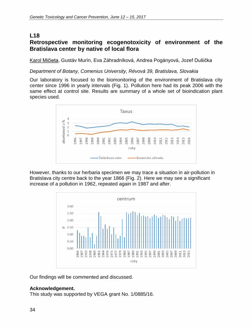
Our laboratory is focused to the biomonitoring of the environment of Bratislava city center since 1996 in yearly intervals (Fig. 1). Pollution here had its peak 2006 with the same effect at control site. Results are summary of a whole set of bioindication plant species used.
However, thanks to our herbaria specimen we may trace a situation in air-pollution in Bratislava city centre back to the year 1866 (Fig. 2). Here we may see a significant increase of a pollution in 1962, repeated again in 1987 and after.
Our findings will be commented and discussed. Acknowledgement. This study was supported by VEGA grant No. 1/0885/16.

Genetic Toxicology and Cancer Prevention, June 12 – 15, 2017
35
L19 Native flora in Bratislava: monitoring of ecogenotoxicity at selected industrial sites
Karol Mičieta, Jozef Dušička, Gustáv Murín
Department of Botany, Comenius University, Révová 39, Bratislava, Slovakia Our laboratory is focused to the biomonitoring of the most affected parts of environment of Bratislava city. We present the results of monitoring – induction factor of ecogenotoxicity - of selected industrial sites in Bratislava (incinerator, chemistry plant Istrochem, rubber plant – Kablo) (see Figure).
Our findings will be commented and discussed. Acknowledgement This study was supported by VEGA grant No. 1/0885/16.
Genetic Toxicology and Cancer Prevention, June 12 – 15, 2017
36
L20 Cannabidiol enhance antitumor effect of 5-FU treatment in colorectal cancer cell lines Alexandra Rejhová1, Alena Opattová, Daniel Slíva, Pavel Vodička Ústav experimentální medicíny AV ČR v.v.i., Vídeňská 1083, 142 20 Praha 4 Colorectal carcinoma (CRC) is a 3rd most most common type of cancer worldwide. After Slovakia and Hungary is the Czech Republic one of the worst affected regions in the world. Coloretal cancer (CRC) therapy using conventional chemotherapeutics, i.e. 5-fluorouracil (5-FU), represents a great burden for the patient's organism. The non-specific action of chemotherapeutics carries a number of side effects. Chemotherapeutics destroy rapidly dividing cells, both tumor cells, but also hair follicle cells, mucous membranes of the oral cavity and gastrointestinal tract, erythrocytes and leukocytes. Therefore increases an interest in natural anticancer compounds. Natural compounds possess anti-tumor effects of various characteristics - antiproliferative, antimetastatic, found in vitro, in vivo or even in clinical trials. Many natural compounds may sensitize to conventional cytotoxic therapy, intensify the combined effect of both administered therapeutics, act cytotoxically only specifically on tumor cells, or affect tumor cells where conventional cytostatics fail. Cannabidiol (CBD) is the most represented non-psychotropic ingredient in Cannabis sativa extract. This plant contains three main classes of bioactive molecules - flavonoids, terpenoids and more than 60 types of cannabinoids. Cannabinoids exhibits many health proficient effects in vitro and in vivo. The aim of our study was to define the effect of CBD on colorectal cancer cells and the interaction between CBD a conventional anticancer chemotherapeutic agent 5-FU in vitro. The tests were taken on human colorectal cancer cell lines HCT116 and HCT116 p53-/- and also non-malignant CRC cell line NCM460. 35 mM CBD decrease CRC cells survival of HCT116 about 32% (p<0.05) and HCT116 p53-/- about 67% (p<0.001). Interestingly, CBD has no effect on non-malignant colorectal cells. Treatment with 2.5 µM concentration of 5-FU alone decreases proliferation of HCT116 after 24 hours to 80%, co-treatment with 10 µM CBD decreases proliferation to 60%. Our results suggest that CBD decreases colorectal cancer cells survival and proliferation and significantly enhances anti-tumor effect of 5-FU. Interestingly, CBD had no effect on non-malignant colorectal cell lines. Interaction of conventional chemotherapeutics with natural compounds introduces a novel aspect in cancer research and therapy. Acklownegement This research was supported by AZV 15-27580A and GAČR P304/15/08239S grants.
Genetic Toxicology and Cancer Prevention, June 12 – 15, 2017
37
L21 Atomic force microscopy: a powerful tool for high-resolution imaging of breast cancer cells Monika Buríková1, Alexandra Poturnayová2,3, Jozef Bizík1, Tibor Hianik3 1Cancer Research Institute, Biomedical Research Center SAS 2 Institute of Biochemistry and Animal Genetics, Center of Biosciences SAS 3Department of Nuclear Physics and Biophysics, Faculty of Mathematics, Physics and Informatics, Comenius University Cancer diseases are among the most severe diagnoses and belong to the most common cause of death in the last decades. In general, tumor cells differ from healthy ones, in that they exhibit increased expression so called tumor-associated antigens, which are useful markers of tumor progression. Such markers include, for example, pre-epidermal growth factor receptor (HER2), typical of tumor cells of breast cancer [1]. The best way of protection against oncologic disease is prevention by early diagnostics. Therefore, development of the methods based on identification of tumor markers is needed. Due to low concentration of tumor cells in early stages of oncologic disease special sensitive methods are required for their detection. Atomic force microscopy (AFM) can be used for this purpose. AFM has opened up new avenues in molecular medicine and biology. This method can image non-conductive cells in their native physiological environment [2], quantitatively analyzes the surface micro-roughness in various physiological conditions and provides interesting information on the structure-function relationship of the cells. In this work we used AFM for analysis of cell morphology, distribution, size and also 3D topography of HER2 negative as well as HER2 positive cancer cell lines. The cells were immobilized at surfaces of glass, gold or mica. Several parameters were used to quantify morphological changes include roughness parameters, skewness and kurtosis. Height profiles were applied to characterize sizes and differences between cell membranes of each cancer cell lines. We compared also effect of fixation and preparation of AFM sample in different conditions („dry “or ‚„wet “technique measurements). We demonstrated that AFM is promising method for distinguishing cancer cells from normal cells and for the detection of tumor markers. Acknowledgement This work was supported by the Slovak Research and Development Agency under the contracts APVV-14-0267 and SK-AT-2015-0004 and by Science Grant Agency VEGA 2/0088/17. References [1] Horber J.K.H., Miles M.J. Scanning probe evolution in biology. Science. 2003; 302: 1002-
1005. [2] Kumar S., Roy S., Microscopy: Science, Technology, Applications and Education, 2010,
500-506.
Genetic Toxicology and Cancer Prevention, June 12 – 15, 2017
38
L22 Epigenetic changes induced by nanomaterials and possible impact on health Božena Smolková1, Alena Gábelová1, Monika Šramková1, Katarína Kozics1, Annamária Srančíková1, Mária Dušinská2 1Cancer Research Institute BMC SAS, Dúbravská cesta 9, 845 05 Bratislava 2 NILU-Norwegian Institute for Air Research, Instituttveien 18, Kjeller, Norway The development of a wide spectrum of nanoscale technologies for treatment, diagnosis, monitoring and control of biological systems, bring along a widespread benefit for patients. Modern nanodrugs allow increase of treatment efficacy, reduction of toxicities; enable targeted delivery of drugs in a tissue-, cell- or organelle-specific manner, enhancement of their pharmaceutical properties or sustained or stimulus-triggered release. Besides their therapeutic potential, nanotechnologies allow also more sensitive diagnostics and imaging. However, their unique features could induce unintended side effects. Main mechanisms of action leading to adverse effects are via oxidative stress, genotoxicity but deregulation of epigenetic machinery might also play important role. Epigenetics, via DNA methylation, histone modifications and interacting regulative non-coding RNAs, allows sophisticated, time and tissue specific control of gene expression in both normal and pathological conditions. An increasing number of studies reporting nanoparticle (NP)-induced epigenetic alterations call for implementation of new testing methods to allow their safety assessment. Despite the large body of evidence for an association between environmental exposure and epigenetic changes, data supporting the link between epigenetic variation and common complex disease phenotypes, except for cancer, several developmental and neurobehavioural disorders, are still missing. Aberrant DNA methylation was also associated with osteoarthritis and cardiovascular disease, pre-eclampsia, rheumatoid arthritis and metabolic disorders. Epigenomic alteration in immune function have been shown in systemic lupus erythematosus, T lymphocyte DNA demethylation contributing to lupus flare severity. The consequences of epigenetic changes induced by exposure to NPs and their causal relationship with complex diseases are still poorly understood. Development of new test guideline recommendations for hazard identification and product safety management, including epigenetic endpoints, will be possible only after a further increase in our understanding of the normal variability of the epigenetic landscape and causality between NP exposure-induced epigenetic alterations and their consequences for the phenotype. Acknowledgement The research leading these results has received funding from European Union´s Horizon 2020 (H2020/2014 - 2020) under grant agreement no. 685817 - HISENTS and by the ERA-NET EuroNanoMed projects GEMNS and INNOCENT.
Genetic Toxicology and Cancer Prevention, June 12 – 15, 2017
39
L23 A multimodular high throughput screening platform for nano-safety assessment Alena Gábelová, Katarína Kozics, Monika Šramková, Annamária Srančíková, Božena Smolková Cancer Research Institute, Biomedical Research Center SAS, Dúbravská cesta 9, 845 05 Bratislava The rapid expansion in the field of nanotechnology and extensive commercialization of nanoscale products have resulted in enhanced human exposure to engineered nanomaterials (ENMs). Taking into account the potential benefits, human exposure to ENMs will increase, primarily in the context of nanomedicine-based diagnostics and therapy, thus the bio-safety of ENMs is a great concern. Current in vitro methods generally utilized in toxicological screening are cost intensive and require time-consuming manual handling to gather enough meaningful toxicological data. Moreover, experimental animals are often exposed to high maximum tolerable doses, which differ from the real-life situation of human being leading to inaccurate predictions. Therefore, the development of a robust high throughput (HTP) screening platform capable of quantitative testing and high content analysis (HCA) approaches are needful for toxicological hazard and risk estimation of ENMs. Microfluidic technology offers an alternative platform for toxicity screening that not only allows to recreate physiologically-relevant in vitro models for nanotoxicity testing, but also provides platforms that deliver an attractive strategy for enhancing the efficiency of hazard profiling. Microfluidic organ-on-a-chips provide a controllable culture microenvironment for living cells in micrometer-sized chambers that imitate the construction of minimal functional units and keep partial function of organs or tissues in vivo. Microfluidic devices could reduce the need for animal testing and become a great platform to replace the conventional screening techniques. The HISENTS project is focused on the development of an innovative multimodular HTP platform for (nano)toxicity screening which include a set of individual chip-based microfluidic tools, each representing a critical physiological function, connected and integrated in a hierarchical vectorial manner by a microfluidic network. Acknowledgement The research leading these results has received funding from European Union´s Horizon 2020 (H2020/2014 - 2020) under grant agreement no. 685817 - HISENTS.
Genetic Toxicology and Cancer Prevention, June 12 – 15, 2017
40
L24 Searching for appropriate in vitro kidney model to measure drug-induced toxicity Annamária Srančíková, Monika Šramková, Katarína Kozics, Božena Smolková, Alena Gábelová Cancer Research Institute BMC SAS, Dúbravská cesta 9, 845 05 Bratislava The kidneys are essential organs in the homeostatic regulation and excretion of waste products from the human body. As majority of heavy metals, toxic chemicals, drug metabolites, including nanomaterials are cleared via the same route of elimination the kidneys are highly susceptible to foreign substances. Nephrotoxicity due to injury of the kidney is the main obstacle in the development of new drugs. In general, drug development failures constitute 19–25% of all cases of acute kidney injury [1]. Based on this fact, there is a great need for more predictive in vitro human kidney models to investigate the absorption, distribution, metabolism, excretion and adverse effects of new potential pharmaceutics. Currently available in vitro 2D renal cell models often lack sufficient physiological relevance and reliability. Organ-on-a-chip technology provides a promising alternative. These microfluidic systems mimic the in vivo microenvironment including fluid shear stress and transepithelial chemical gradients. The kidneys are organs with high degree of tissue heterogeneity that consist from different highly specialized cells. The human embryonic kidney 293 (HEK293) cells are frequently used as a renal model system in in vitro studies. However, the human renal proximal tubule epithelial TH1 cells could be more relevant renal cell model. TH1 cells express alkaline phosphatase and gamma-glutamyltranspeptidase, two brush-border enzymes found in vivo. Here we evaluate the capacity of HEK293 and TH1 cells to detect the genotoxicity of selected chemicals with different mechanisms of action such as chlorpromazine (CPZ), colchicine (COL) and methyl methanesulfonate (MMS). Neither CPZ nor COL induced any significant increase in DNA migration in TH1 and HEK293 cells, as both chemicals are non-genotoxic agents. In contrast, higher level of DNA damage was detected in TH1 cells compared to HEK293 cells after exposure to MMS, a strong alkylating agent. Our preliminary results indicated that TH1 cell line might be a promising in vitro kidney model to predict the genotoxicity of xenobiotics. Moreover, this cell line will be the basis for the development of microfluidic organ-on-a-chip system. Acknowledgement The research leading these results has received funding from European Union`s Horizon 2020 (H2020/2014 - 2020) under grant agreement no. 685817 - HISENTS. References [1] Raina R, Herrera N, Krishnappa V, Sethi SK, Deep A, Kao WM, Bunchman T, Abu-Arja
R Hematopoietic stem cell transplantation and acute kidney injury in children: A comprehensive review. Pediatr Transplant 2017; 21(4).
Genetic Toxicology and Cancer Prevention, June 12 – 15, 2017
41
L25
Use of microRNA in Lung Carcinoma Diagnosis Miroslava Šarlinová1, Martina Krutáková2, Anton Dzian3, Tatiana Matáková1,2, Ľudovít Mušák1, Marián Grendár4, Erika Halašová1,5
1Division of Molecular Medicine, Biomedical Center Martin, Comenius University in Bratislava, Mala Hora 4/C, 03601, Martin, Slovak Republic 2Department of Medical Biochemistry, Comenius University in Bratislava, Jessenius Faculty of Medicine, Martin, Slovak Republic 3Clinic of Thoracic Surgery University Hospital Martin, Comenius University in Bratislava, Kollárova 2, 03601, Martin, Slovak Republic 4Department of Bioinformatics, Biomedical Center Martin, Comenius University in Bratislava, Mala Hora 4/C, 03601, Martin, Slovak Republic 5Department of Medical Biology, Comenius University in Bratislava, Jessenius Faculty of Medicine, Martin, Slovak Republic. Introduction Lung cancer (LC) is one of the most frequently diagnosed malignancies in developed countries. It is one of the leading causes of deaths caused by neoplastic diseases. The prognosis of patients tends to be very negative because it is usually diagnosed only in advanced stages. Lung cancer does not have early warning signals, which would allow the capture of this disease in its early stage (1). It is, therefore, necessary to seek appropriate methods for early diagnosis of this disease. The non-invasive, but sufficiently sensitive method of detection of lung cancer seems to be the use of tissue and tumor specific microRNA molecules (2). MiRNAs are a class of small single-stranded noncoding RNAs with a length of 19-25 nucleotides. They play important regulatory roles in many cellular processes including cell proliferation, differentiation, growth control, and apoptosis (3). These molecules regulate gene expression on the posttranscriptional level by translational repression, mRNA cleavage, or mRNA degradation in various physiological and pathological processes. In addition, some miRNAs can function as oncogenes or tumor suppressors, so they can regulate several genes that play important roles in tumorigenesis. It was found that miRNAs are directly involved in many types of cancer, including lung cancer (4). The aim of this study was to analyze selected types of microRNA molecules that are indicative of lung carcinoma and to continuously monitor changes in their expression in the given disease. We focused on determining the expression of selected miRNAs in peripheral blood of 95 LC patients compared to a group of 100 seemingly healthy individuals. Materials and methods Blood samples of 2.5 ml were obtained from respondents enrolled in the study and total RNA was isolated from the blood. The blood was collected before therapy using PAXgene Blood RNA Tubes for in vitro diagnostic purposes. After blood collection, the tubes were kept for 2 hours at room temperature. The RNA concentration and purity were confirmed by the spectrophotometric ratio using absorbance measurements at
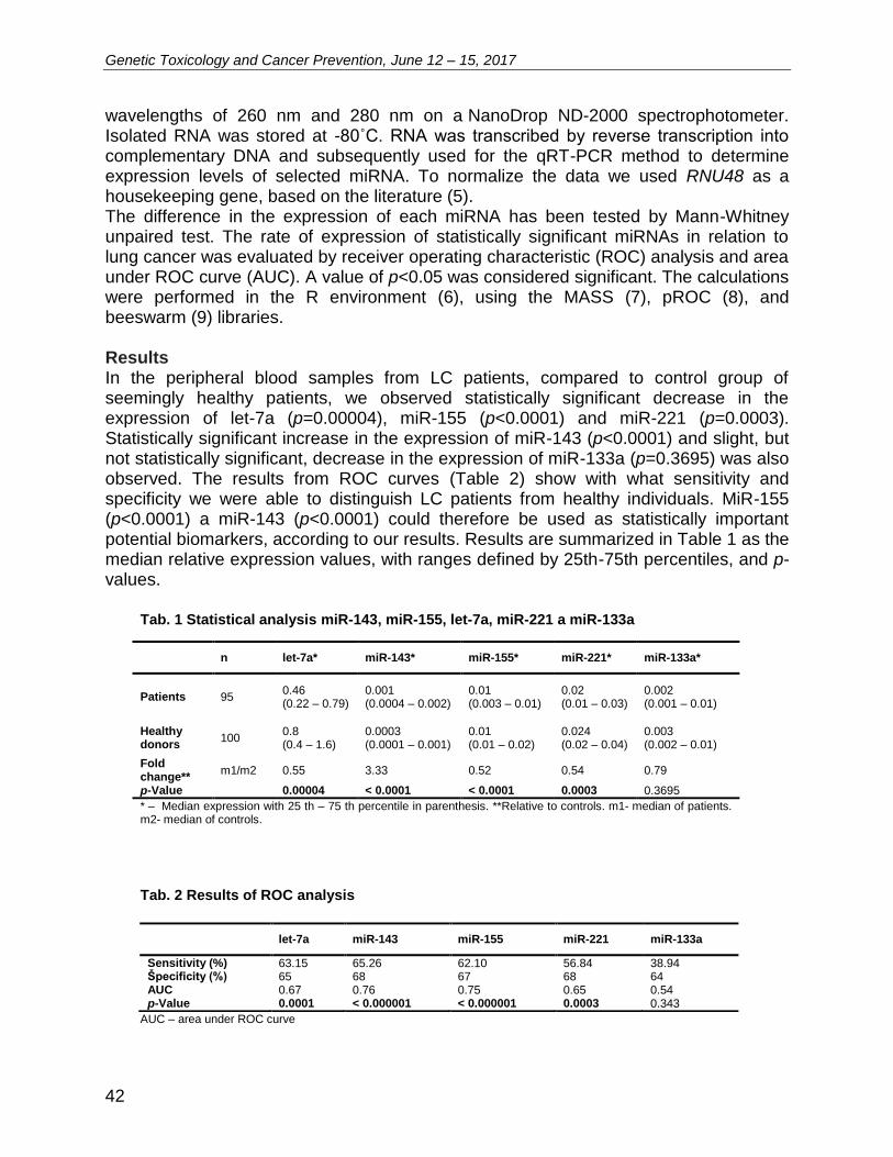
Genetic Toxicology and Cancer Prevention, June 12 – 15, 2017
42
wavelengths of 260 nm and 280 nm on a NanoDrop ND-2000 spectrophotometer. Isolated RNA was stored at -80˚C. RNA was transcribed by reverse transcription into complementary DNA and subsequently used for the qRT-PCR method to determine expression levels of selected miRNA. To normalize the data we used RNU48 as a housekeeping gene, based on the literature (5). The difference in the expression of each miRNA has been tested by Mann-Whitney unpaired test. The rate of expression of statistically significant miRNAs in relation to lung cancer was evaluated by receiver operating characteristic (ROC) analysis and area under ROC curve (AUC). A value of p<0.05 was considered significant. The calculations were performed in the R environment (6), using the MASS (7), pROC (8), and beeswarm (9) libraries. Results In the peripheral blood samples from LC patients, compared to control group of seemingly healthy patients, we observed statistically significant decrease in the expression of let-7a (p=0.00004), miR-155 (p<0.0001) and miR-221 (p=0.0003). Statistically significant increase in the expression of miR-143 (p<0.0001) and slight, but not statistically significant, decrease in the expression of miR-133a (p=0.3695) was also observed. The results from ROC curves (Table 2) show with what sensitivity and specificity we were able to distinguish LC patients from healthy individuals. MiR-155 (p<0.0001) a miR-143 (p<0.0001) could therefore be used as statistically important potential biomarkers, according to our results. Results are summarized in Table 1 as the median relative expression values, with ranges defined by 25th-75th percentiles, and p-values.
Tab. 1 Statistical analysis miR-143, miR-155, let-7a, miR-221 a miR-133a
n let-7a* miR-143* miR-155* miR-221* miR-133a*
Patients 95 0.46 (0.22 – 0.79)
0.001 (0.0004 – 0.002)
0.01 (0.003 – 0.01)
0.02 (0.01 – 0.03)
0.002 (0.001 – 0.01)
Healthy donors
100 0.8 (0.4 – 1.6)
0.0003 (0.0001 – 0.001)
0.01 (0.01 – 0.02)
0.024 (0.02 – 0.04)
0.003 (0.002 – 0.01)
Fold change**
m1/m2 0.55 3.33 0.52 0.54 0.79
p-Value 0.00004 < 0.0001 < 0.0001 0.0003 0.3695
* – Median expression with 25 th – 75 th percentile in parenthesis. **Relative to controls. m1- median of patients. m2- median of controls.
Tab. 2 Results of ROC analysis
let-7a miR-143 miR-155 miR-221 miR-133a
Sensitivity (%) 63.15 65.26 62.10 56.84 38.94 Špecificity (%) 65 68 67 68 64 AUC 0.67 0.76 0.75 0.65 0.54 p-Value 0.0001 < 0.000001 < 0.000001 0.0003 0.343
AUC – area under ROC curve
Genetic Toxicology and Cancer Prevention, June 12 – 15, 2017
43
Conclusion Cancer is one of the most serious diseases around the world. In year 2012, 14.1 million new cases of cancer were diagnosed in the world, and 8.2 million people have suffered from this disease. Up to 13% of all diagnosed malignancies are lung cancer, which is the most widespread and most commonly diagnosed type of cancer. Pulmonary carcinoma is also the most common cause of fatal illness in the world. Tumorigenesis is also regulated by small non-coding RNAs, which are key players in the development of cancer. Endogenous miRNAs in the blood tend to remain stable for a long time and can withstand several repeated freeze-thaw cycles. Growing evidence indicates that cancer cells secrete miRNAs into the systemic circulation. This fact is one of the main reasons for the study of miRNAs as biomarkers in the field of cancer research. In conclusion, we can say that the identification of circulating miRNAs associated with the emergence and the development of lung cancer, would allow earlier detection of the disease at a stage prior to the appearance of clinical symptoms. It can also expand the range of screening methods for identification of individuals at risk and contribute to a better understanding of the role of miRNA. Acknowledgement This work was supported by the project „Biomedical Center Martin“ ITMS code: 26220220187, co-financed from EU sources, by the Ministry of Health of the Slovak Republic under the contract 2012/25-UKMA-2, by the project Competence Center for Research and Development in the Field of Diagnostics and Therapy of Oncological Diseases, ITMS: 26220220153. References [1] Ferro A, Peleteiro B, Malvezzi M, Bosetti C, Bertuccio P, Levi F, Negri E, La Vecchia C and
Lunet N: Worldwide trends in gastric cancer mortality (1980-2011), with predictions to 2015, and incidence by subtype. Eur J Cancer 50(7): 1330-1344, 2014.
[2] Mishra PJ: MicroRNAs as promising biomarkers in cancer diagnostics. Biomark Res 2(19): DOI: 10.1186/2050-7771-2-19, 2014.
[3] Mitchell PS, Parkin RK, Kroh EM, Fritz BR, Wyman SK, Pogosova-Agadjanyan EL, Peterson A, Noteboom J, O'Briant KC, Allen A, Lin DW, Urban N, Drescher CW, Knudsen BS, Stirewalt DL, Gentleman R, Vessella RL, Nelson PS, Martin DB and Tewari M: Circulating microRNAs as stable blood-based markers for cancer detection. Proc Natl Acad Sci USA 105(30): 10513-10518, 2008.
[4] Gee HE, Buffa FM, Camps C, Ramachandran A, Leek R, Taylor M, Patil M, Sheldon H, Betts G, Homer J, West C, Ragoussis J and Harris AL: The small-nucleolar RNAs commonly used for microRNA normalisation correlate with tumour pathology and prognosis. Br J Cancer 104(7): 1168-1177, 2011.
[5] Sarlinova M, Halasa M, Mistuna D, Musak L, Iliev R, Slaby O, Mazuchova J, Valentova V, Plank L, Halasova E: miR-21, miR-221 and miR-150 Are Deregulated in Peripheral Blood of Patients with Colorectal Cancer. Anticancer Res 36(10): 5449-5454, 2016.
[6] R Core Team (2015). R: A language and environment for statistical computing. R Foundation for Statistical Computing, Vienna, Austria. URL https://www.R project.org/.
[7] Venables, W. N. and Ripley, B. D. (2002) Modern Applied Statistics with S. Fourth Edition. Springer, New York. ISBN 0-387-95457-0.

Genetic Toxicology and Cancer Prevention, June 12 – 15, 2017
44
[8] Xavier Robin, Natacha Turck, Alexandre Hainard, Natalia Tiberti, Frederique Lisacek, Jean-Charles Sanchez and Markus Muller (2011). pROC: an open-source package for R and S+ to analyze and compare ROC curves. BMC Bioinformatics, 12, p. 77.
[9] Aron Eklund (2015). beeswarm: The Bee Swarm Plot, an Alternative to Stripchart. R package version 0.2.1. https://CRAN.R project.org/package=beeswarm
Genetic Toxicology and Cancer Prevention, June 12 – 15, 2017
45
L26 Molecular recognition of PTK7 receptors on leukemic T-cells using DNA aptamers Alexandra Poturnayová1,2, Monika Buríková3, Jozef Bizík3, Andreas Ebner4, Michael Leitner4, Tibor Hianik2 1Institute of Biochemistry and Animal Genetics, Center of Biosciences SAS, Bratislava, Slovakia 2Department of Nuclear Physics and Biophysics, Faculty of Mathematics, Physics and Informatics, Comenius University, Bratislava,
Slovakia 3Cancer Research Institute, Biomedical Research Center SAS, Bratislava, Slovakia
4Institute of Biophysics, Johannes Kepler University, Linz, Austria Introduction In the present age of modern medicine, progress and increasing interest about nanooncology sparks revolution in diagnostics and therapy of tumor diseases. It is due to the most recent achievements in the research of these diseases thanks to application of bio- and nanotechnologies. Genomics and early diagnostics are playing a key role in development of precisely targeted therapy for most of the human tumors. Nanooncology, due to discovery of the new biomarkers, contributes to development of more sensitive biosensors for detection of disease in early stage. New diagnostic approaches capable of improving precise detection of tumor cell are decisive for cancer diagnostics. Development of sensitive biosensors based on DNA aptamers is one of possible tool for early detection, like in the case of the circulating tumor cells. DNA and RNA aptamers are single-strand oligonucleotides which are able to form highly specific affinity bond with various target molecules [1]. Their specificity is comparable and in certain cases even higher than for antibodies. In the solution, they form unique structures containing binding site for selected molecules, for example oncomarkers on the cell surface. Because of these properties, aptamers were used as therapeutic, diagnostic, analytic tools and transport systems for targeted drug delivery [2]. Nanoparticles modified by aptamers can be used not only as new imaging tool, but also for quantification and enhancement of oncomarkers detection, for example in blood samples. Materials and methods In this work we used sgc8 DNA aptamer that is specific to protein tyrosine kinase 7 (PTK7) receptors of acute lymphoblastic leukemia cells (ALL). Highly sensitive methods of Single molecule force spectroscopy (SMFS) and thickness shear mode acoustic method (TSM) were used for tracking the interactions between sgc8c aptamer and PTK7 receptors on different cell lines. Results In experiments we used MOLT-4 and Jurkat cells containing in their membranes PTK 7 receptor, which is not expressed on healthy cells of immune system. We used DNA aptamer sgc8c specific to PTK7 receptors. Addition of the cells on the surface of TSM
Genetic Toxicology and Cancer Prevention, June 12 – 15, 2017
46
transducer modified by aptamers in concentration range 50 – 5x10⁵ cells /ml caused the decay in frequency by approx. 97.5 ± 7.6 for Jurkat cells and about -121.7 ± 16.4 Hz for MOLT-4. The aptamer specificity was verified using non-specific aptamer or control cells U266. These interactions were monitored in real time and enabled determination of binding kinetic constants and interaction force between aptamers and monitored receptors. We were able to detect 50 cells/ml. Key role was to determine specific and nonspecific interaction of sgc8 with transmembrane PTK7 receptor on the cell surface. In cooperation with JKU Linz we for the first time used method of simultaneous topography and recognition imaging (TREC) to determine interaction force between aptamers and PTK on leukemic T-cell surface and to determine their surface distribution and surface density [3]. We proved that sgc8c is binding with high probability and selectivity to PTK receptors (41.7±2.5% MOLT-4, 33.4±5.4% Jurkat, 1.1±0.3% U266). Conclusion Results obtained demonstrate that application of sensitive physical techniques such as TSM, SMFS and TREC allowing better understanding of the molecular mechanisms of affinity interactions at the surface of cancer cells and making possible to develop new tools for early diagnostics of cancer diseases. Acknowledgement This work was supported by the Slovak Research and Development Agency under the contracts APVV-14-0267 and SK-AT-2015-0004 and by Science Grant Agency VEGA 2/0088/17. References [1] Ellington a Szostak, Nature 1992, 355, 850–852. [2] Shangguan et al., Proc. Natl. Acad. Sci. USA, 2006, 103, 32, 11838–11843. [3] Leitner et al., Anal. Bioanal. Chem. 2017, 409, 2767-2776.
Genetic Toxicology and Cancer Prevention, June 12 – 15, 2017
47
L27 Decorin-mediated regulation of fibrillin-1 is necessary for renal tissue preservation during obstruction-induced injury Andrea Babelova1,2, Martina Baliova2,3, Wasiliki Tsalastra2, Liliana Schaefer2,4 1Department of Genetics, Cancer Research Institute, BMC SAS, Bratislava, Slovakia 2Department of Internal Medicine D, Münster, Germany 3Laboratory of Neurobiology, Institute of Molecular Biology SAS, Bratislava, Slovakia 4Pharmazentrum Frankfurt, Goethe-University, Frankfurt am Main, Germany Renal fibrosis is a process during which excessive amounts of ECM are cumulated in the kidney tissue which is then replaced by fibrotic tissue. In the end, a healthy and fully functional kidney becomes a small atrophic non-functional kidney. Recently, no therapy is available for the treatment of fibrosis so at this stage only replacement therapy takes over in the form of dialysis or kidney transplantation. Decorin, a small leucine-rich proteoglycan has been thought to play a role in the development of renal fibrosis. We could show that decorin may affect synthesis of the elastic fiber component fibrillin-1 in the kidney. We also were able to identify mechanism by which decorin contributes to fibrillin-1 level during renal injury. We found that decorin is a ligand for insulin-like growth factor-1 (IGF-I) receptor in renal fibroblasts (normal rat kidney fibroblasts, NRK) and by activation of PI3K/Akt signaling pathway including mTOR and S6 kinase is able to enhance synthesis of fibrillin-1. In line with this we have shown that IGF-I stimulated fibrillin-1 synthesis as well. To verify this finding in vivo, unilateral ureteral obstruction (UUO), an animal model of tubulointerstitial injury of the kidney was performed in two months old male DCN -/-, DCN +/+, and C57/BL/6 mice. Fibrillin-1 was considerably reduced due to decorin deficiency despite up-regulation of IGF-I receptor in kidneys from decorin-null mice, as well it was decreased due to administration of mTOR inhibitor rapamycin. Interestingly, a number of studies have shown downregulation of decorin expression in patients with Marfan syndrome, a connective tissue disorder characterized by fibrillin-1 defect. Our results show that fibrillin-1 contributes to the biomechanical properties of Bowman´s capsule and tubules in a model of obstructive nephropathy and that decorin by signaling though IGF-I receptor and PI3K/Akt/mTOR/p70S6K pathway mediates translational regulation of fibrillin-1. These results also suggest that decorin might play important role as a disease modifier in Marfan´s syndrome.

Genetic Toxicology and Cancer Prevention, June 12 – 15, 2017
48
L28 Challenges for micronucleus assay in genetic toxicology Andrea Rossnerova, Tereza Cervena, Pavel Rossner Jr. Department of Genetic Toxicology and Nanotoxicology, Institute of Experimental Medicine, Czech Academy of Sciences, 14220 Prague 4, Czech Republic Micronucleus assay, one of the most commonly used cytogenetic methods in genetic toxicology, is an approach for evaluation of DNA damage (breaks and losses of chromosomes) after exposure to chemicals, including those from the environment, as well as to radiation. The variant of this method, in which cytochalasin-B is used to inhibit cytokinesis, has its roots already in 1985 and has since been applied in almost 1400 studies. Despite significant achievements in many areas of science including genetic toxicology, there are still challenges for future research involving: (A) application of the improved or “new” methodological variants of this assay; (B) new view on the interpretation of the results; and (C) application of this assay in testing of new chemicals and materials including nanoparticles. Regarding the methodological challenges (A), future research should mainly focus on development of: (i) detail analysis of micronuclei content by combination of the classic method with immunostaining or fluorescent in situ hybridization method; (ii) use of appropriate biological material for testing including e.g. 3D models; and (iii) broader application of the automated analysis in very laborious microscopic scoring of micronuclei. Current research focusing on evaluation of DNA damage by this method brings frequently negative or even contradictory results related to the exposure levels. These results should not be ignored, but interpreted in a wider context (B) including: (i) type of exposure; (ii) previous exposure history; and (iii) incorporation of molecular markers into analysis of the data. Micronucleus assay is a broadly utilized approach in testing numerous chemicals, chemical mixtures or new pharmaceutics products. (C) Systematic testing of the wide spectrum of the nanoparticles exposure is another challenge for in vitro and particularly for in vivo biomonitoring studies. This presentation will summarize significant achievements and new avenues of research for all challenges mentioned above. Acknowledgement Supported by the Ministry of Education Youth and Sports CR (#LO1508) and Czech Science Foundation (#16-14631S).
Genetic Toxicology and Cancer Prevention, June 12 – 15, 2017
49
L29 Level of heavy toxic metals, selected biochemical parameters and chromosomal aberrations in welders Ľudovít Mušák1, Martin Petráš1, Miroslava Šarlinová1, Jela Valachová2, Oto Osina2, Erika Halašová1,3
1Biomedical Center Martin, Comenius University (CU) in Bratislava, Jessenius Faculty of Medicine (JFM) in Martin, Malá Hora 11161/4D 036 01 Martin, Slovakia 2Clinic of Occupational Medicine and Toxicology CU JFM and UHM, Martin, Slovakia 3Department of Medical Biology CU JFM, Martin, Slovakia Introduction Welding fumes formed by condensations of vapors, aerosols, dust and gases are the main undesirable harmful factor in welding. Character of toxicity or deleterious effect of a selected substance is manifested by its ability to dissolve or store them in certain organs of the human organism. Easier soluble substances are less harmful. The toxic metals chromium (Cr), nickle (Ni), molybdenum (Mo), manganese (Mn), copper (Cu), lead (Pb), cadmium (Cd) and their compounds are the most toxic substances (Bencko et al., 1995; Moser et al., 2008; Blake, 2010). Stainless steels are resistant to corrosion. To obtain this property, the greatest amount of Cr is added to the steel which has a content in the solid solution greater than 11.5%. The content of Ni may be in the range of 2-6.5%, Mo to 2.5%, and Mn to 1%. The Cr6+ compounds are classified to category 1 by IARC as carcinogen to humans. They are mainly inhaled by the body and many of them cause malignant lung tumors. Their occurrence depends on the dose of inhaled Cr and manifests after a long latent period, with all histological types (Buchancová et al., 2003; Bloom and Brandt, 2008; Halasova et al., 2009). Cr6+ is a considerable clastogen. Chromosome breaks are a result of DNA damage (Sorensen et al., 2007). Nickel causes allergic reactions, reddening of the skin, rashes, chronic bronchitis and decreased lung function. According to IARC, they are also included in category 1 as the carcinogens to humans. In the case of manganese, the triadic compounds are the most toxic to the organism. Chronic poisoning by Mn affects most often the CNS and can lead to permanent disability. It develops with malaise, drowsiness, weakness, emotional disturbances, dizziness, repeated leg cramps or paralysis. Some Mn compounds are among the potential carcinogens (Blake, 2010, Selgrade, 2010). Chromosome aberrations (CA) cause structural and numerical chromosome damage. Chromatide type of aberrations (CTA-type) are formed during S cell cycle phase and are induced by non-radiomimetic agents. Radiomimetics induce aberrations of CTA type in the S and G2 cell cycle phase and chromosome type of aberrations (CSA-type) in the G1 phase (Mateuca et al., 2006). Double-strand breaks are types of chromosomal damage that occur in S cell cycle phase and have the closest relationship to modification to tumor cell (Hagmar et al., 2004, Beyersmann and Hartwig, 2008). Material and methods The toxicity and genotoxicity of heavy toxic metals was monitored in a group of 117 stainless steel welders. The Cr, Ni and Mn values were determined in the blood and
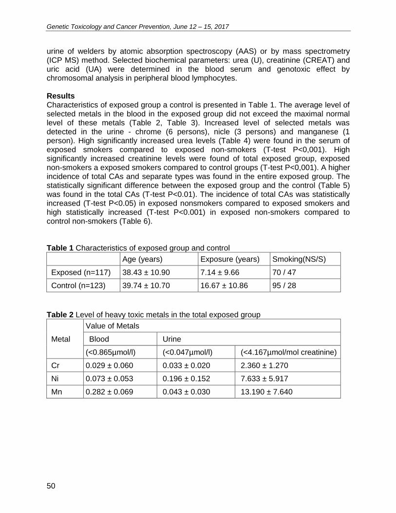
Genetic Toxicology and Cancer Prevention, June 12 – 15, 2017
50
urine of welders by atomic absorption spectroscopy (AAS) or by mass spectrometry (ICP MS) method. Selected biochemical parameters: urea (U), creatinine (CREAT) and uric acid (UA) were determined in the blood serum and genotoxic effect by chromosomal analysis in peripheral blood lymphocytes. Results Characteristics of exposed group a control is presented in Table 1. The average level of selected metals in the blood in the exposed group did not exceed the maximal normal level of these metals (Table 2, Table 3). Increased level of selected metals was detected in the urine - chrome (6 persons), nicle (3 persons) and manganese (1 person). High significantly increased urea levels (Table 4) were found in the serum of exposed smokers compared to exposed non-smokers (T-test P<0,001). High significantly increased creatinine levels were found of total exposed group, exposed non-smokers a exposed smokers compared to control groups (T-test P<0,001). A higher incidence of total CAs and separate types was found in the entire exposed group. The statistically significant difference between the exposed group and the control (Table 5) was found in the total CAs (T-test P<0.01). The incidence of total CAs was statistically increased (T-test P<0.05) in exposed nonsmokers compared to exposed smokers and high statistically increased (T-test P<0.001) in exposed non-smokers compared to control non-smokers (Table 6). Table 1 Characteristics of exposed group and control
Age (years) Exposure (years) Smoking(NS/S)
Exposed (n=117) 38.43 ± 10.90 7.14 ± 9.66 70 / 47
Control (n=123) 39.74 ± 10.70 16.67 ± 10.86 95 / 28
Table 2 Level of heavy toxic metals in the total exposed group
Metal
Value of Metals
Blood Urine
(<0.865µmol/l) (<0.047µmol/l) (<4.167µmol/mol creatinine)
Cr 0.029 ± 0.060 0.033 ± 0.020 2.360 ± 1.270
Ni 0.073 ± 0.053 0.196 ± 0.152 7.633 ± 5.917
Mn 0.282 ± 0.069 0.043 ± 0.030 13.190 ± 7.640
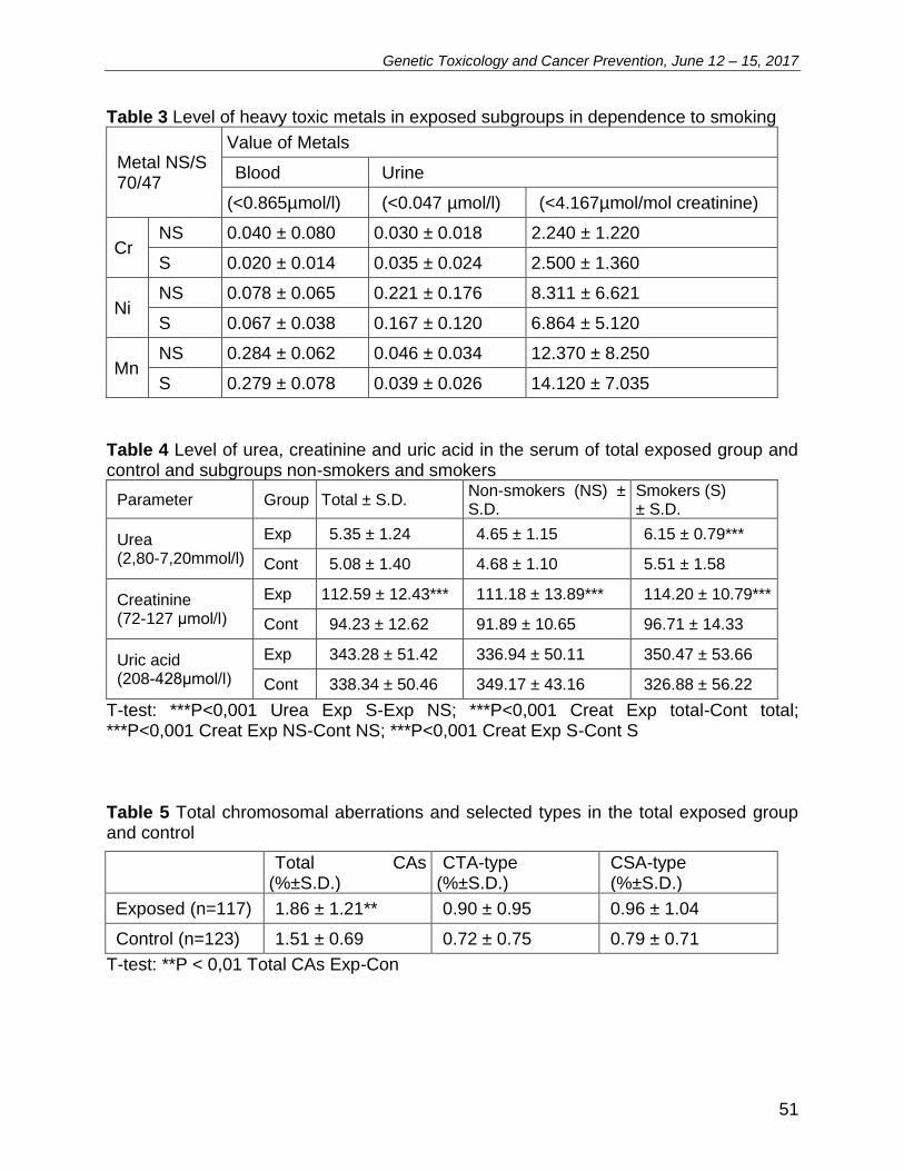
Genetic Toxicology and Cancer Prevention, June 12 – 15, 2017
51
Table 3 Level of heavy toxic metals in exposed subgroups in dependence to smoking
Metal NS/S 70/47
Value of Metals
Blood Urine
(<0.865µmol/l) (<0.047 µmol/l) (<4.167µmol/mol creatinine)
Cr NS 0.040 ± 0.080 0.030 ± 0.018 2.240 ± 1.220
S 0.020 ± 0.014 0.035 ± 0.024 2.500 ± 1.360
Ni NS 0.078 ± 0.065 0.221 ± 0.176 8.311 ± 6.621
S 0.067 ± 0.038 0.167 ± 0.120 6.864 ± 5.120
Mn NS 0.284 ± 0.062 0.046 ± 0.034 12.370 ± 8.250
S 0.279 ± 0.078 0.039 ± 0.026 14.120 ± 7.035
Table 4 Level of urea, creatinine and uric acid in the serum of total exposed group and control and subgroups non-smokers and smokers
Parameter Group Total ± S.D. Non-smokers (NS) ± S.D.
Smokers (S) ± S.D.
Urea (2,80-7,20mmol/l)
Exp 5.35 ± 1.24 4.65 ± 1.15 6.15 ± 0.79***
Cont 5.08 ± 1.40 4.68 ± 1.10 5.51 ± 1.58
Creatinine (72-127 μmol/l)
Exp 112.59 ± 12.43*** 111.18 ± 13.89*** 114.20 ± 10.79***
Cont 94.23 ± 12.62 91.89 ± 10.65 96.71 ± 14.33
Uric acid (208-428μmol/l)
Exp 343.28 ± 51.42 336.94 ± 50.11 350.47 ± 53.66
Cont 338.34 ± 50.46 349.17 ± 43.16 326.88 ± 56.22
T-test: ***P<0,001 Urea Exp S-Exp NS; ***P<0,001 Creat Exp total-Cont total; ***P<0,001 Creat Exp NS-Cont NS; ***P<0,001 Creat Exp S-Cont S Table 5 Total chromosomal aberrations and selected types in the total exposed group and control
Total CAs
(%±S.D.) CTA-type
(%±S.D.) CSA-type (%±S.D.)
Exposed (n=117) 1.86 ± 1.21** 0.90 ± 0.95 0.96 ± 1.04
Control (n=123) 1.51 ± 0.69 0.72 ± 0.75 0.79 ± 0.71
T-test: **P < 0,01 Total CAs Exp-Con
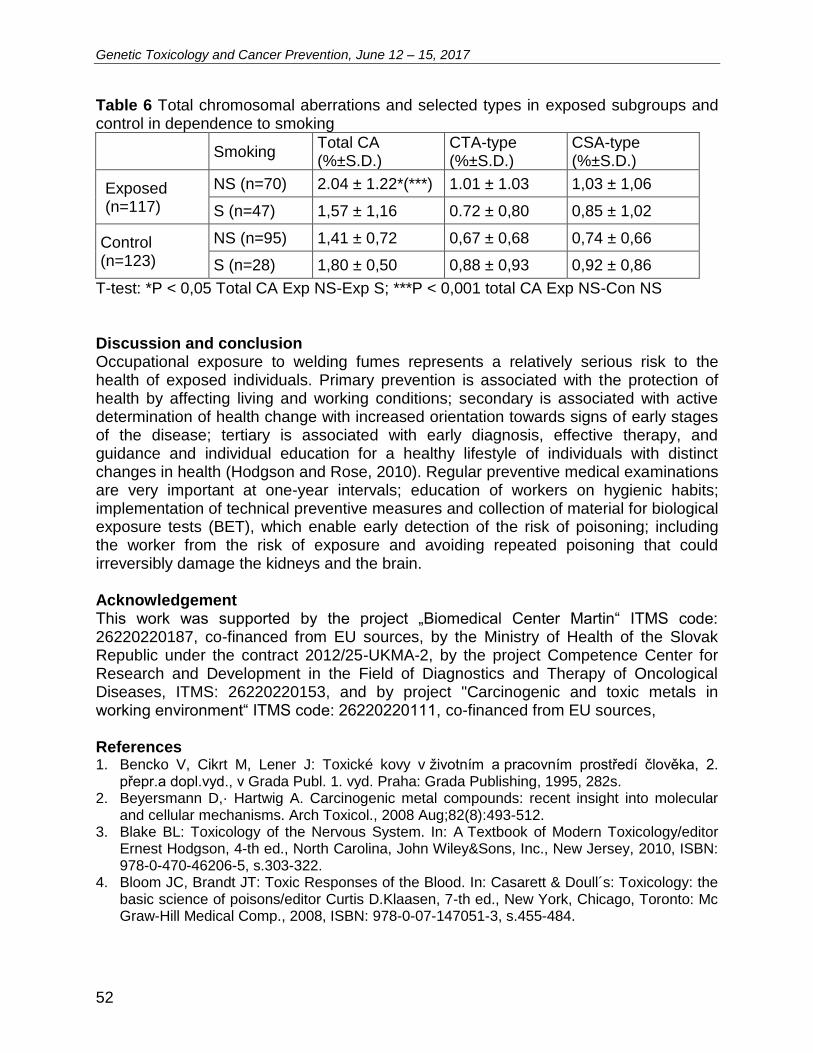
Genetic Toxicology and Cancer Prevention, June 12 – 15, 2017
52
Table 6 Total chromosomal aberrations and selected types in exposed subgroups and control in dependence to smoking
Smoking Total CA (%±S.D.)
CTA-type (%±S.D.)
CSA-type (%±S.D.)
Exposed (n=117)
NS (n=70) 2.04 ± 1.22*(***) 1.01 ± 1.03 1,03 ± 1,06
S (n=47) 1,57 ± 1,16 0.72 ± 0,80 0,85 ± 1,02
Control (n=123)
NS (n=95) 1,41 ± 0,72 0,67 ± 0,68 0,74 ± 0,66
S (n=28) 1,80 ± 0,50 0,88 ± 0,93 0,92 ± 0,86
T-test: *P < 0,05 Total CA Exp NS-Exp S; ***P < 0,001 total CA Exp NS-Con NS Discussion and conclusion Occupational exposure to welding fumes represents a relatively serious risk to the health of exposed individuals. Primary prevention is associated with the protection of health by affecting living and working conditions; secondary is associated with active determination of health change with increased orientation towards signs of early stages of the disease; tertiary is associated with early diagnosis, effective therapy, and guidance and individual education for a healthy lifestyle of individuals with distinct changes in health (Hodgson and Rose, 2010). Regular preventive medical examinations are very important at one-year intervals; education of workers on hygienic habits; implementation of technical preventive measures and collection of material for biological exposure tests (BET), which enable early detection of the risk of poisoning; including the worker from the risk of exposure and avoiding repeated poisoning that could irreversibly damage the kidneys and the brain. Acknowledgement This work was supported by the project „Biomedical Center Martin“ ITMS code: 26220220187, co-financed from EU sources, by the Ministry of Health of the Slovak Republic under the contract 2012/25-UKMA-2, by the project Competence Center for Research and Development in the Field of Diagnostics and Therapy of Oncological Diseases, ITMS: 26220220153, and by project "Carcinogenic and toxic metals in working environment“ ITMS code: 26220220111, co-financed from EU sources, References 1. Bencko V, Cikrt M, Lener J: Toxické kovy v životním a pracovním prostředí člověka, 2.
přepr.a dopl.vyd., v Grada Publ. 1. vyd. Praha: Grada Publishing, 1995, 282s. 2. Beyersmann D,· Hartwig A. Carcinogenic metal compounds: recent insight into molecular
and cellular mechanisms. Arch Toxicol., 2008 Aug;82(8):493-512. 3. Blake BL: Toxicology of the Nervous System. In: A Textbook of Modern Toxicology/editor
Ernest Hodgson, 4-th ed., North Carolina, John Wiley&Sons, Inc., New Jersey, 2010, ISBN: 978-0-470-46206-5, s.303-322.
4. Bloom JC, Brandt JT: Toxic Responses of the Blood. In: Casarett & Doull´s: Toxicology: the basic science of poisons/editor Curtis D.Klaasen, 7-th ed., New York, Chicago, Toronto: Mc Graw-Hill Medical Comp., 2008, ISBN: 978-0-07-147051-3, s.455-484.
Genetic Toxicology and Cancer Prevention, June 12 – 15, 2017
53
5. Buchancová J, Klimentová G, Šulcová M, Fabianová E: Pracovné lekárstvo a toxikológia, 1. vydanie, Martin: Osveta, 2003; 1133s., ISBN 80-8063-113-1, s.317-327.
6. Hagmar L, Stromberg U, Bonassi S, Hansteen IL, Knudsen LE, Lindholm C, Norppa H. Impact of types of lymphocyte chromosomal aberrations on human cancer risk: results from Nordic and Italian cohorts. Cancer res. 2004 Mar;64(6):2258-2263.
7. Halasova E, Matakova T, Kavcova E et al: Human lung cancer and haxavalent chromium exposure. Neuroendocrinol Lett, 2009, 30(suppl 1):182-185.
8. Hodgson E, Rose RL: Metabolism of Toxicants. In: A Textbook of Modern Toxicology/editor Ernest Hodgson, 4-th ed., North Carolina, John Wiley&Sons, Inc., New Jersey, 2010; ISBN: 978-0-470-46206-5, s.115-156.
9. Mateuca R, Lombaert N, Aka PV, Decordier I, Kirsch-Volders M. Chromosomal changes: induction, detection methods and applicability in human biomonitoring. Biochimie. 2006 Nov;88(11):1515-1531.
10. Moser VC, Aschner M, Richardson RJ, Philbert MA: Toxic Responses of the Nervous system. In: Casarett & Doull´s: Toxicology: the basic science of poisons/editor Curtis D.Klaasen, 7-th ed., New York, Chicago... Toronto: Mc Graw-Hill Medical Comp., 2008, ISBN: 978-0-07-147051-3, s.455-484.
11. Selgrade MJK: Immune System. In: A Textbook of Modern Toxicology/editor Ernest Hodgson, 4-th ed., North Carolina, John Wiley&Sons, Inc., New Jersey, 2010, ISBN: 978-0-470-46206-5, s.387-406.
12. Sorensen AR, Thulstrup AM, Hansen J, Ramlau-Hansen CH, Meersohn A, Skytthe A, Bonde JP. Risk of lung cancer according to mild steel and stainless steel welding. Scand J Work Environ Health, 2007 Oct;33(5):379-386.
Genetic Toxicology and Cancer Prevention, June 12 – 15, 2017
54
L30 The results of interconnection of the evidence of professional exposure to genotoxic factors (regex)and cancer registry in the Czech Republic Hana Lehocká1, Ivona Závacká2, Jana Vavrošová2, Vladimír Janout2
1Public Health Institute Ostrava, Czech Republic 2Faculty of Medicine, University of Ostrava, Czech Republic The aim of this study is to analyze the genotoxic risks in the Moravian-Silesian Region in the Czech Republic and assess the significance of genotoxic factors in the etiology of cancer by bringing together the Registry of Occupational Exposure to Genotoxic Factors and the Cancer Registry and compare the rate of detected cancer in persons exposed to genotoxic factors via thein work in the Moravian-Silesian Region with the occurrence of cancer in the population of the Czech Republic. The results show:
a) For the monitored group (748 person) for the period 1996–2008, according to gender, was no statistically significant difference in the incidence of oncological diseases compared to the population of the Czech Republic.
b) But statistically significant diference was found in the cases of oncological diseases in groups according to % AB.C. using the Cytogenetic analysis of human peripheral lymphocytes (CAPL). The highest incidence was in the group with a higher incidence of % AB.C. High values of % AB.C. may predict the development of oncological diseases.
Genetic Toxicology and Cancer Prevention, June 12 – 15, 2017
55
L31 Impact of air pollution to oxidative DNA damage and lipid
peroxidation of mothers and newborns
Antonín Ambrož1, Veronika Vlková1, Pavel Rössner, Jr1, Andrea Rössnerová1, Vlasta Švecová1, Miloš Velemínský, Jr2 and Radim J. Šrám1 1Department of Genetic Toxicology and Nanotoxicology, Institute of Experimental Medicine, AS CR, Prague, Czech Republic 2Ob/Gyn Department, Hospital in Ceske Budejovice, Czech Republic
Respirable particulate matter of aerodynamic diameter < 2.5 µm (PM2.5) is being intensively studied along with carcinogenic polycyclic aromatic hydrocarbons (PAHs) bound to it, including e.g. benzo[a]pyrene (B[a]P). Owing to their small size, PM2.5 particles have the ability to penetrate into the human body via airways. This is why they represent, compared to large particles, an increased health risk. PAHs are produced by incomplete combustion of organic matter. They are widely spread in the environment and some of them can have genotoxic, mutagenic, carcinogenic and embryotoxic effects. PAHs affect organisms through various toxic actions. Oxidative stress has been implicated as an important mechanism of action of PM and PAHs in the human organism. Oxidative damage induced by ROS (reactive oxygen species) may affect any cellular macromolecule. PAHs are able to cross a placenta and cause DNA damage in a developing fetus via maternal exposure. This exposure has been associated with preterm birth, low birth weight along with intrauterine growth restriction and potentially adverse respiratory outcomes. However, final human exposure is a multifactorial issue and can be affected not only by air pollution, but also by socio-economical and others environmental factors. The aim of our study was to analyze the impact of air pollution on oxidative DNA damage [8-oxo-7,8-dihydro-2’-deoxyguanosine (8-oxodG)] and lipid peroxidation [15-F2tisoprostane (15-F2t-IsoP)] in the urine and blood from non-smoking mothers and their newborns from two localities differing in the level of air pollution: Ceske Budejovice (CB), a locality with a relatively clean air and Karvina, a locality with high air pollution. The samples were collected in the summer and winter season. In both sampling periods, the subjects from the Karvina were exposed to significantly higher concentration of air pollutants than subjects from CB (p<0.001). When we separately analyzed the impact of air pollution to oxidative stress in newborns in the polluted region of Karvina, the results of multivariate regression analysis showed PM2.5 concentrations to be a significant predictor for 8-oxodG levels. Exposure to PM2.5 and B[a]P were shown to be a significant predictors of the induction of lipid peroxidation. Our study demonstrates the application of 8-oxodG and 15-F2t-IsoP as biomarkers for measuring the exposure of newborns to air pollution. Our data suggests air pollutants such as PM2.5 and B[a]P have an effect on oxidative damage in newborns in a polluted region. This effect was not seen in mothers.
Acknowledgement: Supported by the GA CR 301-13-13458S.
Genetic Toxicology and Cancer Prevention, June 12 – 15, 2017
56
L32 Analysis of gene expression profile of newborns from districts with
different level of air pollution Katerina Honkova1, Andrea Rossnerova1, Jitka Pavlíkova1, Hans Gmuender2, Vlasta Svecová1, Jana Pulkrabova3, Jana Hajslova3, Milos Veleminsky4, Radim J. Sram1 1Institute of Experimental Medicine Academy of Science of the Czech Republic 2Genedata, Basel, Switzerland 3University of Chemistry and Technology, Prague, Czech Republic 4University of South Bohemia, Ceske Budejovice, Czech Republic The Northern Moravia Region of the Czech Republic (CR) is one of the most polluted region in Europe. This situation was used to study the impact of air pollution to newborns in the exposed district Karvina and control district Ceske Budejovice (CB). The aim was determined differentially expressed transcripts (DETs) and found specific affected biochemical pathways related to exposure of a different environment. The 226 children from Karvina and CB were born on summer 2013 and winter 2014. Sample groups were defined by different level of air pollutants. The highest concentrations were detected in winter 2014 when mothers were exposed to carcinogenic benzo[a]pyrene (5.15 ± 1.62 vs. 1.43 ± 0.47 ng/m3) and PM2.5 (55.35 ± 11.50 vs. 26.39 ± 5.79 μg/m3) and in Karvina and CB, respectively. Maternal and medical questionnaires which provided detail information were obtained. Total RNA from leukocytes of cord blood was isolated. The cRNA was hybridized to Human HT-12 v4 Expression BeadChip. The output of arrays was statistically assessed by used linear models and correlation analysis. We found 3865 DETS which correlated with the exposure data. The affected pathways for winter season in both locations were primary immunodeficienty and neurotrophin signalling pathways. CHD8 gene was positive correlated with exposure of B[a]P, which could be associated with increased risk of autism spectrum disorders. Using of quantitative real-time PCR method, in newborns born in winter season in Karvina we observed down regulation of BDNF, which plays major role in early brain development and its critical for the neurological survival and cognitive development of central nervous system. We also observed lower expression of NF2KB, PIK3CB and SH2B3 in this group. Subjects from Karvina born in summer season has an up-regulation of IL10, which is known anti-inflammatory factor. Our results show that higher concentration of B[a]P and air pollution exposure can affect central nervous system and immune processes. Preliminary results of a comparison these results with the new microarray data in newborns from other locality with an important air pollution exposure will be also presented. Acknowledgement The study is supported by the Strategy AV21 – Qualitas (404-902).

POSTERS

Genetic Toxicology and Cancer Prevention, June 12 – 15, 2017
57
P1 Monitoring the potential genotoxic effect of bisphenol A on eukaryotic cells Ivana Ďurovcová, Marek Puškár, Ivana Útla, Stanislav Kyzek, Dominika Mániková, Zuzana Šestáková, Eliška Gálová, Rui Oliviera, Andrea Ševčovičová 1Department of Genetics, Faculty of Natural Sciences, Comenius University, Ilkovičova 6, Mlynská dolina, Bratislava 842 15, Slovakia, [email protected] 2BMC SAV- Ústav experimentálnej onkológie, Dúbravská cesta 9, 845 05 Bratislava, Slovakia 3University of Minho, Braga, Portugalsko Bisphenol A (BPA) is the main component of the most commonly used plastic products such as plastic bottles, tetra packs, food cans or dental fillings. Recently, it has become a new environmental pollutant. Due to daily exposure of human body to this compound, more and more attention has been paid to its potential adverse health effects. BPA is an endocrine disruptor and it also disrupts the balance between ROS and antioxidant defense system, thus triggering oxidative stress. Our study was aimed at investigation of its possible genotoxic effect on yeast cells (Saccharomyces cerevisiae and Schizosaccharomyces pombe) using several methods including the Comet assay and flow cytometry. Data obtained show that BPA increases intracellular oxidation which corresponds with production of ROS in yeast cells. BPA also affects cell cycle progression in the yeast model Schizosaccharomyces pombe. Moreover, Comet assay revealed that BPA induces DNA damage in S. cerevisiae. To the best of our knowledge this is the first result showing effect of BPA on cell cycle in cells lacking estrogen receptor. Acknowledgement The work was supported by grants APVV-14-0154 and VEGA 1/0053/14

Genetic Toxicology and Cancer Prevention, June 12 – 15, 2017
58
P2 The significant decrease of the expression of RARgamma, TRalpha and TRbeta in thyroid tumour tissue in the group of patients with autoimmune thyroiditis Dana Macejová1, Lucia Toporová1, Ján Podoba2, Július Brtko1
1Institute of Experimental Endocrinology, BMC, Slovak Academy of Sciences, Bratislava, Slovakia 2Department of Endocrinology, St. Elisabeth Institute of Oncology, Bratislava, Slovakia Papillary thyroid carcinomas (PTC) are the most common types of thyroid cancer representing >70% of all thyroid malignancies. PTC occurs more frequently in women, where the most common etiologic factor is radiation, but genetic susceptibility and other factors also contribute to its development (1). The history of the autoimmune thyroiditis (AIT), the most common human autoimmune disease, might be one of the factors contributing in development of the thyroid malignancies in patients (2). Retinoids have shown potential for the inhibition of tumour growth and progression. These effects are mediated through their cognate nuclear receptors (RARs and RXRs). We have already shown that papillary thyroid carcinoma expressed RXRgamma, when compared to non-neoplastic thyroid tissues of the corresponding patients that were lacking expression of RXRgamma or its expression was very low. Moreover, we found significantly increased expression of RARalpha and RARgamma in the overall group of PTC. RARbeta was significantly reduced in the subgroup of classic variant of the papillary carcinoma (3). The objective of this study was to investigate retinoic acid nuclear receptor subtypes RAR/RXR and thyroid hormone receptor pattern in papillary thyroid tumour tissue of the patients with history of AIT in order to compare with those of the patients without AIT and with the non-neoplastic thyroid tissue of the corresponding patients as well. The expression of selected parameters mRNA was examined by real time semi-quantitative RT-PCR. We have found the significant decrease in the expression of RARgamma, TRalpha and TRbeta in tumour tissue in the group of patients with AIT when compared to those of without AIT. These findings offer possibilities for exploitation in clinical oncology, predominantly in the differential diagnosis of thyroid neoplasms. Acknowledgement Supported by the grants APVV-15-0372, APVV-0160-11 and Vega 2/0171/17. References [1.] Lloyd RV, Buehler D, Khanafshar E: Papillary thyroid carcinoma variants. Head Neck
Pathol 5: 51-56, 2011. [2.] Boi F, Minerba L, Lai ML, et al.: Both thyroid autoimmunity and increased serum TSH
are independent risk factors for malignancy in patients with thyroid nodules. J Endocrinol Invest

Genetic Toxicology and Cancer Prevention, June 12 – 15, 2017
59
36:313–320, 2013. [3.] Macejová D, Galbavý S, Podoba J, Bialešová L, Brtko J: mRNA expression pattern of
retinoic acid and retinoid X nuclear receptor subtypes in human thyroid papillary carcinoma. Oncol Rep. 30(5):2371-2378, 2013.
Genetic Toxicology and Cancer Prevention, June 12 – 15, 2017
60
P3 Low temperature plasma effect in combination with hypericin on human lymphocytes Stanislav Kyzek1, Daniel Lovíšek1, Veronika Rubintová1, Terézia Zajíčková1, Jana Feruszová1, Andrea Ševčovičová1, Anna Zahoranová2, Eliška Gálová1 1Department of Genetics, Faculty of Natural Sciences, Comenius University, Ilkovičova 6, Mlynská dolina, Bratislava 842 15, Slovakia, [email protected] 2Department of Experimental Physics, Faculty of Mathematics, Physics and Informatics, Comenius University, Mlynská dolina, Bratislava 842 48, Slovakia Hypericin, a photosensitive pigment isolated from Hypericum perforatum L., has attracted the interest of experts in the field of biology and medicine for decades. Attention is given to its potential anti-retroviral, antidepressant, anti-inflammatory, antineoplastic and antibacterial effects. Nowadays, the scientists are interested mainly in hypericin-mediated photodynamic therapy (PDT), which is a promising anticancer therapy [1]. There is a possibility that hypericin can be activated by low temperature plasma (LTP), which is a source of active species as atomic oxygen, ozone, reactive oxygen (ROS) and nitrogen species (RNS), free electrons, ions, metastables, UV radiation, but any experiments using this design have not been performed yet. The aim of our study was to evaluate a potential genotoxic effect of LTP on human lymphocytes. In the other experiments we try to compare DNA damage after treatment by non-photoactivated hypericin, photoactivated hypericin and hypericin activated by the LTP on human lymphocytes using SCGE (comet assay) method. The comet assay is emethod used for primary damage detection of eukaryotic cell DNA [2]. Th treatment of lymphocytes was performed by the low temperature plasma generated by RPS40 plasma source. It is a small handheld ambient air plasma generator that allows treatment of various flat surfaces and its construction is based on the diffuse coplanar surface barrier discharge. Acknowledgement This work was financially supported by VEGA 1/0904/14 and APVV-14-0154.
Genetic Toxicology and Cancer Prevention, June 12 – 15, 2017
61
P4 Stimulation and inhibition of angiogenesis of the Japanese quail (Coturnix japonica) chorioallantoic membrane model Mariana Máčajová1, Martina Sýkorová2, Ivan Čavarga1,3, Boris Bilčík1
1Institute of Animal Biochemistry and Genetics, CBs SAS, Bratislava, Slovakia 2Department of Animal Physiology and Ethology, Comenius University Bratislava, Slovakia 3St Elizabeth Oncological Institute, Bratislava, Slovakia Pathological angiogenesis, with uncontrolled vessel growth, is an accompanying feature of oncological diseases. The importance of angiogenesis research is based on the understanding of normal processes in the body, in particular the ability to apply acquired knowledge in developing effective therapeutic strategies for treatment. The avian embryo chorioallantoic membrane (CAM) is suitable model for angiogenesis research. Although most studies have been conducted on the chicken CAM model, recently, thanks to many advantages, Japanese quail CAM model is used more often. In our experimental work we used ex ovo technique for testing angiogenic potential of leptin, heparin sulfate and fraxiparine. Heparins play a role in vascular endothelial cell function, and they are able to modulate the activities of angiogenic growth factors. The treatment with low-molecular-weight heparins improves the survival time in cancer patients with those receiving standard heparine. The reason for this may be the different effect on angiogenesis. Fertilized eggs were incubated in a forced draught incubator and at embryonal day (ED) 3 the eggs were opened, the embryos transferred into the six-well tissue culture plates and cultivated until ED7 when the experiments started. On ED7 leptin (5 μg per CAM), heparin sulfat (75 IU per CAM) and fraxiparin (47.5 IU per CAM) in 500 μl PBS were applied to CAM surface. After 24 hours the embryos were fixed for 48 hours, CAM removed, placed on glass slides and photographed. Using ImageJ software we determined the fractal dimension (Df) of the vasculature. Part of the samples from each group was histologically analyzed or used for qRT-PCR for analyzing gene expression of VEGF-A and Quek1. In the leptin group was a significant increase in Df compared to the control group Stimulatory effect of heparin sulfate and inhibitory effect of fraxiparine was observed. Both leptin and heparin sulfate caused a noticeable increase in the CAM thickness compared to the control and fraxiparine groups. There was a greater number of blood vessels and accumulation of fibroblasts. There was no significant impact on gene expression of VEGF-A and Quek1 after treatment, but we observed trends similar to the thickness of CAM and Df. Our results indicate that Japanese quail CAM model is useful tool for the study of antivascular therapy as well as for novel drug testing. Acknowledgement This research was supported by VEGA grant 2/0102/15 and APVV-15-0485.
Genetic Toxicology and Cancer Prevention, June 12 – 15, 2017
62
P5 Bioinformatic study of putative five and six stacked G-quadruplexes occurence in Homo sapiens genome Martin Bartas1, Václav Brázda2, Jiří Červeň1, Václav Karlický3, Vladimír Špunda3, Petr Pečinka1* 1Department of Biology and Ecology/Institute of Environmental Technologies, Faculty of Science, University of Ostrava, Ostrava, Czech Republic 2Institute of Biophysics, Academy of Sciences of the Czech Republic v.v.i., Královopolská 135, 612 65 Brno, Czech Republic 3Department of Physics, Faculty of Science, University of Ostrava, Ostrava, Czech Republic Introduction Common illustrative model of intramolecular G-quadruplexin the DNA is composed of up to four stacked guanine tetrads (Karsisiotis et al., 2011; Rhodes and Lipps, 2015). By conformation we can divide intramolecular G-quadruplexes into the three main classes (parallel, antiparallel and hybrid). One of the best model of intramolecular G-quadruplex forming sequence is (NHE)III1 from c-MYC promoter (Siddiqui-Jain et al., 2002). Three-dimensional structure was solved by NMR showing parallel type of G-quadruplex with three stacked guanine tetrads and three loops (two single -nucleotide long and one double-nucleotide long) (Ambrus et al., 2005). Four stacked intermolecular telomeric G-quadruplex in ciliate Oxytricha nova was characterized by RTG difraction (Haider et al., 2002) and by NMR (Ho et al., 2014). The G-quadruplex was stabilized by five potassium ions. G-quadruplexes could be a potential target for anticancer therapy because they are overrepresented in gene promoter regions (stabilization of G-quadruplex in promoter of oncogene will decrease its transcription) (Balasubramanian and Neidle, 2009). To our best knowledge there is no information about five or more stacked intramolecular G-quadruplex. So we have decided to inspect human genome by exact BLAT search to find out, if somewhere in primary DNA sequence is potentiality to form five stacked G-quadruplexes with one nucleotide spacers (loops). For graphical presentation of our hypothesis see figure 1. Materials and methods We have used BLAT Tool (Kent, 2002) for searching (G)5N1(G)5N2(G)5N3(G)5 and (G)6N1(G)6N2(G)6N3(G)6 in Homo sapiens genome version GRCh38/hg38. For future analysis we accepted only exact (100 %) sequence similarity and same length hits. We conducted separate query for every of eighteen possible combinations supposing single-nucleotides loops (except Gs on loop positions). Using Ensembl release 87 (human database GRCH38) we have inspected chromosome coordinates of some Blat hits to see if they span some interesting locations (such a gene promoters or other regulatory elements) (Zerbino et al., 2015).
*correspondence address: [email protected] (tel.: +420-597092318)
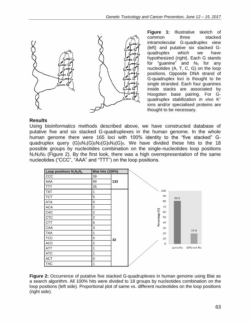
Genetic Toxicology and Cancer Prevention, June 12 – 15, 2017
63
Figure 1: Illustrative sketch of common three stacked intramolecular G-quadruplex view (left) and putative six stacked G-quadruplex which we have hypothesized (right). Each G stands for “guanine” and NS for any nucleotides (A, T, C, G) on the loop positions. Opposite DNA strand of G-quadruplex loci is thought to be single stranded. Each four guanines inside stacks are associated by Hoogsten base pairing. For G-quadruplex stabilization in vivo K+ ions and/or specialised proteins are thought to be necessary.
Results Using bioinformatics methods described above, we have constructed database of putative five and six stacked G-quadruplexes in the human genome. In the whole human genome there were 165 loci with 100% identity to the “five stacked” G-quadruplex query (G)5N1(G)5N2(G)5N3(G)5. We have divided these hits to the 18 possible groups by nucleotides combination on the single-nucleotides loop positions N1N2N3 (Figure 2). By the first look, there was a high overrepresentation of the same nucleotides (“CCC”, “AAA” and “TTT”) on the loop positions.
Loop positions N1N2N3 Blat hits (100%)
CCC 39
133AAA 69
TTT 25
TAT 1
32
TCT 5
ATA 0
ACA 2
CAC 2
CTC 2
CTT 6
CAA 3
TAA 1
TCC 5
ACC 2
ATT 1
ATC 1
ACT 0
TAC 1
Figure 2: Occurrence of putative five stacked G-quadruplexes in human genome using Blat as a search algorithm. All 100% hits were divided to 18 groups by nucleotides combination on the loop positions (left side). Proportional plot of same vs. different nucleotides on the loop positions (right side).
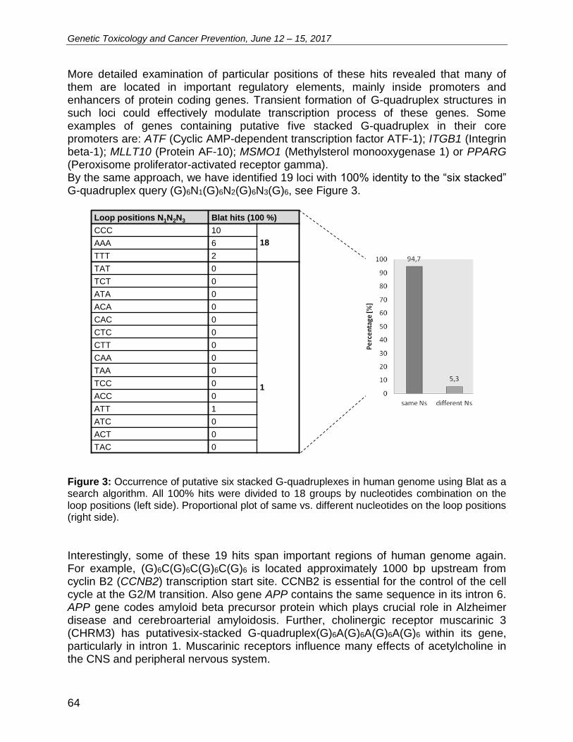
Genetic Toxicology and Cancer Prevention, June 12 – 15, 2017
64
More detailed examination of particular positions of these hits revealed that many of them are located in important regulatory elements, mainly inside promoters and enhancers of protein coding genes. Transient formation of G-quadruplex structures in such loci could effectively modulate transcription process of these genes. Some examples of genes containing putative five stacked G-quadruplex in their core promoters are: ATF (Cyclic AMP-dependent transcription factor ATF-1); ITGB1 (Integrin beta-1); MLLT10 (Protein AF-10); MSMO1 (Methylsterol monooxygenase 1) or PPARG (Peroxisome proliferator-activated receptor gamma). By the same approach, we have identified 19 loci with 100% identity to the “six stacked” G-quadruplex query (G)6N1(G)6N2(G)6N3(G)6, see Figure 3.
Loop positions N1N2N3 Blat hits (100 %)
CCC 10
18AAA 6
TTT 2
TAT 0
1
TCT 0
ATA 0
ACA 0
CAC 0
CTC 0
CTT 0
CAA 0
TAA 0
TCC 0
ACC 0
ATT 1
ATC 0
ACT 0
TAC 0
Figure 3: Occurrence of putative six stacked G-quadruplexes in human genome using Blat as a search algorithm. All 100% hits were divided to 18 groups by nucleotides combination on the loop positions (left side). Proportional plot of same vs. different nucleotides on the loop positions (right side).
Interestingly, some of these 19 hits span important regions of human genome again. For example, (G)6C(G)6C(G)6C(G)6 is located approximately 1000 bp upstream from cyclin B2 (CCNB2) transcription start site. CCNB2 is essential for the control of the cell cycle at the G2/M transition. Also gene APP contains the same sequence in its intron 6. APP gene codes amyloid beta precursor protein which plays crucial role in Alzheimer disease and cerebroarterial amyloidosis. Further, cholinergic receptor muscarinic 3 (CHRM3) has putativesix-stacked G-quadruplex(G)6A(G)6A(G)6A(G)6 within its gene, particularly in intron 1. Muscarinic receptors influence many effects of acetylcholine in the CNS and peripheral nervous system.
Genetic Toxicology and Cancer Prevention, June 12 – 15, 2017
65
Conclusion When three single-residue loops are present in intramolecular G-quadruplex forming sequence, parallel conformation of G-quadruplex according to Hazel et al. (2004) always arises. It was found that intramolecular G-quadruplexes which contain very short loops (less than five nucleotides) are very stable (Guédin et al., 2010). Especially single-nucleotide loops give rise to most stable intramolecular G-quadruplex structures (Bugaut and Balasubramanian, 2008). It is believed that stability of G-quadruplexes increases with increasing numbers of guanin tetrade stacks (Huppert and Balasubramanian, 2005). By means of sophisticated bioinformatic analyses the most frequently loop length combination in putative G-quadruplexes forming sequences was found to be 1 : 1 : 1 (all loops are single nucleotides) (Huppert and Balasubramanian, 2005). At this time there is probably no reliable “wet” lab method to verify if putative five stacked G-quadruplexes give rise as five stacked in vivo. Although antibodies against G-quadruplex are available nowadays, such as BG4 (Biffi et al., 2013), it will be challenge to design G-quadruplex antibodies with specificity to number of guanine tetrade stacks. Acknowledgement: This work was financially supported by the Ministry of Education, Youth and Sports of the Czech Republic in the “National Feasibility Program I”, project LO1208 TEWEP”; EU structural funding Operational Programme Research and Development for innovation, project No. CZ.1.05/2.1.00/19.0388 and by project SGS17/PřF/2016 financed by University of Ostrava. References
[1] Haider, Shozeb, Gary N. Parkinson, and Stephen Neidle. 2002. “Crystal Structure of the Potassium Form of an Oxytricha Nova G-Quadruplex.” Journal of Molecular Biology 320 (2): 189–200.
[2] Ho, Junming, Michael B. Newcomer, Christina M. Ragain, Jose A. Gascon, Enrique R. Batista, J. Patrick Loria, and Victor S. Batista. 2014. “MoD-QM/MM Structural Refinement Method: Characterization of Hydrogen Bonding in the Oxytricha Nova G-Quadruplex.” Journal of Chemical Theory and Computation 10 (11): 5125–5135.
[3] Rhodes, Daniela, and Hans J. Lipps. 2015. “G-Quadruplexes and Their Regulatory Roles in Biology.” Nucleic Acids Research 43 (18): 8627–37. doi:10.1093/nar/gkv862.
[4] Karsisiotis, Andreas Ioannis, NasonMa’aniHessari, Ettore Novellino, GianPieroSpada, Antonio Randazzo, and MateusWebba da Silva. 2011. “Topological Characterization of Nucleic Acid G-Quadruplexes by UV Absorption and Circular Dichroism.” AngewandteChemie International Edition 50 (45): 10645–10648.
[5] Siddiqui-Jain, Adam, Cory L. Grand, David J. Bearss, and Laurence H. Hurley. 2002. “Direct Evidence for a G-Quadruplex in a Promoter Region and Its Targeting with a Small Molecule to Repress c-MYC Transcription.” Proceedings of the National Academy of Sciences 99 (18): 11593–11598.
[6] Ambrus, Attila, Ding Chen, Jixun Dai, Roger A. Jones, and Danzhou Yang. 2005. “Solution Structure of the Biologically Relevant G-Quadruplex Element in the Human c-MYC Promoter. Implications for G-Quadruplex Stabilization.”Biochemistry 44 (6): 2048–58. doi:10.1021/bi048242p.
Genetic Toxicology and Cancer Prevention, June 12 – 15, 2017
66
[7] Hazel, Pascale, Julian Huppert, Shankar Balasubramanian, and Stephen Neidle. 2004. “Loop-Length-Dependent Folding of G-Quadruplexes.” Journal of the American Chemical Society 126 (50): 16405–15. doi:10.1021/ja045154j.
[8] Guédin, Aurore, Julien Gros, PatriziaAlberti, and Jean-Louis Mergny. 2010. “How Long Is Too Long? Effects of Loop Size on G-Quadruplex Stability.” Nucleic Acids Research 38 (21): 7858–68. doi:10.1093/nar/gkq639.
[9] Bugaut, Anthony, and Shankar Balasubramanian. 2008. “A Sequence-Independent Study of the Influence of Short Loop Lengths on the Stability and Topology of Intramolecular DNA G-Quadruplexes.” Biochemistry 47 (2): 689–97. doi:10.1021/bi701873c.
[10] Balasubramanian, Shankar, and Stephen Neidle. 2009. “G-Quadruplex Nucleic Acids as Therapeutic Targets.” Current Opinion in Chemical Biology, Next Generation Therapeutics, 13 (3): 345–53. doi:10.1016/j.cbpa.2009.04.637.
[11] Huppert, Julian L., and Shankar Balasubramanian. 2005. “Prevalence of Quadruplexes in the Human Genome.” Nucleic Acids Research 33 (9): 2908–16. doi:10.1093/nar/gki609.
[12] Kent, W. James. 2002. “BLAT—the BLAST-like Alignment Tool.” Genome Research 12 (4): 656–664.
[13] Zerbino, Daniel R., Steven P. Wilder, Nathan Johnson, Thomas Juettemann, and Paul R. Flicek. 2015. “The Ensembl Regulatory Build.” Genome Biology 16: 56. doi:10.1186/s13059-015-0621-5.
[14] Biffi, Giulia, David Tannahill, John McCafferty, and Shankar Balasubramanian. 2013. “Quantitative Visualization of DNA G-Quadruplex Structures in Human Cells.” Nature Chemistry 5 (3): 182–86. doi:10.1038/nchem.1548.
Genetic Toxicology and Cancer Prevention, June 12 – 15, 2017
67
P6 Antibacterial and antifungal potential of essential oils Mária Bučková, Andrea Puškárová, Domenico Pangallo 1Institute of Molecular biology, Slovak Academy of Sciences, Dúbravska cesta 21, 84551 Bratislava, Slovakia This study was undertaken to determine the in vitro antimicrobial activities of six essential oils (oregano, thyme, clove, arborvitae, lavender and clary sage) in order to pre-select candidates for potential application as broad-spectrum anti-microbial agents for decontamination of indoor environment. Antimicrobial activities (minimum inhibitory concentrations, minimum bactericidal and minimal fungicidal concentrations) were shown against common pathogenic (Escherichia coli, Salmonella typhimurium, Yersinia enterocolitica, Staphylococcus aureus, Listeria monocytogenes, Enterococcus faecalis) as well as environmental bacteria (Bacillus cereus, Arthrobacter protophormiae, Pseudomonas fragi) and fungi (Chaetomium globosum, Penicillium chrysogenum, Cladosporium cladosporoides, Alternaria alternata, Aspergillus fumigatus). Oregano, thyme, clove and arborvitae showed very strong antimicrobial activity against all tested strains at full strength as well as at lower concentrations. Essential oils showed different fungistatic and/or fungicidal activity. Another important aspect regards the oils of lavender and clary sage that didn’t exhibit any antifungal activity against all fungal strains in direct contact phase. Instead, in the vapor phase these EOs showed fungistatic effect, while oregano, thyme, clove and arborvitae exhibited valuable fungicidal activity. Volatile phase effects were found to be more effective on fungal growth. This study provides novel approaches to assess the antimicrobial potential of essential oils in liquid and vapor phase showing also valuable properties of Thuja plicata phenol-free oil. Generally, it is possible to advise the use of EOs for various environmental disinfection strategies, but after an accurate in vitro trial as it was described in this investigation. The data reported in this study demonstrates that the use of essential oils might represent an alternative way to fight microbial contamination. Acknowledgement This work was supported by VEGA Agency, project no. 2/0061/17 "Innovative disinfection strategies: the essential oils effect on microflora and materials of cultural heritage objects".

Genetic Toxicology and Cancer Prevention, June 12 – 15, 2017
68

List of Authors

Genetic Toxicology and Cancer Prevention, June 12 – 15, 2017
69
Ahmed Zubair KL1
Ambrož Antonín L14, L15, L31
Arsenyan Pavel L6
Bábelová Andrea L10, L13, L27
Baliova Martina L27
Bartas Martin P5
Bilčík Boris P4
Bizík Jozef L21, L26
Bobálová Janette L3, L26
Boháč Andrej L7
Brázda Václav P5
Brtko Július L3, P2
Brzicová Táňa L17
Bučková Mária P6
Buríková Monika L21
Ciganek Miroslav L10
Collins Andrew KL3
Čavarga Ivan P4
Červeň Jiří P5
Červená Tereza L14, L24
Dianovský Ján L2
Drážovská Monika L2
Ďurovcová Ivana P1
Dušička Jozef L2, L17, L18
Dušinská Mária KL2, L21
Dvořák Zdeněk L1, L3
Dzian Anton L25
Ebner Andreas L26
El Yamani Naouale KL3
Elje Elisabeth KL3
Elzeinová Fatima L14
Feruszová Jana L4
Gábelová Alena L8, L12, L21, L23, L24
Galdíková Martina L2
Gálová Eliška L4, L7, P1, P3
Gmuender Hans L32
Gregušová Naďa L13
Grendár Marián L25
Hajslova Jana L32
Halašová Erika L25, L29
Hianik Tibor L21, L26
Holečková Beáta L2
Honková Kateřina L32
Hopan František L14
Horák Jiří L14
Horváthová Eva L7,
Hovorka Jan L10
Hubatka František L10
Hunáková Ľuba L3
Chovanec Miroslav L6
Janout Vladimír L30
Karlický Václav P5
Klapáková Martina L7
Kléma Jiří L16
Koneracka Martina L12
Kováčová Simona L2, L16
Kozics Katarina L5
Krpec Kamil L14
Krutáková Martina L25
Genetic Toxicology and Cancer Prevention, June 12 – 15, 2017
70
Kubesa Petr L14
Kulich Pavel L11
Kysela Boris KL1
Kyzek Stanislav L4, P1, P3,
Kyzek Pavel L11
Lacík Igor L8
Lehocká Hana L30
Leitner Michael L26
Líbalová Helena L15
Lovíšek Daniel L4
Lukáčová Patrícia L6
Macejová Dana L3, P2
Máčajová Mariana P4
Machala Miroslav L1, L11
Mániková Dominika L6, P1
Marvanová Soňa L11
Mastihuba Vladimír L7
Mastihubová Mária L7
Mašek Josef L11
Matáková Tatiana L25
Mazancová Petra L8, L9
Medvecká Veronika L4
Mesárošová Monika L5
Mičieta Karol L18, L19
Milcová Alena L14, L15, L16
Murín Gustáv L18, L19
Mušák Ľudovít L25, L29
Neča Jiří L1
Némethová Veronika L8, L9, L10
Novotný Ladislav L3
Oliviera Rui P1
Ondriašová Karolína L4
Opattová Alena L20
Osina Oto L29
Pálková Lenka L1
Pangallo Domenico P6
Pavlíkova Jitka L32
Pečinka Petr P5
Pěnčíková Kateřina L1
Petráš Martin L29
Pikal Petr L16
Podoba Ján P2
Pogányová Andrea L18
Potocká Elena L7
Poturnayová Alexandra L21
Pulkrabova Jana L32
Puškár Marek P1
Puškárová Andrea P6
Rázga Filip L8, L9, L10
Rejhová Alexandra L20
Rendeková Jana L6
Rossner Pavel Jr. L14, L15, L16, L28
Rössnerová Andrea L24, L32, L15
Rubintová Veronika L4
Rundén-Pran Elise KL2
Schaefer Liliana L27
Schmuczerová Jana L1
Schwarzbacherová Viera L2
Sikorová Jitka L6
Skoupý Radim L10
Slíva Daniel L20
Genetic Toxicology and Cancer Prevention, June 12 – 15, 2017
71
Smolková Božena L22, L23, L24
Srančíková Annamária L22, L23, L24
Strapáčová Simona L1
Svitková Barbora L8
Sýkorová Martina P4
Šarlinová Miroslava L25, L29
Šelc Micha L8, L13
Šestáková Zuzana L6, P1
Ševčovičová Andrea L4, L7, P1, P3
Šimečková Pavlína L10
Šiviková Katarína L2
Špunda Vladimír P5
Šrám Radim J. L31, L32
Šramková Monika L11, L22, L23, L24
Švecová Vlasta L14, L31
Topinka Jan L1, L14, L16, L17
Toporová Lucia L3
Tsalastra Wasiliki L27
Tuxworth Richard KL1
Uhrin Filip L4
Útla Ivana P1
Valachová Jela L29
Vavrošová Jana L30
Velemínský Miloš, Jr. L31
Vlasáková Danuša L6
Vlková Veronika L31
Vodička Pavel L20
Vondráček Jan L1
Vrbová Kristýna L15, L16
Zahoranová Anna L4
Záhradníková Eva L18
Zajíčková Terázia P3
Závacká Ivona L30
Závišová Vlasta L12

Genetic Toxicology and Cancer Prevention, June 12 – 15, 2017
72

Sponsors & Exhibitors

73
