genetic studies of paralyzed mutants in ... studies of paralyzed mutants in salmonella. i. genetic...
TRANSCRIPT
GENETIC STUDIES OF PARALYZED MUTANTS IN SALMONELLA. I. GENETIC FINE STRUCTURE OF THE mot LOCI IN
SALMONELLA TYPHIMURIUMI
MASATOSHI ENOMOTO
National Institute of Genetics, Misima, Japan
Received March 1, 1966
structure of bacterial flagella has been extensively studied with the ad- T?ncement of electron microscopy and protein chemistry. However, ques- tions on the function of flagella as locomotor organs, such as the propulsive mech- anism and the energy-supplying system, remain unanswered. This is mainly due to the limitations of available experimental techniques. One method used for attacking the fundamental problems of flagellar movement is a genetic approach with paralyzed mutants, that is mutants which though flagellated are nonmotile. Flagella o i the paralyzed mutant do not differ from those of the wild-type motile cell in either antigenicity, number per bacterium, shape under electron micro- scope (ENOMOTO, unpublished data), or configuration by X-ray diffraction (BEIGHTON, PORTER and STOCKER 1958). Both the wild type and the paralyzed mutants show flagellar antigenic phase variation ( ENOMOTO, unpublished data). These facts suggest that the paralysis is not due to any defect of flagella them- selves, but to a defect of the flagellum-activating mechanism within the bacteria.
Preliminary genetic analysis of the paralyzed mutants of Salmonella typhi- murium was reported by STOCKER, ZINDER and LEDERBERG (1953) and IINO (1958). STOCKER et al. showed that motility of a paralyzed mutant could be re- stored by transduction from the wild type or from another paralyzed mutant. IINO found by transduction experiments with four paralyzed mutants that their mutational sites were contained within a single functional (complementation) unit. For the present investigation, nearly 100 paralyzed mutants were isolated from S. typhimurium. A genetic fine-structure analysis of the motility loci has been carried out by means of complementation, deletion, and two-factor trans- duction tests.
MATERIALS A N D METHODS
Nomenclature: The mutant gene causing paralysis of flagella was designated mot, for motility (IINO 1958). The strain number for the mutants was assigned mechanically according to the order of isolation, and the mutant number was assigned from 201 on for the paralyzed mutants derived from S. typhimurium, TM2. When mutational sites were grouped by complementation tests, a capital letter designating the functional unit was inserted between the gene symbol, mot, and the mutant number (e.g. motA-201). The mutant number was also used to designate the corresponding mutational site.
Contribution No. 607 from the National Institute of Genetics, Misima, Japan.
Genetics 54: 715-i2G September 196B.

716 M. ENOMOTO
Organisms: All of the mutants used in this work were derived from a wild-type strain of Salmonella typhimurium, TM2 (strain LT-2 of ZINDER and LEDERBFXG 1952). Almost all of the paralyzed mutants were isolated by a screening method consisting of a combination of semisolid medium and the "motile-specific'' chi phage (MEYNELL 1961; ENOMOTO and IINO 1963). The paralyzed mutants from SJ597 (mot-269) to SJ637 (mot-300) were isolated by induction with ultraviolet light; others were of spontaneous origin. Strain SJ697, in which HIi is replaced by HIgp of S. dublin, was used for the test of joint transduction of mot and HI. Histidine-requiring mutants were obtained from DR. P. E. HARTMAN, Johns Hopkins University, Maryland. Phage P22 was used for transduction (ZINDER and LEDERBERG 1952). Phage suspensions were prepared as described by STOCKER et al. (1953), and assayed by a modified agar-layer technique (ADAMS 1959).
Media Nutrient broth medium (pH 7.2) was composed of 1% peptone (Kyokuto) and 1% meat extract (Mikuni). Nutrient agar (NA) medium contained 1.5% agar in addition to the nutrient broth. Semisolid nutrient gelatin agar (NGA) medium was prepared by the addition of 0.4% agar and 8% gelatin to the broth medium. The composition of Davis minimal medium (MM) and complete EMB medium was described by LEDERBERG (1950).
Observation of abortive transduction for complementation test: One volume of an overnight broth culture of a paralyzed mutant (approximately 1 x 10⁹ cells per ml) was mixed with an equal volume of phage suspension propagated on a donor strain (approximately 5 to 10 x IO9 plaque forming units per ml), and 0.1 ml of the mixture was inoculated on a NGA plate. After incubation for 15 to 18 hours at 37"C, the plate was examined for the presence of trails under the low-power binocular microscope. In transduction of motility, trails appear if the partial heterozygote produced by abortive transduction has the wild-type, motile, phenotype. A mot- recipient will therefore give trails in transductional crosses with a wild-type donor if its m o r allele is recessive to mot+ ; and also in crosses with a nonidentical mot- mutant if the two mot- mutants can complement one another (LEDERBERG 1956; STOCKER 1956). The number of trails produced in crosses between mot- mutants was examined semiquantitatively and divided into three grades: 100-50%, 50-1%, and less than 1% of the number observed in the control crosses of mot- with the wild-type donor.
Identification of deletion mutants: The paralyzed mutants were incubated on NGA medium overnight at 37°C to identify unstable mutants. Approximately 1 x I O 9 cells of the stable mutants were further incubated on NGA medium for three days. Among the mutant strains producing no swarms after this incubation, those which failed to produce swarms by transduction from more than two other mutants were regarded as deletions.
0.1 ml of the mixture of equal volumes of bacterial and phage suspensions (1 x 10⁸ cells, 5 x 10⁸ plaque forming units per ml) was streaked on each NGA plate. When phages prepared from the wild-type bacteria were used, the mixture was diluted 50-fold after 10 minutes adsorption and streaked. After about 8 hours incubation at 37"C, the number of swarms was counted. Recombination frequency was expressed as a fraction percent of the number of swarms per 108 recipient cells treated with phage grown on a given mot- donor to that treated with phage grown on the wild-type donor. In order to obtain the mutually comparable recombination frequencies, one series of transduction tests was carried out simultaneously with the same recipient culture. At least ten platings were made for each mixture as well as control platings with uninfected bacteria: when revertants appeared, their number was subtracted from that of the motile recombinants. The mean recombination frequencies of two separate experiments were used for the mapping.
Mapping by recombination frequencies in a two-factor cross:
RESULTS
Grouping of paralyzed mutants by complementation tests: Initially, 30 mutants chosen at random were examined in all painvise combinations for abortive trans- duction. All the mutants produced trails in transduction tests with the wild-type donor. By the grouping of noncomplementary mutants, three complementation

SALMONELLA PARALYZED MUTANTS 71 7
groups were established. Representative mutants of each group were further used for grouping the remaining mutants. With 15 exceptions, 97 paralyzed mutants were classified as follows: 52 in motA; 27 in motB; and 3 in motC. Of the remain- ing 15 mutants, mot-292 complemented only motC mutants and was found in later experiments to be a long deletion. The other 14 mutants complemented motC mutants normally. However, they produced weak responses with the repre- sentative mutants of either motA or motB: the number of trails was less than 50% of that in the control crosses. Some of these mutants showed weak responses with both motA and B mutants. In the “weak response,” the decrease in the number of trails was often accompanied by the decrease in the number of minute colonies forming each trail. The similar phenomenon was also observed in partial complementation of fla- mutants (IINO and ENOMOTO 1966). In the partial com- plementation. however, the mutant which produced weak responses with mutants of the same group showed normal responses with other groups. In order to dis- tinguish from partial-complementing mutants, these 14 mutants are, henceforth, designated as weak-complementing mutants. The complementation pattern ob- tained from the semiquantitative experiments among 40 representative mutants including the above 14 mutants is shown in Figure 1 : mutants from mot-257 to 246 belong to motA; 295 to 211 belong to motB; 244 to 279 to motC; and 297 to 262 are weak-complementing mutants. The mutants in each of the three groups complemented all members of other two groups, but did not complement the mutants in the same group, except that they showed partial complementation in some combinations of the same group: less than 1% as compared with the frequency obtained in crosses with the mutants of other groups. One mutant, C-272, when used as a recipient, did not either complement or only complemented weakly motA and B mutants, !whereas as a donor normal complementation with motA and B mutants occurred. The weak response was also observed in trans- duction from the wild-type donor. C-272 seems to carry a factor which suppresses trail production. The 14 weak-complementing mutants could be tentatively sub- divided into two classes, motA (12 mutants) and motB (2: 227,262), from the following observations: a difference in relative number of trails was observed between the crosses with motA and B mutants; motB211 was found to comple- ment normally most of these motA mutants; and mot-227 and -262 were found to be overlapped by the long deletion B-292. The weak-complementing motA mutants also showed weak responses with B-272 and B-262, and with each other in certain combinations.
Transduction between motA and motB mutants always yielded a small number of swarms (wild-type recombinants) as compared with the transductions from the wild-type or motC mutants. This behavior can be explained by assuming the two mot loci A and B are jointly transduced: the mutant allele motA- is transduced into a recipient motB mutant together with the wild-type allele motBf and vice versa.
Phenotypic character of the mot mutants: The mutant strains carrying allele of the three mot genes were phenotypically similar: they were indistinguishable from each other by means of growth tests in minimal medium, flagellar mor-

718 M. ENOMOTO
0 0 0 0 .0 0 0 . .
0 ~ 0 0 0 0 0 0 0 0 0
0 0 0 0 0 :I’ 0 0 0.::::
‘ . . O 0 0 0 0 0 0 0 0 0 0 0 0 0 0 0
0 0 0 o o o o : . : . o 0
0 0 0 0 0 0 0 0 0 0 0 0 0 0 0 0 0 0 0 0 0 0 0 0 0 0 0 0 0 0 0 0 0 0 0 0 0 0 0 0 0 0 0 0
no complementafion 50-100 ?4
.:.:.: c I %
6 . 1-50 %
... not tested
... ... * .
FIGURE 1.-Complementation among mot- mutants. motA group: from 257 to 246; motB group: from 295 to 211; motC group: from 244 to 279; mutants from 297 to 262 are weak-comple- menting ones in which 297-237 are motA, and 227 and 262 are motB.
phology, and distribution of flagella on the cell surface viewed with electron microscopy. All three motC mutants produced minute wild-type colonies at the margin of, or behind, the inoculation site cm NGA medium. This results from the temporary restoration of motility which is probably due to “leaky” character or imperfect block of the mutant ( QUADLING and STOCKER 195 7). However, this phenomenon was not uniquely characteristic of motC mutants, because some mutants of motA and motB also produced such minute colonies. No syntrophic recovery of motility was observed in pairwise mixed cultures in all possible com- binations of the representative mutants of each functional unit.
Joint transduction of mot with other marker genes to test linkage: Mutants A-229, B-231, and C-224 were picked as representative mutants of each func-
SALMONELLA PARALYZED M U T A N T S 71 9
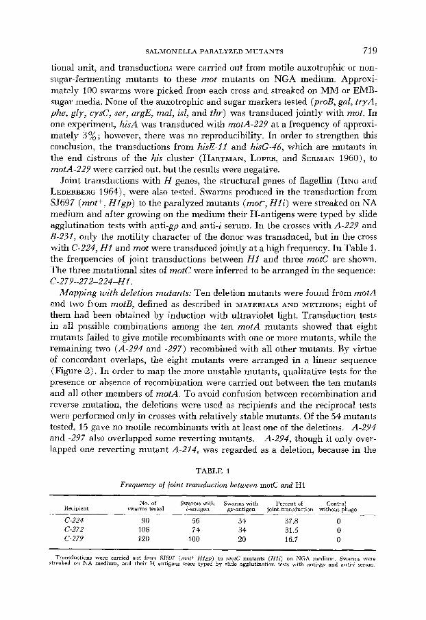
tional unit, and transductions were carried out from motile auxotrophic or non- sugar-fermenting mutants to these mot mutants on NGA medium. Approxi- mately 100 swarms were picked from each cross and streaked on MM or EMB- sugar media. None of the auxotrophic and sugar markers tested (proB, gal, tryA, phe, gly, cysC, ser, argE, mal, isl, and thr) was transduced jointly with mot. In one experiment, hisA was transduced with motA-229 at a frequency of approxi- mately 3%; however, there was no reproducibility. In order to strengthen this conclusion, the transductions from hisE-11 and hisG-46, which are mutants in the end cistrons of the his cluster (HARTMAN, LOPER, and SERMAN 1960), to motA-229 were carried out, but the results were negative.
Joint transductions with H genes, the structural genes of flagellin (IINO and LEDERBERG 1964), were also tested. Swarms produced in the transduction from SJ697 (mot+. H l g p ) to the paralyzed mutants (mot-, H l i ) were streaked on NA medium and after growing on the medium their H-antigens were typed by slide agglutination tests with anti-gp and anti-i serum. In the crosses with A-229 and B-231, only the motility character of the donor was transduced, but in the cross with C-224, H I and mot were transduced jointly at a high frequency. In Table 1. the frequencies of joint transductions between H I and three motC are shown. The three mutational sites of mote were inferred to be arranged in the sequence: C-279-272-224-Hl .
Mapping with deletion mutants: Ten deletion mutants were found from motA and two from motB, defined as described in MATERIALS AND METHODS; eight of them had been obtained by induction with ultraviolet light. Transduction tests in all possible combinations among the ten motA mutants showed that eight mutants failed to give motile recombinants with one or more mutants, while the remaining two (A-294 and -297) recombined with all other mutants. By virtue of concordant overlaps, the eight mutants were arranged in a linear sequence (Figure 2). In order to map the more unstable mutants, qualitative tests for the presence or absence of recombination were carried out between the ten mutants and all other members of motA. To avoid confusion between recombination and reverse mutation, the deletions were used as recipients and the reciprocal tests were performed only in crosses with relatively stable mutants. Of the 54 mutants tested, 15 gave no motile recombinants with at least one of the deletions. A-294 and -297 also overlapped some reverting mutants. A-294, though it only over- lapped one reverting mutant A-214, was regarded as a deletion, because in the
TABLE 1
Frequency of joint transduction between motC and HI
No. of Swarms with Swarms,with Percent of Control Recipient swarms tested I-antigen gp-anbgen joint transduction without phage
C-224 90 56 34 37.8 0 C-272 108 74 34 31.5 0 G-279 120 100 20 16.7 0
Transductions were carried out from SJ697 (mot+ Hfgp) to motC mutants ( H f i ) on NGA medium. Swarms were streaked on NA medium, and their H antigens were typed by slide agglutination tests with anti-gp and anti-i serum.
720 M. ENOMOTO
, : , . : ; : : ; : : : : , : , , . . I . I '
. , , . I
226fSJ4ZllJ
2aMu611) X)J(SJSO) 293fSJS3OJ 2ll215JSIlJ
291fSJS341
111fSJ6011J . : : : , , , ; : ! ; ; , : j : : j , , . , I . , . I
I , I I ' , I +
I * . . . . 29USJ631J . I
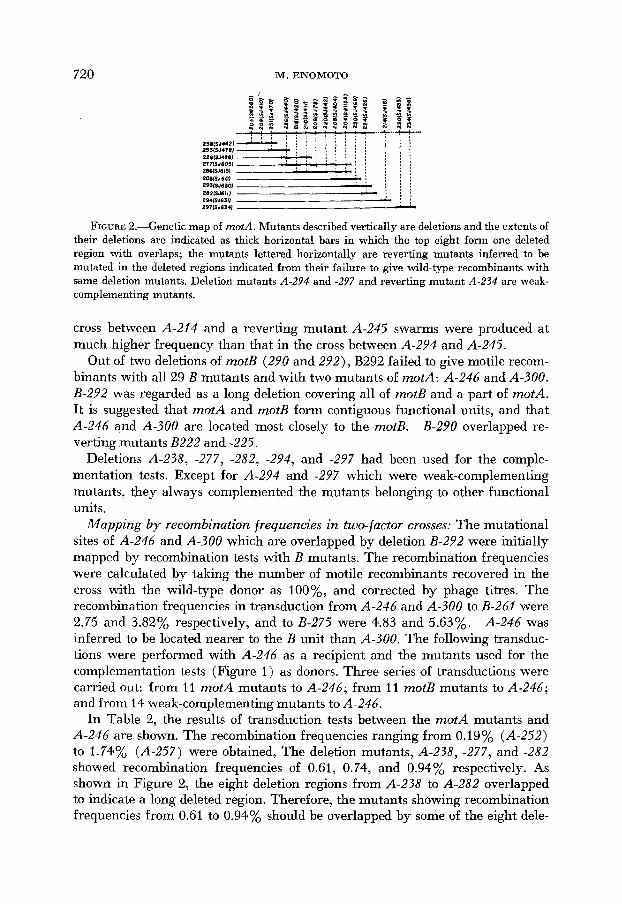
FIGURE 2.-Genetic map of motA. Mutants described vertically are deletions and the extents of their deletions are indicated as thick horizontal bars in which the top eight form one deleted region with overlaps; the mutants lettered horizontally 'are reverting mutants inferred to be mutated in the deleted regions indicated from their failure to give wild-type recombinants with same deletion mutants. Deletion mutants A-294 and -297 and reverting mutant A-234 are weak- complementing mutants.
cross between A-214 and a reverting mutant A-245 swarms were produced at much higher frequency than that in the cross between A-294 and A-245.
Out of two deletions of motB (290 and 292), B292 failed to give motile recom- binants with all 29 B mutants and with two mutants of motA: A-246 and A-300. B-292 was regarded as a long deletion covering all of motB and a part of motA. It is suggested that motA and motB form contiguous functional units, and that A-246 and A-300 are located most closely to the motB. B-290 overlapped re- verting mutants B222 and -225.
Deletions A-238, -277, -282, -294, and -297 had been used for the comple- mentation tests. Except for A-294 and -297 which were weak-complementing mutants, they always complemented the mutants belonging to other functional units.
Mapping by recombination frequencies in truo-factor crosses: The mutational sites of A-246 and A-300 which are overlapped by deletion B-292 were initially mapped by recombination tests with B mutants. The recombination frequencies were calculated by taking the number of motile recombinants recovered in the cross with the wild-type donor as loo%, and corrected by phage titres. The recombination frequencies in transduction from A-246 and A-300 to B-261 were 2.75 and 3.82% respectively, and to B-275 were 4.83 and 5.63%. A-246 was inferred to be located nearer to the B unit than A-300. The following transduc- tions were performed with A-246 as a recipient and the mutants used for the complementation tests (Figure 1) as donors. Three series of transductions were carried out: from 11 motA mutants to A-246; from 11 motB mutants to A-246; and from 14 weak-complementing mutants to A-246.
In Table 2, the results of transduction tests between the motA mutants and A-246 are shown. The recombination frequencies ranging from 0.19% (A-252) to 1.74% (A-257) were obtained, The deletion mutants, A-238, -277, and -282 showed recombination frequencies of 0.61, 0.74, and 0.94% respectively. As shown in Figure 2, the eight deletion regions from A-238 to A-282 overlapped to indicate a long deleted region. Therefore, the mutants showing recombination frequencies from 0.61 to 0.94% should be overlapped by some of the eight dele-

SALMONELLA PARALYZED MUTANTS 72 1
r-. Ln “1
0 p. Y
h U cu
-I-
“1 ::
* “I “1
-t- r. r-. Y
h U cu
VI
7
-I- C? c, Y
h -4 i?
PI L? “1

722 M. ENOMOTO
tions. However, the reverting mutants A-266, -267, and -229 were not overlapped by any of the deletions, though they showed recombination frequencies of 0.64, 0.71, and 0.76% respectively with A-246. This contradiction is probably due to the decrease in recombination frequencies in the crosses with the three deletion mutants.
In Table 3, the results obtained from the crosses between 11 motB mutants and A-246 are presented. In these crosses, recombination frequencies ranged from 0.92 (B-215) to 18.2% (B-211), and these values were fairly high as compared with those shown in Table a. It is not unexpected that the frequency in the cross between mutants in different functional units are higher than that with mutants within the same unit. Mutant B-211 showed highest recombination frequency among motB mutants, and its site was inferred to be farthest from motA. B-211 is the only mutant of motB which complements normally the majority of the weak-complementing motA mutants. This character is perhaps due to the loca- tion of B-221 far from the sites of other 3 mutants.
In recombination tests between weak-complementing mutants and A-246, the tests between two normal complementing mutants (A-259 and B-295) and A-246 were carried out simultaneously to compare the recombination frequencies with those obtained from the other two series of tests (Tables 2, 3 ) . The results are shown in Table 4. The mutational sites of the 12 weak-complementing A mutants were not apparently clustered in any particular region but dispersed randomly in the motA functional unit. A-297 was found to be a deletion overlapping A-230 and A-234. A-297 and A-234 were used for the recombination tests and showed the frequencies of 1.31 and 1.57%, respectively. A reverting mutant A-263 which was not overlapped by A-297 showed a recombination frequency of 1.56%; consequently its mutational site was assigned between A-297 and A-234. This contradiction would be explained if A-297 shows decreased recombination fre- quency on account of the deletion. B-227, which showed a very weak response to motA mutants in the complementation tests, was located nearest to A-246. A-259 and B-295 showed recombination frequencies of 0.42 and 2.91% in this series. These values correspond or are close to the values obtained from the other two series of tests (0.42 and 2.78). Therefore, the recombination frequencies obtained from the cross with the weak-complementing mutants can be compared with those shown in Tables 2 and 3. In Figure 3, the results of the three series of recombination tests are shown. The order of the mutational sites, especially of the adjacent sites, should be considered to be tentative, because the mapping de- pends on the recombination frequencies obtained from the crosses with A-246 only. In order to map precisely, reciprocal transduction tests between other mu- tants are required. However, the approximate location of the mutational sites can be assigned as in Figure 3.
DISCUSSION
The results of the complementation tests showed that the sites of mutations leading to flagellar paralysis were grouped in three functional units: motA, motB, and motC. motA and motB adjoin each other and motC is located apart

v, Y- 3 T 0 h
c
U
U
s 2 ._ P
?
B + s 2 s 2 .$
3
* c
4 *
a s s
0
2
t. 5 z s 8
U
c .U
._ U
E s E 8 2
H
i “ c ?
E ” m a %
4
-1- r.
{ s : 9 ” 5 , 0 4 s a g E ”
v,g
2 s “ 2
- “ 2 ; f ;
2 5 5 g
+I.? 9 2 s s m
3 e 2 2
- - ( D h
2 2 “ N ) - - 2 2 “ 2
$ 5 : d - 2
a h 04 i
q Q ;;? i i
c? U?: 2 3
(Ut.
o m
m i m m i - “ D l
09 q “.
$ 3 i - m
* ( o
f 2 $ 2 2 2
- 0 4 2 6
r ; 2 2 % a g 2
g g E * 9 2 P Q
4 : * a $ $
?# A
8 2 2 O O B 0 2 ; g
p .s 3 !
,--. g 2% & ? E . ,mE - .s +,p* ; g s y c c - s . s g $ ’3 lij $ & p g.n
2 : & p 0 +f+
- h
( U N ) a m m
s e W O
r . q ”
0
2 2
O M
2 m V h a
& g p g g & R F r ; Fr;

724 M. ENOMOTO
B CISTRON A CISTRON
,a' ; 4 A ; ' ! ' ~ " ; " " ; " ' . ; FIGURE 3.-Genetic map of motA and motB. The results of three series of transduction tests
are arranged as follows: crosses between A-246 and motA mutants (top, right) ; between A 2 4 6 and motB mutants (top, left) ; and between A-246 and weak-complementing mutants (bottom). The scale indicates recombination frequencies (percent). The upper and lower maps are com- parable with each other except in respect of close mutational sites (see text). A-238, -277, and -282 of the upper map (cf. Figure 2), and A-294 and -297 of the lower map are deletions.
from them. motC is transduced jointly with HI at a high frequency. Whether motC is located on the his side or try side in relation to H I , and whether motA and motB are located on the same side as motC in relation to HI or the opposite side, remains to be investigated.
The two-factor transduction tests revealed that the representative mutational sites of the motA and motB could be arranged in a linear sequence. In motB, B-227 was located closest to motA and B-211 was farthest from it. B-227 is a weak-com- plementing mutant showing little response to all the motA mutants tested. On the contrary, B-211 complements normally the majority of the weak-comple- menting motA mutants. The behavior of these two mutants suggests that the closer the two mutational sites are, the weaker the complementation.
In order to map the mutational sites by means of two-factor transduction tests, generally all pairwise crosses are carried out and the frequencies of the wild-type recombinants are compared for each pair of mutants (HARTMAN 1956). In the present investigation, fortunately, the mutant A-246 was found to be located near the boundary of the two functional units by virtue of the long deletion B-292, so the mapping has been carried out between A-246 and motA or motB mutants. In order to avoid as many errors as possible, one series of crosses was performed by use of the same recipient culture simultaneously. However, inherent error will occur, such as the correction of the recombination frequency by the potency of transducing phage. Therefore, the accurate order of the mutational sites, espe- cially those of the adjacent mutational sites, should be determined only after reciprocal transduction tests with other mutants.
The mutational sites of 14 weak-complementing mutants were not located in a specific region, but distributed throughout the whole functional unit. They show normal complementation in crosses with the wild-type and motC mutants. Therefore, the character of these mutants is not a result of cytoplasmic defect. These mutants were tentatively assigned to motA' and motB. It is difficult to interpret these mutants by the present genetic data, because 'whether motA and motB control different protein molecules respectively or together control a single protein molecule, are unknown. For the latter the interpretation will be changed according to whether two different polypeptide subunits are elicited by

SALMONELLA PARALYZED MUTANTS 725
separate motA and motB units or one polypeptides is made along the entire A and B region to form a single active protein molecule. The character of these mutants is similar to that of the Dab mutants of the his operon (HARTMAN, HARTMAN, and SERMAN 1960; LOPER et al. 1964) : they do not complement the mutants of either a or b functional unit of hisD.
A biochemical analysis of flagellar movement has not been carried out. It has been suggested that the bond energy of ATP is involved in motility (DE ROBERTIS and PELUFFO 1951 ; DE ROBERTIS and FRANCHI 1952; SHERRIS et al. 1957). How- ever, the ATP content in the motile wild-type cell was not significantly different from that of the paralyzed cells (representatives of motA and motB) (ENOMOTO 1962; unpublished data). The energy required for flagellar propulsion is less than 0.1% of that of the total cell (HERBERT 1951), so that the difference in ATP content may not be detectable. From the finding that (1) ATP and ATPase activity are not detected in flagella detached from both wild-type and paralyzed cells (ENOMOTO 1962; unpublished data), and that (2) the flagellar protein, morphology, and the configuration by X-ray diffraction (BEIGHTON et al. 1958) are not different in the two genotypes, it is proposed that the defect leading to flagellar paralysis is located in the cytoplasm, probably in the basal granules. On the basis of these genetic data with the paralyzed mutants, biochemical investi- gations of flagellar movement are in progress.
The author wishes to express his gratitude to DR. T. IINO for much advice and encouragemenl during the course of this investigation, and to DR. E. M. LFDERBERC for her review of the manuscript. This work was partly supported by a research grant from the National Institute of Allergy and Infectious Diseases (AI-02872), Public Health Service, U.S.A., to T. IINO.
SUMMARY
Ninety-seven paralyzed mutants were divided into three complementation groups by abortive transduction tests: motA with 64; motB with 30; and motC with 3. motA and motB were found to constitute adjoining functional units because of the occurrence of joint transduction of motA and motB, and of a deletion mutation covering the whole motB and a part of motA. mote was transduced jointly with H I , a phase-1 flagellin gene, and the order of the three mutational sites within motC was determined, The order of the representative mutational sites in motA and motB was also determined tentatively by deletion mapping and two-factor transduction tests.
LITERATURE CITED
ADAMS, M. H., 1959 Bacteriophages. (Appendix: Methods of study of bacterial viruses.) Inter-
BEIGHTUN, E., A. M. PORTER, and B. A. D. STOCKER, 1958 X-ray and related studies of the
DEROBERTIS, E., and C. A. PELUFFO, 1951 Chemical stimulation and inhibition of bacterial
DEROBERTIS, E., and C. M. FRANCHI, 1952
science Publishers, New York.
flagella of nonmotile bacteria. Biochim. Biophys. Acta 29: 8-13.
motility studied with a new method. Proc. Soc. Exptl. Biol. Med. 78: 584-589.
of bacterial flagella. J. Appl. Physiol. 23: 161. Macromolecular structure of the contractile protein

726 M. ENOMOTO
ENOMOTO, M., 1962 Grouping of paralyzed mutants in Salmonella. Ann. Rept. Natl. Inst. Genetics (Japan) 13 : 75-76.
ENOMOTO, M., and T. IINO, 1963 Colonial dimorphism in nonmotile Salmonella. J. Bacteriol. 86: 473-477.
HARTMAN, P. E., 1956 Linked loci in the control of consecutive steps in the primary pathway of histidine synthesis in Salmonella typhimurium. Carnegie Inst. Wash. Pub. 612 : 35-61.
HARTMAN, P. E., Z. HARTMAN, and D. SERMAN, 1960 Complementation mapping by abortive transduction of histidine-requiring Salmonella mutants. J. Gen. Microbiol. 22 : 354-368.
HARTMAN, P. E., J. C. L~PER, and D. SERMAN, 1960 Fine structure mapping by complete transduction between histidine-requiring Salmonella mutants. J. Gen. Microbiol. 22 : 323-353.
HERBERT, D., 1951 Hydrodynamic aspects of bacterial locomotion (Abstr.). J. Gen. Microbiol. 5: xx-xxi.
IINO, T., 1958 Cistron test of motility genes in Salmonella. Ann. Rept. Natl. Inst. Genetics (Japan) 9: 96.
IINO, T., and J. LEDERBERG, 1964 Genetics of Salmonella. pp. 111-142. The world problem of Salmonellosis. Junk, The Hague.
IINO, T., and M. ENOMOTO, 1966 Genetical studies of non-flagellate mutants of Salmonella. J. Gen. Microbiol. (In press.)
LEDERBERG, J., 1950 Isolation and characterization of biochemical mutants of bacteria. Methods Med. Res. 3: 5-22. - 1956 Linear inheritance in transducitonal clones. Genetics 41 : 846-871.
LOPER, J. C., M. GRABNER, R. C. STAHL, Z. HARTMAN, and P. E. HARTMAN, 1964 Genes and proteins involved in histidine biosynthesis in Salmonella. Brookhaven Symp. Biol. 17: 15-50.
MEYNELL, E. W., 1961 A phage, @x, which attacks motile bacteria. J. Gen. Microbiol. 25:
QUADLING, C., and B. A. D. STOCKER, 1957 The occurrence of rare motile bacteria in some non-motile Salmonella strains. J. Gen. Microbiol. 17: 424-4.36.
SHERRIS, J. C., N. W. PRESTON, and J. G. SHOESMITH, 1957 The influence of oxygen and arginine on the motility of a strain of Pseudomonas sp. J. Gen. Microbiol. 16: 86-96.
STOCKER, B. A. D., 1956 Abortive transduction of motility in Salmonella; a non-replicated gene transmitted through many generations to a single descendant. J. Gen. Microbiol. 15: 575- 598.
STOCKER, B. A. D., N. D. ZINDER, and J. LEDERBERG, 1953 Transduction of flagellar characters
ZINDER, N. D., and J. LEDERBERG, 1952 Genetic exchange in Salmonella. J. Gen. Microbiol.
253-290.
in Salmonella. J. Gen. Microbiol. 9: 410433.
64: 679-699.