genes involved in formation of structured multicellular ... · these fruiting bodies serve as...
TRANSCRIPT
JOURNAL OF BACTERIOLOGY, June 2004, p. 3970–3979 Vol. 186, No. 120021-9193/04/$08.00�0 DOI: 10.1128/JB.186.12.3970–3979.2004Copyright © 2004, American Society for Microbiology. All Rights Reserved.
Genes Involved in Formation of Structured Multicellular Communitiesby Bacillus subtilis
Steven S. Branda,1 Jose Eduardo Gonzalez-Pastor,2† Etienne Dervyn,3 S. Dusko Ehrlich,3Richard Losick,2 and Roberto Kolter1*
Department of Microbiology and Molecular Genetics, Harvard Medical School, Boston, Massachusetts 021151; Department ofMolecular and Cellular Biology, Harvard University, Cambridge, Massachusetts 021382; and Genetique Microbienne, Institut
National de la Recherche Agronomique, 78352 Jouy en Josas, France3
Received 17 October 2003/Accepted 9 February 2004
The spore-forming bacterium Bacillus subtilis is capable of assembling multicellular communities (biofilms)that display a high degree of spatiotemporal organization. Wild strains that have not undergone domesticationin the laboratory produce particularly robust biofilms with complex architectural features, such as fruiting-body-like aerial projections whose tips serve as preferential sites for sporulation. To discover genes involvedin this multicellular behavior and to do so on a genome-wide basis, we took advantage of a large collection ofmutants which have disruptions of most of the uncharacterized genes in the B. subtilis genome. This collection,which was generated with a laboratory strain, was screened for mutants that were impaired in biofilmformation. This subset of mutated genes was then introduced into the wild strain NCIB 3610 to study theireffects on biofilm formation in liquid and solid media. In this way we identified six genes that are involved inthe development of multicellular communities. These are yhxB (encoding a putative phosphohexomutase thatmay mediate exopolysaccharide synthesis), sipW (encoding a signal peptidase), ecsB (encoding an ABC trans-porter subunit), yqeK (encoding a putative phosphatase), ylbF (encoding a regulatory protein), and ymcA (agene of unknown function). Further analysis revealed that these six genes play different roles in B. subtiliscommunity development.
Formation of biofilms—surface-associated multicellular as-semblages—is an important microbial survival strategy (4, 25).Relative to the rapid, continual, and extreme changes in envi-ronmental conditions that can characterize a planktonic exis-tence, biofilms offer the constituent cells some shelter, en-abling them to establish long-term relationships with eachother and their immediate surroundings. Cells in different re-gions of a developing biofilm experience diverse environmentalconditions, resulting in marked patterns of cellular differenti-ation (e.g., see references 5 and 16). Such spatiotemporal or-ganization is particularly striking in biofilms formed by wildstrains of the spore-forming bacterium Bacillus subtilis. Unlikestrains that have been domesticated by decades of propagationin the laboratory (e.g., B. subtilis 168), which form thin andrelatively undifferentiated biofilms, wild B. subtilis strains formelaborate multicellular communities that display conspicuousarchitectural features, such as fruiting-body-like aerial projec-tions that extend from the surfaces of the biofilm. The tips ofthese fruiting bodies serve as preferential sites for spore for-mation (2).
We are interested in discovering genes that are involved inbiofilm and fruiting body formation. We have focused ourefforts on a relatively undomesticated strain of B. subtilis,NCIB 3610 (hereafter referred to as “3610”), which forms
robust and highly structured biofilms both in liquid and onsolid medium (colonies). When inoculated into a standing cul-ture of minimal medium, 3610 initially grows planktonically asmotile, single cells. The cells then migrate to the air-liquidinterface, where they proliferate as long chains of nonmotilecells. These chains are highly ordered in parallel patterns andare bound together tightly, presumably by an extracellular ma-trix, to form a floating biofilm (pellicle). Growth of the bundledchains continues, giving rise to larger structures that includethe fruiting bodies. Fruiting bodies are also formed on solidmedium, but multicellular development in colonies exhibitsdistinctive features that are not evident in pellicles, such asconcentric rings. Thus, we consider pellicles and colonies ascomplementary settings for the study of B. subtilis biofilms.
Previously we used a “candidate-gene” approach to identifygenes involved in B. subtilis pellicle and colony development(2). Perhaps the most striking result was that genes that regu-late entry into the sporulation pathway (spo0A and spo0H)were required for the initial stages of biofilm development.Other genes identified through this directed approach includedyveQ and yveR, which appear to be involved in the productionof an exopolysaccharide component of the biofilm’s extracel-lular matrix. This matrix acts as the scaffold for biofilm archi-tecture. In addition, genes that mediate production of surfactin(srfAA and sfp) were shown to be required for the erection offruiting bodies. In a parallel study, Hamon and Lazazzera alsoused a candidate-gene approach to analyze solid-surface-asso-ciated submerged biofilms formed by a domesticated B. subtilisstrain. They reported that spo0A and genes involved in theactivation of Spo0A (spo0B, spo0F, and kinC to -E) are re-quired for biofilm formation (13). More recently, transcrip-
* Corresponding author. Mailing address: Department of Microbi-ology and Molecular Genetics, Harvard Medical School, 200 Long-wood Ave., Boston, MA 02115. Phone (617) 432-1776. Fax: (617)738-7664. E-mail: [email protected].
† Present address: Centro de Astrobiología (CSIC-INTA), InstitutoNacional de Tecnica Aeroespacial, 28850 Torrejon de Ardoz, Madrid,Spain.
3970

tional profiling was used to identify transcription factors(Spo0A, Spo0H, LytS, ResE, SigW, YbdK, YcbA, and YfiJ)that affect gene expression during pellicle formation by thedomesticated strain (29). All of these studies have providedmuch information regarding the genetic control of biofilm for-mation by B. subtilis in diverse settings. We now report agenome-wide mutational approach to identify additional genesinvolved in these processes.
To facilitate a screen for genes that play a critical role in B.subtilis biofilm formation, we took advantage of the B. subtilisFunctional Analysis (BFA) mutant collection, which was con-structed by a consortium of laboratories from Europe andJapan. This collection includes mutants in which most B. sub-tilis genes of unknown function were individually disrupted inthe laboratory strain 168 (36, 39). We screened the BFA col-lection for mutants defective in pellicle formation. The mutantgenes were subsequently introduced into 3610 for further anal-ysis. Using this unbiased genetic approach, we identified sixgenes that play important roles in the development of multi-cellular communities.
MATERIALS AND METHODS
Bacterial strains and media. Strain 3610 is B. subtilis NCIB 3610, a prototro-phic strain from our laboratory collection (2), originally obtained from A. L.Sonenshein and the Bacillus Genetic Stock Center, Ohio State University, Co-lumbus. Strain 168 (trpC2) was originally obtained from C. Anagnostopoulos.Media used were LB medium (1% tryptone [Difco], 0.5% yeast extract [Difco],1% NaCl, 1 mM NaOH [14]) and MSgg medium (5 mM potassium phosphate[pH 7], 100 mM morpholinepropane sulfonic acid [pH 7], 2 mM MgCl2, 700 �MCaCl2, 50 �M MnCl2, 50 �M FeCl3, 1 �M ZnCl2, 2 �M thiamine, 0.5% glycerol,0.5% glutamate, 50 �g of tryptophan/ml, 50 �g of phenylalanine/ml [adaptedfrom reference 10]). Media were solidified through addition of agar (Difco) to1.5%, and the plates were allowed to dry at 25°C for 40 h before use. Antibioticconcentrations (final) were erythromycin (EM) at 0.3 �g/ml, erythromycin pluslincomycin (MLS) at 1 and 25 �g/ml, respectively, spectinomycin at 100 �g/ml,and tetracycline at 5 �g/ml for shaken and standing cultures and 10 �g/ml fortransformation and transduction plates, and chloramphenicol at 5 �g/ml.
Screen for pellicle-defective mutants. The BFA mutant collection was con-structed by a consortium of European and Japanese laboratories, and has beendescribed previously (36, 39). In brief, the consortium targeted for disruption thepreviously uncharacterized genes of B. subtilis strain 168. In each case, an inter-nal fragment of the gene of interest was amplified by PCR and cloned into apMUTIN integrating plasmid. The construct was introduced into 168 by trans-formation, selecting for erythromycin resistance (Emr); this resulted in inser-tional mutation of the gene of interest, through a Campbell-type single-crossoverrecombination event, as confirmed by Southern analysis or PCR. Because inte-gration within an operon can lead to polar effects on downstream genes, thepMUTIN plasmid carries an inducible promoter that can be used to driveexpression of downstream genes; we analyzed only mutants that were viablewithout induction of this promoter (i.e., mutants in which the integrated plasmiddid not have polar effects on essential genes). The genes targeted for disruptionin the BFA mutants are listed in the Micado (http://genome.jouy.inra.fr/micado)and BSORF (http://bacillus.genome.ad.jp/bsorf.htm) databases.
For the purposes of our screen, each BFA mutant was used to inoculate 140�l of LB plus EM contained within a well of a 96-well microtiter plate (Falcon35-1172). The microtiter plates were shaken at low speed (160 rpm) at 37°C for16 h, at which point 5 �l of each culture was used to inoculate 1 ml of MSgg plusEM contained within a well of a 48-well microtiter plate (Falcon 35-1178). Thesemicrotiter plates were incubated without shaking at 30°C, and after 60 h ofincubation their pellicles were analyzed by visual inspection. It should be notedthat the addition of antibiotics to standing cultures (as described here and below)had no detectable effect on pellicle formation by any of the mutants analyzed indetail, as indicated by control experiments.
BFA mutants that reproducibly failed to form wild-type pellicles were testedfor their ability to grow in aerated cultures. In these experiments, each mutantwas grown in LB plus EM in a 96-well microtiter plate (as described above), and3 �l of the culture was used to inoculate 3 ml of MSgg plus EM contained withinan 18-ml glass tube that was rolled at 30°C for 16 h. Mutants that did not produce
wild-type pellicles but did grow robustly (to an optical density at 600 nm of � 2.5)in aerated cultures were considered to exhibit a specific defect in pellicle forma-tion.
To confirm that the observed pellicle defect was conferred by the insertionalmutation, genomic DNA isolated from the BFA mutant was reintroduced intoparental strain 168 by transformation, using standard methods (14) and selectingfor the integrated pMUTIN plasmid (i.e., Emr). At least three transformantswere tested for their ability to form pellicles in standing cultures and to growrobustly in aerated cultures, using the methods described above. Specific defectsin pellicle formation exhibited by these second-generation BFA mutants wereattributed to their insertional mutations.
Each insertional mutation associated with a specific defect in pellicle forma-tion in strain 168 was then introduced into strain 3610, via SPP1 phage trans-duction (17) and selection for MLSr. In each case, at least three transductantswere tested for specific defects in pellicle formation. Transductants were alsotested for defects in colony formation: each was grown in 3 ml of LB plus MLSat 37°C for 8 h, and 3 �l of the culture was spotted onto the surface of an MSggplate; after incubation at 30°C for 96 h, the colonies were analyzed by visualinspection.
Deletion mutant construction, analysis, and complementation. Deletion ofgenes of interest from the 168 chromosome and their replacement by an antibi-otic resistance marker were achieved using a long-flanking-homology PCR strat-egy (38). The deletion alleles were then introduced into 3610 by SPP1 phagetransduction.
Deletion mutants were tested for their ability to form pellicles, using thefollowing procedure. Each mutant was grown in 3 ml of LB plus antibiotic at37°C for 8 h, and 3 �l of this culture was used to inoculate 12 ml of MSgg plusantibiotic contained within a well of a six-well microtiter plate; the plate wasincubated at 30°C for 60 h, at which point the pellicles were analyzed by visualinspection. Deletion mutants were also tested for their ability to form colonies,using the method described above.
To complement a deletion mutant, we first amplified by PCR a wild-type copyof the deleted gene, using 168 genomic DNA as the template and flankingprimers that contained restriction sites at their 5� ends. In the case of yhxB, thecoding region plus 407 bp of upstream sequence and 23 bp of downstreamsequence were amplified by using primers amyE::yhxB-1 (TTGGATCCGCGCTGAAAGATGAGGCCGC) and amyE::yhxB-2 (TTGGATCCCAATGACCCAGCTCATACTTG), each of which contains a BamHI site (underlined); in the caseof ylbF, the coding region plus 427 bp of upstream sequence and 60 bp ofdownstream sequence were amplified by using primers amyE::ylbF-1 (TTGGATCCCAGAAGCATTCTCAGCCAGGC) and amyE::ylbF-4 (TTAAGCTTCTCCAATACAATCACCTGCCTG), which contain a BamHI and HindIII site, re-spectively (underlined). Each PCR product was digested with the appropriaterestriction enzyme(s) and ligated into the multiple cloning site of the pDG1662vector (11), using standard methods (27). The plasmid bearing the wild-typeallele was linearized by ClaI digestion and introduced into 168 via transformation(14) and selection for Cmr, which is associated with plasmid integration (11). Thetransformants were then screened for sensitivity to spectinomycin (Spcs), whichis indicative of integration by a double-recombination event (as opposed to aCampbell-type single-crossover recombination event, which is associated withSpcr) (11). To verify that plasmid integration occurred at the amyE locus, thisregion was amplified by PCR using genomic DNA isolated from Cmr Spcs
transformants as the template and flanking primers amyE-1 (GTAAGCGTTAACAAAATTCTCCAG) and amyE-2 (CATTGATGGTTTCTTTCGGTAAG);correct integration was associated with an increase in PCR product size from 2.15kb (amyE) to 4.27 kb (amyE::yhxB) or 3.09 kb (amyE::ylbF). Finally, theamyE::yhxB and amyE::ylbF constructs were introduced into yhxB� and ylbF�mutants, respectively, by transformation (168 background) and transduction(3610 background).
Microscopy and photography. To analyze the initial stages of pellicle forma-tion, the behavior of individual cells was studied. Twelve-milliliter MSgg standingcultures were inoculated as described above, and after incubation at 25°C for36 h a sample of each pellicle was harvested and examined at high-magnification(�1,000) using an Optiphot-2 phase-contrast microscope (Nikon). Similar cul-tures were generated for photography of pellicles. Colonies were grown asdescribed above and photographed at low-magnification (�5) using an SMZ1500stereomicroscope (Nikon) equipped with a charge-coupled device video camerasystem (Optronics Engineering, Goleta, Calif.) and a computer interface.
Computer analyses. Gene structures and annotation, chromosome maps, andother information related to the B. subtilis 168 genome (19) were taken from theSubtiList database (http://genolist.pasteur.fr/SubtiList/). Specific B. subtilis pro-teins were matched with potentially homologous proteins on the basis of se-quence similarity using the BLAST program. For the analysis of YlbF and
VOL. 186, 2004 GENETIC ANALYSIS OF B. SUBTILIS COMMUNITY DEVELOPMENT 3971
YmcA, however, the more sensitive PSI-BLAST program was used instead. Bothprograms can be found at http://www.ncbi.nlm.nih.gov/BLAST/. BLAST andPSI-BLAST also cross-checked query sequences against families of known andpredicted protein domains, using a number of databases, including COG (http://www.ncbi.nlm.nih.gov/COG/), Pfam (http://pfam.wustl.edu/), and SMART(http://smart.embl-heidelberg.de/); the ProDom database (http://prodes.toulouse.inra.fr/prodom/2002.1/html/form.php) was used as an additional source of in-formation about protein domains. Alignments of multiple protein sequenceswere generated using the ClustalW program (http://www.ebi.ac.uk/clustalw/).Gene clusters present in multiple species of bacteria were identified and analyzedusing the Comprehensive Microbial Resource from The Institute for GenomicResearch (http://www.tigr.org/tigr-scripts/CMR2/CMRHomePage.spl).
RESULTS AND DISCUSSION
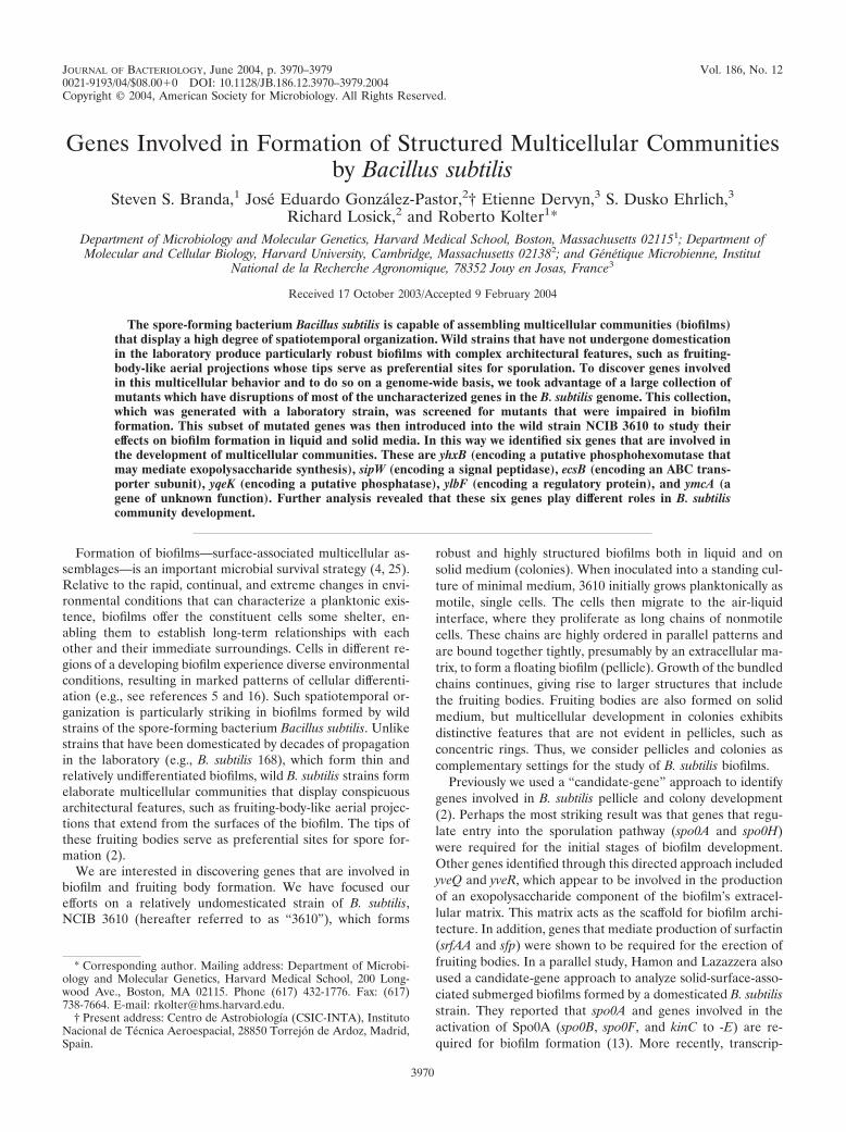
Identification of genes involved in the development of mul-ticellular communities. Genes in the BFA mutant collectionwere individually disrupted by single-reciprocal (Campbell-like) recombination using the insertional vector pMUTIN (36,39). Each mutant was inoculated into standing cultures ofminimal medium and incubated at 30°C for 60 h. Under theseconditions, the parent strain (B. subtilis 168) formed a wrinkledpellicle (see Fig. 1, top left panel). Ninety-four of the 2,105mutants tested produced a pellicle of unusual morphology orno pellicle at all. Seventy of the 94 mutants exhibited plank-tonic growth rates in minimal medium similar to that of 168,indicating that their defects in pellicle formation were not dueto a general growth defect. To determine whether their defectsin pellicle formation were due to pMUTIN insertion, chromo-somal DNA was prepared from each of the 70 mutants andused to transform competent cells of the parental strain; afterselection for the antibiotic resistance gene carried by the vec-tor, the resulting transformants were tested for their ability toform pellicles. Twenty-four of these mutants showed defects inpellicle formation similar to those exhibited by their corre-sponding BFA mutants, while the other 46 showed defects thatwere less severe and/or reproducible than originally observed.Presumably the latter mutants were derived from BFA mutantsthat harbored secondary mutations not linked to their pMUTINinsertions; indeed, others reported that 16 of these BFA mu-tants behaved differently than the reference strain when testedunder a variety of conditions and that these mutant phenotypestypically did not segregate with the pMUTIN insertion in ge-netic backcrosses (http://genome.jouy.inra.fr/micado). The sec-ondary mutations responsible for these phenotypes may haveresulted from the use of nonisogenic versions of strain 168 inthe construction of the BFA mutant collection; however, the 46BFA mutants in question were derived from several differentisolates of 168, which suggests that their pellicle formationdefects may be caused by different secondary mutations.
With regard to the 24 insertional mutations that did conferupon strain 168 defects in pellicle formation, we wonderedwhether they might also affect the ability of wild strains toproduce robust multicellular communities. To investigate thispossibility, each of the mutations was introduced (by phagePBS1-mediated transduction) into strain 3610, and their effectson pellicle formation and colony morphology were assessed.Of these 3610-derived mutants seven showed defects in pellicleformation, two showed defects in colony morphology, andeight showed both types of defects to a limited degree. Incontrast, mutations in seven genes—yhxB, sipW, yqxM, ecsB,yqeK, ylbF, and ymcA—caused a conspicuous defect in both
pellicle formation and colony morphology when introducedinto strain 3610 (Fig. 1 and data not shown). We chose tofurther characterize these seven genes and their roles in thedevelopment of multicellular communities.
Because the insertional mutations were generated by Camp-bell-like recombination, it was possible that they did notcompletely inactivate their target genes (14). To address thisconcern, we deleted the genes of interest from the 168 chro-mosome, replacing each with an antibiotic resistance gene, to
FIG. 1. BFA mutants defective in B. subtilis community develop-ment. Mutants from the BFA collection were isolated on the basis oftheir failure to form wild-type pellicles. The mutant alleles were thenreintroduced into parental strain 168 and also introduced into strain3610. To assay pellicle formation (first and second columns), eachmutant was inoculated at a low density into a standing culture consist-ing of 12 ml of MSgg plus MLS in a microtiter plate well, and thecultures were incubated at 30°C for 60 h without shaking. To assaycolony development (third column), each mutant derived from 3610was grown overnight in a rolled LB plus MLS culture, and a 3-�lsample of the culture was spotted onto MSgg agar and incubated at30°C for 96 h. Bars � 5 mm.
3972 BRANDA ET AL. J. BACTERIOL.
create bona fide null mutations. These null mutations, whichare designated by the symbol “�”, were introduced into 3610by phage transduction (see Materials and Methods). In thefollowing sections we describe the effects of these null muta-tions on pellicle formation and colony morphology in strain3610; whenever necessary we also describe the effects of thenull mutations in strain 168, as well as those of the insertionalmutations in strains 168 and 3610. Note that in all cases eachinsertional mutation and its corresponding null mutation con-ferred identical phenotypes when introduced into a commonparental strain (168 or 3610).
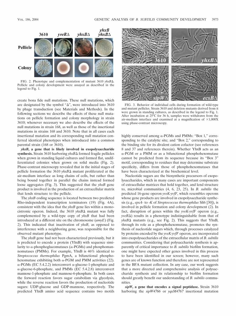
yhxB, a gene that is likely involved in exopolysaccharidesynthesis. Strain 3610 harboring yhxB� formed fragile pellicleswhen grown in standing liquid cultures and formed flat, undif-ferentiated colonies when grown on solid media (Fig. 2).Phase-contrast microscopy revealed that in the initial stages ofpellicle formation the 3610 yhxB� mutant proliferated at theair-medium interface as long chains of cells, but rather thanbeing bound together in parallel the chains instead formedloose aggregates (Fig. 3). This suggested that the yhxB geneproduct is involved in the production of an extracellular matrixthat lends structure to the pellicle.
The yhxB coding sequence is located between two predictedRho-independent transcription terminators (19) (Fig. 4A),consistent with the idea that the yhxB gene lies within a mono-cistronic operon. Indeed, the 3610 yhxB� mutant was fullycomplemented by a wild-type copy of yhxB that had beenintroduced at a different site on the chromosome (amyE) (Fig.2). This indicated that inactivation of yhxB, as opposed tointerference with a neighboring gene, was responsible for theobserved mutant phenotype.
The yhxB gene had not been characterized previously, but itis predicted to encode a protein (YhxB) with sequence simi-larity to �-phosphoglucomutases (�-PGMs) and phosphoman-nomutases (PMMs). For example, YhxB is 46% identical toStreptococcus thermophilus PgmA, a bifunctional phospho-hexomutase exhibiting both �-PGM and PMM activities (22).�-PGMs (EC 5.4.2.2) interconvert �-glucose-1-phosphate and�-glucose-6-phosphate, and PMMs (EC 5.4.2.8) interconvertmannose-1-phosphate and mannose-6-phosphate. In both casesthe forward reaction favors sugar catabolism via glycolysis,while the reverse reaction favors the production of nucleotidesugars: UDP-glucose and GDP-mannose, respectively. Thepredicted YhxB amino acid sequence contains two motifs
highly conserved among �-PGMs and PMMs: “Box 1,” corre-sponding to the catalytic site; and “Box 2,” corresponding tothe binding site for its divalent cation cofactor (see references8 and 37 and references therein). Whether YhxB acts as an�-PGM or a PMM or as a bifunctional phosphohexomutasecannot be predicted from its sequence because its “Box 3”motif, corresponding to residues that may determine substratespecificity, differs from those of phosphohexomutases thathave been characterized at the biochemical level.
Nucleotide sugars are the biosynthetic precursors of exopo-lysaccharides, which in many cases are important componentsof extracellular matrices that hold together, and lend structureto, microbial communities (4, 6, 23, 25). In B. subtilis thepredicted 16-gene operon yveK-yvfF, which resembles operonswhose gene products are involved in exopolysaccharide synthe-sis (e.g., epsA- to -K of Streptococcus thermophilus Sfi6 [30]), isinvolved in pellicle formation and colony development (2). Infact, disruption of genes within the yveK-yvfF operon (e.g.,yveR�) results in a phenotype indistinguishable from that ofyhxB� mutants (e.g., see Fig. 2). This suggests that YhxB,through its role as a phosphohexomutase, promotes the syn-thesis of nucleotide sugars which, through processes catalyzedby proteins encoded by the yveK-yvfF operon, are incorporatedinto exopolysaccharides of the extracellular matrix of B. subtiliscommunities. Considering that polysaccharide synthesis is ap-parently of critical importance to B. subtilis biofilm formation,one might have expected other genes involved in this processto have been identified in our screen; however, many suchgenes are of known function and therefore are not representedin the BFA mutant collection. In any case, our work suggeststhat a more directed and comprehensive analysis of polysac-charide synthesis and its relationship to biofilm formationshould greatly benefit our understanding of B. subtilis commu-nities.
sipW, a gene that encodes a signal peptidase. Strain 3610harboring the sipW4766 or yqxM4767 insertional mutation
FIG. 2. Phenotype and complementation of mutant 3610 yhxB�.Pellicle and colony development were assayed as described in thelegend to Fig. 1.
FIG. 3. Behavior of individual cells during formation of wild-typeand mutant pellicles. Strain 3610 and deletion mutants derived from itwere grown in standing cultures, as described in the legend to Fig. 1.After incubation at 25°C for 36 h, samples were withdrawn from theair-medium interface and examined at a magnification of �1,000Xusing phase-contrast microscopy.
VOL. 186, 2004 GENETIC ANALYSIS OF B. SUBTILIS COMMUNITY DEVELOPMENT 3973
formed pellicles of inconsistent thickness that failed to colo-nize all of the available surface area (Fig. 1 and data notshown). These mutant pellicles were composed of floating flocsthat only loosely interacted with one another, such that phys-ical manipulations which punctured or ripped other pelliclesonly parted the floating flocs. Phase-contrast microscopy re-vealed that the pellicles were composed of aggregates of rela-tively short chains of cells (data not shown); similar resultswere obtained using a deletion mutant (Fig. 3; see below). Thissuggested that the mutants failed to proliferate as long chainsof cells and also failed to produce an extracellular matrixcapable of holding the cells together in parallel alignment.Similarly, the mutants formed flat, undifferentiated colonies onsolid media (Fig. 1 and data not shown), consistent with theidea that they failed to produce a fully functional extracellularmatrix.
The yqxM and sipW genes are the first and second in athree-gene operon whose third member is tasA (31, 33) (Fig.4B). SipW is a type I signal peptidase that is specifically re-quired for the maturation and secretion of its two known en-
dogenous substrates: the protein products of yqxM and tasA(28, 32–34). Thus, there were a number of possible explana-tions for the fact that the yqxM4767 and sipW4766 mutantphenotypes were identical. For instance, the mutant pheno-types might result from: (i) lack of YqxM activity, due todisruption of yqxM or loss of processing by SipW; (ii) lack ofTasA activity, due to polar effects on tasA or loss of processingby SipW; (iii) accumulation of unprocessed TasA protein inthe cytoplasm, due to disruption of, or polar effects on, sipW;(iv) loss of the processing and activity of SipW substrates otherthan YqxM and TasA, due to disruption of, or polar effects on,sipW; or (v) a combination of these effects. To address the thirdpossibility, we deleted the entire yqxM-sipW-tasA operon in the3610 genetic background and found that the resulting mutant(yqxM-sipW-tasA�; referred to as “sipW�”) formed pelliclesand colonies that were indistinguishable from those formed bythe yqxM4767 and sipW4766 mutants (Fig. 3 and 5 and data notshown). These results effectively rule out the third explanationabove. With regard to the remaining explanations, it is inter-esting that Hamon and colleagues have found that sipW is theonly gene in the yqxM-sipW-tasA operon that is required forbiofilm formation by JH642 (13a), a B. subtilis strain derivedfrom 168 (3), which itself is thought to be derived from 3610(D. R. Zeigler, personal communication). This informationwould seem to support the fourth explanation—that loss ofpreprotein processing by SipW, but not lack of YqxM or TasAactivity, accounts for the mutant phenotypes observed—andwe are currently attempting to determine whether SipW playsa similar role in biofilm formation by 3610. In any case, ourresults suggest that SipW and at least some of its substrates arerequired for the structural integrity of the extracellular matrix.
ecsB, a gene encoding the transmembrane subunit of anABC transporter. Strain 3610 harboring ecsB� formed flatpellicles (Fig. 6). Phase-contrast microscopy revealed thatthese mutant pellicles were composed of long chains of cellsthat were bound together rather loosely (Fig. 3). The 3610ecsB� mutant also formed colonies in which certain architec-
FIG. 4. Physical maps of the genes disrupted in BFA mutants ofinterest. Coding regions are represented as horizontal arrows, a ca-nonical promoter element is shown as a small bent arrow, and canon-ical Rho-independent transcriptional terminators are shown as stem-loop symbols. Each insertional mutation is represented by an invertedtriangle bearing the number of its corresponding BFA mutant. Chro-mosomal regions that were deleted and replaced by antibiotic resis-tance markers are represented as horizontal bars—grey if the deletionconferred a defect in community development, checkered if it didn’t.
FIG. 5. Phenotype of mutant 3610 yqxM-sipW-tasA� (sipW�). Pel-licle and colony development were assayed as described in the legendto Fig. 1. The parental strain images (first column) are the same as inFig. 2.
3974 BRANDA ET AL. J. BACTERIOL.

tural features were absent or less prominent than those ob-served in wild-type colonies (Fig. 6).
The ecsB gene is the second in a three-gene operon, ecsABC(20, 21) (Fig. 4C). Thus, the ecsB� mutant allele could poten-tially have polar effects on ecsC. However, deletion of ecsCfrom the 3610 genetic background resulted in a mutant thatformed pellicles and colonies that were indistinguishable fromthose formed by the parental strain (Fig. 6). This suggests thatinactivation of ecsB, and not polar effects on ecsC, was respon-sible for the mutant phenotype observed. However, at this timewe cannot rule out the possibility that it was the combinationof ecsB inactivation and polar effects on ecsC that gave rise tothe mutant phenotype.
The ecsA gene encodes the ATPase subunit and ecsB en-codes the transmembrane subunit of an ABC transporter re-ferred to as Ecs (15, 21). The ecsC gene encodes a protein thatshows no significant sequence similarity to any previously de-scribed ABC transporter component or accessory protein orindeed to any protein of known function. Ecs has been shownto promote the Sec-dependent secretion of several degradativeenzymes, primarily through facilitation of their processing bysignal peptidases (18, 20, 26). Both EcsA and EcsB are re-quired for this function, whereas EcsC is not (18, 20). EcsAand EcsB are also required for efficient sporulation and devel-opment of competence (18 and data not shown), but it is notknown whether this is due to the role of Ecs in protein secre-tion.
Our results indicate that Ecs activity is important for thedevelopment of B. subtilis communities. This effect is likelyindirect; for example, Ecs could promote the maturation andsecretion of protein components of the extracellular matrixand in this way contribute to the structure of pellicles andcolonies. An interesting possibility is that some of the Ecssubstrates might also be SipW substrates. It is conceivable thatEcs and SipW work together to promote the secretion of pro-teins that affect B. subtilis community structure. Indeed, Ecshas been shown to facilitate the processing of a heterologouslyexpressed �-amylase by a different type I signal peptidase (26).Identification of the endogenous substrates of Ecs and SipWshould lead to a better understanding of their roles in B. subtiliscommunity development.
yqeK, a gene that encodes a predicted phosphatase. Strain3610 harboring yqeK� formed pellicles that were initially thinand flat; however, these pellicles eventually became muchthicker, and their flat surfaces were broken by a number ofbroad pits, the inner surfaces of which were covered withfruiting bodies (Fig. 7). This heterogeneity in morphology didnot appear to be due to accumulation of suppressor mutations,as cells harvested from the invaginations and inoculated intofresh standing cultures produced pellicles that were indistin-guishable from their predecessors (data not shown). Phase-contrast microscopy revealed that the mutant pellicles, likethose formed by the parental strain, were composed of longchains of cells bound together in parallel (data not shown). Thecolonies formed by 3610 yqeK� also showed an obvious defectin gross morphology, in that their central regions were flat andglossy (Fig. 7).
The yqeK gene is located within a cluster of eight genes(yqeG to yqeM) that could constitute an operon (19) (Fig. 4D).Thus, the mutant phenotype conferred by yqeK� could poten-tially result from polar effects on downstream genes. However,deletion of the gene located immediately downstream of yqeK(yqeL) from the 3610 genetic background resulted in a mutantthat formed wild-type pellicles and colonies (Fig. 7). This sug-gests that inactivation of yqeK, and not polar effects on down-stream genes, accounts for the defects in community develop-ment exhibited by the 3610 yqeK� mutant.
The yqeG-yqeM gene cluster is well conserved among gram-positive bacterial species. For instance, clusters in which alleight genes are present and in the same order can be found inthe genomes of Staphylococcus aureus MW2 (genes MW1549 toMW1542), Listeria innocua CLIP (genes lin1527 to lin1520),and Oceanobacillus iheyensis HTE831 (genes OB1989 toOB1982). Similar yqeK-containing clusters can be found in thegenomes of six additional gram-positive bacteria, though inthese cases some of the other genes are missing. Aside fromyqeK itself, the only other gene present in all of the 11 clustersanalyzed is nadD. This conservation of physical proximity sug-gests a possible functional relationship between yqeK andnadD, an idea consistent with the fact that the coding regionsof these genes partially overlap in eight of the clusters, includ-ing that of B. subtilis.
The yqeK gene has not been characterized previously, but it
FIG. 6. Phenotypes of mutants 3610 ecsB� and 3610 ecsC�. Pellicleand colony development were assayed as described in the legend toFig. 1. The parental strain images (first column) are the same as in Fig. 2.
FIG. 7. Phenotypes of mutants 3610 yqeK� and 3610 yqeL�. Pelli-cle and colony development were assayed as described in the legend toFig. 1.
VOL. 186, 2004 GENETIC ANALYSIS OF B. SUBTILIS COMMUNITY DEVELOPMENT 3975

is predicted to encode a protein that contains an HD sequencemotif characteristic of metal-dependent phosphatases (1). TheHD phosphatases that have been analyzed at the biochemicallevel are involved in a wide variety of biological processes.Thus, membership in this family reveals little about the bio-logical function of YqeK. Clues regarding the function of yqeKmay come from its physical, and perhaps functional, link tonadD, which encodes the nicotinic acid mononucleotide ad-enylyltransferase (NaMN-AT; EC 2.7.7.18) of B. subtilis (24).NaMN-ATs catalyze the penultimate step in the biosynthesisof NAD (NAD�), the essential cofactor that participates in amultitude of redox reactions. It will be of interest to determinewhether YqeK plays a role in NAD� metabolism, and if so,how abrogation of this function leads to the observed effect onB. subtilis community development.
ylbF, a gene that encodes a positive regulator of competencefactor ComK. Strain 168 harboring the ylbF3233 insertionalmutation failed to form pellicles (Fig. 1). However, when theylbF3233 allele was introduced into the 3610 strain the result-ing mutant formed robust pellicles that featured large, glossywrinkles as well as some flat regions (Fig. 1). Similarly, strain168 harboring ylbF� did not form pellicles (data not shown),whereas 3610 ylbF� formed pellicles that were heterogeneousin morphology (Fig. 8). When samples from 3610 ylbF� pel-licles were streaked to single cells on selective solid mediumand the resulting colonies were used to inoculate standingcultures, each isolate produced pellicles that were homoge-neous in morphology, though that morphology differed fromisolate to isolate (data not shown). These results strongly sug-gested that the 3610 ylbF mutants accumulated suppressormutations that enabled them to form pellicles. Indeed, we havefound that such mutations map to a single genetic locus (D. B.Kearns, F. Chu, S. S. Branda, R. Losick, and R. Kolter, un-published results). It is not yet clear whether similar mutationsaccumulated in the 168 ylbF mutants and, if so, why thesestrains failed to form pellicles. Phase-contrast microscopy re-vealed that the pellicles produced by the 3610 ylbF mutants
were composed of long chains of cells aligned in parallel (datanot shown) and in this respect were indistinguishable fromthose produced by the parental strain (Fig. 3). The coloniesformed by the 3610 ylbF mutants were essentially flat andundifferentiated (Fig. 1 and 8).
The ylbF gene appears to lie within a two-gene operon,ylbFG, that is flanked by two predicted Rho-independent tran-scription terminators (19) (Fig. 4E). Thus, the mutant pheno-type conferred by ylbF� could potentially result from polareffects on ylbG. However, the 3610 ylbF� mutant was fullycomplemented by a wild-type ylbF allele integrated at theamyE locus (Fig. 8 and data not shown). This indicated thatinactivation of ylbF, and not polar effects on ylbG or otherdownstream genes, accounted for the observed mutant pheno-type.
The ylbF gene encodes a protein that promotes the transla-tion and/or stability of ComK (35), a transcription factor thatplays a critical role in the induction of natural competence(12). It seemed possible that YlbF might work in conjunctionwith ComK to coordinate B. subtilis community development.However, deletion of comK from the 3610 genetic backgroundresulted in a mutant that formed wild-type pellicles and colo-nies (Fig. 8). Moreover, a 3610 ylbF� comK� double mutantformed pellicles and colonies that were indistinguishable fromthose formed by 3610 ylbF� (Fig. 8), indicating that the mutantphenotype conferred by ylbF� was not due to misregulation ofComK. Therefore, in contrast to its role in the development ofcompetence, YlbF controls the development of B. subtilis com-munities via a pathway that does not include ComK. It hasbeen suggested that in competence development, YlbF maypromote ComK stability indirectly, through a positive effect onComS, a protein that prevents MecA from targeting ComK fordegradation by the ClpC/ClpP protease (35). This raises thepossibility that YlbF could affect community development via apathway that includes ComS and/or MecA but not ComK orcompetence factors acting upstream of ComS (i.e., ComP andComA), which we and others have shown are not required for
FIG. 8. Phenotypes of mutants 3610 ylbF�, 3610 comK�, and 3610 ylbF� comK� and complementation of 3610 ylbF�. Pellicle and colonydevelopment were assayed as described in the legend to Fig. 1. The parental strain images (first column) are the same as in Fig. 2.
3976 BRANDA ET AL. J. BACTERIOL.

biofilm formation (13 and unpublished results). Alternatively,YlbF may control community development through a pathwaythat is entirely separate from that governing competence. Itshould be noted that YlbF also plays a role in sporulation,though the mechanism of that action remains unknown (35).We anticipate that the means by which YlbF affects B. subtiliscommunity development will be elucidated through identifica-tion of genes whose expression depends upon YlbF and ofproteins which interact with YlbF, as well as through furtherstudy of the suppression of 3610 ylbF mutants.
ymcA, a gene of unknown function. Strain 168 harboringymcA2603 or ymcA� failed to form pellicles (Fig. 1 and datanot shown), whereas introduction of either of these mutationsinto strain 3610 resulted in mutants that formed robust pel-licles of heterogeneous morphology (Fig. 1 and 9). Single cellsisolated from 3610 ymcA� pellicles and grown in standingcultures produced pellicles that were homogeneous in mor-phology, though that morphology differed from isolate to iso-late (data not shown). These results are consistent with theidea that, like 3610 ylbF mutants, the 3610 ymcA mutantsaccumulated suppressor mutations that enabled them to formpellicles. In fact, we have found that such mutations map to thegenetic locus involved in suppression of the 3610 ylbF mutants(Kearns et al., unpublished). Whether comparable mutationsaccumulated in the 168 ymcA mutants remains to be deter-mined. Phase-contrast microscopy revealed that the 3610 ymcAmutant pellicles were composed of long chains of cells alignedin parallel (data not shown). The 3610 ymcA mutants alsoproduced flat, undifferentiated colonies (Fig. 1 and 9). Thus,the ymcA mutants exhibited a complex phenotype essentiallyidentical to that of the ylbF mutants. Importantly, we foundthat a 3610 ylbF� ymcA� double mutant formed pellicles andcolonies that were indistinguishable from those formed by thesingle mutants (Fig. 9). These results suggest that ylbF andymcA act through a common pathway to control the develop-ment of B. subtilis communities.
The ymcA gene appears to lie within a two-gene operon,
ymcBA: canonical �35 and �10 promoter elements are lo-cated upstream of ymcB, and a predicted Rho-independenttranscription terminator lies 7 bp downstream of ymcA (19)(Fig. 4F). Moreover, the gene immediately downstream ofymcA (cotE) is regulated by E (7), which we and others haveshown is not required for biofilm formation (2, 13). Therefore,the ymcA� mutant phenotype almost certainly results frominactivation of ymcA rather than polar effects on downstreamgenes.
The ymcA gene had not been characterized previously, but itis predicted to encode a protein whose sequence resemblesthat of YlbF (19% identity and 51% similarity). However,programs that are designed to group proteins on the basis ofshared domains (COG and ProDom) place YlbF and YmcA inseparate ortholog clusters. This implies that sequence similar-ities between YlbF and YmcA are not concentrated in regionsthat are predicted to be important for protein function. Toexamine this issue more closely, we aligned the amino acidsequences of YlbF and YmcA with those of their respectiveorthologs (48 to 67% identical, with E values of 1 � 10�29 to8 � 10�40) from Bacillus cereus, Bacillus anthracis, and Bacillushalodurans. We then determined whether residues that areshared among all four orthologs are also shared between B.subtilis YlbF and YmcA. We found that of the 149 residues inB. subtilis YlbF, 58 (39%) are conserved in its three orthologs,and of those 58 residues only 10 (17%) are identical to theircorresponding residues in YmcA. Similarly, of the 143 residuesin B. subtilis YmcA, 69 (48%) are conserved in its orthologs,but only 12 (17%) of these are identical to their correspondingresidues in YlbF. Furthermore, the alignments of YlbF andYmcA orthologs revealed that certain regions of these proteinshave been conserved particularly well over the course of evo-lution (e.g., 17 of 21 residues, starting with L16, in YmcA) andthe conserved regions of YlbF do not correspond to those ofYmcA. Thus, despite a fair degree of similarity in amino acidsequence, YlbF and YmcA appear to have different domainstructures and therefore are unlikely to be paralogs.
FIG. 9. Phenotypes of mutants 3610 ymcA� and 3610 ymcA� ylbF�. Pellicle and colony development were assayed as described in the legendto Fig. 1. The parental strain images (first column) are the same as in Fig. 2, and the 3610 ylbF� images (third column) are the same as in Fig. 8.
VOL. 186, 2004 GENETIC ANALYSIS OF B. SUBTILIS COMMUNITY DEVELOPMENT 3977

Aside from YlbF, no other proteins of known or predictedfunction show sequence similarity to YmcA. The gene (ymcB)predicted to share an operon with ymcA encodes a protein thatexhibits strong sequence similarity (44% identity, with an Evalue of 1 � 10�103) to MiaB from Escherichia coli and Sal-monella enterica serovar Typhimurium. MiaB mediates a post-transcriptional modification of a nucleoside residue withintRNAs that read codons with U in the first position (9). Gram-negative bacteria apparently lack ymcA, but in gram-positivebacteria ymcA is often preceded by a gene encoding an MiaBortholog. This physical proximity of the genes could reflect afunctional relationship. This potential relationship, as well asthat between YmcA and YlbF, represents a framework forfurther study of YmcA and its role in B. subtilis communitydevelopment.
In summary, using an unbiased, genome-wide approach weidentified six genes, some of them previously uncharacterized,that play important roles in the development of B. subtiliscommunities. Several of the genes appear to mediate produc-tion of an extracellular matrix that binds together the cells ofthese communities. As discussed above, yhxB likely works withthe yveK-yvfF operon to synthesize exopolysaccharides that areincorporated into the matrix, while sipW and ecsB may pro-mote the secretion of proteins that assemble, modify, or lendstructure to the matrix. Both yhxB and sipW are required forthe basic structural integrity of B. subtilis communities,whereas ecsB is required for formation of their distinctivearchitecture. A fourth gene, yqeK, is predicted to encode aphosphatase, and it too is required for proper formation ofcommunity architecture, though not for production of the ex-tracellular matrix. Finally, ylbF and ymcA appear to work to-gether to regulate community development; whether they af-fect production of the extracellular matrix remains to bedetermined. Thus, the six genes appear to play different rolesin B. subtilis community development.
Considering that the collection screened was composed ofmutants representing only 51% of the genome, it is likely thatmore comprehensive screens will identify additional genes in-volved in community development. Furthermore, screens uti-lizing less domesticated strains such as 3610 should identify stillmore genes, given the observation that mutations can conferdifferent phenotypes when moved from one genetic back-ground to another (Fig. 1). Equally important to our under-standing of structured multicellular communities is the obser-vation that phenotypes varied widely depending on whetherpellicles or colonies were analyzed. This indicates that underdifferent conditions B. subtilis uses different developmentalpathways to form communities. The different pathways doshare some genes, however, as mutation of at least six geneshad profound effects on both pellicles and colonies, as de-scribed above. The relationships between these pathways andgenes, and their implications for community development, willbe the subject of future study.
ACKNOWLEDGMENTS
We thank the BFA consortium for access to their mutant collection,as well as the Ehrlich lab for generous support during the screening ofthe collection. We also thank K. Carniol and A. Driks for strains, B.Lazazzera for communication of unpublished results, J. Shuler and theNikon Imaging Center (Harvard Medical School) for microscopy,
D. Kearns for critical reading of the manuscript, and the Kolter andLosick labs for useful discussions.
This work was supported by National Institutes of Health grantsGM18568 (to R.L.) and GM58213 (to R.K.), and by National ScienceFoundation grant MCB-9727234 (to R.L.). S.S.B. was supported by anAmerican Cancer Society Postdoctoral Fellowship. J.E.G.-P. was sup-ported by a Ministerio de Educacion y Ciencia Postdoctoral Fellow-ship (Spain).
REFERENCES
1. Aravind, L., and E. V. Koonin. 1998. The HD domain defines a new super-family of metal-dependent phosphohydrolases. Trends Biol. Sci. 23:469–472.
2. Branda, S. S., J. E. Gonzalez-Pastor, S. Ben-Yehuda, R. Losick, and R.Kolter. 2001. Fruiting body formation by Bacillus subtilis. Proc. Natl. Acad.Sci. USA 98:11621–11626.
3. Brehm, S. P., F. Le Hegarat, and J. A. Hoch. 1974. Developmental modu-lation of deoxyribonucleic acid-binding proteins of Bacillus subtilis duringsporulation stages. J. Bacteriol. 120:1443–1450.
4. Davey, M. E., and G. A. O’Toole. 2000. Microbial biofilms: from ecology tomolecular genetics. Microbiol. Mol. Biol. Rev. 64:847–867.
5. de Kievit, T. R., R. Gillis, S. Marx, C. Brown, and B. H. Iglewski. 2001.Quorum-sensing genes in Pseudomonas aeruginosa biofilms: their role andexpression patterns. Appl. Environ. Microbiol. 67:1865–1873.
6. de Vuyst, L., and B. Degeest. 1999. Heteropolysaccharides from lactic acidbacteria. FEMS Microbiol. Rev. 23:153–177.
7. Driks, A. 1999. Bacillus subtilis spore coat. Microbiol. Mol. Biol. Rev. 63:1–20.
8. Dwivedi, K., A. F. Post, and S. Bullerjahn. 1996. Cloning and functionalanalysis of the pmmA gene encoding phosphomannomutase from the pho-tosynthetic prokaryote Prochlorothrix hollandica. Biochim. Biophys. Acta1290:210–214.
9. Esberg, B., H.-C. E. Leung, H.-C. T. Tsui, G. R. Bjork, and M. E. Winkler.1999. Identification of the miaB gene, involved in methylthiolation of iso-pentenylated A37 derivatives in the tRNA of Salmonella typhimurium andEscherichia coli. J. Bacteriol. 181:7256–7265.
10. Freese, E., J. E. Heinze, and E. M. Galliers. 1979. Partial purine deprivationcauses sporulation of Bacillus subtilis in the presence of excess ammonia,glucose, and phosphate. J. Gen. Microbiol. 115:193–205.
11. Guerout-Fleury, A.-M., N. Frandsen, and P. Stragier. 1996. Plasmids forectopic integration in Bacillus subtilis. Gene 180:57–61.
12. Hahn, J., A. Luttinger, and D. Dubnau. 1996. Regulatory inputs for thesynthesis of ComK, the competence transcription factor of Bacillus subtilis.Mol. Microbiol. 21:763–775.
13. Hamon, M. A., and B. Lazazzera. 2001. The sporulation transcription factorSpo0A is required for biofilm development in Bacillus subtilis. Mol. Micro-biol. 42:1199–1209.
13a.Hamon, M. A., N. R. Stanley, R. A. Britton, A. D. Grossman, and B. A.Lazazzera. 2004. Identification of Abr-B-regulated genes involved in biofilmformation by Bacillus subtilis. Mol. Microbiol. 52:847–860.
14. Harwood, C. R., and S. M. Cutting. 1990. Molecular biological methods forBacillus. Wiley-Interscience, Chichester, United Kingdom.
15. Higgins, C. F. 2001. ABC transporters: physiology, structure, and mecha-nism—an overview. Res. Microbiol. 152:205–210.
16. Huang, C. T., K. D. Xu, G. A. McFeters, and P. S. Stewart. 1998. Spatialpatterns of alkaline phosphatase expression within bacterial colonies andbiofilms in response to phosphate starvation. Appl. Environ. Microbiol.64:1526–1531.
17. Kearns, D. B., and R. Losick. 2003. Swarming motility in undomesticatedBacillus subtilis. Mol. Microbiol. 49:581–590.
18. Kontinen, V. P., and M. Sarvas. 1988. Mutants of Bacillus subtilis defectivein protein export. J. Gen. Microbiol. 134:2333–2344.
19. Kunst, F., N. Ogasawara, I. Moszer, A. M. Albertini, G. Alloni, V. Azevedo,M. G. Bertero, P. Bessieres, A. Bolotin, S. Borchert, R. Borriss, L. Boursier,A. Brans, M. Braun, S. C. Brignell, S. Bron, S. Brouillet, C. V. Bruschi, B.Caldwell, V. Capuano, N. M. Carter, S.-K. Choi, J.-J. Codani, I. F. Conner-ton, N. J. Cummings, R. A. Daniel, F. Denizot, K. M. Devine, A. Dusterhoft,S. D. Ehrlich, P. T. Emmerson, K. D. Entian, J. Errington, C. Fabret, E.Ferrari, D. Foulger, C. Fritz, M. Fujita, Y. Fujita, S. Fuma, A. Galizzi, N.Galleron, S.-Y. Ghim, P. Glaser, A. Goffeau, E. J. Golightly, G. Grandi, G.Guiseppi, B. J. Guy, K. Haga, J. Haiech, C. R. Harwood, A. Henaut, H.Hilbert, S. Holsappel, S. Hosono, M.-F. Hullo, M. Itaya, L. Jones, B. Joris,D. Karamata, Y. Kasahara, M. Klaerr-Blanchard, C. Klein, Y. Kobayashi, P.Koetter, G. Koningstein, S. Krogh, M. Kumano, K. Kurita, A. Lapidus, S.Lardinois, J. Lauber, V. Lazarevic, S.-M. Lee, A. Levine, H. Liu, S. Masuda,C. Mauel, C. Medigue, N. Medina, R. P. Mellado, M. Mizuno, D. Moestl, S.Nakai, M. Noback, D. Noone, M. O’Reilly, K. Ogawa, A. Ogiwara, B.Oudega, S.-H. Park, V. Parro, T. M. Pohl, D. Portetelle, S. Porwollik, A. M.Prescott, E. Presecan, P. Pujic, B. Purnelle, G. Rapoport, M Rey, S. Reyn-olds, M. Rieger, C. Rivolta, E. Rocha, B. Roche, M. Rose, Y. Sadaie, T. Sato,E. Scanlan, S. Schleich, R. Schroeter, F. Scoffone, J. Sekiguchi, A. Sekowska,S. J. Seror, P. Serror, B.-S. Shin, B. Soldo, A. Sorokin, E. Tacconi, T. Takagi,
3978 BRANDA ET AL. J. BACTERIOL.

H. Takahashi, K. Takemaru, M. Takeuchi, A. Tamakoshi, T. Tanaka, P.Terpstra, A. Tognoni, V. Tosato, S. Uchiyama, M. Vandenbol, F. Vannier, A.Vassarotti, A. Viari, R. Wambutt, E. Wedler, H. Wedler, T. Weitzenegger, P.Winters, A. Wipat, H. Yamamoto, K. Yamane, K. Yasumoto, K. Yata, K.Yoshida, H.-F. Yoshikawa, E. Zumstein, H. Yoshikawa, and A. Danchin.1997. The complete genome sequence of the gram-positive bacterium Ba-cillus subtilis. Nature 390:249–256.
20. Leskela, S., E. Wahlstrom, H.-L. Hyyrylainen, M. Jacobs, A. Palva, M.Sarvas, and V. P. Kontinen. 1999. Ecs, an ABC transporter of Bacillussubtilis: dual signal transduction functions affecting expression of secretedproteins as well as their secretion. Mol. Microbiol. 31:533–543.
21. Leskela, S., V. P. Kontinen, and M. Sarvas. 1996. Molecular analysis of anoperon in Bacillus subtilis encoding a novel ABC transporter with a role inexoprotein production, sporulation, and competence. Microbiology 142:71–77.
22. Levander, F., and P. Radstrom. 2001. Requirement for phosphoglucomutasein exopolysaccharide biosynthesis in glucose- and lactose-utilizing Strepto-coccus thermophilus. Appl. Environ. Microbiol. 67:2734–2738.
23. Mack, D. 1999. Molecular mechanisms of Staphylococcus epidermidis biofilmformation. J. Hosp. Infect. 43:S113–S125.
24. Olland, A. M., K. W. Underwood, R. M. Czerwinski, M.-C. Lo, A. Aulabaugh,J. Bard, M. L. Stahl, W. S. Somers, F. X. Sullivan, and R. Chopra. 2002.Identification, characterization, and crystal structure of Bacillus subtilis nic-otinic acid mononucleotide adenylyltransferase. J. Biol. Chem. 277:3698–3707.
25. O’Toole, G. A., H. B. Kaplan, and R. Kolter. 2000. Biofilm formation asmicrobial development. Annu. Rev. Microbiol. 54:49–79.
26. Pummi, T., S. Leskela, E. Wahlstrom, U. Gerth, H. Tjalsma, M. Hecker, M.Sarvas, and V. P. Kontinen. 2002. ClpXP protease regulates the signalpeptide cleavage of secretory preproteins in Bacillus subtilis with a mecha-nism distinct from that of the Ecs ABC transporter. J. Bacteriol. 184:1010–1018.
27. Sambrook, J., and D. W. Russell. 2001. Molecular cloning: a laboratorymanual. Cold Spring Harbor Laboratory Press, Cold Spring Harbor, N.Y.
28. Serrano, M., R. Zilhao, E. Ricca, A. J. Ozin, C. P. Moran, Jr., and A. O.Henriques. 1999. A Bacillus subtilis secreted protein with a role in endosporecoat assembly and function. J. Bacteriol. 181:3632–3643.
29. Stanley, N. R., R. A. Britton, A. D. Grossman, and B. Lazazzera. 2003.Identification of catabolite repression as a physiological regulator of biofilmformation by Bacillus subtilis by use of DNA microarrays. J. Bacteriol. 185:1951–1957.
30. Stingele, F., J.-R. Neeser, and B. Mollet. 1996. Identification and character-ization of the eps (exopolysaccharide) gene cluster from Streptococcus ther-mophilus Sfi6. J. Bacteriol. 178:1680–1690.
31. Stover, A., and A. Driks. 1999. Control of synthesis and secretion of theBacillus subtilis protein YqxM. J. Bacteriol. 181:7065–7069.
32. Stover, A., and A. Driks. 1999. Regulation of synthesis of the Bacillus subtilistransition-phase, spore-associated antibacterial protein TasA. J. Bacteriol.181:5476–5481.
33. Stover, A., and A. Driks. 1999. Secretion, localization, and antibacterialactivity of TasA, a Bacillus subtilis spore-associated protein. J. Bacteriol.181:1664–1672.
34. Tjalsma, H., A. Stover, A. Driks, G. Venema, S. Bron, and J. M. van Dijl.2000. Conserved serine and histidine residues are critical for activity of theER-type signal peptidase SipW of Bacillus subtilis. J. Biol. Chem. 275:25102–25108.
35. Tortosa, P., M. Albano, and D. Dubnau. 2000. Characterization of ylbF, anew gene involved in competence development and sporulation in Bacillussubtilis. Mol. Microbiol. 35:1110–1119.
36. Vagner, V., E. Dervyn, and S. D. Ehrlich. 1998. A vector for systematic geneinactivation in Bacillus subtilis. Microbiology 144:3097–3104.
37. Videira, P. A., L. L. Cortes, A. M. Fialho, and I. Sa-Correia. 2000. Identifi-cation of the pgmG gene, encoding a bifunctional protein with phosphoglu-comutase and phosphomannomutase activities, in the gellan gum-producingstrain Sphingomonas paucimobilis ATCC 31461. Appl. Environ. Microbiol.66:2252–2258.
38. Wach, A. 1996. PCR-synthesis of marker cassettes with long flanking homol-ogy regions for gene disruptions in S. cerevisiae. Yeast 12:259–265.
39. Yoshida, K., I. Ishio, E. Nagakawa, Y. Yamamoto, M. Yamamoto, and Y.Fujita. 2000. Systematic study of gene expression and transcription organi-zation in the gntZ-ywaA region of the Bacillus subtilis genome. Microbiology146:573–579.
VOL. 186, 2004 GENETIC ANALYSIS OF B. SUBTILIS COMMUNITY DEVELOPMENT 3979