generation of dc from mouse spleen cell cultures in
TRANSCRIPT
Immunology 199584 127-l34
Generation of DC from mouse spleen cell cultures in response to GM-CSF: immunophenotypic and functional analyses
L. LU,* M. HSIEH,* T. B. ORISS,t P. A. MOREL,t T. E. STARZL* A. S. RAO~ & A. w. THOMSON§ Pittsburgh Transplantation Institute and * Departments of Surgery, t Medicine, ~Pathology and §M olecular Genetics and
Biochemistry, University of Pittsburgh, Pittsburgh, Pennsylvania, USA
SUMMARY
In all tissues that have been studied to date, dendritic leucocytes constitute only a small proportion of total cells and are difficult both to isolate .and purify. This study reports on a method for the propagation of large numbers of dendritic cells (DC) from mouse spleen using granulocytemacrophage colony-stimulating factor (GM-CSF) and their characteristics. Within a few days of liquid culture in GM-CSF, BIO BR (H-2\ I-E+) mouse splenocytes formed loosely adherent myeloid cell clusters. Mononuclear progeny released from these clusters at and beyond 4 days exhibited distinct dendritic morphology and strongly expressed leucocyte common antigen (CD45), CD II b, heat-stable antigen, Pgp-I (CD44) and intercellular adhesion molecule-I (ICAM-1: CD54). The intensity of expression of the DC-restricted markers NLDC 145 and 33DI, the macrophage marker F4/80, and FcyRII (CDw32) was low to moderate, whereas the cells were negative for CD3, CD45RA and NKl.l. High and moderate levels, respectively, of cell surface staining for major histocompatibility complex (MHC) class II (I_Ek) and the B7 antigens (counterreceptors of CTLA4, a structural homologue of CD28) were associated with potent stimulation of unprimed, allogeneic T cells (BIO; H_2b, I-E-). DC propagated in a similar fashion from DBA/2 mouse spleen proved to be strong antigen-presenting cells (APC) for MHC-restricted, syngeneic Thelper type 2 (Th2) cell clones specifically responsive to sperm whale myoglobin. Footpad or intravenous injection of GM-CSF-stimulated BIO.BR spleen-derived DC into BIO (H_2b, I-E-) recipients resulted in homing of the allogeneic cells to T -cell-dependent areas of lymph nodes and spleen, where they strongly expressed donor MHC class II antigen 1-2 days later. These findings indicate that cells can be propagated from fresh splenocyte suspensions that exhibit distinctive features of DC. namely morphology, motility, cell-surface phenotype, potent allogeneic and syngeneic APC function and in vivo homing ability. Propagation of DC in this manner from progenitors present in lymphoid tissue provides an alternative and relatively convenient source of high numbers of these otherwise difficult to isolate but functionally important APC.
INTRODUCTION
Dendritic cells (DC) were first isolated from mouse lymphoid tissues during studies on the function of accessory cells in immune responses. l They constitute a minor population of bone marrow-derived leucocytes that is distributed ubiquitously throughout the body. In contrast to macrophages, from which they differ on a number of morphological, phenotypic,
Received 28 April 1994; revised 21 August 1994; accepted 31 August 1994.
Abbreviations: APC. antigen-presenting cells; DC, dendritic cell; GM-CSF, granulocyte-macrophage colony-stimulating factor; MHC, major histocompatibility complex.
Correspondence: Dr A. W. Thomson, Pittsburgh Transplantation Institute, University of Pittsburgh Medical Center, W1544 Biomedical Science Tower, 200 Lothrop Street, Pittsburgh, PA 152l3-2582, USA.
127
enzymatic and behavioural criteria,2-4 mature DC constitutively express high levels of cell-surface major histocompatibility complex (MHC) class II molecules and are the most potent initiators ofT -lymphocyte responses, both in vitro and in vivo.2- 4 DC within secondary lymphoid tissues are present as interdigitating cells in T-cell areas (white pulp of spleen and paracortex of lymph nodes) and in the marginal zone of the spleen.s These cells represent the major MHC class II-positive stimulatory cell type within the spleen. The identity of critical costimulatory signals for resting T cells expressed by mature DC has, however, remained elusive. Recent evidence, based on studies of murine epidermal Langerhans' cells, suggests that upregulation of cell-surface expression of CD80 (formerly termed B7 or BBI), the counter-receptor for the T-cell activation molecule CD28, may correlate with the functional maturation of DC.6
Studies on the properties and functional maturation of DC

128 L. Lu et al.
have been restricted by the rarity of cells that meet criteria for classification as DC « 1 % of unfractionated spleen cells) and by the failure of such cells to survive beyond a few days or to grow in culture. Recently, however, DC progenitors have been propagated in response to granulocyte-macrophage colonystimulating factor (GM-CSF) from precursors present in mouse bone marrow? and blood.s In addition, we have shown that DC lineage cells can be induced to proliferate in response to this cytokine from progenitors present in mouse liver.9 In this study, we report on the propagation of DC from GM-CSF-stimulated progenitors present in unfractionated spleen cell suspensions. We have characterized these DC in terms of their phenotype and function, with particular regard to their role as inducers of T-cell activation.
MATERIALS AND METHODS
Animals Adult 8-l2-week-old male BlO.BR (H_2k, I-E+), C57BLI lOSnJ (BlO, H_2b, I-E-) and DBA/2 (H_2d) mice were purchased from the Jackson Laboratory (Bar Harbor, ME). They were maintained in the specific pathogen-free facility of the University of Pittsburgh Medical Center.
Isolation and propagation of spleen cells Fresh splenocytes were obtained by teasing the spleens under aseptic conditions; single-cell suspensions were prepared using standard procedures. Erythrocytes were removed by osmotic lysis. The nucleated cells were washed twice (5 min at 400g) in RPMI-1640 (Gibco BRL, Grand Island, NY) supplemented with glutamine (2 DlM/ml), non-essential amino acids (0'1 mMI ml), sodium pyruvate (1 mM/ml), 2-mercaptoethanol (20,LlM), antibiotics (penicillin, 100 U Iml; streptomycin, 100 mg/ml) (complete medium) and 5% heat-activated fetal bovine serum (HIFBS; Gibco). To test the capacity of the spleen cells to proliferate in response to GM-CSF, 1-2 x 10⁶ cells were placed in each well of a 24-well plate in 1 ml of complete medium, supplemented with 10% HIFBS and 0'4ng/ml mouse recombinant (r) GM-CSF (R&D Systems, Minneapolis, MN). The cultures were 'fed' with GM -CSF every 48 hr by aspirating 50% of the supernatant, after gentle swirling, and replenishing with an equivalent volume of fresh, GM-CSF-supplemented medium. An objective of these washes was to remove nonadherent granulocytes without dislodging clusters of developing DC that had attached loosely to firmly adherent macrophages.? After 4 days, granulocytes were no longer significant contaminants of the cultures. These were maintained routinely for up to 10 days, at which time non-adherent cells released from growing clusters were harvested. In this manner, approximately 1·5 x 106 DC/spleen were obtained by day 10.
Flow cytometric analysis GM-CSF-stimulated spleen cells were washed in complete medium (2 x 5 min each), then resuspended (5 x lOS/tube) in Hanks' balanced salt solution (HBSS) with 1 % w/v bovine serum albumin (BSA; Sigma, St Louis, MO) and 0'1 % sodium azide (Sigma). They were stained either by direct or indirect immunofluorescence using a panel of monoclonal antibodies (mAb). T lymphocytes were identified using phycoerythrin (pE)-conjugated hamster anti-mouse CD3-e (pharMingen, San Diego, CA). For the detection of B cells, rat anti-mouse B220
(CD45RA; TIB146; ATCC, Rockville, MD) was used, followed by FITC-conjugated AffiniPure goat anti-rat IgG (Jackson Immunoresearch Labs Inc., West Grove, PA). Antileucocyte common antigen (CD45; TIB122; ATCC), antimacrophage antibody (F4/80; HB198; ATCC), anti-heat-stable antigen (JllD; TIB183; ATCC) and antibodies directed against DC-restricted markers (NLDC-145, derived from Kraal et al.,10 and 33DI; TIB227; ATCC; kindly provided by Dr R. M. Steinman, Rockefeller University, New York, NY) were employed to characterize further the lineages of the isolated cells. The presence of natural killer (NK) cells was revealed using antibody against NKl.1 (Dr W. H. Chambers, Department of Pathology, University of Pittsburgh). The expression of accessory molecules was identified using antibodies against the interleukin-2 (IL-2) receptor (CD25; p55, PC6l 5.3; TIB222; ATCC), intercellular adhesion molecule-l (ICAM-l; CD54; Serotec, Indianapolis, IN), Pgp-l glycoprotein (CD44; TIB235; ATCC), CDIlb (MAC-loe unit, Ml/70; TIB128; ATCC) and FcyRII (CDw32; PharMingen). Appropriate FITC-conjugated anti-mouse, anti-rat or anti-hamster secondary antibodies were used. Normal hamster serum or the appropriate rat immunoglobulin isotypes were used as negative controls. Biotin-conjugated mouse anti-mouse I_Ek,d,p,r (PharMingen) was used with FITC-steptavidin (Jackson Immunoresearch Labs) as the secondary reagent. Biotinconjugated mouse IgG2a, together with FITC-streptavidin, was used as a negative control.
To identify counter-receptors of CTLA4 (a structural homologue of CD28), which include CD80 (B7-l) and CD86 (B7-2), the cells were first incubated with either human immunoglobulin (Sigma) as a negative control or with the CTLA4-immunoglobulin fusion protein (2'5 Itg/ml; kindly provided by Dr P. S. Linsley, Bristol Myers Squibb Pharmaceutical Research Institute, Seattle, W A), which contains the extracellular portion of human CTLA4 and a human IgGyl chain. ll They were washed and incubated with FITC-conjugated mouse IgG2a anti-human IgG (Fc; Sigma). Lipopolysaccharide (LPS)-activated splenic B-cell blasts, which express B7/BBl,6 served as positive controls. After staining, the cells were fixed in 1 % v/v paraformaldehyde before analysis in a FACSTAR@ flow cytometer (Becton Dickinson, San Jose, CA). Five thousand events were acquired for each sample.
Immunocytochemistry Cytocentrifuge preparations were stained either directly or indirectly using avidin-biotin-peroxidase complex (ABC)staining procedures. Briefly, specimens were air-dried at room temperature, before fixing in acetone for 5 min. The slides were then washed in phosphate-buffered saline (PBS) and incubated for 1 hr at room temperature with biotinylated mouse IgG2a, anti-mouse I_Ek,d,p,r mAb. For indirect staining, primary rat anti-CD45, anti-NLDC145 or anti-2Al (a gift from Dr R. M. Steinman) mAb were used (1 hr at room temperature) followed by biotinylated mouse IgG F(ab')z anti-rat IgG (Accurate Chemical and Scientific Corporation, Westbury, NY), for 1 hr at room temperature. After three washes of 5 min each in PBS, the slides were incubated with streptavidin-biotin-peroxidase complex (ABC-P; Boehringer Mannheim Corp., Indianapolis, IN) for 30 min at room temperature, and the colour reaction was developed for 6 min using a peroxidase chromogen kit

Generat ion of DC from mouse spleen 129
-It' ""-a ~ ~ • :.. ,
t· ~ ~. -.... ..
r ... 9
w • If ~ , ~
r , .. -oJ •
i~'7: .' . "'tIL~ , r· l-~~' \' _
_ 'N,-.$. I.l.:..,~:c~'
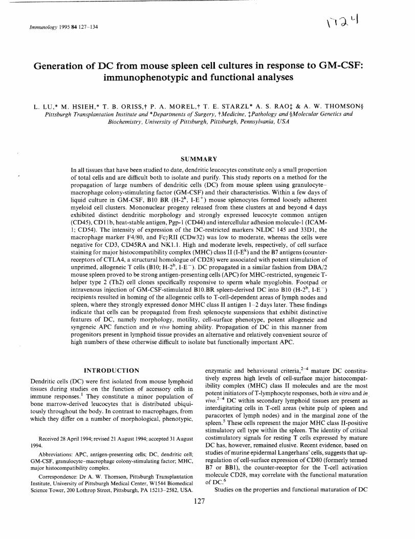
Figure 1. Phase-contrast micrographs illustrating the development and isolation of normal mouse spleen-derived DC in liquid cultures supplemented with GM-CSF. (a) An early aggregate of proliferating, putative DC progenitors (day 4) attached to strongly adherent macrophages and showing typical, loosely adherent cells (arrows) that were released from the aggregates. At 2 and 4 days, nonadherent granulocytes were removed by gentle aspiration of the supernatant and replaced with fresh, GM-CSF-supplemented medium. Magnification, x 100. (b) Higher power view of typical, non-adherent cells with cytoplasmic processes released from aggregates and harvested after 10 days of culture. Magnification, x 400.
Figure 2. Giemsa-stained cytocentrifuge preparation showing cells released from GM-CSF-stimulatcd splccn ccl! aggrcgates (day 10), which exhibit irregular-shaped eccentric nuclei, variable degrees of cytoplasmic vacuolation, absence of prominent granules and distinct cytoplasmic processes. Magnification, x 600. Inset, immunoperoxidasestained preparation, showing a high level of MHC class II (I_Ek) antigen expression on dendritic-shaped cells (also day 10). Magnification, x 600.
(AEC; Biomedia Corp., Foster City, CA). Cells were counterstained lightly with haematoxylin. Controls included the omission of antibody and the use of isotype-matched irrelevant mAb.
Mixed leucocyte cultures To test the immunogenicity of freshly isolated or cultured spleen cells, one-way mixed leucocyte cultures (4 x 10⁵ cells in 200 fll/well) in 96-well, round-bottomed microculture plates (Falcon®; Becton Dickinson Labware, Lincoln Park, NJ) were performed with variable numbers of y-irradiated (20 Gy) allogeneic (BIO.BR) or syngeneic (BIO) spleen-derived cells as stimulators. BIO spleen cells were used as responders, which were T-cell enriched by passage (1 hr at 37°) through a nylon wool column. In some experiments, designed to examine the functional expression ofB7 antigens, CTLA4-immunoglobulin fusion protein (2'5 flg/ml) or human immunoglobulin was added at the start of the cultures. The cells were maintained in complete medium supplemented with 10% HIFBS for
(a) (b) (e)
Figure 3. Immunoperoxidase labelling of cultured cells harvested after 10 days to show additional phenotypic features of released DC progeny. The primary mAb were (a) an li-CD45 (leucocyte common antigen; ATCC; TIBI22), (b) NLDC-145 anti-interdigitating cell, 10 ancl (c) 2AI, which reacts primarily with granules within the cytoplasm of cultured DC in vitro 8 Magnification, x 600.

130 L. Lu et al.
72hr in 5% CO2 in air; for the finalI8hr, lOpl eHjthymidine (eHjTdR; 1 pCi) was added to each well. Cultures were harvested onto glass fibre disks using a multiple cell harvester, and the degree of thymidine incorporation was determined in a liquid scintillation counter. Results were expressed as means (c.p.m.) ± 1 SD.
Stimulation of syngeneic Th2 cell clone The sperm whale myoglobin (SpWMb)-specific T-cell clone, 13.26, was used in proliferation assays with varying numbers of spleen-derived DBA/2 cells in the presence and absence of specific antigen. The T-cell clone 13.26 has been described previouslyl2 and is specific for a SpWMb peptide spanning amino acids 132-147. To test the ability offreshly isolated or cultured spleen cells to stimulate proliferation of the clone, 5 x I04T cells/well were cultured with varying numbers of spleen-derived cells in the presence or absence of Sp WMb peptide 132-147 (20 pg/ml) in 200 pi of medium in 96-well flatbottomed microtitre plates. The cells were incubated for 48 hr at 37°, pulsed with 0'5pCi eHjTdR for an additional 24hr, harvested and counted in a p-scintillation counter.
DC homing GM-CSF-stimulated BlO.BR spleen cells were washed in RPMI-I640 and injected subcutaneously (s.c.) (lor 2·5 x 105
cells in 50 pi) into one hind footpad, or intravenously (i.v.) (1 x 10⁶ in 200 pi) via the lateral tail vein, of normal allogeneic BlO mice. One and 2 days later, the draining popliteal lymph nodes (where appropriate) and spleen were removed and embedded in Tissue-Tek® (OCT Compound; Miles Inc., Elkhart, IN) before freezing at - 70°. Sections (10 Jlffi) were cut using a cryostat microtome at - 30° and melted directly onto slides at room temperature, then air-dried. After brief acetone fixation, immunoperoxidase staining was performed, as described above. Controls included sections of normal recipient strain (BlO) tissues.
RESULTS
Proliferation of spleen cells in response to GM-SCF
Since it has been reported recently that large numbers of DC can be induced to proliferate from mouse blood or bone marrow when cultured with GM_CSF,7,8 we determined whether, using a similar approach, spleen-derived DC could be generated in liquid culture. After 4 days culture of freshly isolated splenocytes, during which time non-adherent granulocytes and mature DC were removed by gentle washes, growth of cell 'clusters' attached to a layer of adherent cells was evident (Fig. Ia); many low buoyant density, dendritic-shaped cells appeared to have been released from the clusters and exhibited cytoplasmic processes (Fig. Ib). With more prolonged culture in GM-CSF, these dendritic-shaped, loosely attached cells continued to detach from the clusters and float in the culture medium. In the absence ofGM-CSF, no clusters were observed and cells did not proliferate. Macrophages and fibroblasts also expanded in the GM-CSF-stimulated cultures, but remained attached firmly to the plastic surface. Floating or loosely adherent, putative DC were harvested by gentle aspiration for various analyses. Approximately 1·5 x 106 of these cells could be recovered consistently at 10 days from cultures derived from
a single spleen (0'7-1 x 10⁸ nucleated cells). This represented an increase of lO-fold over the number of mature DC/spleen that could be isolated in our laboratory using conventional procedures. I,l3
Microscopic and immunophenotypic analysis of GM-CSFstimulated spleen cells
At the microscopic level, most of the cells released from proliferating aggregates of GM-CSF-stimulated cells exhibited typical morphological features of DC, including (in many but not all cells) irregular shape, eccentric nuclei, numerous cytoplasmic dendrites, and absence of prominent cytoplasmic granules (Fig. 2). The cells with dendritic morphology were strongly MHC class 11+ (I_Ek +) (Fig. 2 inset), as described previously for cultured blood-derived DC,8 and expressed CD45 (leucocyte common antigen), the DC-restricted marker NLDC-145 (interdigitating cells) and the DC-restricted granule antigen 2Al (Fig. 3). In order to characterize further the surface phenotype of cells released from proliferating aggregates, flow cytometric analysis was performed after 7-10 days culture in GM-CSF. Staining for cells oflymphoid lineage, including NK cells, was absent. As shown in Fig. 4, the floating cells strongly expressed surface antigens that are known to be associated with mouse DC. These included CD45, heat-stable antigen (J1ID), ICAM-I (CD54), CDIlb (MAC-I) and CD44 (non-polymeric determinant of Pgp-I glycoprotein). In addition, staining of weak to moderate intensity was observed for the DC-restricted markers NLDC-145 and 33Dl and for F4/80 and FCl'RII. F ACS analysis also confirmed that the cells expressed a high intensity of Cell-surface MHC class II (I_Ek). Moreover, they stained for counter-receptors of CTLA4, a structural homologue of CD28 (Fig. 5).
Allostimulatory activity of GM-CSF -propagated spleen DC and the role of B7 antigens
The spleen-derived DC that expressed high levels of cell-surface MHC class II antigen and moderate levels of B7 antigens were potent inducers of 3-day, primary allogeneic T-cell responses (Fig. 6). They were much more effective than freshly isolated spleen cells, although less stimulatory than LPS-activated Bcell blasts at the cell concentrations used (Fig. 7). To examine the functional expression of B7 antigens and other CTLA4 counter-receptors, CTLA4-immunoglobulin fusion protein was added at the start of the cultures. Complete inhibition of the mixed lymphocyte reaction (MLR) induced by the spleenderived DC was observed (Fig. 7), indicating the crucial role of CTLA4 counter-receptors, including costimulatory B7 antigens, in the activation of unprimed T cells by the cultured DC.
Activation of syngeneic T-cell clones
The spleen-derived DC were also potent stimulators of antigenspecific T-cell activation. Significant proliferation of the SpWMb-specific Th2 clone 13.26 was observed with as few as 300 spleen-derived DC, with peak proliferation observed with 3000 stimulator cells/well (Fig. 8). In contrast, freshly isolated spleen cells were much less efficient.

Generation of DC from mouse spleen 131
Rat or hamster mAb
~ c Q) ::>
RatIgG /1
I iii iii
g Mouse mAb u:
NK1.1
.•• MAC·1
{\ NlDC-145/ i".
I
MouselgG
Class II
Fluorescence intensity
Figure 4. Merged FACSCAN@ immunophenotypic profiles of the GM-CSF-stimulated spleen-derived DC released from cell aggregates in liquid culture (day 10) and examined using rat, hamster or mouse mAb as detailed in the Materials and Meth9ds. All data were obtained from the same cell preparation at the same time. The result is representative of three separate experiments performed using cells obtained from 6-1O-day cultures.
In vivo homing of spleen-derived DC
A specialized property of DC is their capacity to 'home' to Tdependent areas of peripheral lymphoid tissues. 14,15 To determine the homing ability of the spleen DC propagated in culture, lO-day GM-CSF-stimulated cells were injected s.c. into one hind footpad or i.v. into allogeneic BlO (I-E-) recipients. One and 2 days later, the mice were killed and cryostat sections of the draining lymph nodes (where appropriate) and spleens
Mouse I .'" IgG '\ tr . j!=.rh .
,tI "1 . {F .: \ '. f
Human IgG
CTLA4-lg ~ Class II
Ir I ~ k I I-E
" , t~ \
\ \
101 102 103 Fluorescence intensity
Figure 5. Flow cytometric analysis of MHC class II (I_Ek) and CTLA4-immunoglobulin counter-receptor expression on GM-CSF-stimulatcd mouse spleen-derived DC released from proliferating aggregates in 10-day cultures. The result is representative of three separate experiments.
'" o x
E ci.
c..i
1000 -{]- Spleen DC
---0-- Spleen DC no antigen
---0- Spleen cells
100 --tr- Spleen cells no antigen
10
O'I+---~~~~--~~~~---+~~~
o 10 Stimulator cells/well (x 103 )
100
Figure 6. Allostimulatory activity of y-irradiatcd, GM-CSF-stimulated BIO.BR mouse spleen-derived DC using naive BIO (I-E-) splenic T cells as responders. The non-adherent cells were harvested from 1 O-day GMCSF -stimulated cultures and set up at various concentrations with 4 x 10⁵ responder T cells. Cultures were maintained for 72 hr; eH]TdR was added 18 hr before harvesting. The MLR-stimulatory activity of freshly isolated allogeneic (B 10.BR) and syngeneic (B I 0) spleen cells is also shown. The results are expressed as mean c.p.m. ± I SD and are representative of three separate experiments.

132 L. Lu et al.
50
• Cells only 40 o +Ig
'" o +CTLA4-lg
~ 30 x
E ci U 20
Media Fresh Bl0 BR LPS-stim BR spleen DC spleen cells B cells
Stimulator cells
Figure 7. Inhibition by CTLA4- immunoglobulin of spleen-derived DC-induced T-cell proliferation in an allogeneic MLR. Unprimed B 10 nylon wool-passed T cells (4 x 10⁵/we ll) were stimulated with 105
B I O.BR spleen-derived DC or Escherichia coli LPS-activated (10 J.lg/ml; 72hr) BIO.BR splenic B cells in the presence of 2·5J.lg/ml CTLA4-immunoglobulin or human immunoglobulin. Results are the means ± I SD of triplicate cultures. Similar results were obtained in three experiments.
were stained with donor-specific mAb to I-Ek As shown in Fig. 9, spleen-derived cells propagated in GM-CSF-supplemented cultures homed almost exclusively to T-cell areas of the recipients' spleens in close proximity to arterioles. Similar observations were made in the draining lymph nodes of footpad-injected mice (data not shown). Moderate to intense I_Ek expression was detected on the spleen-derived cells, many of which also exhibited distinct dendritic morphology. As previously reported for non-lymphoid organ DC,14- 17 the
150 ---0- BR spleen
-<>-- BR spleen DC
--0- B10 spleen
100
'" ~ x
E ci U
50 t
~ :::: -0---
0 10 100
Stimulator cells/well (x 10 3)
Figure 8. Spleen-derived DC efficiently present antigen to a SpWMbspecific Th2 clone, 13.26. The indicated numbers of stimulator cells were incubated with 5 x 10⁴ 13.26 cells in the presence (20 J.lg/ml) or absence of SpWMb peptide 132- 147. The cells were cultured for 72 hr and pulsed with 0·5 J.lCi/well eH]Td R for the last 24 hr. The results are expressed as the mean ± I SD of triplicate wells. A representative of three similar experiments is shown.
I
lal
Ibl
Figure 9. Homing ability of GM-CSF-stimulated BIO.BR spleenderived cells D C (I-E +) released in culture from proliferating cell aggregates (harvested on day 10). The cells (2·5 x 105) were injected s.c (day 0) into one hind footpad of BIO (I-E-) recipients and detec ted by immunohistochemistry in cryostat sections of spleen I or 2 days later. The spleen sections were stained using the ABC peroxidase procedure with donor-specific mouse anti-I-Ek mAb with appropriate controls. (a) Strongly MHC class II-positive cetls (one of which is arrowed) were detected in the periarteriolar lymphatic sheath. Magnification, x 45. (b) At higher magnification, cells with dendritic morphology were readily identified in the T-dependent areas in close proximity to arterioles; day 2. Magnification, x 400.
spleen DC propagated in culture also exhibited a key functional property of this cell lineage-the capacity to home to Tdependent areas of secondary lymphoid tissue and therein to express strong MHC class II cell-surface antigen.
DISCUSSION
Previous estimates of the frequency of the prototypic DC in mouse spleen have been about 0·1-0·3% of nucleated cells, with yields of approximately 2-5 x 10⁵jspleen I 8,19 Similar results are obtained whether the DC are sorted t8 or isolated by more traditional adherencej rosetting methods, 19 which deplete contaminating macrophages. Once isolated, spleen Dare exquisitely dependent on GM-CSF for survival beyond I day of culture. To our knowledge, growth of splenic DC has not been reported in response to GM-CSF, nor has viability been maintained in culture for more than a few days.18 Until recently, it has proved difficult to generate DC in cultures of

Generation of DC from mouse spleen 133
bone marrow,20,21 from which these cells are ultimately derived, and little is known about the properties of progenitors cells of DC in spleen and other tissues.
Within the last 2 years, however, GM-CSF-dependent proliferating DC progenitors that give rise to large numbers of typical, mature DC have been identified in mouse blood,8 bone marrow7 and liver.9 Initially, these studies centred on those murine tissues believed to be relatively deficient in mature DC (e.g. blood and bone marrow). More recently, propagation of DC progenitors for normal mouse liver9 has shown that tissues from which Ia + mature DC can be isolated using conventional procedures22 also contain Ia - progenitors of these cells. In the present study, the use of culture techniques which we have shown to facilitate the growth of DC progenitors from unfractionated, non-parenchymal cells in normal mouse liver,9 also promoted the growth and differentiation of DC progenitors present in fresh spleen cell suspensions. The identity of the cells was confirmed by their morphology, immunophenotypic characteristics, antigen-presenting function and in vivo homing ability, all of which conformed to that of 'classical' murine splenic DC.1-4
Flow cytometric analysis of the GM-CSF-stimulated spleen-derived DC revealed many surface molecules characteristically associated with mouse DC; including leucocyte common antigen (CD45), heat-stable antigen (JlID), ICAMI (CD54), CDllb (MAC-I) and CD44 (non-polymorphic determinant of Pgp-I glycoprotein). In addition, staining of moderate intensity was observed for the DC lineage-restricted markers NLDC-145 (interdigitating cells) and 33DI, for the DC-restricted granule antigen 2AI and for F4/S0 and FcyRiI. Similar observations for each of these markers have recently been made on DC released from proliferating aggregates in cultures of GM-CSF-stimulated mouse blood or bone marrow.7,8 In keeping with these latter findings on circulating or bone marrow DC progenitors, the spleen-derived, GM-CSFstimulated cells expressed very high levels of the MHC class II surface antigen molecule. The cultured splenic DC also expressed moderate intensity of counter-receptors/ligands (B7 antigens) for CTLA4 (the structural homologue of CD2S). Moreover, in blocking experiments, we found that CTLA4 counter-receptors on the cultured DC were required for proliferation of unprimed, allogeneic splenic T cells in primary mixed leucocyte reactions. These findings confirm recent observations of moderate cell-surface immunofluorescence staining and functional expression of B7/BBI (CDSO) on low density, transiently adherent DC-enriched populations isolated from mouse spleen cells suspensions6 using conventional procedures. 23
The intense cell-surface expression of MHC class II (I_Ek) and accompanying staining for B7 antigens was associated with potent antigen-presenting activity of the GM-CSF-stimulated spleen DC, both for allogeneic (I-E-) naive T cells and for antigen-driven proliferation of syngeneic (I_Ad) Th cell clones specifically responsive to sperm whale myoglobin. Recent findings suggest that up-regulation of the expression of B7 antigens on maturing mouse Langerhans' cells is of critical importance in the development of their capacity to activate naive T cells.6
A specialized property of DC is their ability to home to Tcell areas of peripheral lymphoid tissues.8,14,15.17,24 Donorderived DC expressing high levels of alloantigens have previously been identified within the spleens of unmodified
murine allograft recipients, 1-4 days after heart transplantation14 and up to 1 year after liver transplantation. 16 To test the homing capacity of the splenic DC (I-E+) produced in culture, cells were either injected into the footpad or i.v. into unmodified, allogeneic (I-E-) recipients. The precise homing of these MHC class 11+ DC progeny to T-cell areas of the spleen confirmed the lineage of the cells and, together with our in vitro observations, indicated the potential of the GM-CSFcultured cells to act as potent APC in vivo. In vitro, however, cytokines other than GM-CSF, such as tumour necrosis factor-IX (TNF-IX) and IL_425 may also affect their maturation and functional status.
Whilst mature or 'steady state' murine spleen DC (e.g. N4IS+ 'sorted' cells) fail to propagate in vitro in response to cytokines, it appears that GM-CSF-responsive progenitors present in bone marrow,7,26 blood,8liver9 and (as shown in this study) unfractionated suspensions of spleen cells, have the capacity for both macrophage and DC differentiation. Cytokine-driven propagation and differentiation of high numbers of viable DC progenitors from spleen-cell suspensions provides a convenient alternative to traditional labourintensive methods of DC enrichment/sorting where low yields/ viability and/or contamination can prove problematic. The method described also offers opportunities for further molecular analyses of the regulation of growth and differentiation of DC lineage cells, and of the mechanism of action of this cell lineage in the generation and regulation of adoptive immune responses.
ACKNOWLEDGMENTS
This work was supported by National Institute of Health Grants DK29961-14 and AI31427. We thank Dr P. S. Linsley for providing CTLA4-immunoglobulin fusion protein, Dr C. G. McAllister, Mr J. Campbell and Mr R. Lakomy (Pittsburgh Cancer Institute) for conducting the flow cytometric analyses and providing expert advice, Dr Y. Li and Dr V. M. Subbotiil for immunohistological analysis, Dr J. Woo and Dr A. J. Demetris for helpful discussion, and Ms Shelly Conklin for secretarial assistance.
REFERENCES
1. STEINMAN R.M. & COHN Z.A. (1973) Identification of a novel cell type in . peripheral lymphoid organs in mice. I. Morphology, quantitation, tissue distribution. J Exp Med 137, 1142.
2. STEINMAN R.M. (1991) The dendritic cell system and its role in immunogenicity. Annu Rev Immunol9, 271.
3. KNIGHT S.C. (1993) Dendritic cells. In: Clinical Aspects of Immunology (eds P.J. Lachmann, K. Peters, F.S. Rosen & M.J. Walport), Vol. I, 5th edn, p. 481. Blackwell Scientific Publications, Oxford.
4. AusTYN J.M. & WOOD K.J. (1993) Principles of Cellular and Molecular Immunology. Oxford University Press, Oxford.
5. METLAY J.P., WITMER-PACK M.D., AGGER R., CROWLEY M.T., LAWLESS D. & STEINMAN R.M. (1990) The distinct leucocyte integrins of mouse spleen dendritic cells as identified with new hamster monoclonal antibodies. J Exp Med 171,1753.
6. LARSEN C.P., RITCHIE S.C., PEARSON T.C., LINSLEY P.S. & LoWRY R.P. (1992) Functional expression of the costimulatory molecule, B7/BiH, on murine dendritic cell populations. J Exp Med 176, 1215.
7. INABA K., INABA M., ROMANI N. et al. (1992) Generation of large numbers of dendritic cells from mouse bone marrow cultures

134 L. Lu et al.
supplemented with granulocyte/macrophage colony-stimulating factor. J Exp Med 176, 1693.
8. INABA K., STEINMAN R.M., PACK M.W. et al. (1992) Identification of proliferating dendritic cell precursors in mouse blood . .J Exp Med 175, 1157.
9. Lu L., Woo I., RAo A.S. et al. (1994) Propagation of dendritic cell progenitors from normal mouse liver using GM-CSF and their maturational development in the presence of type-I collagen. J Exp MedI79,1823.
10. KRAAL G., BREEL M .. lANSE M. & BRUIN G. (1986) Langerhans cells. veiled cells and interdigitating cells in the mouse recognized by a monoclonal antibody. J Exp Med 163,981.
II. LINSLEY P.S., BRADY W., URNES M., GROSMAIRE L., DAMLE N. & LEDBETTER I.A. (1991) CTLA-4 is a second receptor for the B cell activation antigen B7. J Exp Med 174,561.
12. MOREL P.A, LIVINGSTONE A.M. & FATHMAN C.G. (1987) Correlation of T cell receptor V fJ gene family with MHC restriction. J Exp Med 166, 583.
13. STEINMAN R.M. & COHN Z.A. (1974) Identification of a novel cell type in the peripheral lymphoid organs of mice. II. Functional properties i/l vitro. J Exp Med 139, 380.
14. LARSE:-.i c.P., MORRIS P.I. & AUSTYN I.M. (1990) Migration of dendritic leukocytes from cardiac allografts into host spleens, a novel pathway for initiation of rejection. J Exp Med 171, 307.
15. LARSE:-.i c.P., STEINMAl'-i R.M., WITMER-PACK W., HANKIl'-iS 0.1"., MORRIS P.I. & AUSTYN I.M. (1990) Migration and maturation of Langerhans cells in skin transplants and explants. J Exp Med 172, 1483.
16. QIAN S., DlMETRIS A.1., MURASE N., RAo A.S., FUNG 1.1. & STARZL T.E. (1994) Murine liver allograft transplantation: tolerance and donor cell chimerism. Hepatology 19, 96.
17. THOMSON A.W., Lu L., SUBBOTIN V.M. et al. (1995) 1/1 vitro propagation and homing of liver-derived dendritic cell progenitors to lymphoid tissues of allogeneic recipients. Transplantation (in press).
18. CROWLEY M.T., INABA K., WITMER-PACK M.D., GEZELTER S. & STEINMAN R.M. (1990) Use of the fluorescence activated cell sorter
to enrich dendritic cells from mouse spleen. J Immunol Meth 133, 55.
19. STEINMAN R.M., KAPLAN G., WITMER M.D. & COHN Z.A. (1979) Identification of a novel cell-type in peripheral lymphoid organs of mice. V. Purification of spleen dendritic cells, new surface markers and maintenance in vitro. J Exp Med 149, I.
20. BOWERS W.E. & BERKOWITZ M.R. (1986) Differentiation of dendritic cells in cultures of rat bone marrow cells. J Exp Med 163,872.
21. REID C.D.L., FRYER P.R .. CLIFFORD c., KIRK A., TiKERPAE 1. & KNIGHT S.c. (1990) Identification of hematopoietic progenitors of macrophages and dendritic Langerhans cells (DL-CFU) in human bone marrow and peripheral blood. Blood 76, 1139.
22. Woo 1., Lu L., RAo AS., LI Y., SUBBOTIN V., STARZL T.E. & THOMSON A.W. (1994) Isolation, immunophenotype and allostimulatory activity of murine liver dendritic cells. Transplantation, 58,484.
23. STEIl'iMAN R.M., VAN VOORHIS w.c. & SPALDING D.M. (1986) Dendritic cells. In: Handbook of Experimental Immunology (eds D.M. Weir, L.A. Herzenberg, C. Blackwell & L.A. Herzenbcrg), 4th cdn, p. 49. Blackwell Scientific Publications, Oxford.
24. AusTYN .r.M., KUPIEC-WEGLINSKI M.W., HANKINS D.F. & MORRIS P.I. (1988) Migration pattern of dendritic cells in the mouse. Homing to T cell-dependent areas of spleen, and binding within marginal zone. J Exp Med 167, 646.
25. SALLUSTO F. & LANZAVECCHIA A. (1994) Efficient presentation of soluble antigen by cultured human dendritic cells is maintained by granulocyte/ macrophage colony-stimulating factor plus interleukin-4 and downregulated by tumor necrosis factor IX. J Exp Med 179, 1109.
26. REID C.D.L., STACKPOOLE A, MEAGER A & TIKERPAE I. (1992) Interactions of tumor necrosis factor with granulocytemacrophage colony-stimulating factor and other cytokines in the regulation of dendritic cell growth in vitro from early bipotent CD34 + progenitors in human bone marrow. J Immunol 149,2681.