general surgery - halyard health back to table of contents 1 table of contents general surgery 3...
TRANSCRIPT

Every day without costs you more.
General Surgery Techniques & Clinical Evidence
1h BACK TO TABLE OF CONTENTS
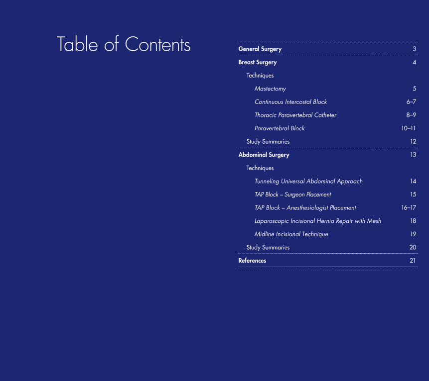
Table of Contents General Surgery 3
Breast Surgery 4
Techniques
Mastectomy 5
Continuous Intercostal Block 6–7
Thoracic Paravertebral Catheter 8–9
Paravertebral Block 10–11
Study Summaries 12
Abdominal Surgery 13
Techniques
Tunneling Universal Abdominal Approach 14
TAP Block – Surgeon Placement 15
TAP Block – Anesthesiologist Placement 16–17
Laparoscopic Incisional Hernia Repair with Mesh 18
Midline Incisional Technique 19
Study Summaries 20
References 21
2h BACK TO TABLE OF CONTENTS
DISCLAIMERS
The disclaimers contained herein pertain to all information included in this booklet. The information provided herein is provided for educational purposes and represents the surgical techniques used by specific doctors. Catheter placements are intended for guidance only and are subject to the individual expertise, experience and school-of-thought of the surgeon placing the catheter. Always refer to the drug manufacturer’s prescribing information when administering any drug with the ON-Q* Pain Relief System. This protocol is not to be construed as a specific recommendation of I-Flow*, LLC.
Cautions• Make sure the catheter is not in a vein or artery. Inadvertent intravascular delivery may result
in systemic toxic effects. Refer to the drug manufacturer’s package insert.
• Patient may experience loss of motor control or feeling at and around the surgical area. Physician should instruct patient on appropriate measures to follow to avoid patient injury.
• Medications used with this system should be administered in accordance with instructions provided by the drug manufacturer. Physician is responsible for prescribing drug based on each patient’s clinical status (e.g., age, body weight, disease state of patient, concomitant medication(s)).
• Vasoconstrictors such as epinephrine or adrenaline are not recommended for continuous infusions.
• Refer to ON-Q* Directions for Use for full instructions on using the ON-Q* Continuous Nerve Block System.
• Consult with surgeon prior to performing block with any surgery that would be prone to compartment syndromes.
Indications For Use• The ON-Q* pump is intended to provide continuous and/or intermittent delivery of
medication (such as local anesthetics or narcotics) to or around surgical wound sites and/or close proximity to nerves for preoperative, perioperative and postoperative regional anesthesia and/or pain management. Routes of administration include: intraoperative site, perineural, percutaneous, and epidural.
• ON-Q* is intended to significantly decrease pain and narcotic use when used to deliver local anesthetics to or around surgical wound sites, or close proximity to nerves, when compared to narcotic only pain management.
Contraindications• ON-Q* is not intended for blood, blood products, lipids, fat emulsions, or Total Parenteral
Nutrition (TPN).
• ON-Q* is not intended for intravascular delivery.
There are inherent risks in all medical devices. Please refer to the product labeling for Indications, Cautions, Warnings, and Contraindications. Failure to follow the product labeling could directly impact patient safety. Physician is responsible for prescribing and administering medications per instructions provided by the drug manufacturer. Refer to www.iflo.com for product safety Technical Bulletins.
3h BACK TO TABLE OF CONTENTS
Management of postoperative pain is a complex challenge facing healthcare professionals in daily clinical practice. Apfelbaum et al., surveyed 250 adults about their postoperative pain following surgery and reported approximately 80% of patients experienced significant pain after surgery. Of these patients, 86% had moderate, severe, or extreme pain and discomfort.1 Inadequate pain control alters the body’s metabolic response and is associated with increased morbidity, that may delay recovery, prolong hospital stays and lead to the development of persistent sensitization manifesting as chronic pain.1, 2
Effective surgical pain treatment involves a multimodal approach, which diminishes the intensity of acute pain. “Current theories propose that a prolonged experience of acute pain in which long-standing changes are seen within and external to the central nervous system (CNS) creates chronic pain with a histological and pathological basis.”2 This approach offers improved postoperative pain control while minimizing the side effects associated with any one therapeutic option.3, 4 In addition to reducing the incidence of chronic pain, effective pain control after surgery may accelerate recovery and facilitate rehabilitation.3, 5
In today’s healthcare environment there is also an increasing emphasis on patient satisfaction scores which influence Medicare reimbursement Hospital Consumer Assessment of Healthcare Providers and Systems (HCAHPS). This includes patient perception of pain.6 Postoperative pain management protocols that minimize adverse events and standardize treatment modalities have added benefits of reducing costs and promoting better patient outcomes.7, 8
The ON-Q* Pain Relief System is a non-narcotic constituent of a multimodal therapeutic approach and is clinically proven to deliver more effective pain management than traditional methods alone, with fewer side effects.9, 10, 11 ON-Q* automatically and continuously delivers local anesthetic via a catheter for targeted pain relief and can be placed by either the surgeon or the anesthesiologist.
General Surgery Techniques & Clinical Evidence
ON-Q* Patient Benefits
f Provides faster recovery – patients can ambulate and start rehabilitation sooner than patients given narcotics alone12
f Patients reported up to a 69% reduction in Visual Analog Scale (VAS) pain scores13, 14
f Increases tissue oxygenation – the improved oxygenation of the wound may enhance wound healing and may prevent postoperative infections15
f Patients reported high postoperative patient satisfaction scores across a wide range of surgical procedures.9, 12, 15, 16
4h BACK TO TABLE OF CONTENTS
Tonya Priestley ON-Q* Patient
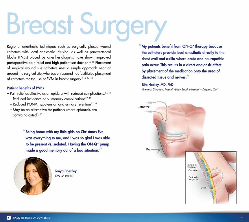
Regional anesthesia techniques such as surgically placed wound catheters with local anesthetic infusion, as well as paravertebral blocks (PVBs) placed by anesthesiologists, have shown improved postoperative pain relief and high patient satisfaction.5, 16 Placement of surgical wound site catheters uses a simple approach near or around the surgical site, whereas ultrasound has facilitated placement of catheters for the use of PVBs in breast surgery.3, 5, 14, 17
Patient Benefits of PVBs• Pain relief as effective as an epidural with reduced complications.17, 18
– Reduced incidence of pulmonary complications17, 19
– Reduced PONV, hypotension and urinary retention17, 18
– May be an alternative for patients where epidurals are contraindicated 5, 20
“ Being home with my little girls on Christmas Eve
was everything to me, and I was so glad I was able
to be present vs. sedated. Having the ON-Q* pump
made a good memory out of a bad situation.”
“ My patients benefit from ON-Q* therapy because
the catheters provide local anesthetic directly to the
chest wall and axilla where acute and neuropathic
pain occur. This results in a direct analgesic effect
by placement of the medication onto the area of
dissected tissue and nerves.”
Rita Hadley, MD, PhDGeneral Surgeon, Miami Valley South Hospital – Dayton, OH
Breast Surgery
Drain
Drain
Drain
Pectoralismajor m.
Pectoralismajor m.
Pectoralisminor m.
Catheter
Drain
Drain
Drain
Pectoralismajor m.
Pectoralismajor m.
Pectoralisminor m.
Catheter
Catheters
5h BACK TO TABLE OF CONTENTS
BREAST SURGERY
Drain
Drain
Drain
Pectoralismajor m.
Pectoralismajor m.
Pectoralisminor m.
Catheter
Drain
Drain
Drain
Pectoralismajor m.
Pectoralismajor m.
Pectoralisminor m.
Catheter
Drain
Drain
Drain
Pectoralismajor m.
Pectoralismajor m.
Pectoralisminor m.
Catheter
Illustrations by Ken X. Probst - 2014
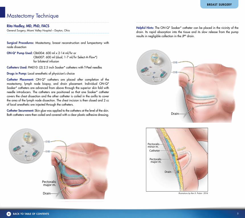
Mastectomy Technique
Rita Hadley, MD, PhD, FACSGeneral Surgery, Miami Valley Hospital – Dayton, Ohio
Surgical Procedures: Mastectomy, breast reconstruction and lumpectomy with node dissection
ON-Q* Pump Used: CB6004: 600 ml x 2-14 ml/hr or CB6007: 600 ml (dual; 1-7 ml/hr Select-A-Flow*) for bilateral infusion
Catheters Used: PM010: (2) 2.5 inch Soaker* catheters with T-Peel needles
Drugs in Pump: Local anesthetic of physician’s choice
Catheter Placement: ON-Q* catheters are placed after completion of the mastectomy, lymph node biopsy, and drain placement. Individual ON-Q* Soaker* catheters are advanced from above through the superior skin fold with needle introducers. The catheters are positioned so that one Soaker* catheter covers the chest dissection and the other catheter is coiled in the axilla to cover the area of the lymph node dissection. The chest incision is then closed and 2 cc of local anesthetic are injected through the catheters.
Catheter Securement: Skin glue was applied to the catheters at the level of the skin. Both catheters were then coiled and covered with a clear plastic adhesive dressing.
Helpful Hints: The ON-Q* Soaker* catheter can be placed in the vicinity of the drain. Its rapid absorption into the tissue and its slow release from the pump results in negligible collection in the JP® drain.

6h BACK TO TABLE OF CONTENTS
Continuous Intercostal Block Technique
Matthew Herren, DO Kurt Stockamp, MDAnesthesiology General Surgery Sacred Heart Hospital – Pensacola, FL
Surgical Procedure: Mastectomy, Breast Reconstruction, soft tissue surgery of the chest. The ON-Q SilverSoaker* catheters are placed to anesthetize T2-T7.
ON-Q* Pump Used: (1) CB6007 600 ml 1-7 ml/hr ON-Q* with Select-A-Flow* (dual site) or (2) CB004 400 ml 2-14 ml/hr ON-Q* with Select-A-Flow*
Tunneler Used: T17X8, 17 GA, 8-inch tunneler or T17X10, 17 GA, 10-inch tunneler
Catheter Used: ON-Q SilverSoaker* catheters 7.5 inch or 5 inch
Drugs in Pump: Local anesthetic of physician’s choice
Pre-incisional Infiltration: 1ml, 1% lidocaine, with or without epinephrine, injected subcutaneously at insertion site
Catheter Placement: Ask the patient to sit with chin to chest on the side of the bed, and let the thoracic area relax towards the practitioner. Locate the tip of scapula, which should be about T7, proceed caudally 3 cm or 3 finger-breadths, palpate for the most prominent rib in this area, and make a mark on the patient. Make sure you are at least 3 cm or 3 finger-breadths away from the spinous process to the mark. This will be your insertion point directly on top of the rib. Repeat process on opposite side if bilateral catheters are needed.
Prep area in standard sterile fashion. Inject 1% lidocaine with epinephrine at the insertion site subcutaneously. Insert a 14 G angiocath needle perpendicular to the rib, until needle tip and catheter are through the skin completely, then direct cephalad until fully inserted. Remove the needle, but leave the angiocath in until ready for insertion of 8 inch tunneler.
CAUTION: Take caution with insertion of needle and subsequent tunneler to avoid placing too deep, which could cause a pneumothorax or organ damage.
This will dilate the insertion point for the tunneler, and decrease the risk of bleeding or leakage versus a knife blade. Remove angiocath and insert tunneler toward rib until sheath pops through subcutaneous layer, then direct cephalad. You can palpate the tunneler as you advance over the ribs until you feel the tip starting to protrude superior to the scapula. As the tunneler moves over the ribs you can also feel some coarse friction against the tunneler. Remove the tunneler, and leave the introducer sheath. The base of the introducer sheath, wing tips, should be against the skin or at least very close.
BREAST SURGERY
3cm3cm
3cm 3cm
3cm3cmMidlineMidline
C7C7
T7 ribT7 rib
Needle Insertion
3cm3cm
3cm 3cm
3cm3cmMidlineMidline
C7C7
T7 ribT7 rib
Patient Positioning
3cm3cm
3cm 3cm
3cm3cmMidlineMidline
C7C7
T7 ribT7 rib
7h BACK TO TABLE OF CONTENTS
BREAST SURGERY
Continuous Intercostal Block Technique (Continued)
Illustrations by Ken X. Probst - 2014
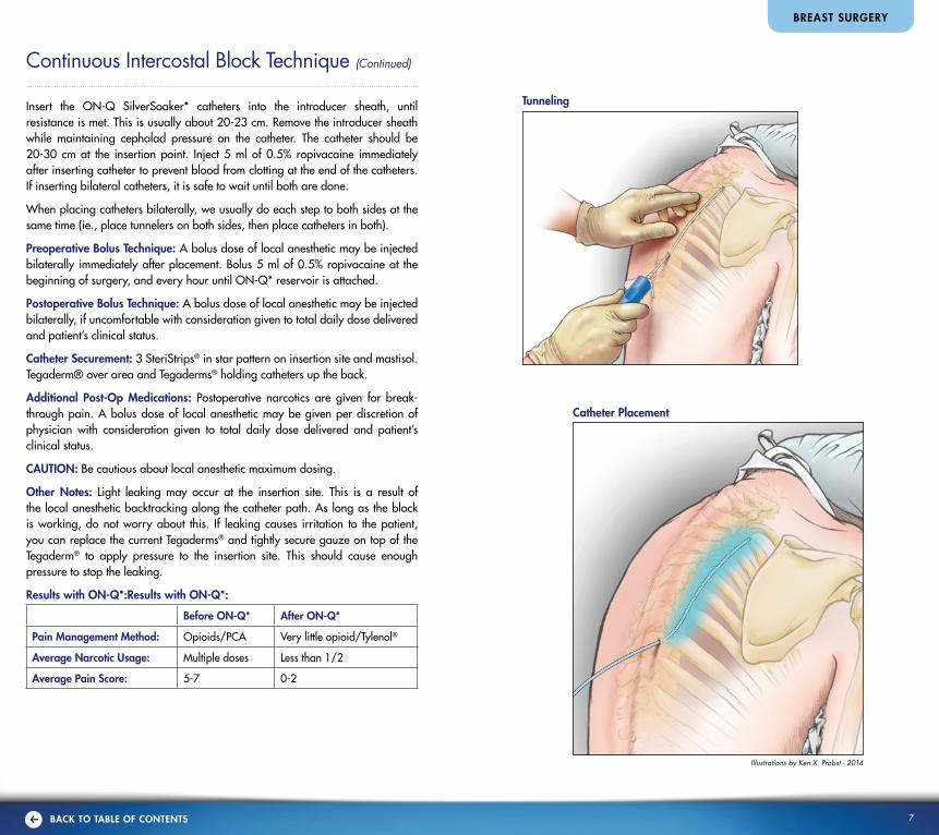
Tunneling
Catheter Placement
3cm3cm
3cm 3cm
3cm3cmMidlineMidline
C7C7
T7 ribT7 rib
3cm3cm
3cm 3cm
3cm3cmMidlineMidline
C7C7
T7 ribT7 rib
Insert the ON-Q SilverSoaker* catheters into the introducer sheath, until resistance is met. This is usually about 20-23 cm. Remove the introducer sheath while maintaining cephalad pressure on the catheter. The catheter should be 20-30 cm at the insertion point. Inject 5 ml of 0.5% ropivacaine immediately after inserting catheter to prevent blood from clotting at the end of the catheters. If inserting bilateral catheters, it is safe to wait until both are done.
When placing catheters bilaterally, we usually do each step to both sides at the same time (ie., place tunnelers on both sides, then place catheters in both).
Preoperative Bolus Technique: A bolus dose of local anesthetic may be injected bilaterally immediately after placement. Bolus 5 ml of 0.5% ropivacaine at the beginning of surgery, and every hour until ON-Q* reservoir is attached.
Postoperative Bolus Technique: A bolus dose of local anesthetic may be injected bilaterally, if uncomfortable with consideration given to total daily dose delivered and patient’s clinical status.
Catheter Securement: 3 SteriStrips® in star pattern on insertion site and mastisol. Tegaderm® over area and Tegaderms® holding catheters up the back.
Additional Post-Op Medications: Postoperative narcotics are given for break-through pain. A bolus dose of local anesthetic may be given per discretion of physician with consideration given to total daily dose delivered and patient’s clinical status.
CAUTION: Be cautious about local anesthetic maximum dosing.
Other Notes: Light leaking may occur at the insertion site. This is a result of the local anesthetic backtracking along the catheter path. As long as the block is working, do not worry about this. If leaking causes irritation to the patient, you can replace the current Tegaderms® and tightly secure gauze on top of the Tegaderm® to apply pressure to the insertion site. This should cause enough pressure to stop the leaking.
Results with ON-Q*:Results with ON-Q*:
Before ON-Q* After ON-Q*
Pain Management Method: Opioids/PCA Very little opioid/Tylenol®
Average Narcotic Usage: Multiple doses Less than 1/2
Average Pain Score: 5-7 0-2
8h BACK TO TABLE OF CONTENTS
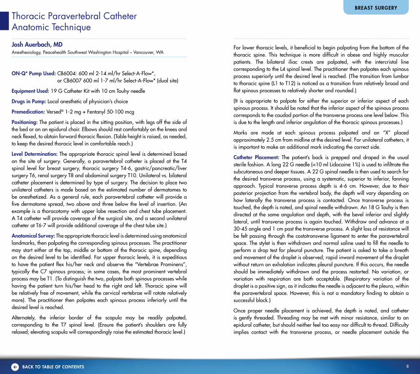
Thoracic Paravertebral Catheter Anatomic Technique
Josh Auerbach, MDAnesthesiology, Peacehealth Southwest Washington Hospital – Vancouver, WA
ON-Q* Pump Used: CB6004: 600 ml 2-14 ml/hr Select-A-Flow*, or CB6007 600 ml 1-7 ml/hr Select-A-Flow* (dual site)
Equipment Used: 19 G Catheter Kit with 10 cm Touhy needle
Drugs in Pump: Local anesthetic of physician’s choice
Premedication: Versed® 1-2 mg + Fentanyl 50-100 mcg
Positioning: The patient is placed in the sitting position, with legs off the side of the bed or on an epidural chair. Elbows should rest comfortably on the knees and neck flexed, to obtain forward thoracic flexion. (Table height is raised, as needed, to keep the desired thoracic level in comfortable reach.)
Level Determination: The appropriate thoracic spinal level is determined based on the site of surgery. Generally, a paravertebral catheter is placed at the T4 spinal level for breast surgery, thoracic surgery T4-6, gastric/pancreatic/liver surgery T6, renal surgery T8 and abdominal surgery T10. Unilateral vs. bilateral catheter placement is determined by type of surgery. The decision to place two unilateral catheters is made based on the estimated number of dermatomes to be anesthetized. As a general rule, each paravertebral catheter will provide a five dermatome spread, two above and three below the level of insertion. (An example is a thoracotomy with upper lobe resection and chest tube placement. A T4 catheter will provide coverage of the surgical site, and a second unilateral catheter at T6-7 will provide additional coverage of the chest tube site.)
Anatomical Survey: The appropriate thoracic level is determined using anatomical landmarks, then palpating the corresponding spinous processes. The practitioner may start either at the top, middle or bottom of the thoracic spine, depending on the desired level to be identified. For upper thoracic levels, it is expeditious to have the patient flex his/her neck and observe the “Vertebrae Prominens”, typically the C7 spinous process; in some cases, the most prominent vertebral process may be T1. (To distinguish the two, palpate both spinous processes while having the patient turn his/her head to the right and left. Thoracic spine will be relatively free of movement, while the cervical vertebrae will rotate relatively more). The practitioner then palpates each spinous process inferiorly until the desired level is reached.
Alternately, the inferior border of the scapula may be readily palpated, corresponding to the T7 spinal level. (Ensure the patient’s shoulders are fully relaxed; elevating scapula will correspondingly raise the estimated thoracic level.)
BREAST SURGERY
For lower thoracic levels, it beneficial to begin palpating from the bottom of the thoracic spine. This technique is more difficult in obese and highly muscular patients. The bilateral iliac crests are palpated, with the intercristal line corresponding to the L4 spinal level. The practitioner then palpates each spinous process superiorly until the desired level is reached. (The transition from lumbar to thoracic spine (L1 to T12) is noticed as a transition from relatively broad and flat spinous processes to relatively shorter and rounded.)
(It is appropriate to palpate for either the superior or inferior aspect of each spinous process. It should be noted that the inferior aspect of the spinous process corresponds to the caudad portion of the transverse process one level below. This is due to the length and inferior angulation of the thoracic spinous processes.)
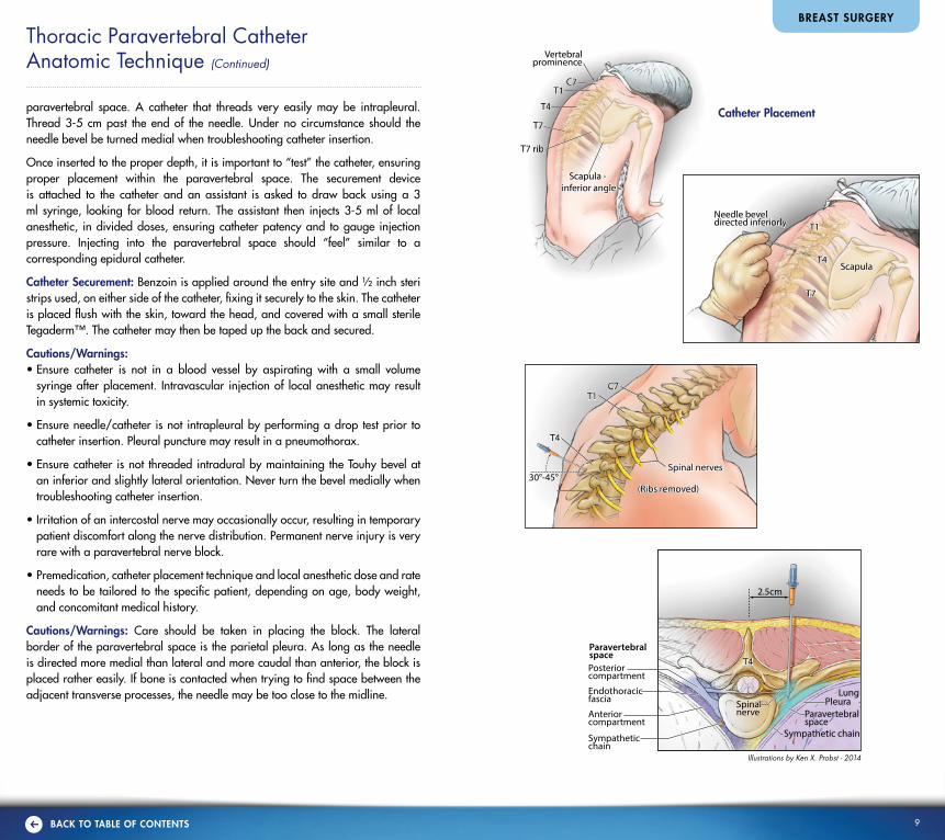
Marks are made at each spinous process palpated and an “X” placed approximately 2.5 cm from midline at the desired level. For unilateral catheters, it is important to make an additional mark indicating the correct side.
Catheter Placement: The patient’s back is prepped and draped in the usual sterile fashion. A long 22 G needle (<10 ml Lidocaine 1%) is used to infiltrate the subcutaneous and deeper tissues. A 22 G spinal needle is then used to search for the desired transverse process, using a systematic, superior to inferior, fanning approach. Typical transverse process depth is 4-6 cm. However, due to their posterior projection from the vertebral body, the depth will vary depending on how laterally the transverse process is contacted. Once transverse process is touched, the depth is noted, and spinal needle withdrawn. An 18 G Touhy is then directed at the same angulation and depth, with the bevel inferior and slightly lateral, until transverse process is again touched. Withdraw and advance at a 30-45 angle and 1 cm past the transverse process. A slight loss of resistance will be felt passing through the costotransverse ligament to enter the paravertebral space. The stylet is then withdrawn and normal saline used to fill the needle to perform a drop test for pleural puncture. The patient is asked to take a breath and movement of the droplet is observed; rapid inward movement of the droplet without return on exhalation indicates pleural puncture. If this occurs, the needle should be immediately withdrawn and the process restarted. No variation, or variation with respiration are both acceptable. (Respiratory variation of the droplet is a positive sign, as it indicates the needle is adjacent to the pleura, within the paravertebral space. However, this is not a mandatory finding to obtain a successful block.)
Once proper needle placement is achieved, the depth is noted, and catheter is gently threaded. Threading may be met with minor resistance, similar to an epidural catheter, but should neither feel too easy nor difficult to thread. Difficulty implies contact with the transverse process, or needle placement outside the
9h BACK TO TABLE OF CONTENTS
2.5cm
ParavertebralspaceParavertebralspace
T7T7
T1T1
T1T1
T4T4
T7T7
T7 ribT7 rib
Scapula - inferior angle
Scapula - inferior angle
Vertebralprominence
Vertebralprominence
C7C7
T4T4ScapulaScapula
C7C7T1T1
30°-45°30°-45°(Ribs removed)(Ribs removed)
Spinal nervesSpinal nerves
T4T4 T4T4
Needle beveldirected inferiorlyNeedle beveldirected inferiorly
Endothoracicfascia
ParavertebralspacePosteriorcompartment
Anteriorcompartment
Sympatheticchain
Sympathetic chainSympathetic chain
LungLungPleuraPleuraSpinal
nerveSpinalnerve
paravertebral space. A catheter that threads very easily may be intrapleural. Thread 3-5 cm past the end of the needle. Under no circumstance should the needle bevel be turned medial when troubleshooting catheter insertion.
Once inserted to the proper depth, it is important to “test” the catheter, ensuring proper placement within the paravertebral space. The securement device is attached to the catheter and an assistant is asked to draw back using a 3 ml syringe, looking for blood return. The assistant then injects 3-5 ml of local anesthetic, in divided doses, ensuring catheter patency and to gauge injection pressure. Injecting into the paravertebral space should “feel” similar to a corresponding epidural catheter.
Catheter Securement: Benzoin is applied around the entry site and ½ inch steri strips used, on either side of the catheter, fixing it securely to the skin. The catheter is placed flush with the skin, toward the head, and covered with a small sterile Tegaderm™. The catheter may then be taped up the back and secured.
Cautions/Warnings:• Ensure catheter is not in a blood vessel by aspirating with a small volume
syringe after placement. Intravascular injection of local anesthetic may result in systemic toxicity.
• Ensure needle/catheter is not intrapleural by performing a drop test prior to catheter insertion. Pleural puncture may result in a pneumothorax.
• Ensure catheter is not threaded intradural by maintaining the Touhy bevel at an inferior and slightly lateral orientation. Never turn the bevel medially when troubleshooting catheter insertion.
• Irritation of an intercostal nerve may occasionally occur, resulting in temporary patient discomfort along the nerve distribution. Permanent nerve injury is very rare with a paravertebral nerve block.
• Premedication, catheter placement technique and local anesthetic dose and rate needs to be tailored to the specific patient, depending on age, body weight, and concomitant medical history.
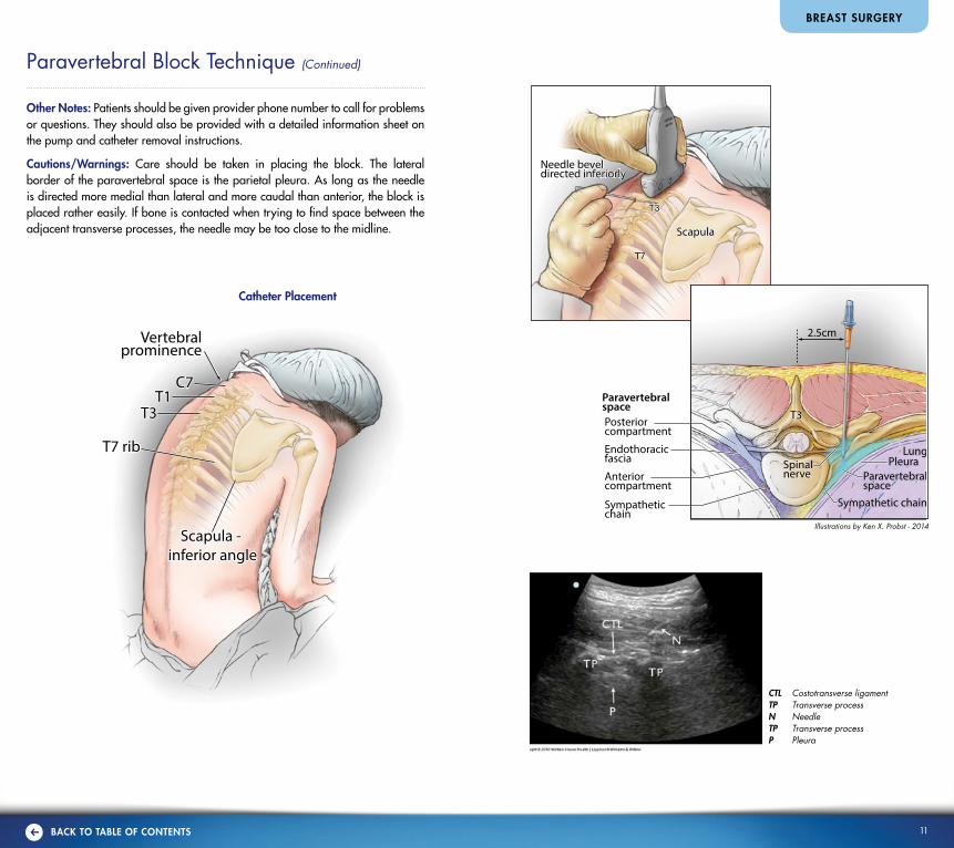
Cautions/Warnings: Care should be taken in placing the block. The lateral border of the paravertebral space is the parietal pleura. As long as the needle is directed more medial than lateral and more caudal than anterior, the block is placed rather easily. If bone is contacted when trying to find space between the adjacent transverse processes, the needle may be too close to the midline.
BREAST SURGERY
Thoracic Paravertebral Catheter Anatomic Technique (Continued)
Illustrations by Ken X. Probst - 2014
Catheter Placement

10h BACK TO TABLE OF CONTENTS
Paravertebral Block Technique
Tanith Graham, MDAnesthesiology, St. Vincent Hospital – Portland, OR
Surgical Procedure: Mastectomy (with & without reconstruction), Thoracotomy, Chest Wall Surgery, Rib fractures
ON-Q* Pump Used: CB6007, 600 ml ON-Q* with Select-A-Flow* 1-7 ml/hr (dual site). Filled to 750 ml
Drugs in Pump: Local anesthetic of physician’s choice
Equipment Used: 20 G Catheter Kit 18-19 G 4-inch Touhy introducer needle Ultrasound Machine
Preoperative Anesthesia: Mild sedation with Midazolam and/or Fentanyl IV
Initial Infiltration: Local anesthetic of physician’s choice in aliquots under ultrasound
Catheter Placement: The patient is placed in the sitting (preferred), lateral, or prone position for bilateral blocks. I advise standard ASA monitoring. C7 is easy to feel as it is more protuberant and is used as the starting place to count down to T2 vertebra. For ease, the T2 spinous process can be palpated and marked in the midline, and just 2.5 cm lateral, a mark can be made over the transverse processes (T1-4). The 10-13 mH ultrasound is placed in a saggital plane over the transverse processes of T1-3. A syringe containing 2% lidocaine with a 25 G needle is used to anesthetize the skin and subcutaneous. At the same time the anesthesiologist can see on ultrasound the direction needed for the larger introducer needle.
Note: For mastectomy procedures enter at T2-4. For thoracotomy or chest wall operations enter at point close to incision. It may be preferable to place catheter after the operation for this reason.
At this point, an 18-19 G Touhy tipped needle is carefully directed in a perpendicular direction with a 10-15 degree tilt of the needle pointing from over the T3 process into the T2-T3 interspace. For orientation purposes, the T3, TP is located just lateral to the T2 spinous process at its most protuberant point. It is important to not let the needle wander in a medial direction to avoid the epidural or SAB spaces. If the needle touches the bone of the transverse process then it should be redirected caudad or cephalad just slightly to clear the transverse process. On ultrasound, the paravertebral space will be found within 1 cm deep to the TP. On ultrasound, a test dose of normal saline is injected to confirm that
BREAST SURGERY
the pleura moves in an anterior direction. At that time the needle can be adjusted slightly if needed. Then in aliquots of 5 ml of local anesthetic, a total of 15 ml is injected per side. Then the catheter is threaded approximately 2.5-5 cm into the paravertebral space. A slight resistance is often felt with threading the catheter, but if blood or CSF is encountered, the catheter should be removed and the catheter placed at a different interspace.
The catheter(s) should be primed with 1 ml of solution of choice to keep the catheter patent after threading. The ON-Q* pump is connected to the catheter just after the operation, and the infusion is started at 2-7 ml/hr. I start my patients at 5 ml/hr, then titrate as needed. If a patient has one-sided pain or an area of lingering pressure, a post-op bolus can be given (5 ml).
Caution: It is important for the clinician to follow the drug manufacturer’s total daily dose recommendations.
Postoperative Bolus Technique: 2 ml every 20 minutes prn, with consideration given to total daily dose delivered and patient’s clinical status
Catheter Securement: Skin mastisol or benzoin, skin glue at catheter entrance, and clear sterile dressing over curled catheter
Intraoperative Anesthesia: General anesthesia or TIVA
Additional Postoperative Pain Medications: Vicodin® or Percocet® although most mastectomy patients use very little
Results with ON-Q*: Excellent pain relief for up to 3 days. Mastectomy patient able to go home same day. All patients receive less narcotic pain meds, and have less sedation, nausea, and itching. Mastectomy patients have noted more mobility and less muscle stiffness.
Before ON-Q* After ON-Q*
Pain Management Method Oxycodone/PCA/Dilaudid®
Hydrocodone/Tylenol®/ Ibuprofen
Average Narcotic Usag 5-10 mg IV 0-1 mg IV
Average Pain Score 2-3 days 0-1 day
11h BACK TO TABLE OF CONTENTS
Other Notes: Patients should be given provider phone number to call for problems or questions. They should also be provided with a detailed information sheet on the pump and catheter removal instructions.
Cautions/Warnings: Care should be taken in placing the block. The lateral border of the paravertebral space is the parietal pleura. As long as the needle is directed more medial than lateral and more caudal than anterior, the block is placed rather easily. If bone is contacted when trying to find space between the adjacent transverse processes, the needle may be too close to the midline.
BREAST SURGERY
Paravertebral Block Technique (Continued)
Illustrations by Ken X. Probst - 2014
Catheter Placement
T1T1T3T3
T7 ribT7 rib
Scapula - inferior angle
Scapula - inferior angle
Vertebralprominence
Vertebralprominence
C7C72.5cm
T7T7
Needle beveldirected inferiorlyNeedle beveldirected inferiorly
T3T3
ScapulaScapula
T3T3
SpinalnerveSpinalnerve Paravertebral
spaceParavertebralspace
Sympathetic chainSympathetic chain
LungLungPleuraPleura
Endothoracicfascia
ParavertebralspacePosteriorcompartment
Anteriorcompartment
Sympatheticchain
2.5cm
T7T7
Needle beveldirected inferiorlyNeedle beveldirected inferiorly
T3T3
ScapulaScapula
T3T3
SpinalnerveSpinalnerve Paravertebral
spaceParavertebralspace
Sympathetic chainSympathetic chain
LungLungPleuraPleura
Endothoracicfascia
ParavertebralspacePosteriorcompartment
Anteriorcompartment
Sympatheticchain
CTL Costotransverse ligamentTP Transverse processN NeedleTP Transverse processP Pleura

12h BACK TO TABLE OF CONTENTS
Study Summary
Heller L, Kowalski A, Wei C, Butler C. Prospective, randomized, double-blind trial of local anesthetic infusion and intravenous narcotic patient-controlled anesthesia pump for pain management after free TRAM flap breast reconstruction. Plast Reconstr Surg 2008;122:1010-18.
STUDY DESIGN: Prospective, randomized, double-blind
NUMBER OF PATIENTS: Treatment group – 23; Control group – 25
SUMMARY: Patients undergoing unilateral skin-sparing mastectomy with transverse rectus abdominis musculocutaneous (TRAM) flap breast reconstruction were randomized to receive two ON-Q Soaker Catheters inserted in the suprapubic area. One catheter was placed in the rectus abdominis muscle defect and the second catheter was placed subcutaneously superficial to the anterior rectus sheath. Patients received either a continuous infusion of 0.375% bupivacaine (treatment group) or normal saline (control group) via an ON-Q dual site 270 ml pump at 2 ml/hr each site. Patients in the treatment group used less PCA narcotic during the first two days compared to the control group, had mean PCA narcotic use 40% less on day 1 (P = 0.036) and 55% less day 2 (P = 0. 053) and were changed from PCA to oral narcotics sooner. Patients in the treatment group were also 3.6 times more likely to have higher satisfaction with their pain control than the control group. There were no significant differences between groups with regard to total narcotic use, length of stay, incidence of narcotic side effects, pain intensity scores or surgical recovery milestones.
ADVERSE EVENTS AND COMPLICATIONS: No technical problems or complications related to the pump or bupivacaine use.
CONCLUSION: Continuous infusion pump systems appear to be a safe and effective method and should be considered for postoperative pain management at the donor site for TRAM flap breast reconstruction patients. System did not eliminate narcotic use. Additional studies are recommended to determine which patients and procedures will benefit most from this method and to evaluate system’s cost effectiveness.
Study Summary
Exadakylos AK, Buggy D, Moriarty C, Mascha E, Sessler D. Can anesthetic technique for primary breast cancer surgery affect recurrence or metastasis? Anesth 2006;105:660-4.
STUDY DESIGN: Retrospective analysis
NUMBER OF PATIENTS: 129 consecutive patients. 50 patients received paravertebral anesthesia (PVB) combined with general anesthesia; 79 patients received general anesthesia (GA) with morphine for postoperative pain control.
SUMMARY: Medical record review of patients who had a mastectomy with axillary clearance or simple mastectomy. PVB group had a catheter placed prior to surgery at the level of T2 or T3 and received a continuous infusion of 0.25% levobupivacaine for 48 hours. GA only group received PCA morphine. Mean follow-up time was 32±5 months. Risk of cancer recurrence was significantly less in PVB group (P = 0.012). Patients in PVB group had lower pain scores at 24 hours. P= 0.04. Potentially regional anesthesia and analgesia may help preserve immune function by decreasing the surgical stress response and decreasing need for opioids.
ADVERSE EVENTS AND COMPLICATIONS: Three patients in the GA group developed infections, compared with two in the PVB group.
CONCLUSION: “Retrospective analysis suggests that paravertebral anesthesia and analgesia for breast cancer surgery reduces the risk of metastasis during the initial years of follow-up. Prospective trials evaluating the effects of regional analgesia and morphine sparing on cancer recurrence seem warranted.”
BREAST SURGERY
Cancer Recurrence and Regional Anesthesia
Early retrospective studies have shown that there may be reduced risk of metastasis of cancer recurrence in the breast 21, 23, 25 and prostate3 with regional anesthesia techniques after surgery. Opioid limiting, effective treatment of postoperative pain could play an important role22, 23 in limiting the metastatic proliferation of cancer cells following oncology surgery. Regional anesthesia/analgesia decreases the neuroendocrine stress response to surgical tissue injury21-23, eliminates or reduces the need for general anesthesia, and minimizes opioid requirement. Prospective, randomized, large-size studies focused on cancers with high risk of recurrence are currently underway to determine if regional anesthesia and analgesic techniques, including opioid sparing therapies, could have potential for clinically reducing the risk of cancer recurrence after oncology surgery.26

13h BACK TO TABLE OF CONTENTS
Janice F. Rafferty, MDColorectal Surgeon,
The Christ Hospital – Cincinnati, OH
UpperMidline
LowerMidline
Paramedian
Subcostal
Transverse
McBurney
Groin
Pfannenstiel
“ I have found that patient comfort levels are much
more easily maintained with the use of ON-Q* as they
progress through their postoperative recovery.”
Kurt Stockamp, MDSacred Heart Hospital – Pensacola, FL
“ Patients who receive a continuous TAP block wake up
with less pain. By reducing the amount of narcotics and
their associated side effects, patients walk, cough and
breathe deeply after surgery, all of which are important
to a good recovery.”
Opioids are frequently used for the treatment of postoperative pain following abdominal surgery (e.g., ventral hernia and bowel procedures, etc.), however their use is often associated with side effects such as PONV, over-sedation, constipation, confusion and ileus which can be especially problematic following abdominal surgery.5, 27, 28, 29
Surgeons can use a simple approach such as placing a catheter to infuse a local anesthetic into the incision site at the end of the procedure, or a surgeon or an anesthesiologist can perform a continuous TAP block. These techniques offer effective pain management and may be alternatives to epidural analgesia, minimizing the potential for adverse side effects.5, 15, 29
Patient Benefits of TAP Block• Significantly better pain scores at rest and while coughing30, 31 • Reduced opioid use and associated side effects 32-34
• High patient satisfaction 30, 33
Abdominal Surgery
14h BACK TO TABLE OF CONTENTS
ABDOMINAL SURGERY
Tunneling Universal Abdominal Approach Technique
Alexander Saba, MDGood Samaritan Hospital, Mercy Mt. Airy Hospital – Cincinnati, OH
ON-Q* Pump Used: PM028-A: 400 ml x 4 ml/hr (dual; 2 ml/site) (2) 5 inch ON-Q* SilverSoaker* catheters ACC03: 8 inch needles
Obese Patients: PM048-A: 400 ml x 4 ml/hr (dual; 2 ml/site) 10 inch ON-Q* SilverSoaker* catheters
Tunneler Used: T17X8: Disposable 17 GA x 8 in Tunneler & Sheaths or T16X12: Disposable 16 GA x 12 in Tunneler & Sheaths
Surgical Procedure: Midline incision procedures (colectomy, laparotomy, open ventral hernia), laparoscopic hand-assisted surgery, laparoscopic ventral hernia.
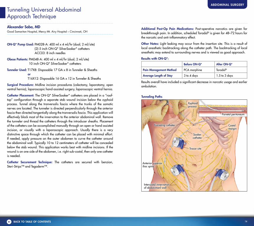
Catheter Placement: The ON-Q* SilverSoaker* catheters are placed in a “roof-top” configuration through a separate stab wound incision below the xyphoid process. Tunnel along the tranversalis fascia where the trunks of the somatic nerves are located. The tunneler is directed perpendicularly through the anterior fascia then directed tangentially along the tranversalis fascia. This application will effectively block most of the innervation to the anterior abdominal wall. Remove the tunneler and thread the catheters through the introducer sheaths. Placement of the catheters can be accomplished manually through an open or hand assisted incision, or visually with a laparoscopic approach. Usually there is a very distinctive space through which the catheter can be placed with minimal effort. If needed, apply pressure on the outer abdomen to curve the catheter around the abdominal wall. Typically 10 to 12 centimeters of catheter will be concealed below the stab wound. This application works best with midline incisions. If the wound is on one side of the abdomen, i.e. right sub-costal, then only one catheter is needed.
Catheter Securement Technique: The catheters are secured with benzion, Steri-Strips™ and Tegaderm™.
Additional Post-Op Pain Medications: Post-operative narcotics are given for breakthrough pain. In addition, scheduled Toradol® is given for 48–72 hours for the narcotic and anti-inflammatory effect.
Other Notes: Light leaking may occur from the insertion site. This is a result of local anesthetic backtracking along the catheter path. The backtracking of local anesthetic may extend to surrounding nerves and is viewed as good approach.
Results with ON-Q*:
Before ON-Q* After ON-Q*
Pain Management Method PCA morphine Toradol®
Average Length of Stay 3 to 4 days 1.5 to 3 days
Results overall have included a significant decrease in narcotic usage and earlier ambulation.
Anterior superior iliac spine
Intercostal innervation of abdonimanl wall
Costal margin
Xyphoid process
Parietal peritoneum
Tunneling catheter
Soakercatheter
Trocar site
Tunneling Paths
15h BACK TO TABLE OF CONTENTS
ABDOMINAL SURGERY
Umbilicus line
Iliac crest
Rectus abdominis
Aponeurosis
Nerves
Transverse abdominisInternal obliqueExternal oblique
Nerves
Costal margin
Rectus abdominis
AponeurosisTransverse abdominisInternal obliqueExternal oblique
Needle Insertion Using In-Plane Technique
Umbilicus line
Iliac crest
Rectus abdominis
Aponeurosis
Nerves
Transverse abdominisInternal obliqueExternal oblique
Nerves
Costal margin
Rectus abdominis
AponeurosisTransverse abdominisInternal obliqueExternal oblique
Umbilicus line
Iliac crest
Rectus abdominis
Aponeurosis
Nerves
Transverse abdominisInternal obliqueExternal oblique
Nerves
Costal margin
Rectus abdominis
AponeurosisTransverse abdominisInternal obliqueExternal oblique
Umbilicus line
Iliac crest
Rectus abdominis
Aponeurosis
Nerves
Transverse abdominisInternal obliqueExternal oblique
Nerves
Costal margin
Rectus abdominis
AponeurosisTransverse abdominisInternal obliqueExternal oblique
Illustrations by Ken X. Probst - 2014
Transversus Abdominis Plane (TAP) Continuous Nerve Block Technique
Rita Hadley, MD, PhD, FACSGeneral Surgery, Miami Valley Hospital – Dayton, Ohio
ON-Q* Pump Used: CB6007: 600 ml (dual; 1-7 ml/hr Select-A-Flow*) for bilateral infusion
Drugs in Pump: Local anesthetic of physician’s choice
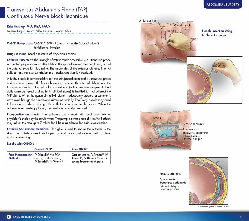
Catheter Placement: The Triangle of Petit is made accessible. An ultrasound probe is oriented perpendicular to the table in the space between the costal margin and the anterior superior iliac spine. The anatomies of the external oblique, internal oblique, and transversus abdominis muscles are clearly visualized.
A Tuohy needle is advanced through the skin just adjacent to the ultrasound probe and advanced toward the fascial boundary between the internal oblique and the transversus muscle. 10-20 ml of local anesthetic, (with consideration given to total daily dose delivered and patient’s clinical status) is instilled to hydrodissect the TAP plane. When the space of the TAP plane is adequately created, a catheter is advanced through the needle and aimed posteriorly. The Tuohy needle may need to be spun or redirected to get the catheter to advance in the space. When the catheter is successfully placed, the needle is carefully removed.
Preoperative anesthesia: The catheters are primed with local anesthetic of physician’s choice by the scrub nurse. The pump is set at a rate of 4 ml/hr. Patients may adjust the rate up to 7 ml/hr for 1 hour as a bolus for pain exacerbation.
Catheter Securement Technique: Skin glue is used to secure the catheter to the skin. The catheters are then looped around twice and secured with a clear, occlusive dressing.
Results with ON-Q*:
Before ON-Q* After ON-Q*
Pain Management Method
IV Dilaudid® via PCA device, oral narcotics, IV Toradol®, IV Tylenol®
Oral narcotics, IV Tylenol®, IV Toradol®, IV Dilaudid® only for severe breakthrough pain
16h BACK TO TABLE OF CONTENTS
Continuous Transversus Abdominis Plane (TAP) Block Technique
Brian Vaughan, MDAnesthesia Associates of Cincinnati, Inc, The Christ Hospital – Cincinnati, OH
ON-Q* Pump Used: Unilateral continuous TAP block: 550-ml, single lumen Select-A-Flow* (CB004) 400 ml x 2-14 ml/hr, fill to 550 ml
Bilateral continuous TAP block: 750-ml, dual lumen Select-A-Flow* (CB6007) 600 ml x 1-7 ml/hr (dual site), fill to 750 ml
Drugs in Pump: Local anesthetic of physician’s choice.
Indication: TAP blocks are indicated for abdominal incisions that are medial to the mid to anterior axillary line, inferior to the costal margin, and at or superior to the inguinal ligament. Midline or near-midline incisions require bilateral placement of TAP blocks.
TAP Block: Analgesia is obtained by using local anesthetic to block pain signal transmission from the intercostal, subcostal, ilioinguinal, and iliohypogastric nerves as they wrap around the abdominal wall in the plane between the transversus abdominis and internal oblique.
Catheter Placement:
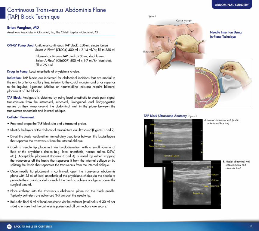
• Prep and drape the TAP block site and ultrasound probe.
• Identify the layers of the abdominal musculature via ultrasound (Figures 1 and 2).
• Direct the block needle either immediately deep to or between the fascial layers that separate the transversus from the internal oblique.
• Confirm needle tip placement via hyrdodissection with a small volume of fluid of the physician’s choice (e.g. local anesthetic, normal saline, D5W, etc.). Acceptable placement (Figures 3 and 4) is noted by either stripping the transversus off the fascia that separates it from the internal oblique or by splitting the fascia that separates the transversus from the internal oblique.
• Once needle tip placement is confirmed, open the transversus abdominis plane with 25 ml of local anesthetic of the physician’s choice via the needle to promote the cranial-caudal spread of the block to achieve analgesia across the surgical wound.
• Place catheter into the transversus abdominis plane via the block needle. Typically catheters are advanced 3-5 cm past the needle tip.
• Bolus the final 5 ml of local anesthetic via the catheter (total bolus of 30 ml per side) to ensure that the catheter is patent and all connections are secure.
ABDOMINAL SURGERY
Needle Insertion Using In-Plane Technique
Iliac crest
Nerves
Rectus abdominis
Transverse abdominisInternal obliqueExternal oblique
Nerves
Costal margin
Rectus abdominis
Transverse abdominisInternal obliqueExternal oblique
Iliac crest
Nerves
Rectus abdominis
Transverse abdominisInternal obliqueExternal oblique
Nerves
Costal margin
Rectus abdominis
Transverse abdominisInternal obliqueExternal oblique
Figure 1
Rectus
A. Lateral abdominal wall (mid to anterior axillary line)
B. Medial abdominal wall (approximately mid clavicular line)
TAP Block Ultrasound Anatomy Figure 2

17h BACK TO TABLE OF CONTENTS
Continuous Transversus Abdominis Plane (TAP) Block Technique (Continued)
ABDOMINAL SURGERY
Catheter Securement Technique: The catheters are secured with Mastisol and dressing of choice.
Post-Op Analgesia: Additional post-op pain medications, including non-narcotic and narcotic-based analgesics may be indicated for supplementary analgesia. With a well functioning continuous TAP block, narcotic use can be greatly reduced compared to a traditional narcotic-based analgesic regimen. For patients who are NPO after surgery, catheters are left in place until bowel function returns (up to post-op day 6) in order to minimize narcotic use. As with any continuous block, the insertion site is monitored daily for any signs of infection.
Other Notes: Leaking at the site, especially in thin patients, may occur. This is a result of local anesthetic tracking along the catheter to the surface. Although the amount of saturation may seem substantial, usually most local anesthetic continues to infuse into the transversus plane and does not affect the quality of the block. Leaking at the site requires dressing reinforcement and does not necessitate removal of the catheter.
TAP Block Ultrasound Post-Injection Figure 4
Iliac crest
Nerves
Rectus abdominis
Transverse abdominisInternal obliqueExternal oblique
Nerves
Costal margin
Rectus abdominis
Transverse abdominisInternal obliqueExternal oblique
Figure 3
Iliac crest
Nerves
Rectus abdominis
Transverse abdominisInternal obliqueExternal oblique
Nerves
Costal margin
Rectus abdominis
Transverse abdominisInternal obliqueExternal oblique
Illustrations by Ken X. Probst - 2014

18h BACK TO TABLE OF CONTENTS
Laparoscopic Incisional Hernia Repair with Mesh Technique
Daniel Tseng, MD FACSGeneral Surgery, Legacy Good Samaritan Hospital – Portland, OR
ON-Q* Pump Used: CB6007: filled with 750ml 600 ml (dual: 7 ml/hr Select-A-Flow*) for bilateral infusion filled to 750 ml.
Catheter Used (depends on incision length): ON-Q* SilverSoaker* Catheters PM040-A,10 inch PM050-A, 7.5 inch PM020-A, 5 inch
Tunneler Used: T11X12, 11 GA x 12 inch T11X 8, 11 GA X 8 inch
Drugs in Pump: Local anesthetic of physician’s choice
Pre-incisional Infiltration: Local anesthetic of physician’s choice
Catheter Placement: Two catheters are placed on either side of the abdomen running vertically from the level of the anterior superior iliac spine to the costal margin. The goal is to place the catheters within the preperitoneal space to allow infusion of local anesthetic to the region. The 10 inch blunt tipped introducer is used for the preperitoneal dissection.
The entry point for catheter placement is typically utilizing the same incisions where the laparoscopic trocars are placed. Typically port placement is at least one 5-mm trocar on the left abdomen and at least one on the right abdomen. One of the left abdominal trocars are removed and the blunt tip introducer is inserted through this incision and tunneled through the pre-peritoneal space. In order to facilitate the dissection, counter-pressure is applied to the anterior abdominal wall to deform it and create a flat plane. This will allow the catheter to glide through the preperitoneal space.
Moving the counter-pressure as the catheter advances is critical to keep the tip of the catheter In the preperitoneal plane. During advancement of the catheter, the tip is visible at all times and should slightly tent up the peritoneum if placed properly. Non-visualization of the tenting of the peritoneum typically indicates that the introducer tip is within the anterior abdominal wall musculature and not within the preperitoneal space.
Once the introducer tip is advanced as far cephalad as it can go (typically costal border) or caudad as far as it can go (typically inguinal ligament) the blunt tipped introducer is removed. This is replaced by one of the 10 inch catheters. During advancement of the catheter, the sheath is slowly retracted which allows the catheter to coil in to the preperitoneal space. The catheter ideally is placed all the way to its hub in order to prevent leakage.
ABDOMINAL SURGERY
Catheter Securement: Clear waterproof dressing applied
Caution/Warning: Avoid placing the tip of the catheter deep into the retroperito-neal space of the pelvis as the local anesthetic may inhibit femoral nerve function resulting in leg weakness. If this occurs, simply remove the catheter.
Illustrations by Ken X. Probst - 2014
Tunneling and Catheter Placement
Cross Section of the Abdomen

19h BACK TO TABLE OF CONTENTS
ABDOMINAL SURGERY
2 cm
Incision
Tunneling
Catheter Placement
Cross Section View
Illustrations by Ken X. Probst - 2014
Midline Incisional Technique
Rita Hadley, MD, PhD, FACSGeneral Surgery, Miami Valley Hospital – Dayton, Ohio
Surgical Procedure: Ventral Hernia, Laparotomy, Colon Resection, and Small Bowel Obstruction
ON-Q* Pump Used: CB6007: 600 ml (dual; 1-7 ml/hr Select-A-Flow*) for bilateral infusion
Tunneler Used: T17X8, 17 G 8-inch tunneler, 5-inch catheter
Catheter Used: ON-Q* SilverSoaker* 5 inch catheter
Drugs in Pump: Local anesthetic of physician’s choice
Catheter Placement: Once the fascia is closed, 30 ml of local anesthetic with consideration given to total daily dose delivered and patient’s status is infiltrated into the area of the fascial repair with a large bore needle. The skin incision is then closed. Two paths parallel to the incision, approximately 2 cm from either side of the incision, are visualized for placement of the catheters. On one side, above the incision, a needle introducer is used to pierce the skin. Through this opening, a tunneling device is directed at a 45 degree angle down toward the fascia. The tunneler is advanced until the tip meets resistance and then is advanced in a plane parallel to the fascia for the length of the tunneler. Above the fascia is an avascular plane through which the tunneler will advance easily. Advance the tunneler until it passes the farthest part of the incision. The stylet of the tunneler is then retracted from the tunneling sheath which is left for placement of the catheter. The SilverSoaker* catheter is then placed through the catheter sheath and advanced until the first tic mark resides at the level of the skin. The sheath is then carefully peeled away and removed from the subcutaneous tissue. This procedure is then repeated on the other side of the incision.
Preoperative Bolus Technique: The catheters are primed with local anesthetic of the physician’s choice by the scrub nurse. The pump is set to run at 4 ml/hr. Patients may adjust up to 7ml/hr for 1 hour for improved pain control as a bolus.
Catheter Securement Technique: Catheters are secured to the skin with skin glue. The catheters are wound twice into a circle and then covered with a clear occlusive dressing.
Results with ON-Q*:
Before ON-Q* After ON-Q*
Pain Management Method
IV Dilaudid® via PCA device, oral narcotics, IV Toradol®, IV Tylenol®
Oral narcotics, IV Dilaudid® only for severe breakthrough pain, IV Tylenol®, IV Toradol®

20h BACK TO TABLE OF CONTENTS
Study Summary
Beaussier M, El-Ayoubi H et al. Continuous Preperitoneal Infusion of Ropivacaine Provides Effective Analgesia and Accelerates Recovery after Colorectal Surgery. Anesthesiology 2007; 107:461-468.
STUDY DESIGN: Prospective, randomized, double-blind, placebo controlled
NUMBER OF PATIENTS: 49 (42 completed study)
SUMMARY: Patients undergoing elective open colorectal surgery were randomized to receive either ropivacaine 0.2% or saline at 10 ml/hr via a multi-holed (Soaker) catheter placed in the preperitoneal space and delivered by the ON-Q pump for 48 hours postoperatively. Patients in the ropivacaine group had significantly lower pain scores at rest during the first 12 hours, and with coughing for 48 hours (P < 0.01). Significantly more morphine was required in the PACU by the control group (P = 0.004) and drug the first 3 days postoperatively (P = 0.0004). Quality of sleep was rated higher in the active group and time to recovery of bowel activity was quicker (P < 0.02).
ADVERSE EVENTS AND COMPLICATIONS: No major complications. Two patients in the ropivacaine group and six in the control group experienced severe postoperative nausea and vomiting (P = NS).
CONCLUSION: Continuous preperitoneal administration of 0.2% ropivacaine at 10 ml/hr for 48 hours after open colorectal procedure reduced morphine consumption, improved pain relief, and accelerated postoperative recovery.
Study Summary
Kadem RV, Field JB. Ultrasound-guided continuous transversus abdominis plane block for abdominal surgery. J Anaesth Clin Pharmacol 2011;27:333-6.
STUDY DESIGN: Prospective, randomized, controlled
NUMBER OF PATIENTS: 20
SUMMARY: Patients undergoing elective abdominal surgery with supra/infra –umbilical incisions were randomized either to the continuous transversus abdominis plane (TAP) block or control group for postoperative pain management. TAP block was performed at the end of surgery using ultrasound -guided technique and a posterior approach for catheter placement. An infusion of 0.2% ropivacaine, 8-10 ml/hr continued for 72 hours via the catheter. Rescue analgesia for both groups was fentanyl PCA. Patients in the TAP group had consistently lower median NRS pain scores, but were only significant on day one with coughing (P = 0.02) and day two at rest (P = 0.04) and with coughing (P = 0.01). Fentanyl use at one hour was 203 µg in the control group and 78 µg in the TAP group (P = 0.03) and at one day was also significantly greater (P = 0.01) in the control group (1237 µg) compared to the TAP group (664 µg). Patient satisfaction ratings were done only in the TAP group as it was a new technique with the following results: Excellent – 3 patients; Satisfied – 4 patients; Poor – 2 Patients.
COMPLICATIONS AND ADVERSE EVENTS: One episode each related to the TAP block: leaking, catheter fell out and disconnection from the filter. There was one episode of PONV in control group; none in the TAP group.
CONCLUSION: Study showed lower pain scores with reduced fentanyl requirements in the TAP group when combined with multimodal analgesia in major abdominal surgery. Larger scale studies are needed.
ABDOMINAL SURGERY

21h BACK TO TABLE OF CONTENTS
23. Biki, B et al. Anesthetic technique for radical prostatectomy surgery. Anesthesiology 2008; 109:108-7.24. Conveney, E et al. Use of paravertebral block anesthesia in the surgical management of breast
cancer. Annals of Surgery 1998; 227:496-501.25. Fraser, S P, et al. Local anaesthetic use in cancer surgery and disease recurrence: role of voltage-
gated sodium channels? British Journal of Anaesthesia 2014; BJA Advance Access published online 7.16.14.
26. https://clinicaltrials.gov/ct2/results?term=cancer+recurrence+and+regional+anesthesia+or+analgesia&Search=Search
27. Wang WW et al. Wound infusion with local anaesthesia after laparotomy: a randomized controlled trial. ANZ J Surg 201080: 794-801.
28. The Joint Commission – Sentinel Event Alert- Safe Use of Opioids in Hospitals. Issue 49; August 8, 2012.
29. Rawal, N. Epidural technique for postoperative pain. Gold standard no more. Reg Anesth and Pain Medicine 2012;37:310-317.
30. Bharti N et al. The efficacy of a novel approach to transversus abdominis plane block for postoperative analgesia after colorectal surgery. Regional Anesthesia and Pain Medicine 2011;112:1504-1508.
31. Kadam RV et al. Ultrasound-guided continuous transverse abdominus plane block for abdominal surgery. Journal of Anaesthesiology and Clinical Pharmacology 2011;27:333-336.
32. McDonnell J et al. The analgesic efficacy of transversus abdominis plane block after abdominal surgery: a prospective randomized controlled trial. Anesthesia and Analgesia 2007;104:193-197.
33. Belavy et al. Ultrasound-guided transversus abdominis plane block for analgesia after caesaren delivery. British Journal of Anaesthesia 2009;103:726-730.
34. Siddiqui M et al. A meta-analysis on clinical effectiveness of transversus abdominis plane block. Journal of Clinical Anesthesia 2011;23:7-14.
References 1. Apfelbaum J et al. Survey suggest postoperative pain tends to be undermanaged. Anesth Analg
2003;97:534-40.2. Voscopoulos C, Lema M. When does acute pain become chronic? Br J Anaesth. 2010; 105 (S1):
i69–i85. 3. Hadzic A (ed). The New York School of Regional Anesthesia: Textbook of Regional Anesthesia and
Pain Management. McGraw-Hill 2007.4. Practice guidelines for acute pain management in the perioperative setting – an updated report by the
American Society of Anesthesiologist Task Force on Acute Pain Management. Anesth 2012;116:248-73.
5. Chelly JE et al. Continuous peripheral nerve blocks in acute pain management. Br J Anaesth 2010;105 (S1): i86–i96.
6. HCAHPS – Hospital Consumer Assessment of Healthcare Providers and Systems, CAHPS Hospital Survery. http://www.hcahpsonline.org/surveyinstrument.aspx . Accessed August 24, 2014.
7. Hanna MN, Gonzalez-Fernandez M, Barrett AD, Williams KA, Pronovost P. American Journal of Medical Quality. Does patient perception of pain control affect patient satisfaction across surgical units in a tertiary hospital. 2012 February 16; Available from: http://ajm.sagepub.com/content/early/2012/02/16/1062860611427769
8. HCAHPS Patient-Level “TOP-BOX” Correlations. Patients-level Pearson correlations of the “top-box” scores of publicity reported HCAHPS measures, for patients discharged between July 2007 and June 2008 (2.2 million completed surveys). Available from: http://www.hcahpsonline.org/executive_insight/Files/Report_HEI_March_2010_Corrs.p
9. White PF, Rawal S, Latham P, et al. Use of a continuous local anesthetic infusion for pain management after median sternotomy. Anesth. 2003;99(4):918-923.
10. Dowling R, Thielmeier K, Ghaly A, Barber D, Boice T, Dine A. Improved pain control after cardiac surgery: results of a randomized, double-blind, clinical trial. J Thorac Cardiovasc Surg.2003;126(5):1271-1278.
11. Forastiere E, Sofra M, Giannarelli D, Fabrizi L, Simone G. Effectiveness of continuous wound infusion of 0.5% ropivacaine by ON-Q pain relief system for postoperative pain management after open nephrectomy. Br J Anaesth. 2008;101(6):841-847.
12. Beaussier M, El’Ayoubi H, Schiffer E, et al. Continuous preperitoneal infusion of ropivacaine provides effective analgesia and accelerates recovery after colorectal surgery. Anesth. 2007;107(3):461-468.
13. Sherwinter DA, Ghaznavi AM, Spinner D, Savel RH, Macura JM, Adler H. Continuous infusion of interperitoneal bupivicaine after laparoscopic surgery: a randomized controlled trial. Obes Surg. 2008;18(12):1581-1586.
14. Klein SM, Grant SA, Greengrass RA, et al. Interscalene brachial plexus block with a continuous catheter insertion system and a disposable infusion pump. Anesth Analg. 2000;91(6):1473-1478.
15. Liu SS, Richman JM, Thirlby RC, Wu CL. Efficacy of continuous wound catheters delivering local anesthetic for postoperative analgesia: a quantitative and qualitative systematic review of randomized controlled trials. J Am Coll Surg. 2006;203(6):914-932.
16. Heller L, Kowalski AM, Wei C, Butler CE. Prospective, randomized, double-blind trial of local anesthetic infusion and intravenous narcotic patient-controlled anesthesia pump for pain management after free TRAM flap breast reconstruction. Plast Reconstr Surg. 2008;122(4):1010-1018.
17. Joshi G et al. A systematic review of randomized trials evaluating regional techniques for postthoracotomy analgesia. Anesth Analg 2008;107:1026-40.
18. Powell ES et al. A prospective, multicenter, observational cohort study of analgesia and outcome after pneumonectomy. Br J Anaesth 2011;106:364-370.
19. Davies R et al. A comparison of the analgesic efficacy and side-effects of paravertebral vs epidural blockade for thoracotomy-a systematic review and meta-analysis of randomized trials. Br J Anaesth 2006;6-9.
20. Mehta Y et al. Comparison of continuous thoracic epidural and paravertebral block for postoperative analgesia after robotic-assisted coronary artery bypass surgery. Ann Cardiac Anaesth 2008;11:91-96.
21. Sessler DL et al. Can regional anesthesia reduce the risk of recurrence after breast cancer? Methodology of a multicenter randomized trial. Contemporary Clinical Trials 2008.
22. Exadakytos AK et al. Can anesthetic technique for primary breast cancer surgery affect recurrence or metastasis? Anesthesiology 2006; 105: 660-664.

Call your ON-Q* representative today for more information: 1-800-448-3569 or visit us at iflo.com
There are inherent risks in all medical devices. Please refer to the product labeling for Indications, Cautions, Warnings, and Contraindications. Failure to follow the product labeling could directly impact patient safety. Physician is responsible for prescribing and administering medications per instructions provided by the drug manufacturer. Refer to www.iflo.com for product safety Technical Bulletins.
A K I M B E R L Y - C L A R K H E A L T H C A R E C O M P A N Y
A K I M B E R L Y - C L A R K H E A L T H C A R E C O M P A N Y
Rx only *Registered Trademark or Trademark of Kimberly-Clark Worldwide, Inc. © 2014 KCWW. All rights reserved.Dilaudid is a registered trademark of Abbott Laboratories • JP is a registered trademark of Jackson-Pratt.• Lock-It Plus is a registered trademark of Smiths Medical. • Percocet is a registered trademark of Endo Pharmaceuticals, Inc. • SteriStrip is a trademark of 3M Healthcare • Tegaderm is a registered trademark of 3M • Toradol is a registered trademark of Hoffman-LaRoche, Inc. • Tylenol is a registered trademark of McNeil Consumer Healthcare Division of McNEIL-PPC, Inc. • Vicodin is a registered trademark of Knoll Pharmaceuticals Co., Inc. • Versed is a registered trademark of Roche Laboratories.MK-00701 09/2014