general statistics second most common complaint after back pain “everyone” has headaches (ha)...
TRANSCRIPT


General Statistics
Second most common complaint after back pain
“Everyone” has headaches (HA) More than 80 million ER visits in U.S. per
year Frequency of HA due to rich nerve supply
and psychological implications of head pain

General Statistics
Nerves responsible for HA have their source from myelinated C fibers and A-delta fibers in cranial nerves V, IX, X, and roots C1, C2, C3
Pain sensitive structures include the eye, ear, paranasal sinuses, large extra and intra cranial arteries, dural sinuses, periosteum of the skull skin, cranial muscles, and the upper cervical spine

Etiologies
Commonly overlooked etiologies include: food, fever, viral, metabolic, withdrawal, and pharmaceutical
International Headache Society Classifications (see attachment)

History: Questions to ask
Character of pain Mode of onset Mode of offset Time of onset Relieving factors Aggravating factors

History: Questions to ask
Precipitating factors Frequency of attacks Duration of attacks Associated symptoms Family history of headache Allergies
Seven danger signals of an ominous headache
A “first” headacheA “first” headache Headache due to exertionHeadache due to exertion Headache with feverHeadache with fever Headache in a drowsy or confused patientHeadache in a drowsy or confused patient

Seven danger signals of an ominous headache
Headache in a patient with nuchal rigidity Headache in a patient with nuchal rigidity or meningeal signsor meningeal signs
Headache in a patient with abnormal Headache in a patient with abnormal physical signsphysical signs
Headache in a patient who “looks ill”Headache in a patient who “looks ill”
Physical Exam
Gait assessment Vital signs Fundoscopic exam Facial symmetry Head & Neck structures Deep tendon reflexes Plantar response Limb strength
Relevant Muscles
Trapezius Sternocleidomastoid Temporalis Occipitofrontalis Suboccipital muscles Masseter

Relevant Muscles
Medial & Lateral Pterygoid Anterior & Posterior Digastric Fascial muscles Splenius Capitis Posterior Cervical musculature Deep Anterior Cervical musculature

Cervical Dysfunction
Upper cervical nerves posses fibers for pain from the lower part of the occipital sinus, vertebral and posterior meningeal arteries, and the dural floor of the posterior fossa (C1, C2, C3)

Differential Diagnosis:Migraine HeadacheEtiology:Etiology: Hereditary component Not correlated with personality types “A” or
neuroses The worsening or migraine that occurs during
periods of intense nervousness, anxiety, and depression is usually due to the superimposition of a tension headache
Vascular spasm followed by vasodilatation

Migraine Headache:Signs & SymptomsClassic MigraineClassic Migraine
Character: throbbing pain Location: hemicranial Associated: preceded with visual
disturbances and less often with hemi-sensory disturbances, hemiparesis, or aphasia

Migraine Headache:Signs & SymptomsClassic & Common MigraineClassic & Common Migraine Character: throbbing pain Location: hemicranial Associated: photophobia and or phonophobia;
tension headache often concomitant Aggravated: red wine, nuts, aged cheese,
chocolate and caffeine containing beverages Risk factor: women are more affected than men

Migraine Headache:Diagnosis & Treatment Response to ergot therapy Drug treatment is widely varied (caffeine,
NSAIDS, barbiturates, narcotics, beta blockers, calcium channel blockers, sedatives, and more…)
Prevention by avoiding predisposing factors, decreasing stress, maintaining sleep regularity
Osteopathic treatment would include stabilizing vasculature and associated concomitant tension headache

Differential Diagnosis:Cluster HeadacheEtiologyEtiology Disturbed hypothalamic biorhythm Excess smoking and drinking may
precipitate via sphenopalatine irritation Hemicranial (unilateral) cranial dysfunction Cervical somatic dysfunction with irritation
of the spinal accessory nerve
Cluster Headache:Signs & Symptoms Character: excruciating pain often stabbing Location: usually near one eye Associated: tearing, flushed face, nasal
congestion, conjunctival congestion (ANS) Risk factor: males affected more than
females Onset: begins at 20 – 40 years of age

Cluster Headache:Signs & Symptoms Attacks last 30 – 90 minutes daily for days
and then disappear for months (Headache “vacation”)
Alcohol can precipitate but only during an active cycle, not during “vacations”
Some are so painful that they can lead to suicide

Cluster Headache: Prevention & Treatment
Drug treatment is widely varied
Osteopathic treatment would include a thorough cranial assessment

Differential Diagnosis:Organic origin, Subarachnoid hemorrhage
EtiologyEtiology
Ruptured aneurysm Arteriovenous malformation Trauma

Differential Diagnosis:Organic origin, Subarachnoid hemorrhage
Signs & SymptomsSigns & Symptoms
Character: full-blown catastrophic headache Location: Holocaine Duration: continuous Associated: photophobia, retinal hemorrhages,
nuchal rigidity, Brudzinski’s sign, Kernig’s sign, obtunded collapse

Differential Diagnosis:Organic origin, Subarachnoid hemorrhage
DiagnosisDiagnosis
CT may show blood and aneurysm Lumbar puncture may show bloody CSF MRI

Differential Diagnosis:Organic origin, MeningitisEtiologyEtiology
Virus Bacteria Fungus Tuberculous

Differential Diagnosis:Organic origin, MeningitisSigns & SymptomsSigns & Symptoms Character: cephalgia is intense, steady, and deep Location: holocranial pain associated with retro-
orbital pain which is aggravated with eye movement
Onset: sub-acute or acute Associated: fever, generalized convulsions, varied
levels of consciousness, nuchal rigidity, Brudzinski and Kernig’s signs
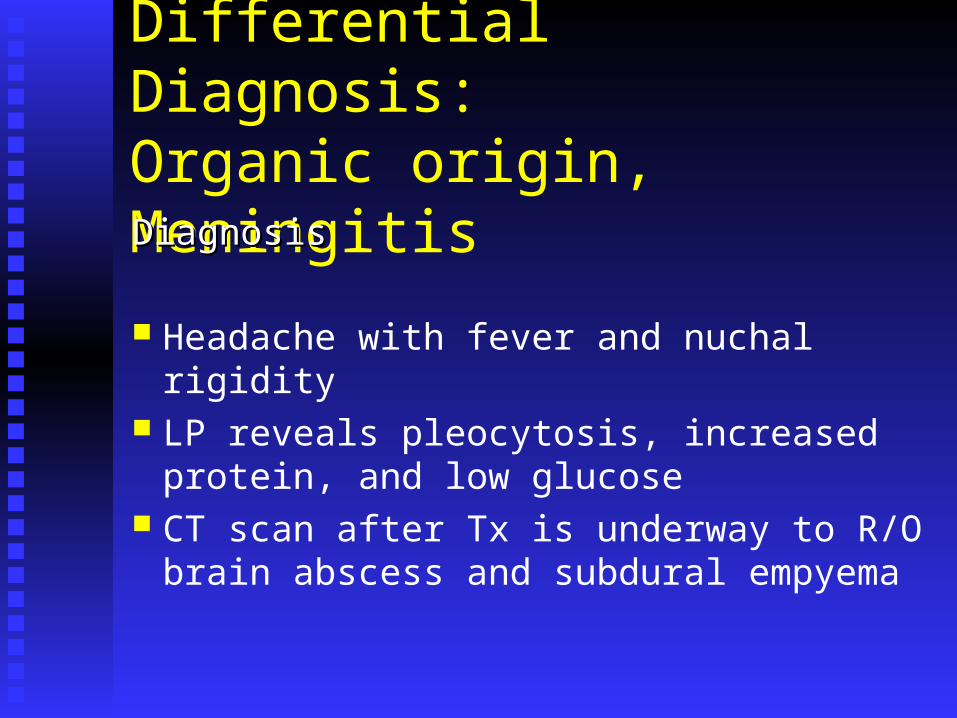
Differential Diagnosis:Organic origin, MeningitisDiagnosisDiagnosis
Headache with fever and nuchal rigidity LP reveals pleocytosis, increased protein,
and low glucose CT scan after Tx is underway to R/O brain
abscess and subdural empyema

Differential Diagnosis:Organic origin, Increased Intracranial pressure
EtiologyEtiology
Increased volume Increased venous pressure Obstruction to flow/absorption of CSF

Differential Diagnosis:Organic origin, Increased Intracranial pressure
Signs & SymptomsSigns & Symptoms
HA is severe HA occur with coughing, sneezing, valsalva effort Associated findings include papilledema,
obtunded, focal neurologic signs & symptoms

Differential Diagnosis:Organic origin, Increased Intracranial pressure
DiagnosisDiagnosis
CT MRI Avoid LP

Differential Diagnosis:Organic origin, Hypertension Usually no HA’s until DBP > 120 mm Hg 3 major causes of acute severe
hypertension: drugs, pheochromocytoma, neurogenic (paraplegia)
Associated findings include: retinopathy, convulsions, confusion or stupor evolving over several days
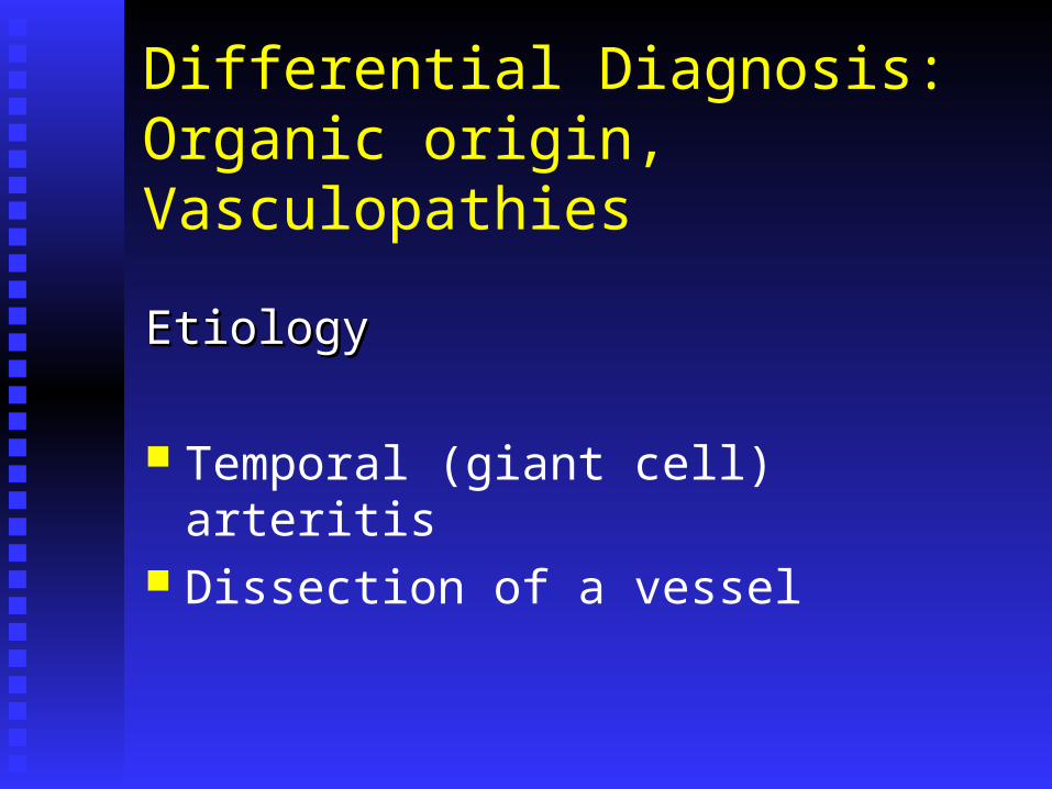
Differential Diagnosis:Organic origin, Vasculopathies
EtiologyEtiology
Temporal (giant cell) arteritis Dissection of a vessel

Differential Diagnosis:Organic origin, Vasculopathies
Signs & Symptoms of Temporal ArteritisSigns & Symptoms of Temporal Arteritis
Character: throbbing and sharp, burning pain Location: focal headache in the temporal or frontal-
occipital region Onset: gradual and progressive Aggravated: headache worse at night and with cold Risk: most common in white females > 50 years old Associated: weight loss, fever, fatigue, polymyalgia
rheumatica, monocular visual loss, jaw claudication
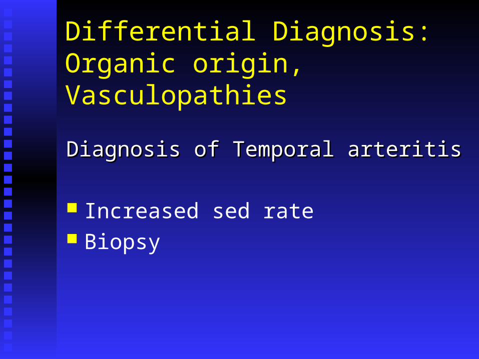
Differential Diagnosis:Organic origin, Vasculopathies
Diagnosis of Temporal arteritisDiagnosis of Temporal arteritis
Increased sed rate Biopsy

Differential Diagnosis:Organic origin, Vasculopathies
Signs & Symptoms (Dissection of vessel)Signs & Symptoms (Dissection of vessel)
Severe, localized HA History of trauma or vigorous exertion Diagnosis with CT

Differential Diagnosis:Organic origin, Acute Purulent Sinusitis Involving the frontal, maxillary, sphenoidal, or
ethmoidal sinuses True “sinus HA” is rare; if present, the patient is
usually very ill, with a severe localized HA for hours or days, PND & tender sinuses; often misdiagnosed as tension HA or common migraine but may have these as concomitant HA
Diagnosis: CT


Differential Diagnosis:Tension HeadacheEtiology
Skeletal componentsSkeletal components
Somatic dysfunctions of the upper cervical unit are going to impinge on the upper cervical nerves which have afferents in the cranium and dura
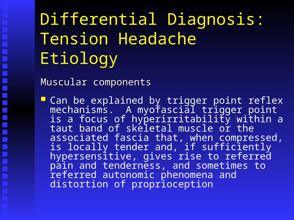
Differential Diagnosis:Tension HeadacheEtiology
Muscular componentsMuscular components
Can be explained by trigger point reflex mechanisms. A myofascial trigger point is a focus of hyperirritability within a taut band of skeletal muscle or the associated fascia that, when compressed, is locally tender and, if sufficiently hypersensitive, gives rise to referred pain and tenderness, and sometimes to referred autonomic phenomena and distortion of proprioception

Differential Diagnosis:Tension HeadacheEtiology
Muscular componentsMuscular components
Trigger points can result directly from ischemia due to chronically tense muscles, acute overload, overwork fatigue, direct trauma, and chilling.
Trigger points can result indirectly from other trigger points (a.k.a. latent trigger points), visceral disease, arthritic joints, and by emotional distress

Differential Diagnosis:Tension HeadacheEtiologySoft tissue componentsSoft tissue components
Ligaments can refer pain to sclerotomes which need to be addressed to completely resolve the somatic dysfunction
LymphaticsLymphatics
Need to free up the thoracic inlet to allow drainage of fluids
Trapezius
The trapezius can have many trigger points but the ones located in the upper fibers are most relevant for cephalgia
Pain referral pattern: Posterolateral aspect of the neck, mastoid process, temple and back of the orbit, and the angle of the jaw

Trapezius
The patient can often be misdiagnosed as having cervical radiculopathy or atypical facial neuralgia. The normally minimal antigravity function of the upper trapezius is overstressed by any position or activity in which the trapezius helps to carry the weight of the arm for a prolonged period
The muscle can also be strained by chronic injury due to overload, carrying a heavy backpack, long telephone calls, and sleeping prone with the head turned to one side

Trapezius
The trapezius can also entrap the greater occipital nerve which enervates the skin of the scalp and the semispinalis capitis muscle

SternocleidomastoidSternal division Pain referral pattern: supra-orbital and
deep within the orbit, occipital ridge, and vertex
Associated autonomic findings: excessive lacrimation, reddening of the conjunctiva, apparent “ptosis,” and visual disturbances
SternocleidomastoidClavicular division
Pain referral pattern: frontal area which extends across the forehead to the other side, and posterior auricular
Associated proprioceptive findings: spatial disorientation
Sternocleidomastoid
The SCM trigger points can be activated by sleeping on two pillows and keeping the neck in a flexed position, or by keeping the neck in an extended position as in painting a ceiling or sitting in the front row of a theater with a high screen or elevated stage. The SCM is often injured in a “whiplash” injury that might occur in an automobile crash.

Temporalis
Pain referral pattern: widely throughout the temple, along the eyebrow, and behind the eye
Temporalis trigger points may be activated by bruxism, direct trauma such as a fall or an impact to the cranium. The temporalis muscle can also be activated secondary to spasm in the masseter muscle

Occipitofrontalis
Frontal division pain referral pattern: upward and over the forehead on the ipsilateral side
Occipital division pain referral pattern: laterally, diffusely over the back of the head and with pain deep in the orbit