genechip expression analysis - department of statistical science
TRANSCRIPT

GENE EXPRESSION MONITORING
GeneChip®
ExpressionAnalysis
Technical Manual
For Research Use Only.
Not for use in diagnostic procedures.
701021 Rev. 4
TrademarksAffymetrix®, GeneChip®, EASI™, ®, , ®, HuSNP™, GenFlex™, Jaguar™, MicroDB™, NetAffx™, CustomExpress™, Flying Objective™ Tools To Take You As Far As Your Vision™, and The Way Ahead™ are trademarks owned or used by Affymetrix, Inc.
GeneArray® is a registered U.S. trademark of Agilent Technologies, Inc.
Enzo is a registered trademark of Enzo Biochem, Inc., and BioArray is a trademark of Enzo Biochem, Inc.
QIAGEN is a registered trademark of QIAGEN GmbH, Hilden Germany.
Limited LicenseEXCEPT AS EXPRESSLY SET FORTH HEREIN, NO RIGHT TO COPY, MODIFY, DISTRIBUTE, MAKE DERIVATIVE WORKS OF, PUBLICLY DISPLAY, MAKE, HAVE MADE, OFFER TO SELL, SELL, USE, OR IMPORT PROBE ARRAYS OR ANY OTHER PRODUCT IS CONVEYED OR IMPLIED WITH THE PROBE ARRAYS, INSTRUMENTS, SOFTWARE, REAGENTS, OR ANY OTHER ITEMS PROVIDED HEREUNDER. EXCEPT FOR CERTAIN ARRAYS AND REAGENTS DESIGNATED AS "ANALYTE SPECIFIC REAGENTS" (SEE APPLICABLE PACKAGE INSERT) WHICH ARE LICENSED FOR USE AS ANALYTE SPECIFIC REAGENTS OR RESEARCH USE, ALL PRODUCTS (INCLUDING THE PROBE ARRAYS, INSTRUMENTS, SOFTWARE, AND REAGENTS) DELIVERED HEREUNDER ARE LICENSED TO BUYER FOR RESEARCH USE ONLY. THIS LIMITED LICENSE PERMITS ONLY THE USE BY BUYER OF THE PARTICULAR PRODUCT(S), IN ACCORDANCE WITH THE WRITTEN INSTRUCTIONS PROVIDED THEREWITH, THAT BUYER PURCHASES FROM AFFYMETRIX (AFX) OR ITS AUTHORIZED REPRESENTATIVE. THE PURCHASE OF ANY PRODUCT(S) DOES NOT BY ITSELF CONVEY OR IMPLY THE RIGHT TO USE SUCH PRODUCT(S) IN COMBINATION WITH ANY OTHER PRODUCT(S). IN PARTICULAR, (I) NO RIGHT TO MAKE, HAVE MADE, OR DISTRIBUTE OTHER PROBE ARRAYS IS CONVEYED OR IMPLIED BY THE PROBE ARRAYS, (II) NO RIGHT TO MAKE, HAVE MADE, IMPORT, DISTRIBUTE, OR USE PROBE ARRAYS IS CONVEYED OR IMPLIED BY THE INSTRUMENTS OR SOFTWARE, AND (III) NO RIGHT TO USE PROBE ARRAYS IN COMBINATION WITH INSTRUMENTS OR SOFTWARE IS CONVEYED UNLESS ALL COMPONENT PARTS HAVE BEEN PURCHASED FROM AFX OR ITS AUTHORIZED REPRESENTATIVE. FURTHERMORE, PROBE ARRAYS DELIVERED HEREUNDER ARE LICENSED FOR ONE (1) TIME USE ONLY AND MAY NOT BE REUSED. THE PRODUCTS DO NOT HAVE FDA APPROVAL. NO PATENT LICENSE IS CONVEYED TO BUYER TO USE, AND BUYER AGREES NOT TO USE, THE PRODUCTS IN ANY SETTING REQUIRING FDA OR SIMILAR REGULATORY APPROVAL OR EXPLOIT THE PRODUCTS IN ANY MANNER NOT EXPRESSLY AUTHOIRZED IN WRITING BY AFX IN ADVANCE.
PROBE ARRAYS, INSTRUMENTS, SOFTWARE, AND REAGENTS ARE LICENSED FOR RESEARCH USE ONLY AND NOT FOR USE IN DIAGNOSTIC PROCEDURES. NO RIGHT TO MAKE, HAVE MADE, OFFER TO SELL, SELL, OR IMPORT OLIGONUCLEOTIDE PROBE ARRAYS OR ANY OTHER PRODUCT IN WHICH AFFYMETRIX HAS PATENT RIGHTS IS CONVEYED BY THE SALE OF PROBE ARRAYS, INSTRUMENTS, SOFTWARE, OR REAGENTS HEREUNDER. THIS LIMITED LICENSE PERMITS ONLY THE USE OF THE PARTICULAR PRODUCT(S) THAT THE USER HAS PURCHASED FROM AFFYMETRIX.
PatentsProducts may be covered by one or more of the following patents and/or sold under license from Oxford Gene Technology: U.S. Patent Nos. 5,445,934; 5,744,305; 6,261,776; 6,291,183; 5,700,637; 5,945,334; 6,346,413; and 6,399,365; and EP 619 321; 373 203 and other U.S. or foreign patents.
Software products may be covered by one or more of the following patents: U.S. Patent Nos. 5,733,729; 5,795,716; 5,974,164; 6,066,454; 6,090,555, 6,185,561 and 6,188,783; and other U.S. or foreign patents.
Fluidics Station products may be covered by U.S. Patent No. 6,114,122; and 6,391,623; 6,422,249; and other U.S. or foreign patents.
Scanner products may be covered by one or more of the following patents: U.S. Patent Nos. 5,578,832; 5,631,734; 5,834,758; 5,936,324; 5,981,956; 6,025,601; 6,141,096; 6,171,793; 6,185,030; 6,201,639; 6,207,960; 6,218,803; 6,225,625; 6,252,236; 6,335,824; 6,403,320; 6,407,858; 6,472,671; and 6,490,533; and other U.S. or foreign patents.
Copyright©1999 - 2003 Affymetrix, Inc. All rights reserved.

Overv
iew
Eu
kary
oti
cP
rok
ary
oti
cF.
S.
Main
ten
an
ce
cp
pen
dic
es
Contents
SECTION 1 Overview
Chapter 1 GeneChip® Expression Analysis Overview 1.1.3
SECTION 2 Eukaryot ic Sample and Array Process ing
Chapter 1 Eukaryotic Target Preparation 2.1.3
Chapter 2 Controls for Eukaryotic Arrays 2.2.3
Chapter 3 Eukaryotic Target Hybridization 2.3.3
Chapter 4 Eukaryotic Arrays: Washing, Staining, and Scanning 2.4.3
SECTION 3 Prokaryot ic Sample and Array Processing
Chapter 1 Prokaryotic Target Preparation 3.1.3
Chapter 2 Preparation of Control Spike-In Transcripts 3.2.3
Chapter 3 Prokaryotic Target Hybridization 3.3.3
Chapter 4 Prokaryotic Arrays:Washing, Staining, and Scanning 3.4.3
SECTION 4
Chapter 1 Fluidics Station Maintenance Procedures 4.1.3
701021 Rev. 4 iii
A

CONTENTS
iv
SECTION 5 Appendices
Appendix A Supplier and Reagent Reference List 5.A.3
Appendix B FAQs & Troubleshooting 5.B.3
Appendix C List of Controls on GeneChip Probe Arrays 5.C.3
Appendix D Technical Bulletins 5.D.3
Appendix E Probe Array Information 5.E.3

Section 1:
GeneChip® Expression Analysis Overview
701022 Rev. 2

Section 1

Overv
iew
Contents
Section 1
Chapter 1 GeneChip® Expression Analysis Overview 1.1.3
701022 Rev. 2
Overv
iew
Section 1, Chapter 1
701023 Rev. 3

Section 1, Chapter 1
Overv
iew
GeneChip® Expression Analysis Overview
Introduction and Objectives . . . . . . . . . . . . . . . . . . . . . . . . . . . . . . . 1.1.4
Explanation of GeneChip® Probe Arrays . . . . . . . . . . . . . . . . . . . . . . . . 1.1.4
GeneChip® Expression Analysis Overview . . . . . . . . . . . . . . . . . . . . . . . 1.1.5
Precautions . . . . . . . . . . . . . . . . . . . . . . . . . . . . . . . . . . . . . . . 1.1.6
Terminology . . . . . . . . . . . . . . . . . . . . . . . . . . . . . . . . . . . . . . . 1.1.7
Interfering Conditions . . . . . . . . . . . . . . . . . . . . . . . . . . . . . . . . . . 1.1.7
Instruments . . . . . . . . . . . . . . . . . . . . . . . . . . . . . . . . . . . . . . . 1.1.7
References . . . . . . . . . . . . . . . . . . . . . . . . . . . . . . . . . . . . . . . . 1.1.7
Limitations . . . . . . . . . . . . . . . . . . . . . . . . . . . . . . . . . . . . . . . 1.1.7
This Chapter Contains:
■ An overview of GeneChip® Expression Analysis.
■ A summary of the procedures covered in the remainder of the manual.
701023 Rev. 3 1.1.3
SECTION 1 GeneChip® Expression Analysis Overview
1.1.4
Introduction and Objectives
Welcome to the Affymetrix GeneChip® Expression Analysis Technical Manual. This manual is a technical guide for using GeneChip expression analysis probe arrays. All protocols included in this manual have been used successfully by scientists at Affymetrix, or have been recommended by our collaborators during the development of particular products. The field of mRNA gene expression monitoring is rapidly evolving and periodic technical updates to this manual will reflect the newest protocols and information for using GeneChip probe arrays. This manual applies to all GeneChip expression products.
As an Affymetrix GeneChip user, your feedback is welcome. Please contact our technical support team with any input on how we can improve this resource.
Explanation of GeneChip® Probe Arrays
GeneChip probe arrays are manufactured using technology that combines photolithography and combinatorial chemistry.1,2 Tens to hundreds of thousands of different oligonucleotide probes are synthesized on each array. Each oligonucleotide is located in a specific area on the array called a probe cell. Each probe cell contains millions of copies of a given oligonucleotide.
Probe arrays are manufactured in a series of cycles. Initially, a glass substrate is coated with linkers containing photolabile protecting groups. Then, a mask is applied that exposes selected portions of the probe array to ultraviolet light. Illumination removes the photolabile protecting groups enabling selective nucleoside phosphoramidite addition only at the previously exposed sites. Next, a different mask is applied and the cycle of illumination and chemical coupling is performed again. By repeating this cycle, a specific set of oligonucleotide probes is synthesized with each probe type in a known location. The completed probe arrays are packaged into cartridges.
During the laboratory procedure described in this manual, biotin-labeled RNA or DNA fragments referred to as the “target” are hybridized to the probe array. The hybridized probe array is stained with streptavidin phycoerythrin conjugate and scanned by the GeneChip® Scanner 3000, or the GeneArray® Scanner. The amount of light emitted at 570 nm is proportional to the bound target at each location on the probe array.

CHAPTER 1 GeneChip® Expression Analysis Overview
Overv
iew
GeneChip® Expression Analysis Overview
The following major steps outline GeneChip expression analysis:
1. Target Preparation
2. Target Hybridization
3. Experiment and Fluidics Station Setup
4. Probe Array Washing and Staining
5. Probe Array Scan
6. Data Analysis
Due to the differences in the RNA species between eukaryotic and prokaryotic organisms, different target labeling protocols have been optimized. Sections 2 and 3 provide detailed protocols for target preparation, hybridization, array washing, and staining for eukaryotic and prokaryotic arrays, respectively. Please refer to the sections in this manual for detailed protocols appropriate for your arrays.
Step 1: Target Preparation
This manual describes procedures for preparing biotinylated target from purified eukaryotic and prokaryotic RNA samples suitable for hybridization to GeneChip expression probe arrays. These procedures are recommendations only. For more information on these procedures, please contact Affymetrix Technical Support at 1-888-DNA-CHIP, or +44 (0)1628 552550 in Europe.
For eukaryotic samples, using protocols in this manual Section 2, double-stranded cDNA is synthesized from total RNA or purified poly-A messenger RNA isolated from tissue or cells. An in vitro transcription (IVT) reaction is then done to produce biotin-labeled cRNA from the cDNA. The cRNA is fragmented before hybridization.
For prokaryotic samples, Section 3 describes a detailed protocol to isolate total RNA followed by reverse transcription with random hexamers to produce cDNA. After fragmentation by DNase I, the cDNA is end-labeled with biotin by terminal transferase.
Step 2: Target Hybridization
A hybridization cocktail is prepared, including the fragmented target, probe array controls, BSA, and herring sperm DNA. It is then hybridized to the probe array during a 16-hour incubation. The hybridization process is described in the respective sections for the different probe array types.
1.1.5
SECTION 1 GeneChip® Expression Analysis Overview
1.1.6
Step 3: Experiment and Fluidics Station Setup
Specific experimental information is defined using Affymetrix® Microarray Suite on a PC-compatible workstation with a Windows 2000 operating system. The probe array type, sample description, and comments are entered in Microarray Suite and saved with a unique experiment name. The fluidics station is then prepared for use by priming with the appropriate buffers. For more information on the fluidics station, refer to the Fluidics Station User’s Guide.
Step 4: Probe Array Washing and Staining
Immediately following hybridization, the probe array undergoes an automated washing and staining protocol on the fluidics station.
Step 5: Probe Array Scan
Once the probe array has been hybridized, washed, and stained, it is scanned. Each workstation running Affymetrix Microarray Suite can control one scanner. The software defines the probe cells and computes an intensity for each cell.
Each complete probe array image is stored in a separate data file identified by the experiment name and is saved with a data image file (.dat) extension.
Review the scanner user’s manual for safety precautions and for more information on using the scanner.
Step 6: Data Analysis
Data is analyzed using the Microarray Suite Expression Analysis window. The .dat image is analyzed for probe intensities; results are reported in tabular and graphical formats. Information on data analysis is provided in the enclosed GeneChip® Expression Analysis: Data Analysis Fundamentals booklet (P/N 701190).
Precautions
1. FOR RESEARCH USE ONLY; NOT FOR USE IN DIAGNOSTIC PROCEDURES.
2. Avoid microbial contamination, which may cause erroneous results.
3. Exercise standard precautions when obtaining, handling, and disposing of potentially carcinogenic reagents.
4. Exercise care to avoid cross contamination of samples during all steps of this procedure, as this may lead to erroneous results.
5. Use powder-free gloves whenever possible to minimize introduction of powder particles into sample or probe array cartridges.
All biological specimens and materials with which they come into contact should be handled as if capable of transmitting infection and disposed of with proper precautions in accordance with federal, state, and local regulations. This includes adherence to the OSHA Bloodborne Pathogens Standard (29 CFR 1910.1030) for blood-derived and other samples governed by this act. Never pipet by mouth. Avoid specimen contact with skin and mucous membranes.

CHAPTER 1 GeneChip® Expression Analysis Overview
Overv
iew
Terminology
Interfering Conditions
Proper storage and handling of reagents and samples is essential for robust performance.
All laboratory equipment used to prepare the target during this procedure should be calibrated and carefully maintained to ensure accuracy, as incorrect measurement of reagents may affect the outcome of the procedure.
Instruments
The Affymetrix GeneChip Expression Analysis Technical Manual is designed for use in a system consisting of a Fluidics Station, a Hybridization Oven 640, and a Scanner.
References
1. Sambrook, J., Fritsch, E.F., Maniatis, T. Molecular Cloning: A Laboratory Manual, v.1 Cold Spring Harbor Laboratory Press, Cold Spring Harbor, NY p 21-52 (1989).
2. See www.affymetrix.com for current GeneChip technology references.
Limitations
■ The results of the assay kit are dependent upon the quality of the input RNA, subsequent proper handling of nucleic acids and other reagents.
■ The results should be evaluated by a qualified individual.
Probes The oligonucleotides on the surface of the probe arrays are called probes because they probe, or interrogate, the sample.
Target The target is the labeled nucleic acid that is being interrogated. It is hybridized to the probes on the array.
Probe Cell Specific areas on the probe array that contain oligonucleotides of a specific sequence.
Wear powder-free gloves throughout procedure. Take steps to minimize the introduction of exogenous nucleases. Water used in the protocols below is molecular biology grade (nuclease free).
Do not store enzymes in a frost-free freezer.
1.1.7

Section 2:
Eukaryotic Sample and Array Processing
701024 Rev. 2

Section 2

Eu
kary
oti
c
Contents
Section 2 Eukaryot ic Sample and Array Processing
Chapter 1 Eukaryotic Target Preparation 2.1.3
Chapter 2 Controls for Eukaryotic Arrays 2.2.3
Chapter 3 Eukaryotic Target Hybridization 2.3.3
Chapter 4 Eukaryotic Arrays: Washing, Staining, and Scanning 2.4.3
701024 Rev. 2

Eu
kary
oti
c
Section 2, Chapter 1
701025 Rev. 3

Section 2, Chapter 1
Eu
kary
oti
c
Eukaryotic Target Preparation
Reagents and Materials Required . . . . . . . . . . . . . . . . . . . . . . . . . . . . 2.1.5
Isolation of RNA . . . . . . . . . . . . . . . . . . . . . . . . . . . . . . . . . . . . 2.1.7
Isolation of RNA from Yeast . . . . . . . . . . . . . . . . . . . . . . . . . . . . . 2.1.7
Isolation of RNA from Arabidopsis. . . . . . . . . . . . . . . . . . . . . . . . . . 2.1.7
Isolation of RNA from Mammalian Cells or Tissues . . . . . . . . . . . . . . . . . 2.1.8
Precipitation of RNA . . . . . . . . . . . . . . . . . . . . . . . . . . . . . . . . . 2.1.8
Quantification of RNA . . . . . . . . . . . . . . . . . . . . . . . . . . . . . . . . 2.1.9
Synthesis of Double-Stranded cDNA from Total RNA. . . . . . . . . . . . . . . . 2.1.10
Step 1: First-strand cDNA Synthesis . . . . . . . . . . . . . . . . . . . . . . . . 2.1.10
Step 2: Second-Strand cDNA Synthesis . . . . . . . . . . . . . . . . . . . . . . 2.1.12
Synthesis of Double-Stranded cDNA from Purified Poly-A mRNA . . . . . . . . . 2.1.13
Step 1: First-Strand cDNA Synthesis . . . . . . . . . . . . . . . . . . . . . . . . 2.1.13
Step 2: Second-Strand cDNA Synthesis . . . . . . . . . . . . . . . . . . . . . . 2.1.14
Cleanup of Double-Stranded cDNA . . . . . . . . . . . . . . . . . . . . . . . . . 2.1.15
Synthesis of Biotin-Labeled cRNA . . . . . . . . . . . . . . . . . . . . . . . . . . 2.1.17
Cleanup and Quantification of Biotin-Labeled cRNA . . . . . . . . . . . . . . . . 2.1.19
Step 1: Cleanup of Biotin-Labeled cRNA . . . . . . . . . . . . . . . . . . . . . 2.1.19
Step 2: Quantification of the cRNA. . . . . . . . . . . . . . . . . . . . . . . . . 2.1.20
Step 3: Checking Unfragmented Samples by Gel Electrophoresis . . . . . . . . . 2.1.21
Fragmenting the cRNA for Target Preparation . . . . . . . . . . . . . . . . . . . . 2.1.21
This Chapter Contains:
■ General Guidelines for extracting RNA from eukaryotic cells or tissues using commercially available reagents and kits
■ Detailed steps for making double-stranded cDNA from extracted RNA.■ Guidelines for producing biotin-labeled antisense cRNA (target) using in vitro
transcription reaction (IVT) and the Enzo® BioArray™ HighYield™ RNA Transcript Labeling Kit
■ Instructions for fragmenting the labeled cRNA target
After completing the procedures described in this chapter, the labeled and fragmented cRNA target is hybridized to the probe array as described in Section 2, Chapter 3.
701025 Rev. 3 2.1.3

SECTION 2 Eukaryotic Sample and Array Processing
2.1.4
6WDUWLQJ�51$�VDPSOHV $SSUR[LPDW([SHULPHQW7LPH
���PLQV
��KU����PLQV
��KUV����PLQV
���PLQV
���PLQV
��KUV
���PLQV
���KUV
��KU����PLQV
�����PLQV
(XNDU\RWLF�7DUJHW�/DEHOLQJ�IRU�*HQH&KLSä �([SUHVVLRQ�$QDO\VL

CHAPTER 1 Eukaryotic Target Preparation
Eu
kary
oti
c
Reagents and Materials Required
The following reagents and materials are recommendations and have been tested and evaluated by Affymetrix scientists. For supplier phone numbers in the U.S. and Europe, please refer to the Supplier and Reagent Reference List, Appendix A, of this manual. Information and part numbers listed are based on U.S. catalog information. Additional reagents needed for the complete analysis are listed in the appropriate chapters. Appendix A contains a master list of all reagents used in this manual.
Total RNA Isolation■ TRIzol Reagent, Invitrogen Life Technologies, P/N 15596-018■ RNeasy Mini Kit, QIAGEN, P/N 74104
Poly-A mRNA Isolation■ Oligotex Direct mRNA Kit (isolation of mRNA from whole cells), QIAGEN,
P/N 72012, 72022, or 72041■ Oligotex mRNA Kit (isolation of mRNA from total RNA), QIAGEN, P/N 70022, 70042, or
70061■ Qiashredder, QIAGEN, P/N 79654 (Required only for use with QIAGEN Oligotex Direct
Kit)■ DEPC-Treated Water, Ambion, P/N 9920
cDNA Synthesis■ GeneChip T7-Oligo(dT) Promoter Primer Kit,
5´ - GGCCAGTGAATTGTAATACGACTCACTATAGGGAGGCGG-(dT)24 - 3´50 µM, HPLC purified, Affymetrix, P/N 900375
■ SuperScript II, Invitrogen Life Technologies, P/N 18064-014 or SuperScript Choice System for cDNA Synthesis, Invitrogen Life Technologies,P/N 18090-019
■ E. coli DNA Ligase, Invitrogen Life Technologies, P/N 18052-019■ E. coli DNA Polymerase I, Invitrogen Life Technologies, P/N 18010-025■ E. coli RNaseH, Invitrogen Life Technologies, P/N 18021-071■ T4 DNA Polymerase, Invitrogen Life Technologies, P/N 18005-025■ 5X Second-strand buffer, Invitrogen Life Technologies, P/N 10812-014■ 10 mM dNTP, Invitrogen Life Technologies, P/N 18427-013■ GeneChip Sample Cleanup Module, Affymetrix, P/N 900371
Synthesis of Biotin-Labeled cRNA
Do not store enzymes in a frost-free freezer.
SuperScript Choice System contains, in addition to SuperScript II Reverse Transcriptase, other reagents for cDNA synthesis. However, not all components provided in the Choice System are used in the GeneChip cDNA synthesis protocol.
RNA Transcript Labeling Kit, Affymetrix, P/N 900182EnzoBioArray
HighYield
2.1.5

SECTION 2 Eukaryotic Sample and Array Processing
2.1.6
IVT cRNA Cleanup and Quantification■ GeneChip Sample Cleanup Module, Affymetrix, P/N 900371■ 10X TBE, Cambrex, P/N 50843
cRNA Fragmentation■ GeneChip Sample Cleanup Module, Affymetrix, P/N 900371
Miscellaneous Reagents■ Absolute ethanol (stored at -20°C for RNA precipitation; store ethanol at room
temperature for use with the GeneChip Sample Cleanup Module)■ 80% ethanol (stored at -20°C for RNA precipitation; store ethanol at room temperature
for use with the GeneChip Sample Cleanup Module)■ SYBR Green II, Cambrex, P/N 50523; or Molecular Probes, P/N S7586 (optional)■ Pellet Paint, Novagen, P/N 69049-3 (optional)■ Glycogen, Ambion, P/N 9510 (optional)■ 3M Sodium Acetate (NaOAc), Sigma-Aldrich, P/N S7899■ Ethidium Bromide, Sigma-Aldrich, P/N E8751■ 1N NaOH■ 1N HCl■ 50 mM MgCl2
■ 0.5M EDTA
Miscellaneous Supplies■ Sterile, RNase-free, microcentrifuge vials, 1.5 mL, USA Scientific, P/N 1415-2600 (or
equivalent)■ Micropipettors, (P-2, P-20, P-200, P-1000), Rainin Pipetman or equivalent■ Sterile-barrier, RNase-free pipette tips (Tips must be pointed, not rounded, for efficient
use with the probe arrays.) Beveled pipette tips may cause damage to the array septa and cause leakage.
■ Mini agarose gel electrophoresis unit with appropriate buffers■ Vacuum filter units (1 liter capacity), VWR Scientific Products, P/N 28199-730■ UV spectrophotometer■ Cooling waterbath

CHAPTER 1 Eukaryotic Target Preparation
Eu
kary
oti
c
Isolation of RNA
Protocols are provided for preparing labeled cRNA from either total RNA or purified poly-A mRNA. We have found that results obtained from samples prepared by both of these methods are similar, but not identical. Therefore, to get the best results we suggest only comparing samples prepared using the same type of RNA material.
Please review precautions and interfering conditions in Section 1.
When using a commercial kit, follow the manufacturer’s instructions for RNA isolation.
Isolation of RNA from Yeast
Total RNA
We have successfully isolated good quality total RNA from yeast cells using a hot phenol protocol described by Schmitt, et al. Nucl Acids Res, 18:3091-3092 (1990).
Poly-A mRNA
Affymetrix recommends first purifying total RNA from yeast cells before isolating poly-A mRNA from total RNA. Good-quality mRNA has been successfully isolated from total RNA using QIAGEN’s Oligotex mRNA Kit. A single round of poly-A mRNA selection provides mRNA of sufficient purity and yield to use as a template for cDNA synthesis. Two rounds of poly-A mRNA selection will result in significantly reduced yield and is not generally recommended.
Isolation of RNA from Arabidopsis
Total RNA
We have been using TRIzol Reagent from Invitrogen Life Technologies to isolate total RNA from Arabidopsis. Please follow the instructions provided by the supplier and, when necessary, use the steps outlined specifically for samples with high starch and/or high lipid content.
Poly-A mRNA
We have successfully isolated Arabidopsis poly-A mRNA using QIAGEN Oligotex products. However, other standard isolation products are likely to be adequate.
The quality of the RNA is essential to the overall success of the analysis. Since the most appropriate protocol for the isolation of RNA can be source dependent, we recommend using a protocol that has been established for the tissues or cells being used. In the absence of an established protocol, we suggest using one of the commercially available kits designed for RNA isolation.
2.1.7
SECTION 2 Eukaryotic Sample and Array Processing
2.1.8
Isolation of RNA from Mammalian Cells or Tissues
Total RNA
We have successfully isolated high-quality total RNA from mammalian cells (such as cultured cells and lymphocytes) using the RNeasy Mini Kit from QIAGEN.
If mammalian tissue is used as the source of RNA, we recommend isolating total RNA with a commercial reagent, such as TRIzol.
Poly-A mRNA
Good-quality mRNA has been successfully isolated from mammalian cells (such as cultured cells and lymphocytes) using QIAGEN’s Oligotex Direct mRNA kit and from total RNA using the Oligotex mRNA kit. If mammalian tissue is used as the source of mRNA, total RNA should be first purified using a commercial reagent, such as TRIzol, and then using a poly-A mRNA isolation procedure or a commercial kit.
Precipitation of RNA
Total RNA
It is not necessary to precipitate total RNA following isolation or cleanup with RNeasy Mini Kit. Adjust elution volumes from the RNeasy column to prepare for cDNA synthesis based upon expected RNA yields from your experiment. Ethanol precipitation is required following TRIzol isolation and hot phenol extraction methods; see methods on page 2.1.9.
For smaller amounts of starting material, please refer to the alternative protocol for target labeling described in Small Sample Target Labeling Assay Version II, available at www.affymetrix.com.
Poly-A mRNA
Most poly-A mRNA isolation procedures will result in dilution of RNA. It is necessary to concentrate mRNA prior to the cDNA synthesis.
If going directly from TRIzol-isolated total RNA to cDNA synthesis, it may be beneficial to perform a second cleanup on the total RNA before starting. After the ethanol precipitation step in the TRIzol extraction procedure, perform a cleanup using QIAGEN RNeasy Mini Kit. Much better yields of labeled cRNA are obtained from the in vitro transcription-labeling reaction when this second cleanup is performed.
Affymetrix recommends starting the cDNA synthesis protocol with a minimum of 0.2 µg poly-A mRNA at a minimum concentration of 0.02 µg/µL, or 5 µg of total RNA at a minimum concentration of 0.5 µg/µL, in order to obtain sufficient quantity of labeled cRNA for target assessment and hybridization to GeneChip expression probe arrays. There are two major advantages to starting with at least the recommended amount of material:
1. Enough material to check sample yield and quality at the various steps of this protocol.
2. Production of enough cRNA for hybridization of the target to multiple probe arrays.

CHAPTER 1 Eukaryotic Target Preparation
Eu
kary
oti
c
Precipitation Procedure
1. Add 1/10 volume 3M NaOAc, pH 5.2, and 2.5 volumes ethanol.*
2. Mix and incubate at -20°C for at least 1 hour.
3. Centrifuge at ≥ 12,000 x g in a microcentrifuge for 20 minutes at 4°C.
4. Wash pellet twice with 80% ethanol.
5. Air dry pellet. Check for dryness before proceeding.
6. Resuspend pellet in DEPC-treated H2O. The appropriate volume for resuspension depends on the expected yield and the amount of RNA required for the cDNA synthesis. Please read ahead to the cDNA synthesis protocol in order to determine the appropriate resuspension volume at this step.
*Addition of Carrier to Ethanol Precipitations
Adding carrier material has been shown to improve the RNA yield of precipitation reactions.
■ Pellet PaintAddition of 0.5 µL of Pellet Paint per tube to nucleic acid precipitations makes the nucleic acid pellet easier to visualize and helps reduce the chance of losing the pellet during washing steps. The pellet paint does not appear to affect the outcome of subsequent steps in this protocol; however, it can contribute to the absorbance at 260 nm when quantifying the mRNA.
■ Glycogen
Addition of 0.5 to 1 µL of glycogen (5 mg/mL) to nucleic acid precipitations aids in visualization of the pellet and may increase recovery. The glycogen does not appear to affect the outcome of subsequent steps in this protocol.
Quantification of RNA
Quantify RNA yield by spectrophotometric analysis using the convention that 1 absorbance unit at 260 nm equals 40 µg RNA per mL.
■ The absorbance should be checked at 260 and 280 nm for determination of sample concentration and purity.
■ The A260/A280 ratio should be close to 2.0 for pure RNA (ratios between 1.9 and 2.1 are acceptable).
2.1.9
SECTION 2 Eukaryotic Sample and Array Processing
2.1.10
Synthesis of Double-Stranded cDNA from Total RNA
This protocol is a supplement to instructions provided in the Invitrogen Life Technologies SuperScript Choice system. Please note the following before proceeding:
■ Read all information and instructions that come with reagents and kits.■ Use the GeneChip T7-Oligo(dT) Promoter Primer Kit1 for priming first-strand cDNA
synthesis in place of the oligo(dT) or random primers provided with the SuperScript Choice kit. The GeneChip T7-Oligo(dT) Promoter Primer Kit provides high-quality HPLC-purified T7-oligo(dT) primer, which is essential for this reaction.
T7-oligo(dT) primer
5´ - GGCCAGTGAATTGTAATACGACTCACTATAGGGAGGCGG-(dT)24 - 3´
Step 1: First-strand cDNA Synthesis
Starting material: High-quality total RNA (5.0 µg - 20.0 µg)
After purification, the RNA concentration is determined by absorbance at 260 nm on a spectrophotometer (one absorbance unit = 40 µg/mL RNA). The A260/A280 ratio should be approximately 2.0, with ranges between 1.9 to 2.1 considered acceptable. We recommend checking the quality of the RNA by running it on an agarose gel prior to starting the assay. The rRNA bands should be clear without any obvious smearing patterns from degradation.
Before starting cDNA synthesis, the correct volumes of DEPC-treated H2O and Reverse Transcriptase (RT) must be determined. These volumes will depend on both the concentration and total volume of RNA that is being added to the reaction.
1. Users who do not purchase the GeneChip T7-Oligo(dT) Promoter Primer Kit may be required to obtain a license under U.S. Patent Nos. 5,569,584, 5,716,785, 5,891,636, 6,291,170, and 5,545,522 or to purchase another licensed kit.
For smaller amounts of starting material, please refer to the alternative protocol for target labeling described in Small Sample Target Labeling Assay Version II, available at www.affymetrix.com.
When using the GeneChip Sample Cleanup Module for the cDNA and IVT cRNA cleanup steps, there is a potential risk of overloading the columns if greater than the recommended amount of starting material is used.
Use Table 2.1.1 and Table 2.1.2 for variable component calculations. Determine the volumes of RNA and SuperScript II RT required in Table 2.1.1, then calculate the amount of DEPC-treated H2O needed in Step 1 Table 2.1.2 to bring the final First-Strand Synthesis volume to 20 µL.
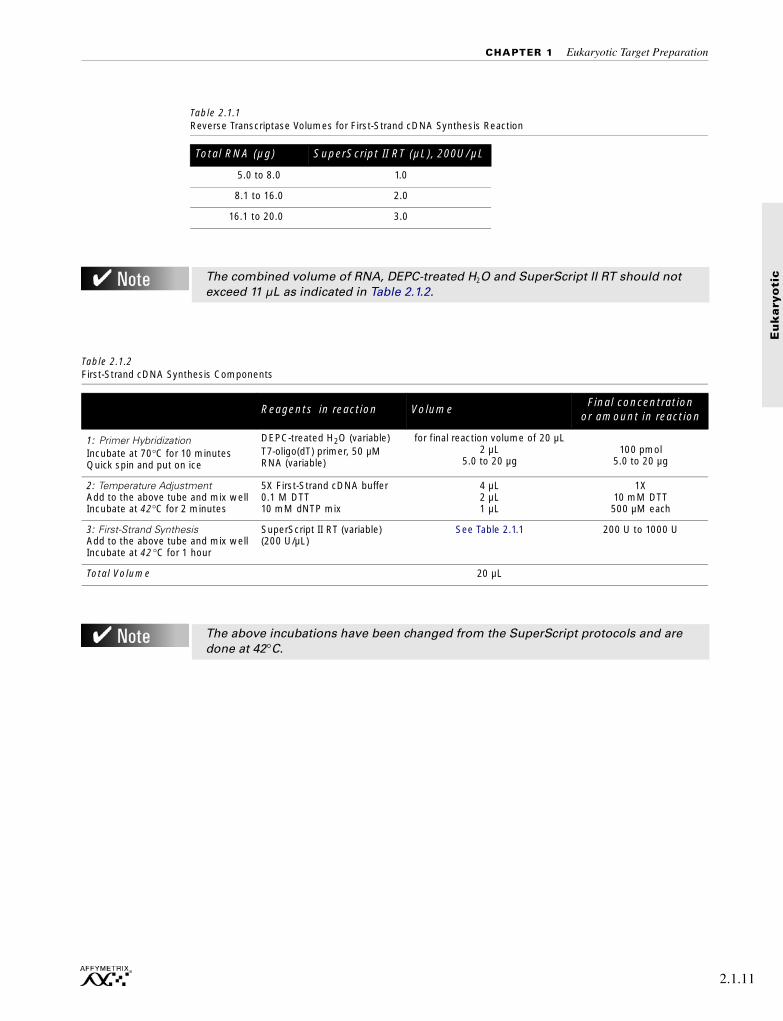
CHAPTER 1 Eukaryotic Target Preparation
Eu
kary
oti
c
.
Table 2.1.1Reverse Transcriptase Volumes for First-Strand cDNA Synthesis Reaction
Total RNA (µg) SuperScript II RT (µL), 200U/µL
5.0 to 8.0 1.0
8.1 to 16.0 2.0
16.1 to 20.0 3.0
The combined volume of RNA, DEPC-treated H2O and SuperScript II RT should not exceed 11 µL as indicated in Table 2.1.2.
Table 2.1.2First-Strand cDNA Synthesis Components
Reagents in reaction VolumeFinal concentration
or amount in reaction
1: Primer Hybridization Incubate at 70°C for 10 minutesQuick spin and put on ice
DEPC-treated H2O (variable) T7-oligo(dT) primer, 50 µMRNA (variable)
for final reaction volume of 20 µL2 µL
5.0 to 20 µg100 pmol
5.0 to 20 µg
2: Temperature AdjustmentAdd to the above tube and mix well Incubate at 42°C for 2 minutes
5X First-Strand cDNA buffer0.1 M DTT10 mM dNTP mix
4 µL2 µL1 µL
1X10 mM DTT
500 µM each
3: First-Strand SynthesisAdd to the above tube and mix wellIncubate at 42 °C for 1 hour
SuperScript II RT (variable)(200 U/µL)
See Table 2.1.1 200 U to 1000 U
Total Volume 20 µL
The above incubations have been changed from the SuperScript protocols and are done at 42°C.
2.1.11

SECTION 2 Eukaryotic Sample and Array Processing
2.1.12
Step 2: Second-Strand cDNA Synthesis
1. Place First-Strand reactions on ice. Centrifuge briefly to bring down condensation on sides of tube.
2. Add to the First-Strand synthesis tube the reagents listed in the following Second-Strand Final Reaction Composition Table (Table 2.1.3).
3. Gently tap tube to mix. Then, briefly spin in a microcentrifuge to remove condensation and incubate at 16°C for 2 hours in a cooling waterbath.
4. Add 2 µL [10 U] T4 DNA Polymerase.
5. Return to 16°C for 5 minutes.
6. Add 10 µL 0.5M EDTA.
7. Proceed to cleanup procedure for cDNA, Cleanup of Double-Stranded cDNA on page 2.1.15, or store at -20°C for later use.
Table 2.1.3 Second-Strand Final Reaction Composition
Component VolumeFinal Concentration or Amount in Reaction
DEPC-treated water 91 µL
5X Second-Strand Reaction Buffer 30 µL 1X
10 mM dNTP mix 3 µL 200 µM each
10 U/µL E. coli DNA Ligase 1 µL 10 U
10 U/µL E. coli DNA Polymerase I 4 µL 40 U
2 U/µL E. coli RNase H 1 µL 2 U
Final Volume 150 µL
CHAPTER 1 Eukaryotic Target Preparation
Eu
kary
oti
c
Synthesis of Double-Stranded cDNA from Purified Poly-A mRNA
This protocol is a supplement to instructions provided in the Invitrogen Life Technologies SuperScript Choice system. Please note the following before proceeding:
■ Read all information and instructions that come with reagents and kits.■ Use the GeneChip T7-Oligo(dT) Promoter Primer Kit2 for priming first-strand cDNA
synthesis in place of the oligo(dT) or random primers provided with the SuperScript Choice kit. The GeneChip T7-Oligo(dT) Promoter Primer Kit provides high-quality HPLC-purified T7-oligo(dT) primer, which is essential for this reaction.
■ It is recommended that each step of this protocol is checked by gel electrophoresis.
T7-oligo(dT) primer
5´ - GGCCAGTGAATTGTAATACGACTCACTATAGGGAGGCGG-(dT)24 - 3´
Step 1: First-Strand cDNA Synthesis
Starting material: High-quality poly-A mRNA (0.2 µg to 2.0 µg).
Before starting cDNA synthesis, the correct volumes of DEPC-treated H2O and Reverse Transcriptase (RT) must be determined. These volumes will depend on both the concentration and total volume of mRNA that is being added to the reaction. For every µg of mRNA, you will need to add 1 µL of SuperScript II RT (200 U/µL). For mRNA quantity ≤ 1 µg, use 1 µL of SuperScript II RT. Synthesis reactions should be done in a polypropylene tube (RNase-free).
2. Users who do not purchase the GeneChip T7-Oligo(dT) Promoter Primer Kit may be required to obtain a license under U.S. Patent Nos. 5,569,584, 5,716,785, 5,891,636, 6,291,170, and 5,545,522 or to purchase another licensed kit.
When using the GeneChip Sample Cleanup Module for the cDNA and IVT cRNA cleanup steps, there is a potential risk of overloading the columns if greater than the recommended amount of starting material is used.
Use Table 2.1.4 for variable component calculations. Determine volumes of mRNA and SuperScript II RT required, and then calculate the amount of DEPC-treated H2O needed in the Primer Hybridization Mix step to bring the final First-Strand Synthesis reaction volume to 20 µL.
2.1.13

SECTION 2 Eukaryotic Sample and Array Processing
2.1.14
Step 2: Second-Strand cDNA Synthesis
1. Place First-Strand reactions on ice. Centrifuge briefly to bring down condensation on sides of tube.
2. Add to the First-Strand synthesis tube the reagents listed in the following Second-Strand Final Reaction Composition Table (Table 2.1.5).
3. Gently tap tube to mix. Then, briefly spin in a microcentrifuge to remove condensation and incubate at 16°C for 2 hours in a cooling waterbath.
4. Add 2 µL [10 U] T4 DNA Polymerase.
5. Return to 16°C for 5 minutes.
6. Add 10 µL 0.5M EDTA.
7. Proceed to cleanup procedure for cDNA, Cleanup of Double-Stranded cDNA on page 2.1.15, or store at -20°C for later use.
Table 2.1.4First-Strand cDNA Synthesis Components
Reagents in Reaction VolumeFinal Concentration
or Amount in Reaction
1: Primer HybridizationIncubate at 70°C for 10 minutesQuick spin and put on ice
DEPC-treated H2O (variable) T7-oligo(dT) primer, 50 µMmRNA (variable)
for final reaction volume of 20 µL2 µL
0.2 to 2 µg100 pmol
0.2 to 2 µg
2: Temperature AdjustmentAdd to the above tube and mix wellIncubate at 37°C for 2 minutes
5X First-Strand cDNA buffer0.1 M DTT10 mM dNTP mix
4 µL2 µL1 µL
1X10 mM
500 µM each
3: First-Strand SynthesisAdd to the above tube and mix well Incubate at 37°C for 1 hour
SuperScript II RT (variable) (200 U/µL)
1 µL per µg mRNA 200 U to 1000 U
Total Volume 20 µL
Table 2.1.5 Second-Strand Final Reaction Composition
Component VolumeFinal Concentration or Amount in Reaction
DEPC-treated water 91 µL
5X Second-Strand Reaction Buffer 30 µL 1X
10 mM dNTP mix 3 µL 200 µM each
10 U/µL E. coli DNA Ligase 1 µL 10 U
10 U/µL E. coli DNA Polymerase I 4 µL 40 U
2 U/µL E. coli RNase H 1 µL 2 U
Final Volume 150 µL
CHAPTER 1 Eukaryotic Target Preparation
Eu
kary
oti
c
Cleanup of Double-Stranded cDNA
Reagents to be Supplied by User
■ Ethanol, 96-100% (v/v)
All other components needed for cleanup of double-stranded cDNA are supplied with the GeneChip Sample Cleanup Module.
1. Add 600 µL cDNA Binding Buffer to the 162 µL final double-stranded cDNA synthesis preparation (page 2.1.10 or 2.1.13). Mix by vortexing for 3 seconds.
2. Check that the color of the mixture is yellow (similar to cDNA Binding Buffer without the cDNA synthesis reaction).
3. Apply 500 µL of the sample to the cDNA Cleanup Spin Column sitting in a 2 mL Collection Tube, and centrifuge for 1 minute at ≥ 8,000 x g (≥ 10,000 rpm).
Discard flow-through.
4. Reload the spin column with the remaining mixture (262 µL) and centrifuge as above.
Discard flow-through and Collection Tube.
5. Transfer spin column into a new 2 mL Collection Tube (supplied). Pipet 750 µL cDNA Wash Buffer onto the spin column. Centrifuge for 1 minute at ≥ 8,000 x g (≥ 10,000 rpm).
Discard flow-through.
6. Open the cap of the spin column and centrifuge for 5 minutes at maximum speed (≤ 25,000 x g). Discard flow-through and Collection Tube.
Place columns into the centrifuge using every second bucket. Position caps over the adjoining bucket so that they are oriented in the opposite direction to the rotation (i.e., if the microcentrifuge rotates in a clockwise direction, orient the caps in a counter-clockwise direction). This avoids damage of the caps.
Centrifugation with open caps allows complete drying of the membrane.
BEFORE STARTING, please note:
■ cDNA Wash Buffer is supplied as a concentrate. Before using for the first time, add 24 mL of ethanol (96-100%), as indicated on the bottle, to obtain a working solution, and checkmark the box on the left-hand side of the bottle label to avoid confusion.
■ All steps of the protocol should be performed at room temperature. During the procedure, work without interruption.
■ If cDNA synthesis was performed in a reaction tube smaller than 1.5 mL, transfer the reaction mixture into a 1.5 or 2 mL microfuge tube (not supplied) prior to addition of cDNA Binding Buffer.
If the color of the mixture is orange or violet, add 10 µL of 3 M sodium acetate, pH 5.0, and mix. The color of the mixture will turn to yellow.
cDNA Wash Buffer is supplied as a concentrate. Ensure that ethanol is added to the cDNA Wash Buffer before use (see IMPORTANT note above before starting).
2.1.15

SECTION 2 Eukaryotic Sample and Array Processing
2.1.16
7. Transfer spin column into a 1.5 mL Collection Tube, and pipet 14 µL of cDNA Elution Buffer directly onto the spin column membrane. Incubate for 1 minute at room temperature and centrifuge 1 minute at maximum speed (≤ 25,000 x g) to elute.
Ensure that the cDNA Elution Buffer is dispensed directly onto the membrane. The average volume of eluate is 12 µL from 14 µL Elution Buffer.
8. An aliquot of the cDNA prepared from isolated poly-A RNA can be analyzed for size distribution and yield on a 1% agarose gel. One microliter of double-stranded cDNA should be sufficient to detect on an agarose gel stained with ethidium bromide. A representative gel is shown in Figure 2.1.1 on page 2.1.22. We do not recommend gel analysis for cDNA prepared from total RNA.
9. After cleanup, please proceed to Synthesis of Biotin-Labeled cRNA on page 2.1.17.
We do not recommend RNase treatment of the cDNA prior to the in vitro transcription and labeling reaction; the carry-over ribosomal RNA does not seem to inhibit the reaction.
Quantifying the amount of double-stranded cDNA by absorbance at 260 nm is not recommended. The primer can contribute significantly to the absorbance. Subtracting the theoretical contribution of the primer based on the amount added to the reaction is not practical.

CHAPTER 1 Eukaryotic Target Preparation
Eu
kary
oti
c
Synthesis of Biotin-Labeled cRNA
1. Enzo® BioArray™ HighYield™ RNA Transcript Labeling Kit3 (Affymetrix, P/N 900182) is used for generating labeled cRNA target. Use the following tables to determine the amount of cDNA used for each IVT reaction. Done properly, each reaction should produce sufficient biotin-labeled target to hybridize to at least two Standard Format (49 Format) GeneChip expression probe arrays in parallel.
The purity and quality of template cDNA is important for high yields of biotin-labeled RNA.
Use only RNase-free water, buffers, and pipette tips.
Store all reagents at -20°C, in a freezer that is not self-defrosting.
Prior to use, centrifuge all reagents briefly to ensure that the components remain at the bottom of the tube.
The product should be used only until the expiration date stated on the label.
3. For Research Use Only. This product is manufactured by ENZO LIFE SCIENCES, INC. for distribution by Affymetrix, Inc. for research purposes only by the end-user and is not intended for diagnostic or therapeutic use. Purchase does not include a license or the right to utilize this product except for research purposes. Purchase does not include the right to distribute or sell this product commercially. As distributed by Affymetrix. Inc., this product may be used only in conjunction with, and is permitted for use only with, Affymetrix GeneChipÆ probe arrays.
Enzo is a registered trademark of Enzo Biochem, Inc. and BioArray is a trademark of Enzo Biochem, Inc.
This product or the use of this product is covered by one or more claims of Enzo patents including, but not limited to, the following: U.S. Patent Nos. 5,328,824; 5,449,767; 5,476,928; 4,711,955 and 4,994,373; EP 0 063 879 B1; EP 0 329 198 B1; DK 171 822 B; Canadian Patent Nos. 1,219,824 and 1,309,672; Japanese Patent Nos. 2,131,266; 1,416,584 and other patents pending.
Table 2.1.6 cDNA in IVT (Total RNA)
Total RNA (µg) Volume of cDNA to use in IVT*
5.0 to 8.0 10 µL
8.1 to 16.0 5 µL
16.1 to 20.0 3.3 µL
* assuming 12 µL was eluted from the column, as previously described.
Table 2.1.7 cDNA in IVT (Poly-A mRNA)
Poly-A mRNA (µg) Volume cDNA to use in IVT*
0.2 - 0.5 10 µL
0.6 - 1.0 8 µL
1 - 2 5 µL
* assuming 12 µL was eluted from the column, as previously described.
2.1.17

SECTION 2 Eukaryotic Sample and Array Processing
2.1.18
2. Add to RNase-free microfuge tubes template cDNA and additions of other reaction components in the order indicated in the following table. Keep reactions at room temperature while additions are made to avoid precipitation of DTT.
3. Carefully mix the reagents and collect the mixture in the bottom of the tube by brief(5 second) microcentrifugation.
4. Immediately place the tube in a 37°C water bath. Incubate for 4 to 5 hours, gently mixing the contents of the tube every 30-45 minutes during the incubation.
5. Store labeled cRNA at -20°C, or -70°C (long-term storage) if not purifying immediately.
6. Proceed to cRNA cleanup procedure, Cleanup and Quantification of Biotin-Labeled cRNA on page 2.1.19
Each GeneChip® Sample Cleanup Module contains 30 cDNA cleanup columns and 30 IVT cRNA cleanup columns. If more than one IVT is carried out from a single cDNA sample and is purified on separate IVT cRNA cleanup columns, there will not be sufficient IVT cRNA columns in each kit for 30 samples.
Table 2.1.8IVT cRNA Labeling
Reagent Volume
Template cDNA Variable. Refer to Table 2.1.6 and Table 2.1.7.
Distilled or deionized water Variable (to give a final reaction volume of 40 µL).
10X HY Reaction Buffer (Vial 1) 4 µL
10X Biotin-Labeled Ribonucleotides (Vial 2)
4 µL
10X DTT (Vial 3) 4 µL
10X RNase Inhibitor Mix (Vial 4) 4 µL
20X T7 RNA Polymerase (Vial 5) 2 µL
Total Volume 40 µL
Overnight incubation may produce shorter products, which is less desirable.

CHAPTER 1 Eukaryotic Target Preparation
Eu
kary
oti
c
Cleanup and Quantification of Biotin-Labeled cRNA
Reagents to be Supplied by User
■ Ethanol, 96-100% (v/v)■ Ethanol, 80% (v/v)
All other components needed for cleanup of biotin-labeled cRNA are supplied with the GeneChip Sample Cleanup Module.
Step 1: Cleanup of Biotin-Labeled cRNA
1. Add 60 µL of RNase-free water to the in vitro transcription reaction and mix by vortexing for 3 seconds.
2. Add 350 µL IVT cRNA Binding Buffer to the sample and mix by vortexing for 3 seconds.
3. Add 250 µL ethanol (96-100%) to the lysate, and mix well by pipetting. Do not centrifuge.
4. Apply sample (700 µL) to the IVT cRNA Cleanup Spin Column sitting in a 2 mL Collection Tube. Centrifuge for 15 seconds at ≥ 8,000 x g (≥ 10,000 rpm). Discard flow-through and Collection Tube.
5. Transfer the spin column into a new 2 mL Collection Tube (supplied). Pipet 500 µL IVT cRNA Wash Buffer onto the spin column. Centrifuge for 15 seconds at ≥ 8,000 x g (≥ 10,000 rpm) to wash. Discard flow-through.
6. Pipet 500 µL 80% (v/v) ethanol onto the spin column and centrifuge for 15 seconds at ≥ 8,000 x g (≥ 10,000 rpm). Discard flow-through.
BEFORE STARTING please note:
■ It is essential to remove unincorporated NTPs, so that the concentration and purity of cRNA can be accurately determined by 260 nm absorbance.
■ DO NOT extract biotin-labeled RNA with phenol-chloroform. The biotin will cause some of the RNA to partition into the organic phase. This will result in low yields.
■ Save an aliquot of the unpurified IVT product for analysis by gel electrophoresis.■ IVT cRNA Wash Buffer is supplied as a concentrate. Before using for the first time, add
20 mL of ethanol (96-100%) as indicated on the bottle to obtain a working solution, and checkmark the box on the left-hand side of the bottle label to avoid confusion.
■ IVT cRNA Binding Buffer may form a precipitate upon storage. If necessary, redissolve by warming in a water bath at 30°C, and then place the buffer at room temperature.
■ All steps of the protocol should be performed at room temperature. During the procedure, work without interruption.
IVT cRNA Wash Buffer is supplied as a concentrate. Ensure that ethanol is added to the IVT cRNA Wash Buffer before use (see IMPORTANT note above before starting).
2.1.19
SECTION 2 Eukaryotic Sample and Array Processing
2.1.20
7. Open the cap of the spin column and centrifuge for 5 minutes at maximum speed(≤ 25,000 x g). Discard flow-through and Collection Tube.
Place columns into the centrifuge using every second bucket. Position caps over the adjoining bucket so that they are oriented in the opposite direction to the rotation (i.e., if the microcentrifuge rotates in a clockwise direction, orient the caps in a counter-clockwise direction). This avoids damage of the caps. Centrifugation with open caps allows complete drying of the membrane.
8. Transfer spin column into a new 1.5 mL Collection Tube (supplied), and pipet 11 µL of RNase-free water directly onto the spin column membrane. Ensure that the water is dispensed directly onto the membrane. Centrifuge 1 minute at maximum speed (≤ 25,000 x g) to elute.
9. Pipet 10 µL of RNase-free water directly onto the spin column membrane. Ensure that the water is dispensed directly onto the membrane. Centrifuge 1 minute at maximum speed (≤ 25,000 x g) to elute.
For subsequent photometric quantification of the purified cRNA, we recommend dilution of the eluate between 1:100 fold and 1:200 fold.
Step 2: Quantification of the cRNA
Use spectrophotometric analysis to determine the cRNA yield. Apply the convention that 1 absorbance unit at 260 nm equals 40 µg/mL RNA.
■ Check the absorbance at 260 nm and 280 nm to determine sample concentration and purity.
■ Maintain the A260/A280 ratio close to 2.0 for pure RNA (ratios between 1.9 and 2.1 are acceptable).
For quantification of cRNA when using total RNA as starting material, an adjusted cRNA yield must be calculated to reflect carryover of unlabeled total RNA. Using an estimate of 100% carryover, use the formula below to determine adjusted cRNA yield:
adjusted cRNA yield = RNAm - (total RNAi)(y)
RNAm = amount of cRNA measured after IVT (µg)total RNAi = starting amount of total RNA (µg)y = fraction of cDNA reaction used in IVT
Example: Starting with 10 µg total RNA, 50% of the cDNA reaction is added to the IVT, giving a yield of 50 µg cRNA. Therefore, adjusted cRNA yield = 50 µg cRNA - (10 µg total RNA) (0.5 cDNA reaction) = 45.0 µg.
Use adjusted yield in Eukaryotic Target Hybridization on page 2.3.3.
The minimum concentration for purified cRNA is 0.6 µg/µL before starting the following fragmentation reaction in "Fragmenting the cRNA for Target Preparation" on page 2.1.21.
Please refer to Table 2.3.1 on page 2.3.7 for the amount of cRNA required for one array hybridization experiment. The amount varies depending on the array format. Please refer to the specific probe array package insert for information on the array format.
CHAPTER 1 Eukaryotic Target Preparation
Eu
kary
oti
c
Step 3: Checking Unfragmented Samples by Gel Electrophoresis
Gel electrophoresis of the IVT product is done to estimate the yield and size distribution of labeled transcripts. Parallel gel runs of unpurified and purified IVT product can help determine the extent of a loss of sample during the cleanup process.
■ Run 1% of each sample on a 1% agarose gel.■ Mix RNA (samples or markers) with loading dye and heat to 65°C for 5 minutes before
loading on the gel. ■ Ethidium bromide can be used to visualize the RNA in the gel.
Alternatively, gels can be stained with SYBR Green II at a 1:10,000 dilution in 1X TBE buffer. SYBR Green II stains single-stranded RNA with greater sensitivity than ethidium bromide, but it requires a special photographic filter available from Molecular Probes to photograph stained bands.
■ As an option, run a denaturing gel to obtain a more accurate estimation of the RNA size distribution. Please refer to Figure 2.1.1 for the typical size distribution of unfragmented cRNA.
Fragmenting the cRNA for Target Preparation
5X Fragmentation Buffer is supplied with the GeneChip Sample Cleanup Module.
Fragmentation of cRNA target before hybridization onto GeneChip probe arrays has been shown to be critical in obtaining optimal assay sensitivity.
Affymetrix recommends that the cRNA used in the fragmentation procedure be sufficiently concentrated to maintain a small volume during the procedure. This will minimize the amount of magnesium in the final hybridization cocktail. The cRNA must be at a minimum concentration of 0.6 µg/µL. Fragment an appropriate amount of cRNA for hybridization cocktail and gel analysis (see Section 2, Chapter 3, Table 2.3.1).
1. Add 2 µL of 5X Fragmentation Buffer for every 8 µL of RNA plus H2O. The fragmentation buffer has been optimized to break down full-length cRNA to 35 to 200 base fragments by metal-induced hydrolysis.
The final concentration of RNA in the fragmentation mix can range from 0.5 µg/µL to 2 µg/µL. The following table shows an example of a fragmentation mix for a 20 µg cRNA sample at a final concentration of 0.5 µg/µL.
For fragmentation, use adjusted cRNA concentration, as described in Step 2: Quantification of the cRNA on page 2.1.20.
Example for 0.5 µg/µL final concentration:
2. Incubate at 94°C for 35 minutes. Put on ice following the incubation.
Table 2.1.9 Example of Fragmentation Reaction
Component Volume
20 µg cRNA 1 to 21 µL
5X Fragmentation Buffer
8 µL
RNase-free water to 40 µL
2.1.21

SECTION 2 Eukaryotic Sample and Array Processing
2.1.22
3. Save an aliquot for gel analysis.
At least 1 µg fragmented cRNA is needed if using ethidium bromide for staining the gel. Less RNA can be used with SYBR Green II staining. See Step 3: Checking Unfragmented Samples by Gel Electrophoresis on page 2.1.21, for information regarding gel electrophoresis. The standard fragmentation procedure should produce a distribution of RNA fragment sizes from approximately 35 to 200 bases. An example of a gel with cRNA samples before and after fragmentation is shown below.
4. Store undiluted, fragmented sample RNA at -20°C until ready to perform the hybridization, as described in Section 2, Chapter 3.
Figure 2.1.1 Monitoring of target preparation by agarose gel electrophoresis
Gib
co B
RL
1 Kb
DN
A L
adde
r
purif
ied
cRN
A –
500
ng
Am
bion
RN
A C
entu
ry M
arke
r
1636
1018
506, 517
200
100
doub
le-s
tran
ded
cDN
A–3
00 n
gfr
agm
ente
d cR
NA
– 1
µg

Eu
kary
oti
c
Section 2, Chapter 2
701026 Rev. 3

Section 2, Chapter 2
Eu
kary
oti
c
Controls for Eukaryotic Arrays
Reagents and Materials Required . . . . . . . . . . . . . . . . . . . . . . . . . . . . 2.2.5
Hybridization Control Kit . . . . . . . . . . . . . . . . . . . . . . . . . . . . . . . . 2.2.7
Poly-A Spike Controls . . . . . . . . . . . . . . . . . . . . . . . . . . . . . . . . . 2.2.7
This Chapter Contains:
■ General guidelines for producing controls for eukaryotic arrays.
After completing the procedures described in this chapter, the control transcripts are combined in variable concentrations before adding to the target hybridization mix, as explained in Section 2, Chapter 3.
701026 Rev. 3 2.2.3

CHAPTER 2 Controls for Eukaryotic Arrays
Eu
kary
oti
c
Reagents and Materials Required
The following reagents and materials are recommendations and have been tested and evaluated by Affymetrix scientists. For supplier phone numbers in the U.S. and Europe, please refer to the Supplier and Reagent Reference List, Appendix A, of this manual. Information and part numbers listed are based on U.S. catalog information. Additional reagents needed for the complete analysis are listed in the appropriate chapters. Appendix A contains a master list of all reagents used in this manual.
GeneChip® Eukaryotic Hybridization Control Kit (Complete Kit)■ Affymetrix, P/N 900299 (30 reactions) or P/N 900362 (150 reactions)
Poly-A Spike Controls■ pGIBS-lys ATCC 87482
■ pGIBS-phe ATCC 87483
■ pGIBS-thr ATCC 87484
■ pGIBS-trp ATCC 87485
■ pGIBS-dap ATCC 87486
2.2.5
CHAPTER 2 Controls for Eukaryotic Arrays
Eu
kary
oti
c
Hybridization Control Kit
Each commercially available eukaryotic probe array contains probe sets for several prokaryotic genes as controls. These probe sets are readily identified by the AFFX prefix in the probe set name. The .chp data for these control probe sets can be examined in the Summary Report File (.rpt).
Control Oligo B2
Control Oligo B2 hybridizes to features along the outer edge of all expression arrays and to the checkerboard pattern in each corner. These predefined patterns provide signals for the Affymetrix® Microarray Suite software to perform automatic grid alignment during image analysis. They can also be used to align the grid manually. The fluorescence intensities for Control Oligo B2 are not used for analyzing data.
A 60X stock of the B2 oligo is provided as part of the GeneChip Eukaryotic Hybridization Control Kit (P/N 900299 or 900362, for 30 or 150 reactions, respectively), or can be purchased alone (P/N 900301). Please refer to the instructions in Section 2, Chapter 3 for detailed information on including the B2 oligo in preparing the hybridization cocktail.
Biotinylated Hybridization Controls: bioB, bioC, bioD, and cre
BioB, bioC, and bioD are genes of the biotin synthesis pathway from the bacteria E. coli, and cre is the recombinase gene from P1 bacteriophage. A ready-prepared mixture of these biotinylated controls at staggered concentrations can be added with labeled eukaryotic cRNA samples to hybridize onto GeneChip probe arrays. Signal intensities obtained on these genes provide information on how well the hybridization, washing, and staining procedures have performed.
Affymetrix provides a kit that contains a 20X pre-mixed control reagent (P/N 900299 or 900362) and the final concentrations in the hybridization cocktail are 1.5 pM, 5 pM, 25 pM, and 100 pM for the four transcripts bioB, bioC, bioD, and cre, respectively.
Poly-A Spike Controls
Five poly-A-tailed control clones encoding B. subtilis genes (dap, thr, trp, phe, lys) are cloned into pBluescript as an Xho I to Not I insert, 5´ to 3´, respectively.
Poly-A-tailed constructs (dap, thr, trp, phe, lys)
These clones can be cut with different restriction enzymes to produce template DNA for either sense strand RNA synthesis or antisense RNA synthesis. The antisense control RNA for each B. subtilis gene is synthesized from linearized plasmid using T7 RNA polymerase with biotinylated nucleotides. The sense RNA for each B. subtilis gene is synthesized from linearized plasmid using T3 RNA polymerase with unlabeled nucleotides. For detailed preparation of sense RNA controls, please refer to Section 3, Chapter 2.
T3 5’
Xho I Not I
AAAAA 3’ T7
2.2.7

SECTION 2 Eukaryotic Sample and Array Processing
2.2.8
The antisense strand B. subtilis RNA controls are used as described above for bioB, bioC, and bioD genes. The sense strand RNA controls can be spiked into samples during mRNA preparation to monitor the efficiency of target preparation, hybridization, wash, and stain.
Bacteria containing these recombinant plasmids can be obtained from the American Type Culture Collection (ATCC). See Reagents and Materials Required on page 2.2.5 for details.

Eu
kary
oti
c
Section 2, Chapter 3
701027 Rev. 3

Section 2, Chapter 3

Eu
kary
oti
c
Eukaryotic Target Hybridization
Reagents and Materials Required . . . . . . . . . . . . . . . . . . . . . . . . . . . . 2.3.5
Reagent Preparation . . . . . . . . . . . . . . . . . . . . . . . . . . . . . . . . . . . 2.3.6
Eukaryotic Target Hybridization . . . . . . . . . . . . . . . . . . . . . . . . . . . . 2.3.7
This Chapter Contains:
■ Detailed steps for preparing the eukaryotic hybridization mix containing labeled targetand control cRNA.
■ Instructions for hybridizing the target mix to a eukaryotic GeneChip® probe array.
After completing the procedures described in this chapter, the hybridized probe array is ready for washing, staining, and scanning, as detailed in Section 2, Chapter 4.
701027 Rev. 3 2.3.3

CHAPTER 3 Eukaryotic Target Hybridization
Eu
kary
oti
c
Reagents and Materials Required
The following reagents and materials are recommendations and have been tested and evaluated by Affymetrix scientists. For supplier phone numbers in the U.S. and Europe, please refer to the Supplier and Reagent Reference List, Appendix A, of this manual. Information and part numbers listed are based on U.S. catalog information. Additional reagents needed for the complete analysis are listed in the appropriate chapters. Appendix A contains a master list of all reagents used in this manual.
■ Water, Molecular Biology Grade, BioWhittaker Molecular Applications / Cambrex, P/N 51200
■ Acetylated Bovine Serum Albumin (BSA) solution (50 mg/mL), Invitrogen Life Technologies, P/N 15561-020
■ Herring Sperm DNA, Promega Corporation, P/N D1811
■ GeneChip Eukaryotic Hybridization Control Kit, Affymetrix, P/N 900299 (30 reactions) orP/N 900362 (150 reactions), contains Control cRNA and Control Oligo B2
■ Control Oligo B2, 3 nM, Affymetrix, P/N 900301 (can be ordered separately)
■ 5M NaCl, RNase-free, DNase-free, Ambion, P/N 9760G
■ MES Free Acid Monohydrate SigmaUltra, Sigma-Aldrich, P/N M5287
■ MES Sodium Salt, Sigma-Aldrich, P/N M5057
■ EDTA Disodium Salt, 0.5M solution (100 mL), Sigma-Aldrich, P/N E7889
Miscellaneous Reagents■ Surfact-Amps 20 (Tween-20), 10%, Pierce Chemical, P/N 28320
Miscellaneous Supplies■ Hybridization Oven 640, Affymetrix, P/N 800139 (110V) or 800139 (220V)
■ Sterile, RNase-free, microcentrifuge vials, 1.5 mL, USA Scientific,P/N 1415-2600 (or equivalent)
■ Micropipettors, (P-2, P-20, P-200, P-1000), Rainin Pipetman or equivalent
■ Sterile-barrier pipette tips and non-barrier pipette tips
■ Heatblock
2.3.5

SECTION 2 Eukaryotic Sample and Array Processing
2.3.6
Reagent Preparation
12X MES Stock
(1.22M MES, 0.89M [Na+])
For 1,000 mL:
70.4g MES-free acid monohydrate
193.3g MES Sodium Salt
800 mL of Molecular Biology Grade water
Mix and adjust volume to 1,000 mL.
The pH should be between 6.5 and 6.7. Filter through a 0.2 µm filter.
2X Hybridization Buffer
(Final 1X concentration is 100 mM MES, 1 M [Na+], 20 mM EDTA, 0.01% Tween 20)
For 50 mL:
8.3 mL of 12X MES Stock
17.7 mL of 5 M NaCl
4.0 mL of 0.5 M EDTA
0.1 mL of 10% Tween 20
19.9 mL of water
Store at 2°C to 8°C, and shield from light
Do not autoclave. Store at 2°C to 8°C, and shield from light.
Discard solution if yellow.

CHAPTER 3 Eukaryotic Target Hybridization
Eu
kary
oti
c
Eukaryotic Target Hybridization
Please refer to the table below for the necessary amount of cRNA for appropriate probe array format. These recipes take into account that it is necessary to make extra hybridization cocktail due to a small loss of volume (10-20 µL) during each hybridization.
1. Mix the following for each target, scaling up volumes for hybridization to multiple probe arrays.
2. Equilibrate probe array to room temperature immediately before use.
3. Heat the hybridization cocktail to 99°C for 5 minutes in a heat block.
4. Meanwhile, wet the array by filling it through one of the septa (see Figure 2.3.1 for location of the probe array septa) with appropriate volume 1X Hybridization Buffer using a micropipettor and appropriate tips (Table 2.3.2).
5. Incubate the probe array filled with 1X Hybridization Buffer at 45°C for 10 minutes with rotation.
Table 2.3.1Hybridization Cocktail for Single Probe Array*
Component49 Format (Standard) /
64 Format Array100 Format (Midi) Array
400 Format (Micro) Array /169 Format (Mini) Array
Final Concentration
Fragmented cRNA ** 15 µg 10 µg 5 µg 0.05 µg/µL
Control Oligonucleotide B2 (3 nM)
5 µL 3.3 µL 1.7 µL 50 pM
20X Eukaryotic Hybridization Controls (bioB, bioC, bioD, cre)
15 µL 10 µL 5 µL 1.5, 5, 25 and 100 pM respectively
Herring Sperm DNA(10 mg/mL)
3 µL 2µL 1 µL 0.1 mg/mL
Acetylated BSA (50 mg/mL)
3 µL 2 µL 1 µL 0.5 mg/mL
2X Hybridization Buffer 150 µL 100 µL 50 µL 1X
H2O to final volume of 300 µL to final volume of 200 µL
to final volume of 100 µL
Final volume 300 µL 200 µL 100 µL
*Please refer to specific probe array package insert for information on array format.
**Please see Section 2, Chapter 1, page 2.1.20, for amount of adjusted fragmented cRNA to use when starting from total RNA.
It is imperative that frozen stocks of 20X GeneChip Eukaryotic Hybridization Control is heated to 65°C for 5 minutes to completely resuspend the cRNA before aliquotting.
It is important to allow the arrays to equilibrate to room temperature completely. Specifically, if the rubber septa are not equilibrated to room temperature, they may be prone to cracking, which can lead to leaks.
It is necessary to use two pipette tips when filling the probe array cartridge: one for filling and the second to allow venting of air from the hybridization chamber.
2.3.7

SECTION 2 Eukaryotic Sample and Array Processing
2.3.8
6. Transfer the hybridization cocktail that has been heated at 99°C, in step 3, to a 45°C heat block for 5 minutes.
7. Spin hybridization cocktail(s) at maximum speed in a microcentrifuge for 5 minutes to remove any insoluble material from the hybridization mixture.
8. Remove the buffer solution from the probe array cartridge and fill with appropriate volume (Table 2.3.2 on page 2.3.8) of the clarified hybridization cocktail, avoiding any insoluble matter at the bottom of the tube.
9. Place probe array into the Hybridization Oven, set to 45°C.
Avoid stress to the motor; load probe arrays in a balanced configuration around the axis. Rotate at 60 rpm.
10. Hybridize for 16 hours.
During the latter part of the 16-hour hybridization, proceed to Section 2, Chapter 4 to prepare reagents required immediately after completion of hybridization.
Table 2.3.2Probe Array Cartridge Volumes
Array Hybridization Volume Total Fill Volume
49 Format (Standard) 200 µL 250 µL
64 Format 200 µL 250 µL
100 Format (Midi) 130 µL 160 µL
169 Format (Mini) 80 µL 100 µL
400 Format (Micro) 80 µL 100 µL
Figure 2.3.1 GeneChip® Probe Array

Eu
kary
oti
c
Section 2, Chapter 4
701028 Rev. 3

Section 2, Chapter 4
Eu
kary
oti
c
Eukaryotic Arrays: Washing, Staining, and Scanning
Reagents and Materials Required . . . . . . . . . . . . . . . . . . . . . . . . . . . . 2.4.5
Reagent Preparation . . . . . . . . . . . . . . . . . . . . . . . . . . . . . . . . . . . 2.4.6
Experiment and Fluidics Station Setup . . . . . . . . . . . . . . . . . . . . . . . . . 2.4.7
Step 1: Defining File Locations . . . . . . . . . . . . . . . . . . . . . . . . . . . . 2.4.7
Step 2: Entering Experiment Information. . . . . . . . . . . . . . . . . . . . . . . 2.4.7
Step 3: Preparing the Fluidics Station. . . . . . . . . . . . . . . . . . . . . . . . . 2.4.8
Probe Array Wash and Stain . . . . . . . . . . . . . . . . . . . . . . . . . . . . . . 2.4.9
Washing and Staining Procedure 1: Single Stain for Eukaryotic Targets . . . . . . 2.4.9
Washing and Staining Procedure 2: Antibody Amplification Stain for Eukaryotic Targets . . . . . . . . . . . . . . . . . . . . . . . . . . . . . . . . . 2.4.13
Probe Array Scan . . . . . . . . . . . . . . . . . . . . . . . . . . . . . . . . . . . 2.4.18
Handling the GeneChip® Probe Array . . . . . . . . . . . . . . . . . . . . . . . 2.4.18
Scanning the Probe Array . . . . . . . . . . . . . . . . . . . . . . . . . . . . . . 2.4.19
Shutting Down the Fluidics Station . . . . . . . . . . . . . . . . . . . . . . . . . . 2.4.20
Customizing the Protocol . . . . . . . . . . . . . . . . . . . . . . . . . . . . . . . 2.4.21
This Chapter Contains:
■ Instructions for using the Fluidics Station 400 and 450/250 to automate the washing and staining of eukaryotic GeneChip® expression probe arrays.
■ Instructions for scanning probe arrays using the GeneArray® Scanner or the GeneChip® Scanner 3000.
After completing the procedures described in this chapter, the scanned probe array image (.dat file) is ready for analysis, as explained in the enclosed GeneChip Expression Analysis: Data Analysis Fundamentals booklet (P/N 701190).
701028 Rev. 3 2.4.3

CHAPTER 4 Eukaryotic Arrays: Washing, Staining, and Scanning
Eu
kary
oti
c
Reagents and Materials Required
The following reagents and materials are recommendations and have been tested and evaluated by Affymetrix scientists. For supplier phone numbers in the U.S. and Europe, please refer to the Supplier and Reagent Reference List, Appendix A, of this manual. Information and part numbers listed are based on U.S. catalog information. Additional reagents needed for the complete analysis are listed in the appropriate chapters. Appendix A contains a master list of all reagents used in this manual.
■ Water, Molecular Biology Grade, BioWhittaker Molecular Applications / Cambrex, P/N 51200
■ Distilled water, Invitrogen Life Technologies, P/N 15230147
■ Acetylated Bovine Serum Albumin (BSA) solution (50 mg/mL), Invitrogen Life Technologies, P/N 15561-020
■ R-Phycoerythrin Streptavidin, Molecular Probes, P/N S-866
■ 5M NaCl, RNase-free, DNase-free, Ambion, P/N 9760G
■ PBS, pH 7.2, Invitrogen Life Technologies, P/N 20012-027
■ 20X SSPE (3M NaCl, 0.2M NaH2PO4, 0.02 M EDTA), BioWhittaker Molecular Applications / Cambrex, P/N 51214
■ Goat IgG, Reagent Grade, Sigma-Aldrich, P/N I 5256
■ Anti-streptavidin antibody (goat), biotinylated, Vector Laboratories, P/N BA-0500
■ 10% surfact-Amps20 (Tween-20), Pierce Chemical, P/N 28320
■ Bleach (5.25% Sodium Hypochlorite), VWR Scientific, P/N 21899-504 (or equivalent)
Miscellaneous Supplies■ Sterile, RNase-free, microcentrifuge vials, 1.5 mL, USA Scientific, P/N 1415-2600
(or equivalent)
■ Micropipettors, (P-2, P-20, P-200, P-1000), Rainin Pipetman (or equivalent)
■ Sterile-barrier pipette tips and non-barrier pipette tips
■ Tygon Tubing, 0.04″ inner diameter, Cole-Parmer, P/N H-06418-04
■ Tough-Spots, Label Dots, USA Scientific, P/N 9185
2.4.5
SECTION 2 Eukaryotic Sample and Array Processing
2.4.6
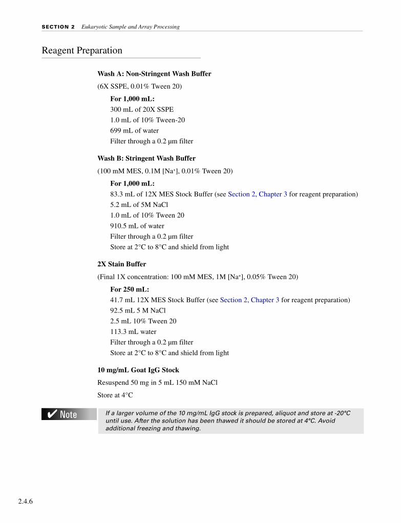
Reagent Preparation
Wash A: Non-Stringent Wash Buffer
(6X SSPE, 0.01% Tween 20)
For 1,000 mL:
300 mL of 20X SSPE
1.0 mL of 10% Tween-20
699 mL of water
Filter through a 0.2 µm filter
Wash B: Stringent Wash Buffer
(100 mM MES, 0.1M [Na+], 0.01% Tween 20)
For 1,000 mL:
83.3 mL of 12X MES Stock Buffer (see Section 2, Chapter 3 for reagent preparation)
5.2 mL of 5M NaCl
1.0 mL of 10% Tween 20
910.5 mL of water
Filter through a 0.2 µm filter
Store at 2°C to 8°C and shield from light
2X Stain Buffer
(Final 1X concentration: 100 mM MES, 1M [Na+], 0.05% Tween 20)
For 250 mL:
41.7 mL 12X MES Stock Buffer (see Section 2, Chapter 3 for reagent preparation)
92.5 mL 5 M NaCl
2.5 mL 10% Tween 20
113.3 mL water
Filter through a 0.2 µm filter
Store at 2°C to 8°C and shield from light
10 mg/mL Goat IgG Stock
Resuspend 50 mg in 5 mL 150 mM NaCl
Store at 4°C
If a larger volume of the 10 mg/mL IgG stock is prepared, aliquot and store at -20°C until use. After the solution has been thawed it should be stored at 4°C. Avoid additional freezing and thawing.

CHAPTER 4 Eukaryotic Arrays: Washing, Staining, and Scanning
Eu
kary
oti
c
Experiment and Fluidics Station Setup
Step 1: Defining File Locations
Before working with Affymetrix® Microarray Suite, it is important to define where the program stores and looks for files.
1. Launch Microarray Suite from the workstation and select Tools → Defaults → File Locations from the menu bar.
2. The File Locations window displays the locations of the following files:
■ Probe Information (library files, mask files)
■ Fluidics Protocols (fluidics station scripts)
■ Experiment Data (.exp, .dat, .cel, and .chp files are all saved to location selected here)
3. Verify that all three file locations are set correctly and click OK.
Contact Affymetrix Technical Support if you have any questions regarding this procedure.
Step 2: Entering Experiment Information
To wash, stain, and scan a probe array, an experiment must first be defined in Microarray Suite.
1. Select Run → Experiment Info from the menu bar. Alternatively, click the New Experiment icon on the tool bar.
⇒ The Experiment Information dialog box appears allowing the experiment name to be defined along with several other parameters, such as probe array type, sample description, and comments.
2. Type in the Experiment Name.
3. In the Probe Array Type box, click the arrow and select the probe array type from the drop-down list.
Experiment name and probe array type are required. Complete as much of the other information as desired. The protocol information at the bottom of the dialog box is exported to the experiment information dialog box after the hybridization and scan are completed.
4. Save the experiment by selecting Save.
The name of the experiment is used by Microarray Suite to access the probe array type and data for the sample while it is being processed. Data files generated for the sample are automatically labeled to correspond to the experiment name. Microarray Suite automatically fills in the Protocol section of this dialog box with information on array processing from the fluidics station.
5. Close the Experiment Information dialog box.
2.4.7

SECTION 2 Eukaryotic Sample and Array Processing
2.4.8
Step 3: Preparing the Fluidics Station
The Fluidics Station 400, 450, or 250 is used to wash and stain the probe arrays. It is operated using Microarray Suite.
Setting Up the Fluidics Station
1. Turn on the Fluidics Station using the toggle switch on the lower left side of the machine.
2. Select Run → Fluidics from the menu bar.
⇒ The Fluidics Station dialog box appears with a drop-down list for selecting the experiment name for each of the fluidics station modules. A second drop-down list is accessed for choosing the Protocol for each of the fluidics station modules.
Priming the Fluidics Station
Priming ensures that the lines of the fluidics station are filled with the appropriate buffers and the fluidics station is ready for running fluidics station protocols.
Priming should be done:
■ when the fluidics station is first started.
■ when wash solutions are changed.
■ before washing, if a shutdown has been performed.
■ if the LCD window instructs the user to prime.
1. To prime the fluidics station, select Protocol in the Fluidics Station dialog box.
2. Choose Prime or Prime_450 for the respective modules in the Protocol drop-down list.
3. Change the intake buffer reservoir A to Non-Stringent Wash Buffer, and intake buffer reservoir B to Stringent Wash Buffer.
4. Click Run for each module to begin priming.
Refer to the Fluidics Station User’s Guide for instructions on connecting and addressing multiple fluidics stations.

CHAPTER 4 Eukaryotic Arrays: Washing, Staining, and Scanning
Eu
kary
oti
c
Probe Array Wash and Stain
Affymetrix offers two staining protocols: 1) the single stain protocol for eukaryotic targets (page 2.4.9), and 2) a signal amplification protocol for eukaryotic targets (page 2.4.13). Please use the Antibody Amplification Washing and Staining Protocol for all arrays with probe cells of 24 µm or smaller.
1. After 16 hours of hybridization, remove the hybridization cocktail from the probe array and set it aside in a microcentrifuge vial. Store on ice during the procedure or at -80°C for long-term storage.
2. Fill the probe array completely with the appropriate volume of Non-Stringent Wash Buffer (Wash A), as given in Table 2.3.2 on page 2.3.8.
Washing and Staining Procedure 1: Single Stain for Eukaryotic Targets
Preparing the SAPE Stain Solution
Streptavidin Phycoerythrin (SAPE) should be stored in the dark at 4°C, either foil-wrapped or kept in an amber tube. Remove SAPE from refrigerator and tap the tube to mix well before preparing stain solution. Do not freeze SAPE. Always prepare the SAPE stain solution immediately before use.
For each probe array to be stained, combine the following components in a microcentrifuge vial:
If necessary, at this point, the probe array can be stored at 4°C for up to 3 hours before proceeding with washing and staining. Equilibrate the probe array to room temperature before washing and staining.
Volumes needed will be the same for all fluidic protocols. This procedure takes approximately 75 minutes to complete.
Table 2.4.1 SAPE Solution Mix
Components Volume Final Concentration
2X MES Stain Buffer 300.0 µL 1X
50 mg/mL acetylated BSA 24.0 µL 2 mg/mL
1 mg/mL Streptavidin Phycoerythrin (SAPE) 6.0 µL 10 µg/mL
DI H20 270.0 µL —
Total 600 µL
2.4.9

SECTION 2 Eukaryotic Sample and Array Processing
2.4.10
If you are using the Fluidics Station 450/250:
Washing and Staining the Probe Array
1. In the Fluidics Station dialog box on the workstation, select the correct experiment name in the drop-down Experiment list.
⇒ The probe array type appears automatically.
2. In the Protocol drop-down list, select the specific single stain protocol to control the washing and staining of the probe array format being used: Table 2.4.2.
3. Choose Run in the Fluidics Station dialog box to begin the washing and staining. Follow the instructions on the LCD window on the fluidics station.
If you are unfamiliar with inserting and removing probe arrays from the fluidics station modules, please refer to the appropriate Fluidics Station User’s Guide, or Quick Reference Card (P/N 08-0072 for the FS-400 Fluidics Station or P/N 08-0093 for the FS-450/250 Fluidics Stations).
4. Insert the appropriate probe array into the designated module of the fluidics station while the cartridge lever is in the down, or eject, position. When finished, verify that the cartridge lever is returned to the up, or engaged, position.
5. Remove any microcentrifuge vial remaining in the sample holder of the fluidics station module(s) being used.
6. Place the microcentrifuge vial containing the SAPE stain solution into the indicated sample holder 1.
7. Press down on the needle lever to snap needles into position and to start the run.
The run begins. The Fluidics Station dialog box at the workstation terminal and the LCD window display the status of the washing and staining as they progress.
Table 2.4.2 Fluidics Protocols - Single Stain for Eukaryotic Targets (protocols for the Fluidics Station 450/250 will have _450 as a suffix).
49 Format (Standard) 64 Format
EukGE-WS1a
a. EukGE-WS1_450 for the FS-450/250
169 Format (Mini)
Mini_euk1b
b. Mini_euk1_450 for the FS-450/250
Post Hyb Wash #1 10 cycles of 2 mixes/cycle with Wash Buffer A at 25°C
10 cycles of 2 mixes/cycle with Wash Buffer A at 25°C
Post Hyb Wash #2 4 cycles of 15 mixes/cycle with Wash Buffer B at 50°C
8 cycles of 15 mixes/cycle with Wash Buffer B at 50°C
Stain Stain the probe array for 30 minutes in SAPE solution at 25°C
Stain the probe array for 10 minutes in SAPE solution at 25°C
Final Wash10 cycles of 4 mixes/cycle with Wash Buffer A at 25°C. The holding temperature is 25°C
10 cycles of 4 mixes/cycle with Wash Buffer A at 30°C. The holding temperature is 25°C
• Wash Buffer A = non-stringent wash buffer• Wash Buffer B = stringent wash buffer

CHAPTER 4 Eukaryotic Arrays: Washing, Staining, and Scanning
Eu
kary
oti
c
8. At the end of the run, or at the appropriate prompt, remove the microcentrifuge vials and replace with three empty microcentrifuge vials.
9. Remove the probe arrays from the fluidics station modules by first moving the cartridge lever to the down position.
10. Check the probe array window for large bubbles or air pockets.
■ If bubbles are present, proceed to Table 2.4.3.
■ If the probe array has no large bubbles, it is ready to scan on the GeneArray® Scanner or the GeneChip® Scanner 3000. Pull up on the cartridge lever to engage the washblock and proceed to Probe Array Scan on page 2.4.18.
If you do not scan the arrays right away, keep the probe arrays at 4°C and in the dark until ready for scanning.
If there are no more samples to hybridize, shut down the fluidics station following the procedure outlined in the section, Shutting Down the Fluidics Station on page 2.4.20.
If you are using the Fluidics Station 400:
Washing and Staining the Probe Array
1. In the Fluidics Station dialog box on the workstation, select the correct experiment name in the drop-down Experiment list. The probe array type will appear automatically.
2. In the Protocol drop-down list, select the specific single stain protocol to control the washing and staining of the probe array format being used: Table 2.4.2.
3. Choose Run in the Fluidics Station dialog box to begin the washing and staining. Follow the instructions on the LCD window on the fluidics station.
If you are unfamiliar with inserting and removing probe arrays from the fluidics station modules, please refer to the Fluidics Station 400 User’s Guide, Fluidics Station 400 Video In-Service CD (P/N 900374), or Quick Reference Card (P/N 08-0072).
For proper cleaning and maintenance of the fluidics station, including the bleach protocol, refer to Section 4, Fluidics Station Maintenance Procedures.
Table 2.4.3If Bubbles are Present
Return the probe array to the probe array holder. Engage the washblock by gently pushing up on the cartridge lever to the engage, or closed, position.The fluidics station will drain the probe array and then fill it with a fresh volume of the last wash buffer used. When it is finished, the LCD window displays EJECT CARTRIDGE again, remove the probe array and inspect it again for bubbles. If no bubbles are present, it is ready to scan. Proceed to Probe Array Scan on page 2.4.18.If several attempts to fill the probe array without bubbles are unsuccessful, the array should be filled with Wash A (non-stringent buffer) manually, using a micropipette. Excessive washing will result in a loss of signal intensity.
2.4.11

SECTION 2 Eukaryotic Sample and Array Processing
2.4.12
4. Insert the appropriate probe array into the designated module of the fluidics station while the cartridge lever is in the EJECT position. When finished, verify that the cartridge lever is returned to the ENGAGE position.
5. Remove any microcentrifuge tube remaining in the sample holder of the fluidics station module(s) being used.
6. Place the microcentrifuge tube containing the SAPE stain solution into the sample holder, verifying that the metal sampling needle is in the tube with its tip near the bottom.
⇒ The Fluidics Station dialog box and the LCD window display the status of the washing and staining as they progress. When the wash is complete, the LCD window displays the message EJECT CARTRIDGE.
7. At the end of the run, or at the appropriate prompt, remove microcentrifuge tube containing stain and replace with an empty microcentrifuge tube.
8. Remove the probe arrays from the fluidics station modules by first moving the cartridge lever to the EJECT position.
9. Check the probe array window for large bubbles or air pockets.
■ If bubbles are present, proceed to Table 2.4.3.
■ If the probe array has no large bubbles, it is ready to scan on the GeneChip® Scanner 3000 or GeneArray® Scanner. ENGAGE washblock and proceed to Probe Array Scan on page 2.4.18.
If you do not scan the arrays right away, keep the probe arrays at 4°C and in the dark until ready for scanning.
If there are no more samples to hybridize, shut down the fluidics station following the procedure outlined in the section, Shutting Down the Fluidics Station on page 2.4.20.
For proper cleaning and maintenance of the fluidics station, including the bleach protocol, refer to Section 4, Fluidics Station Maintenance Procedures.
Table 2.4.4If Bubbles are Present
Return the probe array to the probe array holder. Latch the probe array holder by gently pushing it up until a light click is heard. Engage the washblock by firmly pushing up on the cartridge lever to the ENGAGE position.The fluidics station will drain the probe array and then fill it with a fresh volume of the last wash buffer used. When it is finished, the LCD window displays EJECT CARTRIDGE again, remove the probe array and inspect it again for bubbles. If no bubbles are present, it is ready to scan. Proceed to Probe Array Scan on page 2.4.18.If several attempts to fill the probe array without bubbles are unsuccessful, the array should be filled with Wash A (non-stringent buffer) manually, using a micropipette. Excessive washing will result in a loss of signal intensity.

CHAPTER 4 Eukaryotic Arrays: Washing, Staining, and Scanning
Eu
kary
oti
c
Washing and Staining Procedure 2: Antibody Amplification Stain for Eukaryotic Targets
This protocol is recommended for use with probe arrays with probe cells of 24 µm or smaller. This procedure takes approximately 90 minutes to complete.
Preparing the Staining Reagents
Prepare the following reagents. Volumes given are sufficient for one probe array.
SAPE Stain Solution
Streptavidin Phycoerythrin (SAPE) should be stored in the dark at 4°C, either foil-wrapped or kept in an amber tube. Remove SAPE from refrigerator and tap the tube to mix well before preparing stain solution. Do not freeze SAPE. Always prepare the SAPE stain solution immediately before use.
Mix well and divide into two aliquots of 600 µL each to be used for stains 1 and 3, respectively.
Antibody Solution
Table 2.4.5 SAPE Solution Mix
Components Volume Final Concentration
2X MES Stain Buffer 600.0 µL 1X
50 mg/mL acetylated BSA 48.0 µL 2 mg/mL
1 mg/mL Streptavidin Phycoerythrin (SAPE) 12.0 µL 10 µg/mL
DI H20 540.0 µL —
Total 1200 µL
Table 2.4.6 Antibody Solution Mix
Components Volume Final Concentration
2X MES Stain Buffer 300.0 µL 1X
50 mg/mL acetylated BSA 24.0 µL 2 mg/mL
10 mg/mL Normal Goat IgG 6.0 µL 0.1 mg/mL
0.5 mg/mL biotinylated antibody 3.6 µL 3 µg/mL
DI H20 266.4 µL —
Total 600 µL
2.4.13

SECTION 2 Eukaryotic Sample and Array Processing
2.4.14
If you are using the Fluidics Station 450/250:
Washing and Staining the Probe Array
1. In the Fluidics Station dialog box on the workstation, select the correct experiment name from the drop-down Experiment list.
⇒ The Probe Array Type appears automatically.
2. In the Protocol drop-down list, select the appropriate antibody amplification protocol to control the washing and staining of the probe array format being used.
3. Choose Run in the Fluidics Station dialog box to begin the washing and staining. Follow the instructions in the LCD window on the fluidics station.
If you are unfamiliar with inserting and removing probe arrays from the fluidics station modules, please refer to the appropriate Fluidics Station User’s Guide, or Quick Reference Card (P/N 08-0093 for the FS-450/250 fluidics stations).
4. Insert the appropriate probe array into the designated module of the fluidics station while the cartridge lever is in the, down, or eject, position. When finished, verify that the cartridge lever is returned to the up, or engaged, position.
Table 2.4.7 Fluidics Protocols - Antibody Amplification for Eukaryotic Targets (protocols for the Fluidics Station 450/250 will have _450 as a suffix).
49 Format (Standard) /64 Format
EukGE-WS2a
a. EukGE-WS2_450 for the FS-450/250
100 Format (Midi)
Midi_euk2b
b. Midi_euk2_450 for the FS-450/250
169 Format (Mini) /400 Format (Micro)
Micro_1 / Mini_euk2c
c. Micro_1_450 / Mini_euk2_450 for the FS-450/250
Post Hyb Wash #1
10 cycles of 2 mixes/cycle with Wash Buffer A at 25°C
10 cycles of 2 mixes/cycle with Wash Buffer A at 30°C
10 cycles of 2 mixes/cycle with Wash Buffer A at 25°C
Post Hyb Wash #2
4 cycles of 15 mixes/cycle with Wash Buffer B at 50°C
6 cycles of 15 mixes/cycle with Wash Buffer B at 50°C
8 cycles of 15 mixes/cycle with Wash Buffer B at 50°C
Stain Stain the probe array for 10 minutes in SAPE solution at 25°C
Stain the probe array for 5 minutes in SAPE solution at 35°C
Stain the probe array for 10 minutes in SAPE solution at 25°C
Post Stain Wash
10 cycles of 4 mixes/cycle with Wash Buffer A at 25°C
10 cycles of 4 mixes/cycle with Wash Buffer A at 30°C
10 cycles of 4 mixes/cycle with Wash Buffer A at 30°C
2nd Stain Stain the probe array for 10 minutes in antibody solution at 25°C
Stain the probe array for 5 minutes in antibody solution at 35°C
Stain the probe array for 10 minutes in antibody solution at 25°C
3rd Stain Stain the probe array for 10 minutes in SAPE solution at 25°C
Stain the probe array for 5 minutes in SAPE solution at 35°C
Stain the probe array for 10 minutes in SAPE solution at 25°C
Final Wash
15 cycles of 4 mixes/cycle with Wash Buffer A at 30°C. The holding temperature is 25°C
15 cycles of 4 mixes/cycle with Wash Buffer A at 35°C. The holding temperature is 25°C
15 cycles of 4 mixes/cycle with Wash Buffer A at 35°C. The holding temperature is 25°C
• Wash Buffer A = non-stringent wash buffer• Wash Buffer B = stringent wash buffer

CHAPTER 4 Eukaryotic Arrays: Washing, Staining, and Scanning
Eu
kary
oti
c
5. Remove any microcentrifuge vial remaining in the sample holder of the fluidics station module(s) being used.
6. If prompted to “Load Vials 1-2-3,” place the three experiment sample vials (the microcentrifuge vials) into the sample holders 1, 2, and 3 on the fluidics station.
■ Place one vial containing 600 µL of streptavidin phycoerythrin (SAPE) solution mix in sample holder 1.
■ Place one vial containing 600 µL of anti-streptavidin biotinylated antibody in sample holder 2.
■ Place one vial containing 600 µL of streptavidin phycoerythrin (SAPE) solution in sample holder 3.
■ Press down on the needle lever to snap needles into position and to start the run.
The run begins. The Fluidics Station dialog box at the workstation terminal and the LCD window display the status of the washing and staining as they progress.
7. At the end of the run, or at the appropriate prompt, remove the microcentrifuge vials and replace with three empty microcentrifuge vials.
8. Remove the probe arrays from the fluidics station modules by first pressing down the cartridge lever to the eject position.
9. Check the probe array window for large bubbles or air pockets.
■ If bubbles are present, proceed to Table 2.4.8.
■ If the probe array has no large bubbles, it is ready to scan on the GeneArray® Scanner or the GeneChip® Scanner 3000. Pull up on the cartridge lever to engage washblock and proceed to Probe Array Scan on page 2.4.18.
If you do not scan the arrays right away, keep the probe arrays at 4°C and in the dark until ready for scanning.
If there are no more samples to hybridize, shut down the fluidics station following the procedure outlined in the section, Shutting Down the Fluidics Station on page 2.4.20.
For proper cleaning and maintenance of the fluidics station, including the bleach protocol, refer to Section 4, Fluidics Station Maintenance Procedures.
Table 2.4.8If Bubbles are Present
Return the probe array to the probe array holder. Engage the washblock by gently pushing up on the cartridge lever to the engaged, or closed, position.The fluidics station will drain the probe array and then fill it with a fresh volume of the last wash buffer used. When it is finished, the LCD window will display EJECT CARTRIDGE. Again, remove the probe array and inspect it for bubbles. If no bubbles are present, it is ready to scan. Proceed to Probe Array Scan on page 2.4.18.If several attempts to fill the probe array without bubbles are unsuccessful, the array should be filled with Wash A (non-stringent buffer) manually, using a micropipette. Excessive washing will result in a loss of signal intensity.
2.4.15

SECTION 2 Eukaryotic Sample and Array Processing
2.4.16
If you are using the Fluidics Station 400:
Washing and Staining the Probe Array
1. In the Fluidics Station dialog box on the workstation, select the correct experiment name in the drop-down Experiment list. The probe array type will appear automatically.
2. In the Protocol drop-down list, select the appropriate antibody amplification protocol to control the washing and staining of the probe array format being used: Table 2.4.2.
3. Choose Run in the Fluidics Station dialog box to begin the washing and staining. Follow the instructions on the LCD window on the fluidics station.
4. If you are unfamiliar with inserting and removing probe arrays from the fluidics station modules, please refer to the Fluidics Station 400 User’s Guide, Fluidics Station 400 Video In-Service CD (P/N 900374), or Quick Reference Card (P/N 08-0072).
5. Insert the appropriate probe array into the designated module of the fluidics station while the cartridge lever is in the EJECT position. When finished, verify that the cartridge lever is returned to the ENGAGE position.
6. Remove any microcentrifuge vial remaining in the sample holder of the fluidics station module(s) being used.
7. When the LCD window indicates, place the microcentrifuge vial containing 600 µL of streptavidin phycoerythrin (SAPE) stain solution into the sample holder. Verify that the metal sampling needle is in the vial with its tip near the bottom.
8. When the LCD window indicates, replace the microcentrifuge vial containing the streptavidin stain with a microcentrifuge vial containing antibody stain solution into the sample holder, making sure that the metal sampling needle is in the vial with its tip near the bottom.
9. When the LCD window indicates, replace the microcentrifuge vial containing the antibody stain solution with a microcentrifuge vial containing 600 µL of streptavidin phycoerythrin (SAPE) stain solution into the sample holder. Verify that the metal sampling needle is in the vial with its tip near the bottom.
⇒ The Fluidics Station dialog box and the LCD window display the status of the washing and staining as they progress. When the wash is complete, the LCD window displays the message EJECT CARTRIDGE.
10. At the end of the run, or at the appropriate prompt, remove microcentrifuge vial containing stain and replace with an empty microcentrifuge vial.
11. Remove the probe arrays from the fluidics station modules by first moving the probe array holder lever to the EJECT position.
12. Check the probe array window for large bubbles or air pockets.
■ If bubbles are present, proceed to Table 2.4.9.
■ If the probe array has no large bubbles, it is ready to scan on the GeneChip® Scanner 3000 or GeneArray® Scanner. ENGAGE wash block and proceed to Probe Array Scan on page 2.4.18.

CHAPTER 4 Eukaryotic Arrays: Washing, Staining, and Scanning
Eu
kary
oti
c
If you do not scan the arrays right away, keep the probe arrays at 4°C and in the dark until ready for scanning.
If there are no more samples to hybridize, shut down the fluidics station following the procedure outlined in the section, Shutting Down the Fluidics Station on page 2.4.20.
For proper cleaning and maintenance of the fluidics station including the bleach protocol, refer to Section 4, Fluidics Station Maintenance Procedures.
Table 2.4.9If Bubbles are Present
Return the probe array to the probe array holder. Latch the probe array holder by gently pushing it up until a light click is heard. Engage the wash block by firmly pushing up on the cartridge lever to the ENGAGE position.The fluidics station will drain the probe array and then fill it with a fresh volume of the last wash buffer used. When it is finished, the LCD window displays EJECT CARTRIDGE again, remove the probe array and inspect it again for bubbles. If no bubbles are present, it is ready to scan. Proceed to Probe Array Scan on page 2.4.18.If several attempts to fill the probe array without bubbles are unsuccessful, the array should be filled with Wash A (non-stringent buffer) manually, using a micropipette. Excessive washing will result in a loss of signal intensity.
2.4.17

SECTION 2 Eukaryotic Sample and Array Processing
2.4.18
Probe Array Scan
The scanner is also controlled by Affymetrix® Microarray Suite. The probe array is scanned after the wash protocols are complete. Make sure the laser is warmed up prior to scanning by turning the laser on at least 15 minutes before use (if you are using the Agilent GeneArray® Scanner) or 10 minutes (if you are using the Affymetrix® GeneChip® Scanner 3000). If probe array was stored at 4°C, warm to room temperature before scanning. Refer to the Microarray Suite online help and the appropriate scanner user’s manual for more information on scanning.
Handling the GeneChip® Probe Array
Before you scan the probe array, follow the directions in this section on handling the probe array. If necessary, clean the glass surface of probe array with a non-abrasive towel or tissue before scanning. Do not use alcohol to clean glass.
Before scanning the probe array cartridge, apply Tough-Spots™ to each of the two septa on the probe array cartridge to prevent the leaking of fluids from the cartridge during scanning.
1. On the back of the probe array cartridge, clean excess fluid from around septa.
2. Carefully apply one Tough-Spots to each of the two septa. Press to ensure that the spots remain flat. If the Tough-Spots do not apply smoothly, that is, if you observe bumps, bubbles, tears, or curled edges, do not attempt to smooth out the spot. Remove the spot and apply a new spot. See Figure 2.4.1.
The scanner uses a laser and is equipped with a safety interlock system. Defeating the interlock system may result in exposure to hazardous laser light.
You must have read, and be familiar with, the operation of the scanner before attempting to scan a probe array. Please refer to the Microarray Suite User’s Guide (P/N 08-0081) or to the GeneChip® Scanner 3000 quick reference card (P/N 08-0075).
Apply the spots just before scanning. Do not use them in the hyb process.

CHAPTER 4 Eukaryotic Arrays: Washing, Staining, and Scanning
Eu
kary
oti
c
3. Insert the cartridge into the scanner and test the autofocus to ensure that the Tough-Spots do not interfere with the focus. If you observe a focus error message, remove the spot and apply a new spot. Ensure that the spots lie flat.
Scanning the Probe Array
1. Select Run → Scanner from the menu bar. Alternatively, click the Start Scan icon in the tool bar.
⇒ The Scanner dialog box appears with a drop-down list of experiments that have not been run.
2. Select the experiment name that corresponds to the probe array to be scanned.
A previously run experiment can also be selected by using the Include Scanned Experiments option box. After selecting this option, previously scanned experiments appear in the drop-down list.
3. By default, for the GeneArray® Scanner only, after selecting the experiment the number [2] is displayed in the Number of Scans box to perform the recommended 2X image scan. For the GeneChip® Scanner 3000, only one scan is required.
4. Once the experiment has been selected, click the Start button.
⇒ A dialog box prompts you to load an array into the scanner.
5. If you are using the GeneArray® Scanner, click the Options button to check for the correct pixel value and wavelength of the laser beam.
■ Pixel value = 3 µm
■ Wavelength = 570 nm
If you are using the GeneChip Scanner 3000, pixel resolution and wavelength are preset and cannot be changed.
Figure 2.4.1 Applying Tough-Spots™ to the probe array cartridge
2.4.19
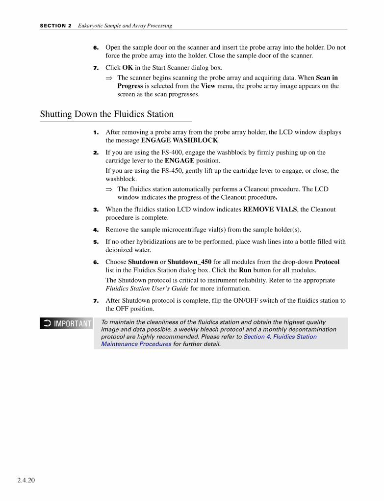
SECTION 2 Eukaryotic Sample and Array Processing
2.4.20
6. Open the sample door on the scanner and insert the probe array into the holder. Do not force the probe array into the holder. Close the sample door of the scanner.
7. Click OK in the Start Scanner dialog box.
⇒ The scanner begins scanning the probe array and acquiring data. When Scan in Progress is selected from the View menu, the probe array image appears on the screen as the scan progresses.
Shutting Down the Fluidics Station
1. After removing a probe array from the probe array holder, the LCD window displays the message ENGAGE WASHBLOCK.
2. If you are using the FS-400, engage the washblock by firmly pushing up on the cartridge lever to the ENGAGE position.
If you are using the FS-450, gently lift up the cartridge lever to engage, or close, the washblock.
⇒ The fluidics station automatically performs a Cleanout procedure. The LCD window indicates the progress of the Cleanout procedure.
3. When the fluidics station LCD window indicates REMOVE VIALS, the Cleanout procedure is complete.
4. Remove the sample microcentrifuge vial(s) from the sample holder(s).
5. If no other hybridizations are to be performed, place wash lines into a bottle filled with deionized water.
6. Choose Shutdown or Shutdown_450 for all modules from the drop-down Protocol list in the Fluidics Station dialog box. Click the Run button for all modules.
The Shutdown protocol is critical to instrument reliability. Refer to the appropriate Fluidics Station User’s Guide for more information.
7. After Shutdown protocol is complete, flip the ON/OFF switch of the fluidics station to the OFF position.
To maintain the cleanliness of the fluidics station and obtain the highest quality image and data possible, a weekly bleach protocol and a monthly decontamination protocol are highly recommended. Please refer to Section 4, Fluidics Station Maintenance Procedures for further detail.

CHAPTER 4 Eukaryotic Arrays: Washing, Staining, and Scanning
Eu
kary
oti
c
Customizing the Protocol
There may be times when the fluidics protocols need to be modified. Modification of protocols must be done before downloading the protocol to the fluidics station. Protocol changes will not affect runs in progress. For more specific instructions, refer to the Microarray Suite online help.
1. Select Tools → Edit Protocol from the menu bar.
⇒ The Edit Protocol dialog box appears.
2. Select the protocol to be changed from the Protocol Name drop-down list.
⇒ The name of the protocol is displayed in the Protocol Name box. The conditions for that protocol are displayed on the right side of the Edit Protocol dialog box.
3. Select the item to be changed and input the new parameters as needed, keeping the parameters within the ranges shown below in Table 2.4.1.
4. To return to the default values for the protocol selected, click the Defaults button.
5. After all the protocol conditions are modified as desired, change the name of the edited protocol in the Protocol Name box.
6. Click Save, then close the dialog box.
Enter 0 (zero) for hybridization time if hybridization step is not required. Likewise, enter 0 (zero) for the stain time if staining is not required. Enter 0 (zero) for the number of wash cycles if a wash solution is not required.
Table 2.4.1Valid Ranges for Wash/Stain Parameters
Parameter Valid Range
Wash Temperature for A1, B, A2, or A3 (°C) 15 to 50
Number of Wash Cycles for A1, B, A2, or A3 0 to 99
Mixes / Wash cycle for A1, B, A2, or A3 15 to 50
Stain Time (seconds) 0 to 86399
Stain Temperature (°C) 15 to 50
Holding Temperature (°C) 15 to 50
• Wash A1 corresponds to Post Hyb wash #1 in Tables 2.4.2 and 2.4.7.• Wash B corresponds to Post Hyb wash #2 in Tables 2.4.2 and 2.4.7.• Wash A2 corresponds to Post Stain Wash in Tables 2.4.2 and 2.4.7.• Wash A3 corresponds to Final Wash in Tables 2.4.2 and 2.4.7.
If the protocol is saved without entering a new Protocol Name, the original protocol parameters will be overwritten.
2.4.21
Section 3:
Prokaryotic Sample and Array Processing
701029 Rev. 3

Section 3

Pro
kary
oti
c
Contents
Section 3 Prokaryot ic Sample and Array Processing
Chapter 1 Prokaryotic Target Preparation 3.1.3
Chapter 2 Preparation of Control Spike-In Transcripts 3.2.3
Chapter 3 Prokaryotic Target Hybridization 3.3.3
Chapter 4 Prokaryotic Arrays:Washing, Staining, and Scanning 3.4.3
701029 Rev. 3

Pro
kary
oti
c
Section 3, Chapter 1
701030 Rev. 3

Section 3, Chapter 1

Pro
kary
oti
c
Prokaryotic Target Preparation
Reagents and Materials Required . . . . . . . . . . . . . . . . . . . . . . . . . . . . 3.1.5
Reagent Preparation . . . . . . . . . . . . . . . . . . . . . . . . . . . . . . . . . . . 3.1.6
Total RNA Isolation . . . . . . . . . . . . . . . . . . . . . . . . . . . . . . . . . . . 3.1.7
cDNA Synthesis . . . . . . . . . . . . . . . . . . . . . . . . . . . . . . . . . . . . . 3.1.8
Step1: cDNA Synthesis . . . . . . . . . . . . . . . . . . . . . . . . . . . . . . . . 3.1.8
Step 2: Removal of RNA . . . . . . . . . . . . . . . . . . . . . . . . . . . . . . . 3.1.9
Step 3: Purification and Quantitation of cDNA. . . . . . . . . . . . . . . . . . . . 3.1.9
cDNA Fragmentation . . . . . . . . . . . . . . . . . . . . . . . . . . . . . . . . . 3.1.10
Terminal Labeling . . . . . . . . . . . . . . . . . . . . . . . . . . . . . . . . . . . 3.1.11
This Chapter Contains:
This chapter describes the assay procedures recommended for use with the GeneChip® P. aeruginosa Genome Array and the GeneChip® E. coli Antisense Genome Array. The assay utilizes reverse transcriptase and random hexamer primers to produce DNA complementary to the RNA. The cDNA products are then fragmented by DNase I and labeled with terminal transferase and biotinylated ddUTP at the 3’ termini.
This protocol is presented as a recommendation only, and has not been validated by Affymetrix.
701030 Rev. 3 3.1.3
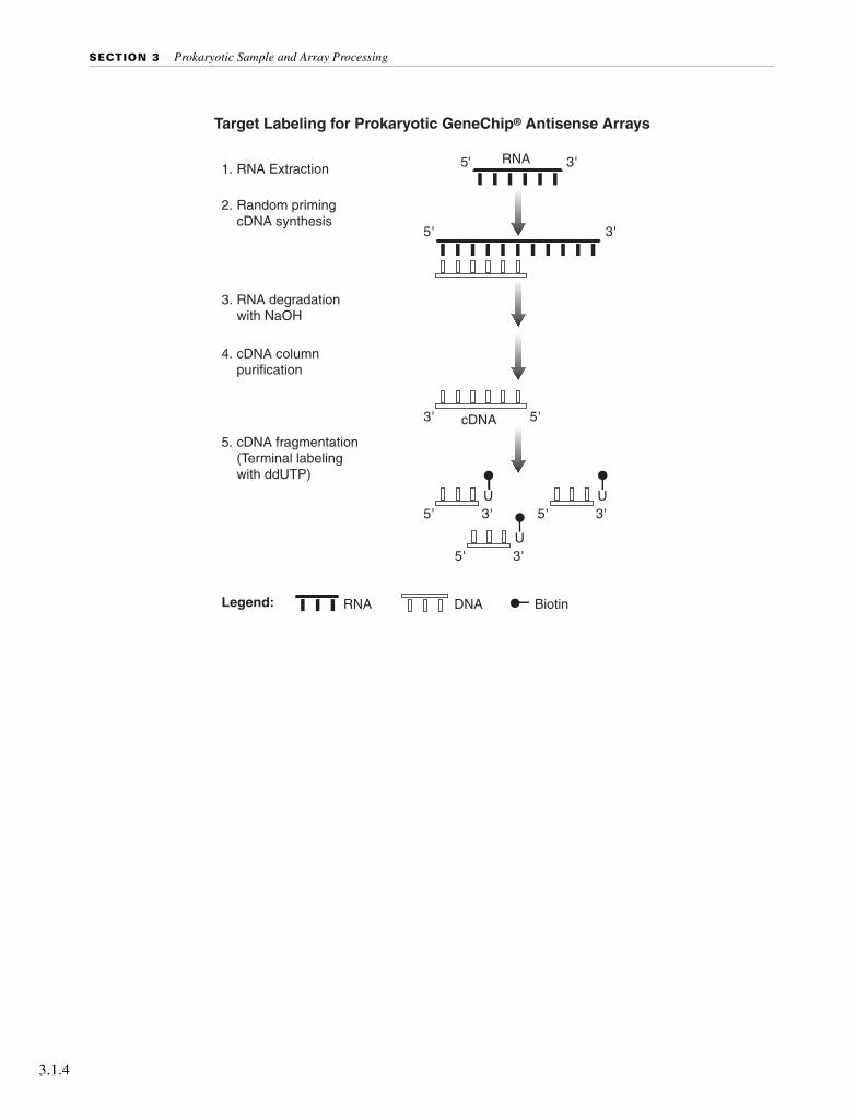
SECTION 3 Prokaryotic Sample and Array Processing
3.1.4
Target Labeling for Prokaryotic GeneChip® Antisense Arrays
1. RNA Extraction
2. Random priming cDNA synthesis
3. RNA degradation with NaOH
4. cDNA column purification
5' 3'
RNA DNA Biotin
5'
5' 3'
5'
3'
3'
5. cDNA fragmentation (Terminal labeling with ddUTP)
Legend:
RNA
cDNA
U5' 3'
U
5' 3'U

CHAPTER 1 Prokaryotic Target Preparation
Pro
kary
oti
c
Reagents and Materials Required
The following reagents and materials are recommendations and have been tested and evaluated by Affymetrix scientists. For supplier phone numbers in the U.S. and Europe, please refer to the Supplier and Reagent Reference List, Appendix A, of this manual. Information and part numbers listed are based on U.S. catalog information. Additional reagents needed for the complete analysis are listed in the appropriate chapters. Appendix A contains a master list of all reagents used in this manual.
Labeling■ dATP, dCTP, dGTP, dTTP, 100 mM, Amersham Pharmacia Biotech, P/N 27-2035-01
■ Random Primers, 3 µg/µL, Invitrogen Life Technologies, P/N 48190-011
■ SuperScript II Reverse Transcriptase, Invitrogen Life Technologies, P/N 18064-071
■ SUPERase•In™, Ambion, P/N 2696
■ Nuclease-free Water, Ambion, P/N 9930
■ NaOH, 1N solution, VWR Scientific Products, P/N MK469360
■ HCl, 1N solution, VWR Scientific Products, P/N MK638860
■ QIAquick PCR Purification Kit, QIAGEN, P/N 28104
■ 10X One-Phor-All Buffer, Amersham Pharmacia Biotech, P/N 27-0901-02
■ Deoxyribonuclease I (DNase I), Amersham Pharmacia Biotech, P/N 27-0514-01
■ Enzo® BioArray™ Terminal Labeling Kit, Affymetrix, P/N 900181
■ EDTA, 0.5M, pH 8.0, Invitrogen Life Technologies, P/N 15575-020
Gel-Shift Assay■ Novex XCell SureLock™ Mini-Cell, Invitrogen Life Technologies, P/N EI0001
■ 4-20% TBE Gel, 1.0 mm, 12 well, Invitrogen Life Technologies, P/N EC62252
■ Sucrose Gel Loading Dye, 5X, Amresco, P/N E-274
■ 10X TBE Running Buffer
■ SYBR Gold, Molecular Probes, P/N S-11494
■ 10 bp and 100 bp DNA ladder, Invitrogen Life Technologies, P/N 10821-015 and 15628-019, respectively
■ ImmunoPure NeutrAvidin, Pierce Chemical, P/N 31000
■ 1M Tris, pH 7.0, Ambion, P/N 9850G
■ PBS, pH 7.2, Invitrogen Life Technologies, P/N 20012-027
Prepare a mix of all 4 dNTPs at a final concentration of 10 mM each following the instructions below.
3.1.5

SECTION 3 Prokaryotic Sample and Array Processing
3.1.6
Reagent Preparation
10 mM dNTP mix
For 1000 µL:
100 µL 100 mM dATP
100 µL 100 mM dCTP
100 µL 100 mM dGTP
100 µL 100 mM dTTP
600 µL Nuclease-free H2O
Store at -20°C in a non-frost-free freezer.
75 ng/µL Random Primers
For 1000 µL:
25 µL 3 µg/µL Random Primers
975 µL Nuclease-free H2O
Store at -20°C in a non-frost-free freezer.
2 mg/mL NeutrAvidin
Resuspend 10 mg NeutrAvidin in 5 mL PBS solution. Store at 4°C.

CHAPTER 1 Prokaryotic Target Preparation
Pro
kary
oti
c
Total RNA Isolation
As starting material for the cDNA synthesis procedure, total RNA can be isolated by using standard procedures for bacterial RNA isolation or various commercial RNA isolation kits.
For Pseudomonas aeruginosa and E. coli, we have successfully used the QIAGEN® RNeasy Mini Purification Kit. Caution should be used to minimize chromosomal DNA contamination during the isolation, due to the high sensitivity of the assay. It is suggested that no more than 1 X 10⁹ cells are applied to a single purification column. Also, use the lysozyme at a concentration of 1 mg/mL, and not the recommended 400 µg/mL. Additional DNase I treatment may be required to eliminate DNA contamination when the bacterial culture is grown at high density.
After purification, RNA concentration is determined by absorbance at 260 nm on a spectrophotometer (1 absorbance unit = 40 µg/mL RNA). The A260/A280 ratio should be approximately 2.0, with ranges between 1.8 to 2.1 considered acceptable. We recommend checking the quality of RNA by running it on an agarose gel prior to starting the assay. The 23S and 16S rRNA bands should be clear without any obvious smears. Any indication of the presence of chromosomal DNA contamination (high molecular weight bands or smears on the gel) would require additional DNase treatment before proceeding to cDNA synthesis.
Figure 3.1.1 Typical RNA preparation from E. coli
Lane 1 - 1 µg Sample 1Lane 2 - 1 µg Sample 2Lane 3 - 1 µg Sample 3Lane 4 - RNA Size Markers
3.1.7
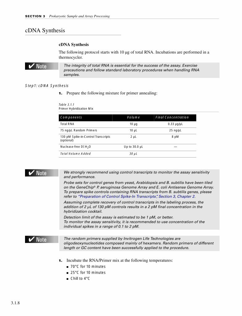
SECTION 3 Prokaryotic Sample and Array Processing
3.1.8
cDNA Synthesis
cDNA Synthesis
The following protocol starts with 10 µg of total RNA. Incubations are performed in a thermocycler.
Step1: cDNA Synthesis
1. Prepare the following mixture for primer annealing:
1. Incubate the RNA/Primer mix at the following temperatures:
■ 70°C for 10 minutes
■ 25°C for 10 minutes
■ Chill to 4°C
The integrity of total RNA is essential for the success of the assay. Exercise precautions and follow standard laboratory procedures when handling RNA samples.
Table 3.1.1Primer Hybridization Mix
Components Volume Final Concentration
Total RNA 10 µg 0.33 µg/µL
75 ng/µL Random Primers 10 µL 25 ng/µL
130 pM Spike-in-Control Transcripts (optional)
2 µL 8 pM
Nuclease-free DI H2O Up to 30.0 µL —
Total Volume Added 30 µL
We strongly recommend using control transcripts to monitor the assay sensitivity and performance.
Probe sets for control genes from yeast, Arabidopsis and B. subtilis have been tiled on the GeneChip® P. aeruginosa Genome Array and E. coli Antisense Genome Array. To prepare spike controls containing RNA transcripts from B. subtilis genes, please refer to “Preparation of Control Spike-In Transcripts”, Section 3, Chapter 2.
Assuming complete recovery of control transcripts in the labeling process, the addition of 2 µL of 130 pM controls results in a 2 pM final concentration in the hybridization cocktail.
Detection limit of the assay is estimated to be 1 pM, or better. To monitor the assay sensitivity, it is recommended to use concentration of the individual spikes in a range of 0.1 to 2 pM.
The random primers supplied by Invitrogen Life Technologies are oligodeoxynucleotides composed mainly of hexamers. Random primers of different length or GC content have been successfully applied to the procedure.

CHAPTER 1 Prokaryotic Target Preparation
Pro
kary
oti
c
2. Prepare the reaction mix for cDNA synthesis. Briefly centrifuge the reaction tube to collect sample at the bottom and add the cDNA synthesis mix from Table 3.1.2 to the RNA/primer hybridization mix.
3. Incubate the reaction at the following temperatures:
■ 25°C for 10 minutes
■ 37°C for 60 minutes
■ 42°C for 60 minutes
■ Inactivate SuperScript II at 70°C for 10 minutes
■ Chill to 4°C
Step 2: Removal of RNA
1. Add 20 µL of 1N NaOH and incubate at 65°C for 30 minutes.
2. Add 20 µL of 1N HCl to neutralize.
Step 3: Purification and Quantitation of cDNA
1. Use QIAquick Column to clean up the cDNA synthesis product (for detailed protocol, see QIAquick PCR Purification Kit Protocols provided by the supplier). Elute the product with 40 µL of EB Buffer (supplied with QIAquick kit).
2. Quantify the purified cDNA product by 260 nm absorbance (1.0 A260 unit = 33 µg/mL of single-stranded DNA).
Table 3.1.2 cDNA Synthesis Components
Components Volume Final Concentration
RNA/Primer hybridization mix (from previous step)
30 µL
5X 1st Strand Buffer 12 µL 1X
100 mM DTT 6 µL 10 mM
10 mM dNTPs 3 µL 0.5 mM
SUPERase•In (20 U/µL) 1.5 µL 0.5 U/µL
SuperScript II (200 U/µL) 7.5 µL 25 U/µL
Total Volume 60 µL
Typical yields of cDNA are 3 to 7 µg. A minimum of 1.5 µg of cDNA is required for subsequent procedures to obtain sufficient material to hybridize onto the array and to perform necessary quality control experiments.
3.1.9

SECTION 3 Prokaryotic Sample and Array Processing
3.1.10
cDNA Fragmentation
1. Prepare the following reaction mix:
2. Incubate the reaction at 37°C for 10 minutes.
3. Inactivate DNase I at 98°C for 10 minutes.
4. The fragmented cDNA is applied directly to the terminal labeling reaction. Alternatively, the material can be stored at -20°C for later use.
Table 3.1.3 Fragmentation Reaction
Components Volume Concentration
10X One-Phor-All Buffer 5 µL 1X
cDNA 40 µL 3-7 µg
DNase I (see note below) X µL 0.6 U/µg of cDNA
Nuclease-free H2O Up to 50 µL -
Total Volume 50 µL
Dilute DNase I to 0.6 U/µL in 1X One-Phor-All Buffer. Prepare fresh dilution each time immediately before use.
It is anticipated that DNase I enzyme activity may vary from lot to lot. A titration assay is strongly recommended for each new lot of enzyme to determine the dosage of the DNase I (unit of DNase I per µg of cDNA) to be used in the fragmentation reaction. 0.6 U for each µg of cDNA can be used as a starting point for the titration.
To examine the fragmentation result, load ~200 ng of the product on a 4% to 20% acrylamide gel and stain with SYBR Gold. The majority of the fragmented cDNA should be in the 50 to 200 base-pairs range.
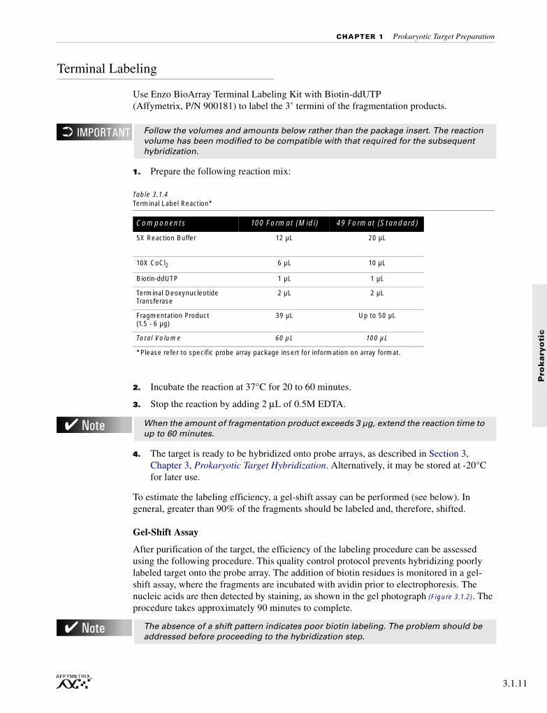
CHAPTER 1 Prokaryotic Target Preparation
Pro
kary
oti
c
Terminal Labeling
Use Enzo BioArray Terminal Labeling Kit with Biotin-ddUTP (Affymetrix, P/N 900181) to label the 3’ termini of the fragmentation products.
1. Prepare the following reaction mix:
2. Incubate the reaction at 37°C for 20 to 60 minutes.
3. Stop the reaction by adding 2 µL of 0.5M EDTA.
4. The target is ready to be hybridized onto probe arrays, as described in Section 3, Chapter 3, Prokaryotic Target Hybridization. Alternatively, it may be stored at -20°C for later use.
To estimate the labeling efficiency, a gel-shift assay can be performed (see below). In general, greater than 90% of the fragments should be labeled and, therefore, shifted.
Gel-Shift Assay
After purification of the target, the efficiency of the labeling procedure can be assessed using the following procedure. This quality control protocol prevents hybridizing poorly labeled target onto the probe array. The addition of biotin residues is monitored in a gel-shift assay, where the fragments are incubated with avidin prior to electrophoresis. The nucleic acids are then detected by staining, as shown in the gel photograph (Figure 3.1.2). The procedure takes approximately 90 minutes to complete.
Follow the volumes and amounts below rather than the package insert. The reaction volume has been modified to be compatible with that required for the subsequent hybridization.
Table 3.1.4 Terminal Label Reaction*
Components 100 Format (Midi) 49 Format (Standard)
5X Reaction Buffer 12 µL 20 µL
10X CoCl2 6 µL 10 µL
Biotin-ddUTP 1 µL 1 µL
Terminal Deoxynucleotide Transferase
2 µL 2 µL
Fragmentation Product (1.5 - 6 µg)
39 µL Up to 50 µL
Total Volume 60 µL 100 µL
*Please refer to specific probe array package insert for information on array format.
When the amount of fragmentation product exceeds 3 µg, extend the reaction time to up to 60 minutes.
The absence of a shift pattern indicates poor biotin labeling. The problem should be addressed before proceeding to the hybridization step.
3.1.11
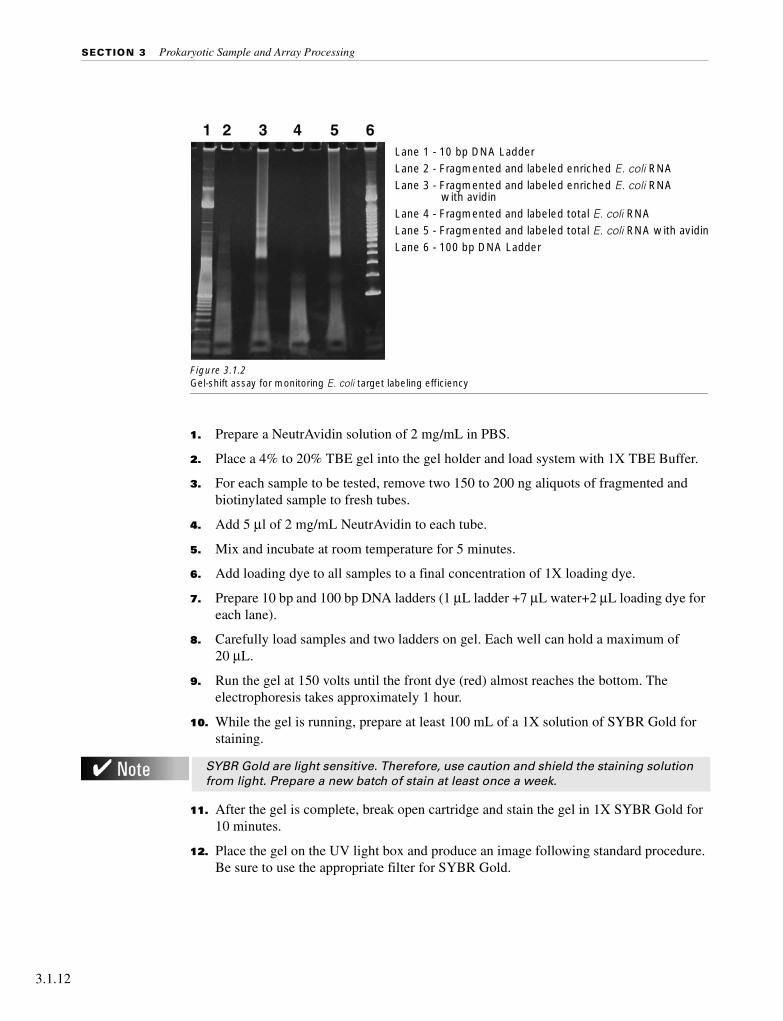
SECTION 3 Prokaryotic Sample and Array Processing
3.1.12
1. Prepare a NeutrAvidin solution of 2 mg/mL in PBS.
2. Place a 4% to 20% TBE gel into the gel holder and load system with 1X TBE Buffer.
3. For each sample to be tested, remove two 150 to 200 ng aliquots of fragmented and biotinylated sample to fresh tubes.
4. Add 5 µl of 2 mg/mL NeutrAvidin to each tube.
5. Mix and incubate at room temperature for 5 minutes.
6. Add loading dye to all samples to a final concentration of 1X loading dye.
7. Prepare 10 bp and 100 bp DNA ladders (1 µL ladder +7 µL water+2 µL loading dye for each lane).
8. Carefully load samples and two ladders on gel. Each well can hold a maximum of 20 µL.
9. Run the gel at 150 volts until the front dye (red) almost reaches the bottom. The electrophoresis takes approximately 1 hour.
10. While the gel is running, prepare at least 100 mL of a 1X solution of SYBR Gold for staining.
11. After the gel is complete, break open cartridge and stain the gel in 1X SYBR Gold for 10 minutes.
12. Place the gel on the UV light box and produce an image following standard procedure. Be sure to use the appropriate filter for SYBR Gold.
Figure 3.1.2 Gel-shift assay for monitoring E. coli target labeling efficiency
SYBR Gold are light sensitive. Therefore, use caution and shield the staining solution from light. Prepare a new batch of stain at least once a week.
Lane 1 - 10 bp DNA LadderLane 2 - Fragmented and labeled enriched E. coli RNALane 3 - Fragmented and labeled enriched E. coli RNA
with avidinLane 4 - Fragmented and labeled total E. coli RNALane 5 - Fragmented and labeled total E. coli RNA with avidinLane 6 - 100 bp DNA Ladder

Pro
kary
oti
c
Section 3, Chapter 2
701031 Rev. 3

Section 3, Chapter 2

Pro
kary
oti
c
Preparation of Control Spike-In Transcripts
Overview . . . . . . . . . . . . . . . . . . . . . . . . . . . . . . . . . . . . . . . . 3.2.4
Reagents and Materials Required . . . . . . . . . . . . . . . . . . . . . . . . . . . . 3.2.5
Bacterial Plasmid DNA Preparation . . . . . . . . . . . . . . . . . . . . . . . . . . 3.2.7
Linearization of Plasmid DNA Preparation . . . . . . . . . . . . . . . . . . . . . . . 3.2.7
Purification of Linearized Plasmid DNA . . . . . . . . . . . . . . . . . . . . . . . . 3.2.7
Preparing the Control Transcript Mix . . . . . . . . . . . . . . . . . . . . . . . . . . 3.2.9
In vitro Transcription (IVT) to Produce Control Sense Transcripts . . . . . . . . . . 3.2.8
This Chapter Contains:
■ Detailed steps for producing full-length control spike sense RNA.
After completing the procedures described in this chapter, the control sense transcripts can be added to purified prokaryotic RNA samples prior to enrichment and labeling procedure as described in Section 3, Chapter 1.
701031 Rev. 3 3.2.3

SECTION 3 Prokaryotic Sample and Array Processing
3.2.4
Overview
This chapter describes protocols used to generate sense RNA controls from B. subtilis genes. These control transcripts can be spiked into P. aeruginosa or E.coli total RNA used for target preparation at a predetermined concentration to monitor labeling, hybridization, and staining efficiency.
To be used as control for assay performance, GeneChip® P. aeruginosa and E. coli Antisense Genome Arrays contain probe sets with sequences of dap, thr, phe, and lys genes from B. subtilis. These genes have been cloned into Stratagene pBluescript as an Xho I to Not I insert, 5´ to 3´ respectively (see Section 2, Chapter 2, Controls for Eukaryotic Arrays).
pGIBS-lysATCC 87482
pGIBS-pheATCC 87483
pGIBS-thrATCC 87484
pGIBS-dapATCC 87486
These clones can be digested with the Not I restriction enzyme to produce linear template DNA for the subsequent in vitro transcription (IVT) to produce sense RNA by T3 RNA polymerase as control molecules.
Bacteria containing these recombinant plasmids can be obtained from the American Type Culture Collection (ATCC).
T3 5’
Xho I Not I
3’ T7

CHAPTER 2 Preparation of Control Spike-In Transcripts
Pro
kary
oti
c
Reagents and Materials Required
The following reagents and materials are recommendations and have been tested and evaluated by Affymetrix scientists. For supplier phone numbers in the U.S. and Europe, please refer to the Supplier and Reagent Reference List, Appendix A, of this manual. Information and part numbers listed are based on U.S. catalog information. Additional reagents needed for the complete analysis are listed in the appropriate chapters. Appendix A contains a master list of all reagents used in this manual.
■ Expression Control Clones, American Type Culture Collection (ATCC)
■ pGIBS-lys ATCC 87482
■ pGIBS-phe ATCC 87483
■ pGIBS-thr ATCC 87484
■ pGIBS-dap ATCC 87486
■ Not I restriction Endonuclease, New England BioLabs, P/N R0189S
■ Phase Lock Gel, Brinkmann Instruments, P/N 955 15 415■ Phenol/chloroform/isoamyl alcohol, Ambion, P/N 9732
■ MEGAscript T3 Kit, Ambion, P/N 1338
Miscellaneous Reagents■ NaAcetate (NaOAc), 3M
■ Absolute Ethanol
■ 80% Ethanol
■ RNeasy Mini Kit, QIAGEN, P/N 74104
■ TE, 1X, BioWhittaker Molecular Applications / Cambrex, P/N 16-013B
3.2.5
CHAPTER 2 Preparation of Control Spike-In Transcripts
Pro
kary
oti
c
Bacterial Plasmid DNA Preparation
1. Grow E. coli bacterial cultures containing recombinant plasmids according to established protocols (a minimum 50 mL of culture volume is recommended).
2. Prepare plasmid DNA from overnight cultures using standard procedures or commercial kits.
We have obtained reliable results using QIAGEN Plasmid Kits for plasmid DNA isolation.
Linearization of Plasmid DNA Preparation
1. In a 50 µL reaction volume, digest 10 µg of plasmid with the restriction enzyme, Not I, according to the enzyme manufacturer’s recommendations.
2. Analyze 50 ng of the uncut and linearized plasmid by gel electrophoresis on a 1% agarose gel. Complete digestion of the plasmid is required for IVT. Repeat restriction enzyme digestion, if necessary.
Purification of Linearized Plasmid DNA
Purify the linearized plasmid from restriction enzymes and potential RNase contaminants before proceeding to IVT using the following Phase Lock Gel (PLG)-phenol/chloroform extraction procedure.
Phase Lock Gels form an inert sealed barrier between the aqueous and organic phases of phenol/chloroform extractions. The solid barrier allows more complete recovery of the sample (aqueous phase) and minimizes interface contamination of the sample. PLG’s are sold as pre-measured aliquots in 1.5 mL tubes to which sample and phenol/chloroform are directly added.
1. Pellet the Phase Lock Gel (1.5 mL tube with PLG I-heavy) in a microcentrifuge at≥ 12,000 x g for 20 seconds.
2. Dilute the linearized plasmid to final volume of 150 µL with TE and add equal volume of (25:24:1) Phenol:chloroform:isoamyl alcohol (saturated with 10 µM tris-HCl, pH 8.01 / 1 mM EDTA). Vortex.
3. Transfer the mix to the PLG tube and microcentrifuge at ≥ 12,000 x g for 2 minutes.
4. Transfer the top aqueous phase to a new 1.5 mL tube.
5. Add 0.1 volumes (15 µL) of 3M NaOAc and 2.5 volumes (375 µL) of absolute ethanol to the samples. Vortex.
6. Immediately centrifuge at ≥ 12,000 x g in a microcentrifuge at room temperature for 20 minutes.
7. Carefully remove supernatant.
8. Wash pellet with 0.5 mL of 80% ethanol, then centrifuge at ≥ 12,000 x g at room temperature for 5 minutes.
3.2.7
SECTION 3 Prokaryotic Sample and Array Processing
3.2.8
9. Remove the supernatant very carefully and air dry the pellet.
10. Resuspend DNA pellet in 15 µL of RNase-free water.
11. Quantify the DNA by absorbance at 260 nm (50 µg/mL of DNA for 1 absorbance unit at 260 nm).
In vitro Transcription (IVT) to Produce Control Sense Transcripts
Use MEGAscript T3 Kit for the IVT reaction.
1. To make up the reaction mix, follow the procedures in the instruction manual provided by Ambion.
2. Incubate the reaction for 4 hours at 37°C.
3. Clean up the reaction product with RNeasy mini column.
4. Quantify the transcript by absorbance at 260 nm (40 µg/mL RNA = 1 absorbance unit at 260 nm).
The quality of DNA template can be monitored by the A260/A280 ratio, which should be between 1.8 and 2.0 for pure DNA.
No tracer is involved in this assay.
It is recommended to examine the quality and integrity of the IVT product on an agarose gel. The expected IVT product sizes are shown in Table 3.2.1.
Aliquot and freeze the IVT transcripts at -80°C. Avoid repeated freeze / thaw cycles.

CHAPTER 2 Preparation of Control Spike-In Transcripts
Pro
kary
oti
c
Preparing the Control Transcript Mix
1. Prepare stock solutions for each of the five transcripts at 650 pM for each transcript. Use Table 3.2.1 to calculate the amount of transcript needed.
2. Mix equal volumes of the 650 pM stocks for all four transcripts for a final concentration of 130 pM for each transcript.
3. Apply 2 µL of the transcript mix with each 5-10 µg of total RNA prior to the cDNA synthesis procedure (as described in Section 3, Chapter 1, Prokaryotic Target Preparation). Final concentration applied on the array for the control transcripts would be 2 pM, assuming 100% recovery during the cDNA synthesis and labeling process.
Table 3.2.1 Conversions for Preparing Control Transcript Mix
Control RNA Size (kb) Molecular Weight pmoles / µg
lys 1 330,000 3.03
phe 1.32 435,600 2.30
dap 1.84 607,200 1.65
thr 1.98 653,400 1.53
Different concentrations of transcript stock can be prepared to generate “staggered” concentrations for different transcripts to monitor the dynamic range of the assay.
Aliquot and freeze the IVT transcripts at -80°C. Avoid repeated freeze / thaw cycles.
3.2.9
Pro
kary
oti
c
Section 3, Chapter 3
701032 Rev. 3

Section 3, Chapter 3

Pro
kary
oti
c
Prokaryotic Target Hybridization
Reagents and Materials Required . . . . . . . . . . . . . . . . . . . . . . . . . . . . 3.3.5
Reagent Preparation . . . . . . . . . . . . . . . . . . . . . . . . . . . . . . . . . . . 3.3.6
Prokaryotic Target Hybridization . . . . . . . . . . . . . . . . . . . . . . . . . . . . 3.3.7
This Chapter Contains:
This chapter contains detailed steps for preparing the hybridization mix, and instructions for hybridizing the target mix to the GeneChip® P. aeruginosa Genome Array and GeneChip® E. coli Antisense Genome Array. The hybridized probe array is then ready for washing, staining, and scanning as detailed on page 3.4.3.
701032 Rev. 3 3.3.3

CHAPTER 3 Prokaryotic Target Hybridization
Pro
kary
oti
c
Reagents and Materials Required
The following reagents and materials are recommendations and have been tested and evaluated by Affymetrix scientists. For supplier phone numbers in the U.S. and Europe, please refer to the Supplier and Reagent Reference List, Appendix A, of this manual. Information and part numbers listed are based on U.S. catalog information. Additional reagents needed for the complete analysis are listed in the appropriate sections. Appendix A contains a master list of all reagents used in this manual.
■ Water, Molecular Biology Grade, BioWhittaker Molecular Applications / Cambrex, P/N 51200
■ Acetylated Bovine Serum Albumin (BSA) solution, 50 mg/mL, Invitrogen Life Technologies, P/N 15561-020
■ Herring Sperm DNA, Promega Corporation, P/N D1811
■ Micropure Separator, Millipore, P/N 42512 (optional)
■ Control Oligo B2, 3 nM, Affymetrix, P/N 900301 (can be ordered separately)
■ NaCl, 5M, RNase-free, DNase-free, Ambion, P/N 9760G
■ MES Free Acid Monohydrate SigmaUltra, Sigma-Aldrich, P/N M5287
■ MES Sodium Salt, Sigma-Aldrich, P/N M5057
■ EDTA Disodium Salt, 0.5M solution (100 mL), Sigma-Aldrich, P/N E7889
Miscellaneous Reagents■ Tough-Spots, Label Dots, USA Scientific, P/N 9185 (optional)
■ 100% DMSO, Sigma-Aldrich, P/N D2650
■ Surfact-Amps X-100 (Tween-20), 10%, Pierce Chemical, P/N 28320
Miscellaneous Supplies■ Hybridization Oven 640, Affymetrix, P/N 800138 (110V) or 800139 (220V)
■ Sterile, RNase-free, microcentrifuge vials, 1.5 mL, USA Scientific,P/N 1415-2600 (or equivalent)
■ Micropipettors, (P-2, P-20, P-200, P-1000), Rainin Pipetman or equivalent
■ Sterile-barrier pipette tips and non-barrier pipette tips
3.3.5
SECTION 3 Prokaryotic Sample and Array Processing
3.3.6
Reagent Preparation
12X MES Stock (1.22M MES, 0.89M [Na+])
For 1,000 mL:
70.4g MES free acid monohydrate
193.3g MES Sodium Salt
800 mL of Molecular Biology Grade water
Mix and adjust volume to 1,000 mL.
The pH should be between 6.5 and 6.7. Filter through a 0.2 µm filter.
2X Hybridization Buffer (50 mL)
(final 1X concentration is 100mM MES, 1M [Na+], 20 mM EDTA, 0.01% Tween 20)
For 50 mL:
8.3 mL of 12X MES Stock
17.7 mL of 5M NaCl
4.0 mL of 0.5M EDTA
0.1 mL of 10% Tween 20
19.9 mL of water
Store at 2°C to 8°C, and shield from light.
Do not autoclave, store at 2°C to 8°C, and shield from light. Discard solution if yellow.

CHAPTER 3 Prokaryotic Target Hybridization
Pro
kary
oti
c
Prokaryotic Target Hybridization
After determining that the fragmented cDNA is labeled with biotin, prepare the hybridization solution mix. The minimum amount of cDNA product required for target hybridization is 1 µg. The solution is stable for approximately 6 to 8 hours at 4°C. The following protocol can be used for freshly prepared or frozen hybridization cocktail. Re-use of prokaryotic sample has not been thoroughly tested and, therefore, is not recommended.
1. Prepare the following hybridization solution mix.
2. Equilibrate probe array to room temperature immediately before use.
3. Add the indicated amount of hybridization solution mix to the probe array. Refer to specific probe array package insert for information on array format.
4. Place probe array in the hybridization oven set at the temperatures indicated below.
■ P. aeruginosa 50°C
■ E. coli Antisense 45°C
5. Avoid stress to the motor; load probe arrays in a balanced configuration around axis. Rotate at 60 rpm.
6. Hybridize for 16 hours.
During the latter part of the 16-hour hybridization, proceed to Section 3, Chapter 4, Prokaryotic Arrays: Washing, Staining, and Scanning to prepare reagents required immediately after completion of hybridization.
Table 3.3.1 Hybridization Cocktail for Single Probe Array*3
Components 100 Format (Midi) 49 Format (Standard)Final
Concentration
2X MES Hybridization Buffer 65 µL 100.0 µL 1X
3 nM B2 Control Oligo 2.2 µL 3.3 µL 50 pM
10 mg/mL Herring Sperm DNA 1.3 µL 2.0 µL 0.1 mg/mL
50 mg/mL BSA 1.3 µL 2.0 µL 0.5 mg/mL
100% DMSO 9.2 µL - 7%
Fragmented and Labeled cDNA 51 µL Up to 92.7 µL 1.5 – 6.0 µg
Molecular Biology Grade Water To a final volume of 130 µL To a final volume of 200 µL
Total Volume 130 µL 200 µL
*Please refer to specific probe array package insert for information on array format.
It is important to allow the arrays to normalize to room temperature completely. Specifically, if the rubber septa are not equilibrated to room temperature, they may be prone to cracking, which leads to leaks.
It is necessary to use two pipette tips when filling the probe array cartridge: one for filling and the second to allow venting of air from the hybridization chamber.
The hybridization temperature of 50°C is higher than that used for other expression assays. The increased hybridization temperature is required due to the high GC content of P. aeruginosa.
3.3.7

Pro
kary
oti
c
\
Section 3, Chapter 4
701033 Rev. 3

Section 3, Chapter 4

Pro
kary
oti
c
Prokaryotic Arrays:Washing, Staining, and Scanning
Reagents and Materials Required . . . . . . . . . . . . . . . . . . . . . . . . . . . . 3.4.5
Reagent Preparation . . . . . . . . . . . . . . . . . . . . . . . . . . . . . . . . . . . 3.4.6
Experiment and Fluidics Station Setup . . . . . . . . . . . . . . . . . . . . . . . . . 3.4.7
Step 1: Defining File Locations . . . . . . . . . . . . . . . . . . . . . . . . . . . . 3.4.7
Step 2: Entering Experiment Information. . . . . . . . . . . . . . . . . . . . . . . 3.4.7
Step 3: Preparing the Fluidics Station. . . . . . . . . . . . . . . . . . . . . . . . . 3.4.8
Probe Array Wash and Stain . . . . . . . . . . . . . . . . . . . . . . . . . . . . . . 3.4.9
Probe Array Scan . . . . . . . . . . . . . . . . . . . . . . . . . . . . . . . . . . . 3.4.15
Handling the GeneChip® Probe Array . . . . . . . . . . . . . . . . . . . . . . . 3.4.15
Scanning the Probe Array . . . . . . . . . . . . . . . . . . . . . . . . . . . . . . 3.4.16
Shutting Down the Fluidics Station . . . . . . . . . . . . . . . . . . . . . . . . . . 3.4.17
Customizing the Protocol . . . . . . . . . . . . . . . . . . . . . . . . . . . . . . . 3.4.18
This Chapter Contains:
■ Instructions for using the Fluidics Station 400 or 450/250 to automate the washing and staining of GeneChip P. aeruginosa and GeneChip E. coli Antisense Genome Arrays.
■ Instructions for scanning probe arrays using the GeneArray® Scanner or the GeneChip® Scanner 3000.
After completing the procedures described in this chapter, the scanned probe array image (.dat file) is ready for analysis, as explained in the enclosed GeneChip Expression Analysis: Data Analysis Fundamentals booklet (P/N 701190).
701033 Rev. 3 3.4.3
CHAPTER 4 Prokaryotic Arrays: Washing, Staining, and Scanning
Pro
kary
oti
c
Reagents and Materials Required
The following reagents and materials are recommendations and have been tested and evaluated by Affymetrix scientists. For supplier phone numbers in the U.S. and Europe, please refer to the Supplier and Reagent Reference List, Appendix A, of this manual. Information and part numbers listed are based on U.S. catalog information. Additional reagents needed for the complete analysis are listed in the appropriate sections. Appendix A contains a master list of all reagents used in this manual.
■ Water, Molecular Biology Grade, BioWhittaker Molecular Applications / Cambrex, P/N 51200
■ Distilled water, Invitrogen Life Technologies, P/N 15230-147
■ Acetylated Bovine Serum Albumin (BSA) solution, 50 mg/mL, Invitrogen Life Technologies, P/N 15561-020
■ R-Phycoerythrin Streptavidin, Molecular Probes, P/N S-866
■ NaCl, 5M, RNase-free, DNase-free, Ambion, P/N 9760G
■ PBS, pH 7.2, Invitrogen Life Technologies, P/N 20012-027
■ 20X SSPE (3 M NaCl, 0.2M NaH2PO4, 0.02M EDTA), BioWhittaker Molecular Applications / Cambrex, P/N 51214
■ Goat IgG, Reagent Grade, Sigma-Aldrich, P/N I 5256
■ Anti-streptavidin antibody (goat), biotinylated, Vector Laboratories, P/N BA-0500
■ 10% surfact-Amps20 (Tween-20), Pierce Chemical, P/N 28320
■ Bleach (5.25% Sodium Hypochlorite), VWR Scientific, P/N 21899-504 (or equivalent)
■ ImmunoPure Streptavidin, Pierce Chemical, P/N 21125
Miscellaneous Supplies■ Sterile, RNase-free, microcentrifuge vials, 1.5 mL, USA Scientific, P/N 1415-2600 (or
equivalent)
■ Micropipettors, (P-2, P-20, P-200, P-1000), Rainin Pipetman (or equivalent)
■ Sterile-barrier pipette tips and non-barrier pipette tips
■ Tygon Tubing, 0.04″ inner diameter, Cole-Parmer, P/N H-06418-04
■ Tough-Spots, Label Dots, USA Scientific, P/N 9185
3.4.5

SECTION 3 Prokaryotic Sample and Array Processing
3.4.6
Reagent Preparation
Wash A: Non-Stringent Wash Buffer
(6X SSPE, 0.01% Tween-20)
For 1,000 mL:
300 mL of 20X SSPE
1.0 mL of 10% Tween-20
699 mL of water
Filter through a 0.2 µm filter.
Store at room temperature.
Wash B: Stringent Wash Buffer
(100 mM MES, 0.1M [Na+], 0.01% Tween 20)
For 1,000 mL:
83.3 mL of 12 X MES Stock Buffer (see Section 3, Chapter 3 for reagent preparation)
5.2 mL of 5M NaCl
1.0 mL of 10% Tween 20
910.5 mL of water
Filter through a 0.2 µm filter
Store at 2°C to 8°C and shield from light.
2X Stain Buffer
(final 1X concentration: 100 mM MES, 1M [Na+], 0.05% Tween 20)
For 250 mL:
41.7 mL 12X MES Stock Buffer (see Section 3, Chapter 3)
92.5 mL 5M NaCl
2.5 mL 10% Tween 20
113.3 mL water
Filter through a 0.2 µm filter.
Store at 2°C to 8°C and shield from light.
10 mg/mL Goat IgG Stock
Resuspend 50 mg in 5 mL 150 mM NaCl.
Store at 4°C.
1 mg/mL Streptavidin Stock
Resuspend 5 mg in 5 mL of PBS.
Store at 4°C.

CHAPTER 4 Prokaryotic Arrays: Washing, Staining, and Scanning
Pro
kary
oti
c
Experiment and Fluidics Station Setup
Step 1: Defining File Locations
Before working with Affymetrix® Microarray Suite it is important to define where the program stores and looks for files.
1. Launch Microarray Suite from the workstation and select Tools → Defaults → File Locations from the menu bar.
The File Locations window displays the locations of the following files:
■ Probe Information (library files, mask files)
■ Fluidics Protocols (fluidics station scripts)
■ Experiment Data (.exp, .dat, .cel, and .chp files are all saved to location selected here)
2. Verify that all three file locations are set correctly and click OK.
Contact Affymetrix Technical Support if you have any questions regarding this procedure.
Step 2: Entering Experiment Information
To wash, stain, and scan a probe array, an experiment must first be defined in Microarray Suite.
1. Select Run → Experiment Info from the menu bar. Alternatively, click the New Experiment icon on the tool bar.
⇒ The Experiment Information dialog box appears, allowing the experiment name to be defined along with several other parameters, such as probe array type, sample description, and comments.
2. Type in the Experiment Name.
3. In the Probe Array Type box, click the arrow and select the experiment name as indicated below:
■ P.aeruginosa select Pae_G1a from the drop-down list
■ E.coli select Ecoli_Antisense from the drop-down list.
Experiment name and probe array type are required. Complete as much of the other information as desired. The protocol information at the bottom of the dialog box is imported to the experiment information dialog box after the hybridization and scan have been completed.
4. Save the experiment by selecting Save.
The name of the experiment is used by Microarray Suite to access the probe array type and data for the sample while it is being processed. Data files generated for the sample are automatically labeled to correspond to the experiment name. Microarray Suite automatically fills in the Protocol section of this dialog box with information on array processing from the fluidics station.
5. Close the Experiment Information dialog box.
3.4.7

SECTION 3 Prokaryotic Sample and Array Processing
3.4.8
Step 3: Preparing the Fluidics Station
The Fluidics Station 400, 450, or 250 is used to wash and stain the probe arrays. It is operated using Microarray Suite.
Setting Up the Fluidics Station
1. Turn on the Fluidics Station using the switch on the lower left side of the machine.
2. Select Run → Fluidics from the menu bar.
The Fluidics Station dialog box appears with a drop-down list for selecting the experiment name for each of the fluidics station modules. A second list is accessed for choosing the Protocol for each of the four fluidics station modules.
Priming the Fluidics Station
Priming ensures that the lines of the fluidics station are filled with the appropriate buffers and the fluidics station is ready for running fluidics station protocols.
Priming should be done:
■ When the fluidics station is first started
■ When wash solutions are changed
■ Before washing if a shutdown has been performed
■ If the LCD window instructs the user to prime
1. Select Protocol in the Fluidics Station dialog box.
2. Choose Prime or Prime_450 for the respective modules in the Protocol drop-down list.
3. Change the intake buffer reservoir A to Non-stringent Wash Buffer and intake buffer reservoir B to Stringent Wash Buffer.
4. Click Run for each module to begin priming.
Refer to the appropriate GeneChip® Fluidics Station User’s Guide for instructions on connecting and addressing multiple fluidics stations.

CHAPTER 4 Prokaryotic Arrays: Washing, Staining, and Scanning
Pro
kary
oti
c
Probe Array Wash and Stain
Following hybridization, the wash and stain procedures are carried out by the Fluidics Station. A modified FlexMidi_euk2v3 fluidic script (FlexMidi_euk2v3_450, if you are using the FS-450) is used for the GeneChip P. aeruginosa Genome Array, and the ProkGE-WS2 fluidic script (ProkGE-WS2_450, if you are using the FS-450) is used for the GeneChip E. coli Antisense Genome Array. The procedures take approximately 75 and 90 minutes, respectively, to complete. The use of streptavidin in the first part of the stain procedure enhances the overall signal.
Preparing the Staining Reagents
1. Prepare the following stain and wash solutions the day of the procedure. The solutions are stable for approximately 6 to 8 hours at 4°C. Volumes given are sufficient for one probe array.
Streptavidin Phycoerythrin (SAPE) should be stored in the dark at 4° C, either foil wrapped or kept in an amber tube. Remove SAPE from refrigerator and tap the tube to mix well before preparing stain solution.
Do not freeze SAPE. Always prepare the SAPE stain solution immediately before use.
Table 3.4.1 Streptavidin Solution Mix - Vial 1
Components Volume Final Concentration
2X MES Stain Buffer 300.0 µL 1X
50 mg/mL BSA 24.0 µL 2 mg/mL
1 mg/mL Streptavidin 6.0 µL 10 µg/mL
DI H20 270.0 µL —
Total Volume 600 µL
3.4.9
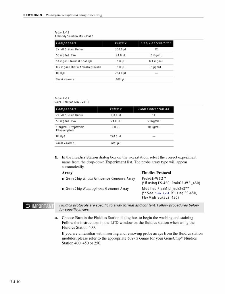
SECTION 3 Prokaryotic Sample and Array Processing
3.4.10
2. In the Fluidics Station dialog box on the workstation, select the correct experiment name from the drop-down Experiment list. The probe array type will appear automatically.
Array Fluidics Protocol■ GeneChip E. coli Antisense Genome Array ProkGE-WS2 *
(*if using FS-450, ProkGE-WS_450)
■ GeneChip P. aeruginosa Genome Array Modified FlexMidi_euk2v3**(**See Table 3.4.4. If using FS-450, FlexMidi_euk2v3_450)
3. Choose Run in the Fluidics Station dialog box to begin the washing and staining. Follow the instructions in the LCD window on the fluidics station when using the Fluidics Station 400.
If you are unfamiliar with inserting and removing probe arrays from the fluidics station modules, please refer to the appropriate User’s Guide for your GeneChip® Fluidics Station 400, 450 or 250.
Table 3.4.2 Antibody Solution Mix - Vial 2
Components Volume Final Concentration
2X MES Stain Buffer 300.0 µL 1X
50 mg/mL BSA 24.0 µL 2 mg/mL
10 mg/mL Normal Goat IgG 6.0 µL 0.1 mg/mL
0.5 mg/mL Biotin Anti-streptavidin 6.0 µL 5 µg/mL
DI H20 264.0 µL —
Total Volume 600 µL
Table 3.4.3 SAPE Solution Mix - Vial 3
Components Volume Final Concentration
2X MES Stain Buffer 300.0 µL 1X
50 mg/mL BSA 24.0 µL 2 mg/mL
1 mg/mL Streptavidin Phycoerythrin
6.0 µL 10 µg/mL
DI H20 270.0 µL —
Total Volume 600 µL
Fluidics protocols are specific to array format and content. Follow procedures below for specific arrays
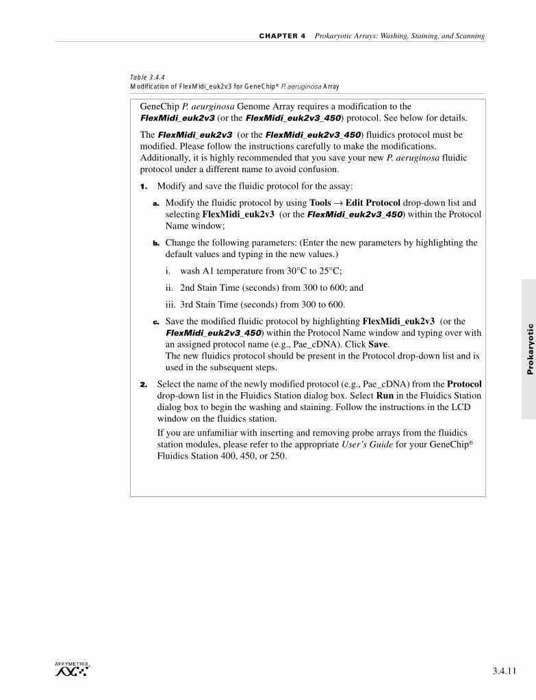
CHAPTER 4 Prokaryotic Arrays: Washing, Staining, and Scanning
Pro
kary
oti
c
Table 3.4.4 Modification of FlexMidi_euk2v3 for GeneChip® P. aeruginosa Array
GeneChip P. aeurginosa Genome Array requires a modification to the FlexMidi_euk2v3 (or the FlexMidi_euk2v3_450) protocol. See below for details.
The FlexMidi_euk2v3 (or the FlexMidi_euk2v3_450) fluidics protocol must be modified. Please follow the instructions carefully to make the modifications. Additionally, it is highly recommended that you save your new P. aeruginosa fluidic protocol under a different name to avoid confusion.
1. Modify and save the fluidic protocol for the assay:
a. Modify the fluidic protocol by using Tools → Edit Protocol drop-down list and selecting FlexMidi_euk2v3 (or the FlexMidi_euk2v3_450) within the Protocol Name window;
b. Change the following parameters: (Enter the new parameters by highlighting the default values and typing in the new values.)
i. wash A1 temperature from 30°C to 25°C;
ii. 2nd Stain Time (seconds) from 300 to 600; and
iii. 3rd Stain Time (seconds) from 300 to 600.
c. Save the modified fluidic protocol by highlighting FlexMidi_euk2v3 (or the FlexMidi_euk2v3_450) within the Protocol Name window and typing over with an assigned protocol name (e.g., Pae_cDNA). Click Save.The new fluidics protocol should be present in the Protocol drop-down list and is used in the subsequent steps.
2. Select the name of the newly modified protocol (e.g., Pae_cDNA) from the Protocol drop-down list in the Fluidics Station dialog box. Select Run in the Fluidics Station dialog box to begin the washing and staining. Follow the instructions in the LCD window on the fluidics station.
If you are unfamiliar with inserting and removing probe arrays from the fluidics station modules, please refer to the appropriate User’s Guide for your GeneChip® Fluidics Station 400, 450, or 250.
3.4.11
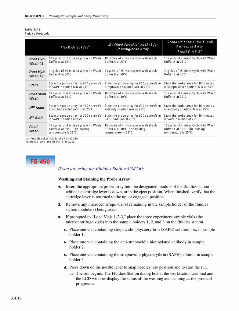
SECTION 3 Prokaryotic Sample and Array Processing
3.4.12
If you are using the Fluidics Station 450/250:
Washing and Staining the Probe Array
1. Insert the appropriate probe array into the designated module of the fluidics station while the cartridge lever is down, or in the eject position. When finished, verify that the cartridge lever is returned to the up, or engaged, position.
2. Remove any microcentrifuge vial(s) remaining in the sample holder of the fluidics station module(s) being used.
3. If prompted to “Load Vials 1-2-3,” place the three experiment sample vials (the microcentrifuge vials) into the sample holders 1, 2, and 3 on the fluidics station.
a. Place one vial containing streptavidin phycoerythrin (SAPE) solution mix in sample holder 1.
b. Place one vial containing the anti-streptavidin biotinylated antibody in sample holder 2.
c. Place one vial containing the streptavidin phycoerythrin (SAPE) solution in sample holder 3.
d. Press down on the needle lever to snap needles into position and to start the run.
⇒ The run begins. The Fluidics Station dialog box at the workstation terminal and the LCD window display the status of the washing and staining as the protocol progresses.
Table 3.4.5 Fluidics Protocols
FlexMidi_euk2v3a Modified FlexMidi_euk2v3 for P. aeruginosa Array
Standard Format for E. coli Antisense Array
ProkGE-WS-2b
Post Hyb Wash #1
10 cycles of 2 mixes/cycle with Wash Buffer A at 30°C
10 cycles of 2 mixes/cycle with Wash Buffer A at 25°C
10 cycles of 2 mixes/cycle with Wash Buffer A at 25°C
Post Hyb Wash #2
4 cycles of 15 mixes/cycle with Wash Buffer B at 50°C
4 cycles of 15 mixes/cycle with Wash Buffer B at 50°C
4 cycles of 15 mixes/cycle with Wash Buffer B at 45°C
Stain Stain the probe array for 600 seconds in SAPE Solution Mix at 25°C
Stain the probe array for 600 seconds in Streptavidin Solution Mix at 25°C
Stain the probe array for 10 minutes in Streptavidin Solution. Mix at 25°C.
Post Stain Wash
10 cycles of 4 mixes/cycle with Wash Buffer A at 30°C
10 cycles of 4 mixes/cycle with Wash Buffer A at 30°C
10 cycles of 4 mixes/cycle with Wash Buffer A at 30°C
2nd StainStain the probe array for 300 seconds in antibody solution mix at 25°C
Stain the probe array for 600 seconds in antibody solution mix at 25°C
Stain the probe array for 10 minutes in antibody solution. Mix at 25°C.
3rd StainStain the probe array for 300 seconds in SAPE Solution at 25°C
Stain the probe array for 600 seconds in SAPE Solution at 25°C
Stain the probe array for 10 minutes in SAPE Solution at 25°C.
Final Wash
15 cycles of 4 mixes/cycle with Wash Buffer A at 30°C. The holding temperature is 25°C.
15 cycles of 4 mixes/cycle with Wash Buffer A at 30°C. The holding temperature is 25°C.
15 cycles of 4 mixes/cycle with Wash Buffer A at 30°C. The holding temperature is 25°C.
a. FlexMidi_euk2v_450 for the FS-450/250b. prokGE_ws2_450 for the FS-450/250

CHAPTER 4 Prokaryotic Arrays: Washing, Staining, and Scanning
Pro
kary
oti
c
4. When the protocol is complete, the LCD window displays the message EJECT CARTRIDGE.
5. Remove the probe arrays from the fluidics station modules by first pressing down the cartridge lever to the eject position.
6. Lift up on the needle lever to disengage the needles from the microcentrifuge vials. Remove the three empty microcentrifuge vials from the needle holders.
7. Check the probe array window for large bubbles or air pockets.
■ If bubbles are present, refer to Table 3.4.6.
■ If the probe array has no large bubbles, it is ready to scan on the GeneArray® Scanner, or the GeneChip® Scanner 3000. Pull up on the cartridge lever to close the washblock and proceed to Probe Array Scan on page 3.4.15.
8. If there are no more samples to hybridize, shut down the fluidics station following the procedure in Shutting Down the Fluidics Station on page 3.4.17.
9. Keep the probe arrays at 4°C and in the dark until ready for scanning.
10. Lift up on the cartridge lever to close the washblock.
If you are using the Fluidics Station 400:
Washing and Staining the Probe Array
1. Insert the appropriate probe array into the designated module of the fluidics station while the cartridge lever is in the EJECT position. When finished, verify that the cartridge lever is returned to the ENGAGE position.
2. Remove any microcentrifuge tube remaining in the sample holder of the fluidics station module(s) being used.
3. Place a microcentrifuge tube containing the streptavidin solution into the sample holder, making sure that the metal sampling needle is in the tube with its tip near the bottom
⇒ The Fluidics Station dialog box and the LCD window display the status of the washing and staining as they progress.
For proper cleaning and maintenance of the fluidics station, including the bleach protocol, refer to Section 4, Fluidics Station Maintenance Procedures.
Table 3.4.6If bubbles are present
Return the probe array to the probe array holder. Engage the washblock by gently pushing up on the cartridge lever to the engage position.The fluidics station will drain the probe array and then fill it with a fresh volume of the last wash buffer used. When it is finished, the LCD window will display EJECT CARTRIDGE. Again, remove the probe array and inspect it for bubbles. If no bubbles are present, it is ready to scan. Proceed to Probe Array Scan on page 3.4.15.If several attempts to fill the probe array without bubbles are unsuccessful, the array should be filled with Wash A (non-stringent buffer) manually, using a micropipette. Excessive washing will result in a loss of signal intensity.
3.4.13

SECTION 3 Prokaryotic Sample and Array Processing
3.4.14
4. When the LCD window indicates, replace the microcentrifuge tube containing the streptavidin stain with a microcentrifuge tube containing antibody stain solution into the sample holder, making sure that the metal sampling needle is in the tube with its tip near the bottom.
5. When the LCD window indicates, replace the microcentrifuge tube containing antibody solution with the microcentrifuge tube containing the SAPE solution.
6. When the protocol is complete, the LCD window displays the message EJECT CARTRIDGE.
7. Remove microcentrifuge tube containing stain and replace with an empty microcentrifuge tube.
8. Remove the probe arrays from the fluidics station modules by first moving the cartridge lever to the EJECT position.
9. Check the probe array window for large bubbles or air pockets.
■ If bubbles are present, refer to Table 3.4.7.
■ If the probe array has no large bubbles, it is ready to scan on the GeneChip Scanner 3000 or the GeneArray® Scanner. ENGAGE washblock and proceed to Probe Array Scan on page 3.4.15.
If you do not scan the arrays right away, keep the probe arrays at 4°C and in the dark until ready for scanning.
If there are no more samples to hybridize, shut down the fluidics station following the procedure outlined in the section, Shutting Down the Fluidics Station, on page 4.17.
For proper cleaning and maintenance of the fluidics station, including the bleach protocol, refer to Section 4, Fluidics Station Maintenance Procedures.
Table 3.4.7If bubbles are present
Return the probe array to the probe array holder. Latch the probe array holder by gently pushing it up until a light click is heard. Engage the washblock by firmly pushing up on the cartridge lever to the ENGAGE position.The fluidics station will drain the probe array and then fill it with a fresh volume of the last wash buffer used. When it is finished, the LCD window displays EJECT CARTRIDGE again, remove the probe array and inspect it again for bubbles. If no bubbles are present, it is ready to scan. Proceed to Probe Array Scan on page 3.4.15.If several attempts to fill the probe array without bubbles are unsuccessful, the array should be filled with Wash A (non-stringent buffer) manually, using a micropipette. Excessive washing will result in a loss of signal intensity.

CHAPTER 4 Prokaryotic Arrays: Washing, Staining, and Scanning
Pro
kary
oti
c
Probe Array Scan
The scanner is also controlled by Affymetrix Microarray Suite. The probe array is scanned after the wash protocols are complete. Make sure laser is warmed up prior to scanning by turning the laser on at least 15 minutes before use (if you are using the Agilent GeneArray® Scanner) or 10 minutes (if you are using the GeneChip® Scanner 3000). If probe array was stored at 4°C, warm to room temperature before scanning. Refer to the Microarray Suite online help and the appropriate scanner user’s manual for more information on scanning.
Handling the GeneChip® Probe Array
Before you scan the probe array, follow the directions in this section on handling the probe array. If necessary, clean the glass surface of probe array with a non-abrasive towel or tissue before scanning. Do not use alcohol to clean glass.
Before scanning the probe array cartridge, follow this procedure to apply Tough-Spots™ to the probe array cartridge to prevent the leaking of fluids from the cartridge during scanning.
1. On the back of the probe array cartridge, clean excess fluid from around septa.
2. Carefully apply one Tough-Spot to each of the two septa. Press to ensure that the spots remain flat. If the Tough-Spots do not apply smoothly; that is, if you observe bumps, bubbles, tears or curled edges, do not attempt to smooth out the spot. Remove the spot and apply a new spot. See Figure 3.4.1.
The scanner uses a laser and is equipped with a safety interlock system. Defeating the interlock system may result in exposure to hazardous laser light.
You must have read and be familiar with the operation of the scanner before attempting to scan a probe array. Please refer to the Microarray Suite User’s Guide (P/N 08-0081) or to the GeneChip® Scanner 3000 quick reference card (P/N 08-0075).
Apply the spots just before scanning. Do not use them in the hyb process.
3.4.15

SECTION 3 Prokaryotic Sample and Array Processing
3.4.16
3. Insert the cartridge into the scanner and test the autofocus to ensure that the Tough-Spots do not interfere with the focus. If you observe a focus error message, remove the spot and apply a new spot. Ensure that the spots lie flat.
Scanning the Probe Array
1. Select Run → Scanner from the menu bar. Alternatively, click the Start Scan icon in the tool bar.
⇒ The Scanner dialog box appears with a drop-down list of experiments that have not been run.
2. Select the experiment name that corresponds to the probe array to be scanned.
A previously run experiment can also be selected by using the Include Scanned Experiments option box. After selecting this option, previously scanned experiments appear in the drop-down list.
3. By default, for the Agilent® GeneArray® Scanner only, after selecting the experiment the number [2] is displayed in the Number of Scans box to perform the recommended 2X image scan. For the GeneChip® Scanner 3000, only one scan is required.
4. Once the experiment has been selected, click the Start button.
⇒ A dialog box prompts you to load a sample into the scanner.
5. If you are using the GeneArray® Scanner, click the Options button to check for the correct pixel value and wavelength of the laser beam.
■ Pixel value = 3 µm
■ Wavelength = 570 nm
If you are using the GeneChip Scanner 3000, pixel resolution and wavelength are preset and cannot be changed.
Figure 3.4.1 Applying Tough-Spots™ to the probe array cartridge

CHAPTER 4 Prokaryotic Arrays: Washing, Staining, and Scanning
Pro
kary
oti
c
6. Open the sample door on the scanner and insert the probe array into the holder. Do not force the probe array into the holder. Close the sample door of the scanner. If you are using the GeneChip Scanner 3000, do not attempt to close the door by hand. The door closes automatically through the User Interface when start scan is selected or the scanner goes into stand-by mode.
7. Click OK in the Start Scanner dialog box.
⇒ The scanner begins scanning the probe array and acquiring data. When Scan in Progress is Selected from the View menu, the probe array image appears on the screen as the scan progresses.
Shutting Down the Fluidics Station
1. After removing a probe array from the probe array holder, the LCD window displays the message ENGAGE WASHBLOCK.
2. If you are using the FS-400, engage the washblock by firmly pushing up on the cartridge lever to the ENGAGE position.
If you are using the FS-450, gently lift up the cartridge lever to engage, or close, the washblock.
⇒ The fluidics station automatically performs a Cleanout procedure. The LCD window indicates the progress of the Cleanout procedure.
3. When the fluidics station LCD window indicates REMOVE VIALS, the Cleanout procedure is complete.
4. Remove the sample microcentrifuge vial(s) from the sample holder(s).
5. If no other hybridizations are to be performed, place wash lines into a bottle filled with deionized water.
6. Select Shutdown or Shutdown_450 for all modules from the drop-down Protocol list in the Fluidics Station dialog box. Click the Run button for all modules.
The Shutdown protocol is critical to instrument reliability. Refer to the appropriate Fluidics Station User’s Guide for more information.
7. After Shutdown protocol is complete, flip the ON/OFF switch of the fluidics station to the OFF position.
To maintain the cleanliness of the fluidics station and obtain the highest quality image and data possible, a weekly bleach protocol and a monthly decontamination protocol are highly recommended. Please refer to Section 4, Fluidics Station Maintenance Procedures for further detail.
3.4.17
SECTION 3 Prokaryotic Sample and Array Processing
3.4.18
Customizing the Protocol
There may be times when the fluidic protocols need to be modified. Modification of protocols must be done before downloading the protocol to the fluidics station. Protocol changes will not affect runs in progress. For more specific instructions, refer to the Microarray Suite online help.
1. Select Tools → Edit Protocol from the menu bar.
2. In the Edit Protocol dialog box under Protocol Name, click the arrow to open a list of protocols. Click the protocol to be changed.
⇒ The name of the protocol is displayed in the Protocol Name text box. The conditions for that protocol are displayed on the right side of the Edit Protocol dialog box.
3. Select the item to be changed and input the new parameters as needed, keeping parameters within the ranges shown below in Table 3.4.8.
4. To return to the default values for the protocol selected, click the Defaults button.
5. Once all the protocol conditions are modified as desired, change the name of the edited protocol in the Protocol Name box.
6. Click Save, then close the dialog box.
Enter 0 (zero) for hybridization time if hybridization step is not required. Likewise, enter 0 (zero) for the stain time if staining is not required. Enter 0 (zero) for the number of wash cycles if a wash solution is not required.
Table 3.4.8Valid Ranges for Wash/Stain Parameters
Parameter Valid Range
Wash Temperature for A1, B, A2, or A3 (°C) 15 to 50
Number of Wash Cycles for A1, B, A2, or A3 0 to 99
Mixes / Wash Cycle for A1, B, A2, or A3 15 to 50
Stain Time (seconds) 0 to 86,399
Stain Temperature (°C) 15 to 50
Holding Temperature (°C) 15 to 50
• Wash A1 corresponds to Post Hyb Wash #1 in Table 3.4.6.• Wash B corresponds to Post Hyb Wash #2 in Table 3.4.6.• Wash A2 corresponds to Post Stain Wash in Table 3.4.6.• Wash A3 corresponds to Final Wash in Table 3.4.6.
If the protocol is saved without entering a new Protocol Name, the original protocol parameters will be overwritten.

Section 4:
Fluidics Station Maintenance Procedures
701040 Rev. 2

Section 4

F.S
. M
ain
ten
an
ce
Contents
Section 4
Chapter 1 Fluidics Station Maintenance Procedures 4.1.3
701040 Rev. 2

F.S
. M
ain
ten
an
ce
Section 4, Chapter 1
701041 Rev. 3
Section 4, Chapter 1

F.S
. M
ain
ten
an
ce
Fluidics Station Maintenance Procedures
Weekly Fluidics Station Cleanout. . . . . . . . . . . . . . . . . . . . . . . . . . . . 4.1.4
Bleach Protocol . . . . . . . . . . . . . . . . . . . . . . . . . . . . . . . . . . . . 4.1.4
Monthly Fluidics Station Decontamination Protocol . . . . . . . . . . . . . . . . . 4.1.10
This Section Contains:
■ A weekly fluidics station bleach protocol.
■ A monthly fluidics station decontamination protocol.
701041 Rev. 3 4.1.3

SECTION 4 Fluidics Station Maintenance Procedures
4.1.4
Weekly Fluidics Station Cleanout
A cleaning protocol is recommended for fluidics station maintenance if the antibody staining procedure is used. Choose Bleach or Bleach_450 for all modules from the drop-down list in the Fluidics Station dialog box. Click the Run button for all modules and follow LCD instructions.
Bleach Protocol
This protocol is designed to eliminate any residual SAPE-antibody complex that may be present in the fluidics station tubing and needles. We recommend running this protocol at least once a week.
1. Prepare 1 liter of 0.525% sodium hypochlorite solution using distilled water. Shake well.
2. Cut tubing.
If you are using the Fluidics Station 450/250:
If you are using the FS-450 or FS-250, cut three pieces of tubing with each piece at least 2.5-3 feet in length (Tygon tubing, 0.04”) for each module of each fluidics station, for a total of 6 tubes (for the FS-250) or 12 tubes (for the FS-450). These can be reused for subsequent BLEACH runs.
If you are using the Fluidics Station 400:
If you are using the FS-400, cut four pieces of tubing at least 2.5-3 feet in length.
3. Place all three wash lines (these are not the tubing on the needles but the supply lines from the reagent bottles on the side of the station) of each fluidics station in 1 liter of distilled water.
4. Choose Fluidics from the Run menu. Alternatively, click the down arrow Protocol list on the toolbar.
5. Choose Bleach or Bleach_450 for the respective modules in the Protocol drop-down list.
6. Disengage the washblock for each module by pressing down on the cartridge lever.
Each fluidics station with four modules requires at least 500 mL of the 0.525% sodium hypochlorite solution.
The BLEACH protocol requires at least 550 mL of distilled water.

CHAPTER 1 Fluidics Station Maintenance Procedures
F.S
. M
ain
ten
an
ce
7. Connect tubing to needles.
If you are using the Fluidics Station 450/250:
If you are using the FS-450/250, connect one end of the plastic tubing to each of the three needles. The proper technique is to press down on the cartridge lever until the needles extend a convenient distance from the module, then slip the tube on as you hold the cartridge lever down with the free hand as shown in Figure 4.1.2. Take care not to bend or break the needles.
If you are using the Fluidics Station 400:
If you are using the FS-400, connect one end of the plastic tubing to each needle at the bottom of each module.
Figure 4.1.1 Disengaged washblocks showing cartridge levers in the down position.
Temperature will ramp up to 50°C.
Wash blocks disengagedwith cartridge levers down
4.1.5

SECTION 4 Fluidics Station Maintenance Procedures
4.1.6
8. Insert the other ends into 0.525% sodium hypochlorite solution (at least 500 mL for all four modules) as shown in Figure 4.1.3.
Figure 4.1.2 Inserting tubes on the needles. Take care not to break or bend the needles.
Remove cartridges before you start the bleach protocol.

CHAPTER 1 Fluidics Station Maintenance Procedures
F.S
. M
ain
ten
an
ce
Ensure that all the tube ends remain immersed in the bleach solution by tamping down on the tubes using a dowel or similar object as shown in Figure 4.1.4.
Figure 4.1.3 The tubes extending from the modules to the bleach bottle. Note that the probe array cartridges must be removed before the protocol can begin.
4.1.7

SECTION 4 Fluidics Station Maintenance Procedures
4.1.8
9. Remove any probe array cartridges and engage the washblock as shown in Figure 4.1.5. The fluidics station will begin the protocol, begin to empty the lines, and perform three cleaning cycles of 10 rinses each using bleach solution.
Figure 4.1.4 Tamp down on the tubes to ensure that the ends remain immersed in the solution.
For ease of handling, band the tubes together using a rubber band.

CHAPTER 1 Fluidics Station Maintenance Procedures
F.S
. M
ain
ten
an
ce
10. When the fluidics station LCD window displays Remove Tube from Needles, carefully remove tubing from each module needle by pushing the tubing down with your fingers while holding the needle with the other.
11. Load empty microcentrifuge vials onto each module.
The fluidics station will empty the lines and run three cycles with three rinses each. In addition, the fluidics station will rinse the needle 20 times, twice using distilled water, then bring the temperature back to 25°C and drain the lines with air.
12. The LCD display will read CLEANING DONE.
Figure 4.1.5 Remove probe array cartridges and pull up on the cartridge lever to engage the washblock and begin the protocol.
Do not pull the tube out, as this may damage the needle in the process.
4.1.9
SECTION 4 Fluidics Station Maintenance Procedures
4.1.10
Monthly Fluidics Station Decontamination Protocol
To maintain your Fluidics Station in the best possible working condition, we recommend that the following decontamination protocol be performed on your fluidics station at least once a month, in addition to the weekly cleaning described above. The protocol requires approximately 2 hours to run.
This protocol ensures that all of the tubing associated with the station is kept thoroughly clean. Keeping this tubing as clean as possible ensures that array images will be optimized and high-quality results will be obtained.
1. Prepare 2 liters of 0.525% sodium hypochlorite solution using distilled water. Mix well.
2. Place all three wash lines of the fluidics station in 1 liter of 0.525% sodium hypochlorite solution.
3. Run the Prime protocol (page 2.4.8) on all four modules with wash lines in 0.525% sodium hypochlorite solution (instead of wash buffers A and B).
4. Run the Shutdown protocol (page 2.4.20) on all four modules with wash lines in 0.525% sodium hypochlorite solution (instead of distilled water).
5. Follow Bleach Protocol (as described on page 4.1.4) with the following change in Step 3: place the three wash lines of the fluidics station in 1 liter of 0.525% sodium hypochlorite solution instead of distilled water.
6. Change intake tubing and peristaltic tubing, if required (as described in the Fluidics Station 400 User’s Guide or the Fluidics Station 450/250 User’s Guide).
7. Run the Bleach protocol (Fluidics Station 400) or the Bleach_450 protocol (Fluidics Station 450) with three wash lines of the fluidics station in distilled water.
8. Run the Prime protocol (Fluidics Station 400) or the Prime_450 protocol (Fluidics Station 450) with wash lines in distilled water (instead of wash buffers A and B).
9. Run the Shutdown protocol (Fluidics Station 400) or the Shutdown_450 protocol (Fluidics Station 450) with wash lines in distilled water.
10. Run the Prime protocol (Fluidics Station 400) or the Prime_450 protocol (Fluidics Station 450) with wash lines in distilled water (instead of wash buffers A and B).
11. Run the Shutdown protocol (Fluidics Station 400) or the Shutdown_450 protocol (Fluidics Station 450) with distilled water.
At the end of each step, the fluidics station will indicate a ‘ready’ status. The fluidics station should not be used until this entire procedure (steps 1-11) is complete.

Section 5:
Appendices
701042 Rev. 2

Section 5

pp
en
dic
es
Contents
Section 5 Appendices
Appendix A Supplier and Reagent Reference List 5.A.3
Appendix B FAQs & Troubleshooting 5.B.3
Appendix C List of Controls on GeneChip Probe Arrays 5.C.3
701042 Rev. 2
A

pp
en
dic
es
Section 5, Appendix A
701043 Rev. 3
A

Section 5, Appendix A

pp
en
dic
es
Supplier and Reagent Reference List
Affymetrix Technical Support . . . . . . . . . . . . . . . . . . . . . . . . . . . . . 5.A.4
Supplier Contact Information . . . . . . . . . . . . . . . . . . . . . . . . . . . . . . 5.A.5
Reagents and Materials Quick List . . . . . . . . . . . . . . . . . . . . . . . . . . . 5.A.7
Reagent List . . . . . . . . . . . . . . . . . . . . . . . . . . . . . . . . . . . . . . . 5.A.9
701043 Rev. 3 5.A.3
A

SECTION 5 Appendices
5.A.4
Affymetrix Technical Support
Affymetrix provides technical support via phone or e-mail. To contact Affymetrix Technical Support:
Affymetrix Inc.3380 Central ExpresswaySanta Clara, CA 95051USA
Tel: 1-888-362-2447 (1-888-DNA-CHIP)Fax: 1-408-731-5441E-mail: [email protected]
Affymetrix UK LtdVoyager, Mercury Park,Wycombe Lane, Wooburn Green,High Wycombe HP10 0HHUnited Kingdom
Tel: +44 (0)1628 552550Fax: +44 (0)1628 552585E-mail: [email protected]
Affymetrix Japan, K.K.Mita NN Bldg16 Floor, 4-1-23 Shiba,Minato-ku, Tokyo 108-0014Japan
Tel: +81-(0)3-5730-8200Fax: +81-(0)3-5730-8201E-mail: [email protected]
www.affymetrix.com

APPENDIX A Supplier and Reagent Reference List
pp
en
dic
es
Supplier Contact Information
Supplier U.S. United Kingdom France Germany
Ambionwww.ambion.com
(800) 888-8804 +44 (0)1993 706 500 +33 (0)1 42 53 14 53 +49 (0)692 88082
Amersham/Pharmacia Biotechwww.apbiotech.com
(800) 323-9750 +44 (0)800 515 313 +33 (0)1 69 35 67 00 +49 (0)761 49030
Amrescowww.amresco-inc.com
(800) 448-4442 +44 (0)1582 745 000 +33 (0)4 70 03 88 55 +49 (0) 551 506860
ATCCwww.atcc.org
(800) 638-6597 U.S. (703) 365-2700
BioWhittaker Molecular Applications / Cambrexwww.cambrex.com
(800) 341-1574 +44 (0)1189 795 234 (Belgium) +32 8-732-1611
Brinkmann Instrumentswww.brinkmann.com
(800) 421-9988 see web site for nearest distributor
Cambrexwww.cambrex.com
(800) 341-1574 see web site for nearest distributor
Cole-Parmerwww.coleparmer.com
(800) 323-4340 +44 (0)1815 747 556 +33 (0)3 88 67 14 14 +49 (0)785 17069
CLONTECHwww.clontech.com
(800) 662-CLON +44 (0)1256 476 500 +33 (0)1 34 60 24 24 +49 (0)622 134170
Epicentre Technologieswww.epicentre.com
(800) 284-8474 +44 (0)1223 366 500 +33 (0)1 30 46 39 00 +49 (0)515 29020
Eppendorf - 5 Primewww.5prime.com
(800) 533-5703(303) 440-3705
Invitrogen Life Technologieswww.invitrogen.com
(800) 955-6288 00 800 5345 5345 (Toll-free for Europe)
Millipore Corpwww.millipore.com
(800) 645-5476 +44 (0)1923 816 375 +33 (0)1 30 12 70 00 +49 (0)619 64940
Molecular Probeswww.probes.com
(541) 465-8300 +44 (0)1223 316 855 +33 (0)4 70 03 88 55 +49 (0)551 371062
New England Biolabswww.neb.com
(800) 632-5227 +44 (0)800 318 846 +33 (0)1 34 60 24 24 +49 (0)800 2465 227
Novagenwww.novagen.com
(800) 207-0144 +44 (0)800 622 935 +33 (0)1 30 46 39 00 +49 (0)800 6931 000
Operon Technologiesoligos.qiagen.com
(800) 688-2248 00800-67 673377
Pierce Chemicalwww.piercenet.com
(800) 874-3723 +44 (0)1244 382 525 +33 (0)4 70 03 88 55 +49 2241 96850
Promega Corporationwww.promega.com
(800) 356-9526 +44 (0)800 378 994 +33 (0)8 00 48 79 99 +49 (0)621 85010
Proligowww.gensetoligos.com
(800) 995-0308 +33 (0)1 43 56 59 00
QIAGENwww.qiagen.com
(800) 426-8157 +44 (0)1293 422 911 +33 (0)1 60 92 09 20 +49 (0)210 3892 230
5.A.5
A

SECTION 5 Appendices
5.A.6
Rainin www.rainin.com
(800) 472-4646 +44 (0)1582 456 666 see web site for nearest distributor
Roche Molecular Biochemical biochem.roche.com
(800) 262-1640 +44 (0)1273 480 444 +33 (0)4 76 76 30 87 +49 (0)621 75985 68
Sigma-Aldrichwww.sigma-aldrich.com
(800) 325-3010 +44 (0)1202 733 114 +33 (0)4 74 82 28 88 +49 (0)896 5131130
USA Scientificwww.usascientific.com
(800) 522-8477 U.S. (352)-237-6288
Vector Laboratorieswww.vectorlabs.com
(800) 227-6666 +44 (0)1733 237 999 44 86 22 75 +49 (0)9342 39499 or 0800 253 9472
VWR Scientific Productswww.vwrsp.com
(800) 932-5000(908) 757-4045
Supplier U.S. United Kingdom France Germany

APPENDIX A Supplier and Reagent Reference List
pp
en
dic
es
r Kit
r 16
t
Reagents and Materials Quick List
Reagent Vendor P/NVolume per Rxn
Volume in Kit Rxns pe
First-Strand cDNA Synthesis
T7-Oligo(dT) Primer, 50 µM Affymetrix 900375 2 µL 300 uL 150
DEPC-Water Ambion 9920 Variable 500 µL ---
5x First-Strand cDNA Buffer Invitrogen Supplied with SS II RT 4 µL 1 mL 250
0.1M DTT Invitrogen Supplied with SS II RT 2 µL 250 µL 125
10 mM dNTP Mix1 Invitrogen 18427-013 1 µL 100 µL 100
SuperScript II RT Invitrogen 18064-014 1, 2, or 3 µL 50 µL (10,000 U) 50, 25, o
Second-Strand cDNA Synthesis
DEPC-Water Ambion 9920 91 µL 500 mL 5,494
5x Second-Strand cDNA Buffer Invitrogen 10812-014 30 µL 500 µL 16
10 mM dNTP Mix1 Invitrogen 18427-013 3 µL 100 µL 33
E. coli DNA Ligase Invitrogen 18052-019 1 µL 10 µL (100 U) 10
E. coli DNA Polymerase I Invitrogen 18010-025 4 µL 100 µL (1,000 U) 25
E. coli RNase H Invitrogen 18021-071 1 µL 60 µL (120 U) 60
T4 DNA Polymerase Invitrogen 18005-025 2 µL 50 µL (250 U) 25
0.5M EDTA Invitrogen 15575-020 10 µL 400 mL (4 X 100) 40,000
cDNA Cleanup
GeneChip® Sample Cleanup Module2 Affymetrix 900371 --- --- 30
IVT Reaction
Enzo® BioArrayô RNA Labeling Kit Affymetrix 900182 --- --- 10
Distilled or Deionized Water --- --- --- --- ---
cRNA Cleanup
GeneChip Sample Cleanup Module2 Affymetrix 900371 --- --- 30
cRNA Fragmentation
GeneChip Sample Cleanup Module3 Affymetrix 900371 --- --- >30
1 10 mM dNTP Mix used for both 1st and 2nd Strand reactions, for a total of 4 µL per reaction. Each tube of dNTP Mix contains 100 µL, so each tube is sufficienfor 25 reactions.
2 Note: The GeneChip Sample Cleanup Module contains reagents and columns for both the cDNA and the IVT cRNA cleanup.
3 Note: 5x Fragmentation Buffer is included in the GeneChip Sample Cleanup Module.
5.A.7
A

SECTION 5 Appendices
5.A.8
).)
r Kit
Hybridization Cocktail (Following quantities are for 49 Format (Standard) Arrays (see Table 2.3.1 for details on other array formats
GeneChip® Eukaryotic Hybridization Control Kit4
Affymetrix 900299 or900362
--- --- 30150
Herring Sperm DNA Promega D1811 3 µL 1 mL 333
Acetylated BSA5 Invitrogen 15561-020 3 µL 3 mL (3 X 1) 1,000
2x MES Hybridization Buffer6 --- --- 150 µL 50 mL 333
Mol. Bio. or DEPC-Water --- --- --- --- ---
Stain Reagents (Following quantities are for the Antibody Amplification Staining Protocol)
2x MES Stain Buffer7 --- --- 900 µL 250 mL 277
DI water --- --- 806.4 µL --- ---
Acetylated BSA5 Invitrogen 15561-020 72 µL 3 mL (3 X 1) 41
Streptavidin Phycoerythrin (SAPE) Molecular Probes
S-866 12 µL 1 mL 83
Normal Goat IgG Sigma I5256 6 µL 1 mL 166
Biotinylated Antibody Vector Labs
BA-0500 3.6 µL 1 mL 277
4 The GeneChip Eukaryotic Hybridization Control Kit contains 20x Eukaryotic Hybridization Controls and Oligo B2.
5 Total Acetylated BSA used for hybridization and stain reagents is 75 µL. Each order of BSA contains 3 mL, so each order is sufficient for 40 samples.
6 See page 2.3.6, for 50 mL. preparation.
7 See page 2.3.6, for 250 mL. preparation.
Reagent Vendor P/NVolume per Rxn
Volume in Kit Rxns pe
APPENDIX A Supplier and Reagent Reference List
pp
en
dic
es
Reagent List
AAcetic Acid, Glacial, Sigma-Aldrich, P/N A6283Acetylated Bovine Serum Albumin (BSA) solution, 50 mg/mL, Invitrogen Life Technologies,
P/N 15561020Ammonium Acetate, 7.5 M, Sigma-Aldrich, P/N A2706Anti-streptavidin antibody (goat), biotinylated, Vector Laboratories, P/N BA-0500Antibody (goat), Anti-streptavidin, biotinylated, 0.5 mg, Vector Laboratories, P/N BA-0500Antibody, IgG, Goat, Reagent Grade, 50 mg, Sigma-Aldrich, P/N I5256γ-S-ATP, 20 µmoles, Roche Molecular Biochemical, P/N 1162306
BBleach (5.25% Sodium Hypochlorite), VWR Scientific Products, P/N 21899-504Bovine Serum Albumin (BSA) solution, 50 mg/mL, Acetylated, Invitrogen Life Technologies,
P/N 15561020
CCHROMA SPIN-100 Columns in Swing Bucket Format, CLONTECH, P/N K1302-1Control Oligo B2, 30 nM, Affymetrix, P/N 900301
DdATP, dCTP, dGTP, dTTP, Amersham Pharmacia Biotech, P/N 27-2035-01Deoxyribonuclease I (DNase I), Amersham Pharmacia Biotech, P/N 27-0514-01DMSO, Hybrid-Max®, Sigma-Aldrich, P/N D2650DNA, Herring Sperm, Promega Corporation, P/N D1811DNA Ligase, E. coli, Invitrogen Life Technologies, P/N 18052-019DNA Polymerase, E. coli, Invitrogen Life Technologies, P/N 18010-025DNA Polymerase, T4, Invitrogen Life Technologies, P/N 18005-025dNTP, 10 mM, Invitrogen Life Technologies, P/N 18427-013DTT, 100 mM, Epicentre Technologies, P/N M4410K (supplied with MMLV Reverse Transcriptase)
EEDTA Disodium Salt, 0.5 M solution, 100 mL, Sigma-Aldrich, P/N E7889EDTA, 0.5 M, pH 8.0, Invitrogen Life Technologies, P/N 15575-020Enzo® BioArray™ HighYield™ RNA Transcript Labeling Kit, Affymetrix, P/N 900182Ethidium Bromide, Sigma-Aldrich, P/N E8751Expression Control Clones, American Type Culture Collection (ATCC)
pGIKS-bioB ATCC 87487pGIKS-bioC ATCC 87488pGIKS-bioD ATCC 87489pGIKS-cre ATCC 87490pGIBS-lys ATCC 87482pGIBS-phe ATCC 87483
5.A.9
A

SECTION 5 Appendices
5.A.10
pGIBS-thr ATCC 87484pGIBS-trp ATCC 87485pGIBS-dap ATCC 87486
GGeneChip® Eukaryotic Hybridization Control Kit, Affymetrix, P/N 900299Glycogen, Ambion, P/N 9510Glycogen, 20 mg/mL, Roche Molecular Biochemical, P/N 901393Goat IgG, Reagent Grade, Sigma-Aldrich, P/N I5256
HHCl, 1N solution, VWR Scientific Products, P/N MK638860Herring Sperm DNA, Promega Corporation, P/N D1811Hybridization Oven 640, Affymetrix, P/N 800139
IImmunoPure® NeutrAvidin, Pierce Chemical, P/N 31000ImmunoPure® Streptavidin, Pierce Chemical, P/N 21125
L10 bp and 100 bp ladder, Invitrogen Life Technologies, P/N 10821-015 and15628-019, respectively
MMagnesium acetate (MgOAc), Sigma-Aldrich, P/N M2545MasterPure™ RNA Purification Kit, Epicentre Technologies, P/N MCR85102MEGAscript T3 Kit, Ambion, P/N 1338MEGAscript T7 Kit, Ambion, P/N 1334MES Free Acid Monohydrate SigmaUltra, Sigma-Aldrich, P/N M5287MES Sodium Salt, Sigma-Aldrich, P/N 5057MMLV Reverse Transcriptase, New England BioLabs, P/N M0253LMMLV Reverse Transcriptase Buffer, 10X, New England BioLabs, P/N M0253LMOPS, Sigma-Aldrich, P/N M3183
NNaCl, 5 M, RNase-free, DNase-free, Ambion, P/N 9760GNaOH, 1N Solution, VWR Scientific Products, P/N MK469360Novex XCell SureLock™ Mini-Cell, Invitrogen Life Technologies, P/N EI0001Nuclease-free Water, Ambion, P/N 9930Nucleotides, labeled, Biotin-11-CTP and Biotin-16-UTP, Enzo, P/N 42818 (CTP) and
P/N 42814 (UTP)Nucleotides, Biotin-11-CTP, Sigma-Aldrich, P/N B7048Nucleotides, Biotin-16-UTP, Roche Molecular Biochemicals, P/N 1388908Nucleotides, Biotin-16-UTP, Sigma-Aldrich, P/N B6923

APPENDIX A Supplier and Reagent Reference List
pp
en
dic
es
OOligo B2, Control, Control Oligo for the antisense probe array, HPLC purified
5´ -bio GTCGTCAAGATGCTACCGTTCAGGA- 3 ´Oligotex Direct mRNA Kit, QIAGEN, P/N 72012, 72022, or 72041Oligotex mRNA Kit, QIAGEN, P/N 70022, 70042, or 70061
PPBS, pH 7.2, Invitrogen Life Technologies, P/N 20012-027Pellet Paint, Novagen, P/N 69049-3 PEO-Iodoacetyl-Biotin, 50 mg, Pierce Chemical, P/N 21334ZZPhase Lock Gel, Brinkmann Instruments, P/N 955 15 415Phenol/chloroform/isoamyl alcohol, Ambion, P/N 9732 Phycoerythrin-Streptavidin, Molecular Probes, P/N S-866Polynucleotide Kinase, T4, New England BioLabs, P/N 201LPotassium acetate (KOAc), Sigma-Aldrich, P/N P5708Primer, T7– (dT)24, (Genset Corp), HPLC purified
5´ - GGCCAGTGAATTGTAATACGACTCACTATAGGGAGGCGG-(T)24 - 3´
QQiashredder, QIAGEN, P/N 79654
RR-Phycoerythrin Streptavidin, Molecular Probes, P/N S-866Ribonuclease H (RNase H), E. coli, Epicentre Technologies, P/N R0601KRNA/DNA Mini Column Kit, QIAGEN, P/N 14123RNase H, E. coli, Invitrogen Life Technologies, P/N 18021-021, or Epicentre Technologies,
P/N R0601KRNeasy Mini Kit, QIAGEN, P/N 74104, 74106
SSecond-strand buffer, 5X, Invitrogen Life Technologies, P/N 10812-014Sodium Acetate, 3 M, pH 5.2, Sigma-Aldrich, P/N S7899Sodium Hypochlorite, Sigma-Aldrich, P/N 7681-52-9SSPE, 20X, BioWhittaker Molecular Applications / Cambrex, P/N 51214Streptavidin, ImmunoPure®, Pierce Chemical, P/N 21125Sucrose Gel Loading Dye, 5X, Amresco, P/N E-274SUPERase•In™, Ambion, P/N 2696SuperScript II RT, Invitrogen Life Technologies, P/N 18064-071SuperScript Choice system, Invitrogen Life Technologies, P/N 18090-019SYBR Gold, Molecular Probes, P/N S-11494SYBR Green II, Cambrex, P/N 50523, or Molecular Probes, P/N S7586
5.A.11
A

SECTION 5 Appendices
5.A.12
TTBE, 10X, Cambrex, P/N 50843TBE Gel (4-20%), 1.0 mm, 12 well, Invitrogen Life Technologies, P/N EC62252TE, 1X, BioWhittaker Molecular Applications / Cambrex, P/N 51235Tough-Spots, Label Dots, USA Scientific, P/N 9185Tris pH 7.0, 1M, Ambion, P/N 9850GTrizma Base, Sigma-Aldrich, P/N T1503TRIzol Reagent, Invitrogen Life Technologies, P/N 15596-018Tubes, Sterile, RNase-free, microcentrifuge, 1.5 mL, USA Scientific, P/N 1415-2600Tubing, Tygon, 0.04” inner diameter, Cole Parmer, P/N H-06418-04Tween-20, 10%, Pierce Chemical, P/N 28320
VVacuum filter units 1 liter capacity, 0.20 µm or 0.45 µm, Corning, P/N 25988-1L
WWater, DEPC-Treated, Ambion, P/N 9902Water, Distilled, Invitrogen Life Technologies, P/N 15230147Water, Molecular Biology Grade, BioWhittaker Molecular Applications / Cambrex, P/N 51200
pp
en
dic
es
Section 5, Appendix B
701044 Rev. 3
A

Section 5, Appendix B
pp
en
dic
es
FAQs & Troubleshooting
FAQs
This section contains frequently asked questions related to GeneChip® expression analysis.
Sample Preparation
What is the minimum amount of total RNA I can use for each microarray experiment?
We currently recommend 5 µg of total RNA for our standard eukaryotic expression arrays. Reducing the amount of starting material used in the standard assay may result in a subsequent decrease in sensitivity. Additionally, a Technical Note: GeneChip Eukaryotic Small Sample Target Labeling Assay Version II is available on www.affymetrix.com describing a small-sample target-labeling protocol and reporting results from Affymetrix using this protocol on reduced starting material. Contact Affymetrix Technical Support for any questions concerning this alternative protocol.
What is the least amount of labeled eukaryotic cRNA target I can put on an array?
You should always use the recommended quantity of cRNA described in this manual. Please refer to Table 2.3.1 for detailed instructions on the amount of cRNA needed for different array formats. Although there is a tolerance for some variation in quantity, we have found that hybridization of significantly less cRNA results in reduced sensitivity, particularly for low-copy transcripts.
How long can I store my eukaryotic cRNA target after its first hybridization?
Assuming no RNase contamination, cRNA targets can be stored for at least one year at -80°C without significant loss of signal intensity. The fact that the cRNA is fragmented prior to hybridization reduces the effects of any subsequent degradation.
What parameters should I use to QC my GeneChip® probe array data?
Quality assessments are critical in obtaining highly reproducible GeneChip probe array results. QC procedures should be performed at various key checkpoints:
1. RNA sample quality: As described in this manual, the quality of starting RNA is very important. Ratio of 260/280 absorbance values, as well as appearance of samples by gel electrophoresis, are suggested methods to detect any degradation of your RNA samples.
2. Target labeling: Various QC protocols described in this manual can be employed at different stopping points of the assay. For example, gel electrophoresis after cDNA synthesis (if using poly-A mRNA as starting material), after cRNA synthesis, and after fragmentation is helpful in estimating quantity and size distribution. Spectrophotometric measurements are also important after cRNA synthesis. Low
701044 Rev. 3 5.B.3
A
SECTION 5 Appendices
5.B.4
cRNA yield can be a sensitive indicator of problematic labeling procedures and/or starting material. You may also want to experiment with using real-time PCR analysis on house-keeping genes after each of these reactions to monitor the efficiency of each step.
3. GeneChip array image and basic data analysis. Routine QC parameters to monitor include visual array inspection, background, scaling factor, noise, 3’/5’ GAPDH and Actin ratios, and % Present calls.
Can I hybridize samples to an array from a species other than the organism for which the array was designed?
Affymetrix has not validated the use of GeneChip expression arrays with alternate species. Although there may be high homology between different species, the sequence differences may be sufficient to interfere with hybridization, and more importantly, data interpretation. However, some customers have explored this approach. The following publication is an example of this type of study. Please note that this reference is listed for the convenience of our customers and is not endorsed or supported by Affymetrix.
Kayo, T., Allison, D.B., Weindruch, R., Prolla, T.A. Influences of aging and caloric restriction on the transcriptional profile of skeletal muscles from rhesus monkeys. Proceedings of the National Academy of Sciences of the USA 98:5093-5098 (2001).
When I follow your recommended protocol of isolating total RNA from mammalian tissues, first using TRIzol reagents, then with RNeasy columns, I sometimes see a reduced recovery off the RNeasy columns.
TRIzol reagents and RNeasy columns are based on very different principles for nucleic acids purification. RNeasy columns exclude certain contaminants that may give rise to a falsely higher spectrophotometric reading, including carried-over phenol and transcripts shorter than 200 nucleotides in length. These shorter transcripts include the 5S rRNA and tRNA molecules that may account for 10% or more of the total RNA isolated.
To verify that the RNA of interest has been cleaned up efficiently during column purification, it may be helpful to run aliquots of your samples on a gel or perform some gene-specific real-time PCR quantitation. In addition, you can estimate how much total RNA you anticipate to recover since the yield is highly dependent on tissue type. These reference numbers can be obtained through your own experience or can be found in published literature, for example, the RNeasy Mini HandBook (www.qiagen.com/literature/handbooks/rna/rnamini/1016272HBRNY_062001WW.pdf).
If you continue to observe significant loss of material on RNeasy columns, please contact QIAGEN Technical Support directly.

APPENDIX B FAQs & Troubleshooting
pp
en
dic
es
Does the GeneChip® Sample Cleanup Module generate comparable results relative to the previously recommended phenol/chloroform extraction for cDNA purification?
Highly concordant results have been obtained during our product development process by comparing global array hybridization results obtained from samples cleaned up with both protocols. The concordance was determined based on the overall signal intensity, as well as the qualitative calls. However, due to the different mechanisms associated with each cleanup procedure, there will be minor differences in the data obtained. For example, cDNA cleanup column reduces the recovery of fragments of 100 nucleotides or less, whereas these fragments are retained in the phenol/chloroform method. However, we do believe these differences are minor in magnitude. Customers are encouraged to perform their own comparisons and analysis to determine when to adopt the Sample Cleanup Module into their laboratories.
Hybridization, Washing, and Staining
What happens if the hybridization time is extended beyond 16 hours?
The standard gene expression hybridization time is 14-16 hours at 45°C. At high temperatures or with longer incubation times, the sample will evaporate. Loss of sample is undesirable for several reasons:
1. Low volume of hybridization solution in the probe array can lead to dry spots that will show up as uneven hybridization and, thus, compromise data.
2. Sample loss compromises the possibility of repeating the experiment with the identical sample.
3. Sample evaporation can lead to changes in the salt concentration of the solution, which can affect the stringency conditions for hybridization.
How many times can I scan an array before the data is affected?
It is always best to capture the data on the initial scan. Scanning bleaches the fluorophore and will result in reduction in signal intensity of 10-20% with each scan of the GeneArray® Scanner and 3-5% with each scan of the GeneChip® Scanner 3000. Therefore, subsequent scans will not give signals equivalent to the initial scan.
How often do I need to do maintenance on the fluidics station?
With normal use (e.g., 20 arrays/module/week), we recommend the following schedule: Every week, the needle bleaching protocol (i.e., “Bleach” fluidic protocol) should be performed; on a monthly basis, the full-fluidics bleaching protocol (i.e., “Monthly Decontamination” protocol) should be performed and the peristaltic-pump tubing replaced. Please refer to Section 4, Fluidics Station Maintenance Procedures, for more detail.
5.B.5
A

SECTION 5 Appendices
5.B.6
What fluidic script do I use?
The appropriate fluidic script is specific to the array format and the organism (eukaryotic or prokaryotic)), and the model of Fluidics Station being used. Scripts for FS-450 and FS-250 are identified by a ‘_450’ suffix. Information on the array format and appropriate script is contained in the package insert that comes with each array package. Please refer to the hybridization protocols in the respective sections of this manual for more detail.
Is there a possibility of contaminating the fluidics station with RNase when gene expression, genotyping, and health management applications are being performed on a shared station?
It is extremely important to change the vials each time a sample is removed or loaded onto a probe array. This prevents cross-contamination, as well as sample loss. RNase contamination is not an issue with gene expression applications due to the fact that the cRNA sample is fragmented prior to hybridization and is removed prior to array processing on the fluidics station.
I have a bubble in the array. How do I get rid of it?
After the final wash on the fluidics station, if the door is still open, place the array in the probe array holder and close the door. The fluidics module will automatically run a drain and fill protocol with buffer A. If one cycle does not remove the bubble, repeat the process and try again. If this doesn’t work or the door has already been closed, manually drain the array and refill with buffer A.
What are the safe stopping points in the assay?
It is safe to stop work after each of the major steps in the sample preparation process: first-strand cDNA synthesis, second-strand synthesis, IVT, fragmentation, or after preparing the hybridization cocktail. If possible, work with extracted RNA samples immediately rather than freezing them. Although it is common practice to use stored, frozen RNA samples in the process, eliminating freeze-thaws will most likely yield higher quality cRNA.

APPENDIX B FAQs & Troubleshooting
pp
en
dic
es
Data Analysis
I have observed on occasion that multiple _at probe sets are mapped to the same gene but give different expression results. How do I reconcile the difference?
There are various reasons why this happens. With increasing knowledge of the genome, the unique probe sets (_at probe sets) that were initially designed may turn out to represent subclusters that have collapsed into a single cluster in a later design. Therefore, it may seem that multiple “unique” _at probe sets now correspond to a single gene.
Different results from the probe sets could be observed due to the following reasons:
1. They represent splice variants or may cross-hybridize to different members that belong to a highly similar gene family or transcripts with different poly-A sites
2. One probe set is more 5’ than the other
3. One probe set is better designed than the other
In these cases, it is important to use the resources available on the NetAffx™ Analysis Center (www.affymetrix.com) to understand if any of the above scenarios apply. Other expression analysis techniques may also be used to confirm which probe set reflects the transcript level more accurately.
What 3’/5’ ratio for control genes, for example GAPDH and Actin, should I anticipate to obtain on GeneChip probe arrays?
In addition to the conventional probe sets designed to be within the most 3’ 600 bases of a transcript, additional probe sets in the 5’ region and middle portion (M) of the transcript have also been selected for certain housekeeping genes, including GAPDH and Actin. Signal intensity ratio of the 3’ probe set over the 5’ probe set is often referred to as the 3’/5’ ratio. This ratio gives an indication of the integrity of your starting RNA, efficiency of first-strand cDNA synthesis, and/or in vitro transcription of cRNA. The signal of each probe set reflects the sequence of the probes and their hybridization properties. A 1:1 molar ratio of the 3’ to 5’ transcript regions will not necessarily give a signal ratio of 1.
There is no single threshold cutoff to assess sample quality for all of the diverse organisms and tissues. This is due to the presence of different isoforms of these house-keeping genes and their different expression patterns in various tissues and organisms. Although we routinely refer to a threshold ratio of less than 3 for the most common tissues, such as mammalian liver and brain, this may not be applicable to all situations. It may be more appropriate to document the 3’/5’ ratios within a particular study and flag the results that deviate, therefore representing an unusual sample that deserves further investigation.
5.B.7
A

SECTION 5 Appendices
5.B.8
Can results from different laboratories and different times be compared with each other directly, and how do you control the variables in this type of experiment?
Array results can potentially be compared directly. However, it is important to check the following important elements before doing so:
1. Experimental design strategy should be the same at various sites.
2. Identical target labeling protocols should be followed, and yields from cDNA and IVT reactions should be within the same range as specified for that study.
3. Scanners are adjusted to the same PMT setting.
4. Same algorithm parameters are used.
5. Similar results from 3’/5’ ratios, background, noise, and scaling factors. Check arrays for scratches and even hybridization/staining.
6. Comparability of results obtained from different operators should be evaluated before including their results in the same study.
Affymetrix® Microarray Suite (MAS) is on the C: drive, which is low on space. How can I create more room on the hard drive?
The library and data files can be moved to another drive, then deleted from the C: drive. After moving the files, remember to change your library file default settings in MAS to the appropriate directory by clicking on the Tools tab, and then select Defaults in the drop-down menu, then File locations tab in the Defaults window.
What is the difference between scaling and normalization when I scale or normalize my data to all genes on the array?
With scaling, you select an arbitrary target intensity and scale the average intensity of all genes (minus the highest 2% and lowest 2% Signal values) on each array within a data set to that number. This enables you to compare multiple arrays within a data set. The scaling factor remains the same for a particular array as long as you use the same arbitrary target intensity for scaling. Scaling can be performed independent of the comparison analysis.
On the other hand, normalization can only be done when performing a comparison analysis. It compares an experimental array with a baseline array and normalizes the average intensity of all genes (minus the highest 2% and lowest 2% Signal values) of the experimental array to the corresponding average intensity of the baseline array when running a comparison analysis in MAS. The normalization factor for a particular array changes when you change the comparison baseline array.
How important is it to evaluate the value of the Scaling Factor between different arrays?
Scaling Factor is the multiplication factor applied to each Signal value on an array. A Scaling Factor of 1.0 indicates that the average array intensity is equal to the Target Intensity. Scaling Factors will vary across different samples and there are no set guidelines for any particular sample type. However, if they differ by too much within a set of experiments, approximately 3-fold or more, this indicates wide variation in the .dat files. Therefore, the analyzed data (in the .chp file) should be treated with caution.

APPENDIX B FAQs & Troubleshooting
pp
en
dic
es
Should I always anticipate the hybridization controls, bioB, bioC, bioD, and cre, to be called as Present?
The four transcripts are added to the hybridization cocktail at staggered concentrations. At 1.5 pM, bioB is at the detection limit for most expression arrays and is anticipated to be called Present at least 70% of the time. In contrast, the other controls should be called Present all of the time, with increasing Signal values (bioC, bioD, and cre, respectively). Absent calls, or relatively low Signal values, indicate a potential problem with the hybridization reaction or subsequent washing and staining steps. Check to see if the hybridization cocktail was prepared correctly, if the recommended hybridization temperature and Fluidic Protocol were used, and make sure the SAPE staining solution did not deteriorate.
Other than qualitative calls and Signal values, the 3’/5’ ratio data for these controls are not as informative since they do not relate to the quality of the samples and data.
What does high background mean?
A high background implies that impurities, such as cell debris and salts, are binding to the probe array in a nonspecific manner, and that these substances are fluorescing at 570 nm (the scanning wavelength). This nonspecific binding causes a low signal-to-noise ratio (SNR), meaning that genes for transcripts present at very low levels in the sample may be incorrectly called as Absent. High background creates an overall loss of sensitivity in the experiment.
What are masks?
Masks are rarely used features in MAS. There are three types of mask files:
Image mask files: You may want to use an image mask if there is a large visible aberration on an image. You define the image mask based on the physical location of the image. Probe pairs included in the mask are excluded from the analysis. Image masks are associated with a given .dat/.cel file and cannot be used on other images.
Probe mask files: Probe masks are defined by the probe set and probe pair number. Probe pairs included in this type of probe mask are excluded from the analysis when the probe mask is used. Probe masks can be applied across a data set. For a detailed description, please refer to Affymetrix Microarray Suite User’s Guide (P/N 701099).
A second type of probe mask defines a select group of probe sets that can be used in normalization or scaling. Please refer to Affymetrix Microarray Suite User’s Guide where this type of probe set mask file is described.
If I realign the grid, how do I create a new .cel file?
If manual adjustment of the grid is necessary, the corresponding .cel file present at the time of adjustment will no longer be a valid representation of the realigned image data. Microarray Suite automatically detects this situation either on initial reopening of the readjusted .dat file or during the analysis process. Once the readjusted .dat file is opened, the .cel file is automatically created. The user does not need to carry out any overt steps to accomplish this.
5.B.9
A

SECTION 5 Appendices
5.B.10
How do I add additional probe sets in the .rpt file?
Use the Report Settings dialog on the short cut menu in Microarray Suite to open the Expression Report. You may add any probe sets desired by simply typing in the probe set name(s) you wish to add (this can also be accomplished by cutting and pasting from a text file). Keep in mind that the probe set name must be entered exactly as it appears in the analysis file, including the suffixes, such as “12345_s_at”.
Why can I not analyze data files stored on a CD?
Files in CD-ROM format are copied to the hard drive in read-only mode. MAS requires that this attribute be removed. To do this, open NT Explorer and select the file(s) you copied from the CD. Click the right mouse button and select Properties. Clear the Read-only check box near the bottom of the Properties screen and click OK.
How can the mismatch probe cell have a higher intensity than its corresponding perfect match probe cell?
There could be a number of causes for this. It is possible that this probe sequence has high homology with another unknown sequence, resulting in a high mismatch-to-perfect match ratio. Another possibility is a mutation or set of mutations in the sequence of the target transcript, which causes specific binding to the Mismatch. Regardless of the cause, the built-in redundancy using multiple probe pairs to represent a single sequence on the probe array mitigates any significant impact on the final interpretation of the data.
There are too many files showing in the file window in Microarray Suite. What can I do?
By placing files for projects in their own directories and changing the default settings for data in Microarray Suite appropriately, you can manage large numbers of files.
In addition, with the Windows 2000 operating system, users can specify their own directory defaults in Microarray Suite while logging on and create their own directories for data. To do so, each user should have a unique logon name and organize files in subdirectories, for example, by project, user, date, or lab. Each user can then set the data default to a subdirectory of choice.
Experimental Design
Which is greater, sample or assay variability?
Sample variability, which arises mainly from biological heterogeneity, is certainly higher than assay variability, and has been estimated to be at least 10-fold greater. We recommend that researchers run multiple samples per data point to account for sample-to-sample variability. In addition, carefully design the experiment in order to minimize potential variation associated with the samples.

APPENDIX B FAQs & Troubleshooting
pp
en
dic
es
Troubleshooting
Problem Likely Cause Solution
Sample Quality
High 3’/5’ ratio Most often caused by degradation of the RNA during the isolation process.
Start with a fresh sample and minimize the possibility of RNase activity. Look for the presence of Ribosomal RNA bands on a non-denaturing agarose gel.
Low cRNA yield Low RNA quality, which interferes with reverse transcription and subsequent labeling.
It sometimes helps to do a Trizol-based isolation followed by cleanup with an RNeasy column. For samples with a high lipid content, such as brain, use procedures to reduce the lipid content prior to the reverse transcription reaction.
Enzo® BioArray™ HighYield™ RNA Transcript Labeling Kit
Apparent insufficient volume in reagent tubes
The reagent tubes are opened before centrifugation.
The small volume may be expelled by opening. The tubes should be centrifuged briefly before use to ensure that reagents remain at the bottom of the tube.
Precipitation in the reaction buffer
After many freeze-thaw cycles, a precipitate may form.
Centrifuge briefly to remove precipitate before use. The precipitate formation does not interfere with the reaction.
Low yield Poor quality template. Check starting material quality.
Loss in enzyme activity. Repeat IVT.
Incorrect DTT concentration due to DTT precipitation prior to addition to the IVT reaction.
Repeat IVT.
Reaction temperature is not set appropriately. Repeat IVT.
Image / Array Quality
Low or absent Oligo B2 hybridization
Addition of control Oligo B2 and hybridization, washing or staining.
Make sure that the Control Oligo B2 has been added to the hybridization cocktail at the correct concentration. Also, check the makeup of the hybridization buffer, the stain solution, and hybridization temperature.
Dim Corners In need of fluidics maintenance. Bleach the fluidics as recommended and change the peristaltic pump tubing. If the problem persists, call Affymetrix Technical Support.
Dim Arrays Hybridization problems. Check the signal from control Oligo B2 to see if the signals are also weak. If it appears to be a hybridization issue, check all hybridization reagents and equipment settings before running another assay. Test arrays can be useful for troubleshooting this issue.
Sample preparation problems. Re-check each of the quality control procedures recommended in the manual, such as absorbance measurement and running an aliquot on gel, to ensure that there is no significant loss of sample during target preparation due to manipulation of the sample or RNase contamination.Also see above for “low cRNA yield”.
Leaking septa Leaking septa are most often created during the array filling with a pipette.
Be sure to use pipette tips without a beveled end. When filling the arrays, be careful to push the pipette tip straight through the septum and maintain a constant perpendicular angle during filling and draining of the array.
5.B.11
A

SECTION 5 Appendices
5.B.12
Software Problems
In Microarray Suite (MAS), I received the error message, “Could not find the .cif file.”
The default path for the library files in MAS is incorrect.
Set the correct path for the library files.
The library files for those specific arrays are not installed on the computer.
Install the library files for that array, making sure to check the box appropriate for that array during the installation process.
The probe array type is missing from the pull-down menu when creating an .exp file.
The default path for the library files in MAS is incorrect.
Set the correct path for the library files.
The library files for those specific arrays are not installed on the computer.
Install the library files for that array, making sure to check the box appropriate for that array during the installation process.
The fluidics protocols are missing from the pull-down menu in the Fluidics control window.
The default path for the protocol files in MAS is incorrect.
Check that the location of the fluidics files on the hard drive corresponds to the default protocol path in MAS.
The library files are not installed on the computer.
Install the library files, making sure the protocols are in the same directory as the default path set in MAS.
After putting the computer on the network, the probe array descriptions are not available and a SQL error message appears.
When networking computers, the name of the computer is often changed to correspond to an organization’s standard conventions. This results in a breakdown of the connection between MAS and the Microsoft Data Engine (MSDE).
After the computer is renamed, uninstall MAS and MSDE and reinstall MAS.
Microarray Suite is on the C: drive and it’s filling up.
The library and protocol files can be moved (or dragged) to another, larger drive. Remember to change the default path for the library and protocol files in MAS, and modify this path for each log in name. In addition, GeneChip data should always be stored locally on the largest available drive on the workstation.
The gene descriptions show up for some users and not for others.
This is a result of different security settings between users and administrators of the workstation.
Call Affymetrix Technical Support for information on how to change the registry to correct this.
Problem Likely Cause Solution

pp
en
dic
es
Section 5, Appendix C
701045 Rev. 3
A

Section 5, Appendix C

pp
en
dic
es
List of Controls on GeneChip Probe Arrays
Table 5.C.1Control Genes on GeneChip® probe arrays
Array TypeOrigin of Organism
Control Gene Name Utility for GeneChip® Experiments
Associated Affymetrix Products
Eukaryotic Arrays
synthetic B2 Oligo Grid alignment. Control Oligo B2, P/N 900301
Section 2, Chapter 2 and Chapter 3
E. coli bioBbioCbioD
Antisense biotinylated cRNA are used as hybridization controls.
GeneChip Eukaryotic Hybridization Control Kit, P/N 900299
Section 2, Chapter 3P1 Bacteriophage cre
B. subtilis dapthrtrpphelys
Poly-A-tailed sense RNA can be produced by IVT and spiked into isolated RNA samples as controls for the labeling and hybridization process. The spikes can also be used to estimate assay sensitivity.
N/A
Section 2, Chapter 2
Prokaryotic Arrays
synthetic B2 Oligo Grid alignment. Control Oligo B2, P/N 900301
Section 3, Chapter 3
B. subtilis dapthrtrpphelys
Sense RNA can be produced by IVT and spiked into purified sample RNA as control for the labeling and hybridization process. The spikes can also be used to estimate assay sensitivity.
N/A
Section 3, Chapter 2 and Chapter 3
701045 Rev. 3 5.C.3
A

Registration
To automatically receive updates to the Expression Analysis Technical Manual, please register on our web site at: www.affymetrix.com/support/technical/expression_registration.affx