gaze estimation using sclera and iris …cheung/doc/thesis-prashanth.pdf · university of kentucky...
TRANSCRIPT

University of KentuckyUKnowledge
University of Kentucky Master's Theses Graduate School
2011
GAZE ESTIMATION USING SCLERA ANDIRIS EXTRACTIONPrashanth Rao PeriketiUniversity of Kentucky, [email protected]
This Thesis is brought to you for free and open access by the Graduate School at UKnowledge. It has been accepted for inclusion in University ofKentucky Master's Theses by an authorized administrator of UKnowledge. For more information, please contact [email protected].
Recommended CitationPeriketi, Prashanth Rao, "GAZE ESTIMATION USING SCLERA AND IRIS EXTRACTION" (2011). University of KentuckyMaster's Theses. Paper 145.http://uknowledge.uky.edu/gradschool_theses/145

ABSTRACT OF THESIS
GAZE ESTIMATION USING SCLERA AND IRIS EXTRACTION
Tracking gaze of an individual provides important information in understanding the
behavior of that person. Gaze tracking has been widely used in a variety of applications
from tracking consumers gaze fixation on advertisements, controlling human-computer
devices, to understanding behaviors of patients with various types of visual and/or
neurological disorders such as autism. Gaze pattern can be identified using different
methods but most of them require the use of specialized equipments which can be
prohibitively expensive for some applications. In this dissertation, we investigate the
possibility of using sclera and iris regions captured in a webcam sequence to estimate
gaze pattern. The sclera and iris regions in the video frame are first extracted by using an
adaptive thresholding technique. The gaze pattern is then determined based on areas of
different sclera and iris regions and distances between tracked points along the irises. The
technique is novel as sclera regions are often ignored in eye tracking literature while we
have demonstrated that they can be easily extracted from images captured by low-cost
camera and are useful in determining the gaze pattern. The accuracy and computational
efficiency of the proposed technique is demonstrated by experiments with human
subjects.
Key words: gaze, sclera, threshold, optical flow and lucas-kanade algorithm.
Multimedia elements used: JPEG (.jpg).
Prashanth Rao Periketi.
08/01/2011

GAZE ESTIMATION USING SCLERA AND IRIS EXTRACTION
By
Prashanth Rao Periketi
Dr. Sen Ching Samson Cheung.
Associate Professor
Dr. Zhi David Chen.
Director General of Graduate Studies

RULES FOR THE USE OF THESIS
Unpublished theses submitted for the Master‟s degree and deposited in the University of
Kentucky Library are as a rule open for inspection, but are to be used only with due
regard to the rights of the authors. Bibliographical references may be noted, but
quotations or summaries of parts may be published only with the permission of the
author, and with the usual scholarly acknowledgments.
Extensive copying or publication of the thesis in whole or in part also requires the
consent of the Dean of the Graduate School of the University of Kentucky.
A library that borrows this thesis for use by its patrons is expected to secure the signature
of each user
Name Date

THESIS
Prashanth Rao Periketi
The Graduate School
University of Kentucky
2011

GAZE ESTIMATION USING SCLERA AND IRIS EXTRACTION
THESIS
A thesis submitted in partial fulfillment of the requirements for the degree of the
Master of Science in Electrical Engineering in the College of Engineering at the
University of Kentucky
By
Prashanth Rao Periketi
Lexington, Kentucky
Director: Dr. Sen Ching Samson Cheung, Professor of Electrical and Computer
Engineering, Lexington, Kentucky
2011
Copyright ©
Prashanth Rao Periketi 2011
iv
TABLE OF CONTENTS
Table of Contents..……….……………………………………………………………….iv
List of Figures.…………………………………………………………………………....vi
Chapter One: Introduction
1.1. Background…………….………………………………………………………....1
1.2. Problems with existing techniques……………………………………………….1
1.3. Proposed Methods..…….………………………………………………………...3
1.4. Outline of Thesis.....…….………………………………………………………...3
Chapter Two: Related Work
2.1. Introduction………………………………………………………………………5
2.2. Literature Review………………………………………………………………...6
Chapter Three: Feature Extraction
3.1. Calibration............……………………………………………………………… 9
3.2. Selection and tracking of points..........…………………………………………..9
3.3. Thresholding in different color spaces.......……………………………………..10
3.3.1. RGB.............……………………………………………………………..10
3.3.2. HSV.............……………………………………………………………..11
3.3.3. Lab...............……………………………………………………………..12
3.3.4. YUV.............……………………………………………………………..12
3.3.5. YCbCr..........……………………………………………………………..13
3.3.6. NTSC...........……………………………………………………………..13
3.4. Adaptive thresholding technique........…………………………………………..14
Chapter Four: Gaze Estimation
4.1. Using sclera area and distance between tracking Points along iris axis...............18
4.2. Using sclera and iris areas......…………………………………………………..20
Chapter Five: Experimental Results
5.1. Procedure.......……………………………………………………………...........22

v
5.2. Results....................……………………………………………………………..23
5.2.1. Advantages and Disadvantages of methods……………………………..24
Chapter Six: Conclusions
6.1. Contributions and Limitations…………………………………………………..28
References............................……………………………………………………………..29
Vita.....................................................................................................................................32

vi
LIST OF FIGURES
Fig1: Basic design model...................................................................................................2
Fig2: Embedding a Rectangular Mask on eye....................................................................2
Fig3: Calibration and tracking of Points.............................................................................9
Fig4: Test Image for performing thresholding in different color spaces...........................10
Fig5: Sclera and other pixel values in RGB color space along different channels............11
Fig6: Sclera and other pixel values in HSV color space along different channels............11
Fig7: Sclera and other pixel values in Lab color space along different channels..............12
Fig8: Sclera and other pixel values in YUV color space along different channels...........12
Fig9: Sclera and other pixel values in YCbCr color space along different channels........13
Fig10: Sclera and other pixel values in NTSC color space along different channels.......13
Fig11: Histogram of a gray-level image with dark objects on light background..............14
Fig12: Histogram of gray-level distribution of an image..................................................15
Fig13: Sclera Areas of each eye.........................................................................................18
Fig14: Variation of sclera areas with respect to different points on the screen.................19
Fig15: Variation of distance between tracking points along iris axis with respect to
different points on screen...................................................................................................20
Fig16: Sclera and iris areas................................................................................................20
Fig17: Variation of iris areas with respect to different points on the screen.....................21
Fig18: Experimental set-up................................................................................................22
Fig19: Experimental results for gaze estimation...............................................................25
Fig20: Results of sclera and iris extraction using adaptive thresholding...........................26
Fig21: Results using global and adaptive thresholding.....................................................27

1
1. INTRODUCTION
1.1.Background:
Gaze estimation is the process of measuring either the point of gaze or the motion of an
eye relative to the head. The device to measure gaze is called eye tracker [1]. Tracking
eye gaze of an individual has numerous applications in many domains. Gaze tracking is a
powerful tool for the study of real time cognitive processing and information transfer-as
human attention can be deduced in many cases by following gaze to the object of interest.
Latest human-computer interacting devices use person‟s gaze as input to the computer in
the same way as using a mouse. Gaze estimation has also found usage in the auto
industry for monitoring driver vigilance, based on driver‟s gaze patterns [2]. Eye gaze
also has applications in the medical field, for example studying the behaviors of patients
suffering from neurological, communication and vision disorders like pervasive
developmental disorder and autism spectrum disorder. Studies show that during a typical
interaction with another person, rather than focusing on the eyes, children with ASD
(Autism Spectrum Disorder) tend to focus more on mouth, bodies and other objects at the
scene [3-5]. Thus the gaze pattern could serve as a useful tool for early detection of ASD.
1.2.Problems with existing Technique:
Most of the accurate eye tracking devices available today are expensive because they
require high quality cameras and specially designed equipments like wearing custom
designed glasses, high speed machines etc. Some of the devices require trained
individuals in collecting the raw data and for conducting experiments. The techniques
they use mostly concentrate on the reflection of the IR light on the pupil and track its
position using mathematical analysis. In our thesis, we tried to show that the iris and
sclera can also be used for gaze estimation and they can be detected easily. The objective
of this thesis is to devise a simple web-cam based eye tracking algorithm that can provide
reasonably good accuracy. The basic design of the proposed algorithm in shown in fig1,
2:

2
Fig1: Basic design model
Fig2: Embedding a rectangular mask on an eye (Blue points indicate calibration points
and green points indicate tracking points).
Select calibration points
Points are tracked using
LK Algorithm
Embed a rectangular mask on the eye
using calibrated and tracking points
Apply adaptive thresholding
technique for extracting features.
Select Tracking points
Gaze Estimation Using Sclera Area and
distance between tracking
points along iris axis
Using Sclera and Iris
Areas

3
1.3.Proposed Methods:–
First by selecting one point on each eye (basically a point on pupil of each eye) from the
camera image, these points are tracked for the entire duration of video sequence. We use
Lucas – Kanade optical flow estimation algorithm [22] for tracking these points. Lucas-
Kanade algorithm is a two-frame differential algorithm in which the optical flow is
assumed to be constant in a small neighborhood around the center point under
consideration at any given point of time. The principal advantage of Lucas-Kanade
algorithm over other tracking algorithms is its robustness to noise. Furthermore it does
not yield a very high density of motion vectors unlike other algorithms.
Assuming that the head is stationary, gaze pattern is estimated by extracting sclera and
iris regions from the tracked eyes. There are a number of technical difficulties in
extracting the sclera region including varying illumination, a large variety of shapes and
sizes as well as blinking of eyes. In this dissertation, the sclera and iris of an eye are
extracted by using a novel adaptive thresholding technique. In the proposed adaptive
thresholding technique, we use a small spatial mask covering the entire eye. We then
calculate the mean of all the pixels in the image/video in the neighborhood of the center
pixel encompassed by the mask and compare it with the actual intensity of the center
pixel. If the center pixel or desired pixel intensity is greater than the mean then that pixel
is determined to be a pixel in sclera region and the intensity of that pixel is changed to
white (say equal to 255), otherwise it is considered as an iris pixel and intensity is made
equal to zero (i.e. to black). The sclera region can also be extracted by using thresholding
in different color space, by using color-based Bayes decision thresholds [10] and by using
a modified time adaptive self-organizing map (TASOM) based active contour models
(ACMs) [11]. However the results obtained using our adaptive thresholding technique are
superior to other methods since it is less affected by illumination and also it is
computationally efficient.
1.4. Outline of Thesis:–
Finally, we give a brief tour of the material in this dissertation .The dissertation starts in
Chapter 2 with explanation of gaze estimation by providing literature review of related

4
work in this field. Chapter 3 covers Feature Extraction, which is an important part of the
dissertation. This includes topics such as calibration (3.1), selection and tracking of
points (3.2), adaptive thresholding technique (3.3), thresholding in different color spaces
(3.4) and why adaptive thresholding is used for feature extraction (3.5). In Chapter 4, we
cover gaze estimation techniques using (i) sclera area with distance between tracking
points along iris axis (4.1) and (ii) sclera and iris areas (4.2). In Chapter 5 we discuss
about experimental procedure (5.1) and results (5.2). Chapter 6 covers the conclusion,
explaining the things which we have done better using our technique and also mentions
the steps for improving the accuracy and efficiency of the system.

5
2. RELATED WORK
2.1. Introduction:–
Even though significant research has been done on estimating the gaze pattern, eye
tracking remains a challenging task because of the individuality of eyes, occlusion and
light conditions [2]. The ideal eye tracking device should satisfy the following usability
requirements [8, 9]:
a) Offer an unobstructed field of view with good access to the face and head.
b) Make no contact with the subject.
c) Meet the practical challenge of being capable of artificially stabilizing the retinal
image if necessary.
d) Possess an accuracy of at least one percent or few minutes of arc.
e) Offer a resolution of 1 arc.sec-1
, and thus be capable of detecting the smallest
changes in eye position; resolution is limited only by instrumental noise.
f) Possess a real time response to allow physiological maneuvers.
g) Measure all three degrees of angular rotation and be insensitive to ocular
translation.
h) Be easily extended to binocular recording.
i) Be compatible with head and body recordings.
j) Be easy to use on a variety of subjects.
All the above requirements are desirable but not all of them are prerequisites for
acceptable eye tracking interfaces. Requirement (j) would be nice, as it would enable a
greater freedom of movement for the user, allow one eye to be closed and generally yield
more reliable tracking data, it is not essential for the average usage. The current

6
techniques of today can be classified into three types based on the way they make contact
with the subject. They are:
(i) Measuring the reflection of light that is shone onto the eye.
(ii) Measuring the electric potential of the skin around the eyes.
(iii) Applying a special contact lens that facilitates tracking of its position.
Techniques belonging to type 1 make least contact with the user while type 3 includes
techniques that make the most contact. However, all the techniques require some form of
calibration before usage and also frequent recalibration during use.
2.2. Literature Review:–
Modern gaze estimation techniques consist of the following main categories:
i. Electro-Oculography: Method in which we measure skin‟s electric potential
differences of electrodes placed around the eye.
ii. Scleral Contact Lens/Search coil: Method involves attaching a mechanical or
optical reference object mounted on contact lens which is then worn directly on
the eye and employs a wire coil embedded in a Scleral contact lens which is then
measured moving through an electro-magnetic field.
iii. Photo-Oculography (POG) or Video-Oculography (VOG): This category groups
together a wide variety of eye movement recording techniques involving the
measurement of distinguishable features of the eyes under rotation/translation and
corneal reflections of a closely situated directed light source (often infra-red).
iv. Video-based Combined Pupil/Corneal Reflection: In this method, corneal
reflection of light source (typically infra-red) is measured relative to the location
of pupil center and corneal reflections are called as Purkinje reflections or images
[6]. The gaze estimation methods also include feature based estimation methods
and appearance based methods [2]. Feature based gaze estimation methods
include extracting the local features such as contours, eye corners and reflections
from eye images. Gaze tracking can be also estimated using appearance based
methods as described in [7].

7
The first three categories of techniques are usually quite intrusive, requiring physical
contact with the subject and explicit intervention from the operator. The last category of
techniques determines where a user is looking from the appearance of the user‟s eye. Due
to the ambiguity of appearance of the eyes due to head movement, it is important to also
obtain a reliable estimation of the head pose. Head pose estimation is nothing but the
ability to infer the orientation of person‟s head with respect to the view of the camera.
The authors of [12] give a detailed survey report of all the head pose estimation
techniques and also the difficulties in head pose estimation. In challenging environments
where head pose is difficult to estimate, an alternative solution is to rely on head-
mounted devices. Head-mounted devices have at least two advantages over most
techniques that track distance, they track the light rays that actually enter the eye and one
tracker can cover the entire room. The dual purkinje–image tracking technique can in fact
record the user‟s accommodation of focus i.e., how far away the user is looking.
However, this technique also requires the user‟s head to stay still in relation to the
measuring equipment and one way of doing this is by using a head–mounting device.
There are a myriad of video-based techniques in analyzing the gaze pattern. In [13], the
authors use eye segmentation, pupil ellipse fitting and sclera detection to extract relevant
eye features from an image of a user which will be used as training data for two neural
networks to estimate gaze‟s horizontal and vertical coordinates. In sub pixel gaze
tracking [14], the authors propose algorithms for detecting the inner eye corner and center
of an iris in sub-pixel accuracy and construct a 2-D linear mapping from the vector
between eye corner and iris center to the gaze angle. Gaze directions in successive frames
are calculated by interpolation. The gaze of a user can also be determined by using
human sclera of eyes as mentioned in [15], [16]. In [15], the sclera of the eyes is detected
by using a statistical model based on Bayes decision rule to detect the color of sclera of
an eye. However, this system lacks efficiency. In [16], the sclera is detected by using
time-adaptive self-organizing map (TASOM)-based active contour models (ACMs) for
detecting the boundaries of human eye sclera and tracking its moments in sequence of
images. This method is insensitive to global intensity changes and illumination changes
but it is sensitive to occlusion. Also some of the gaze estimation techniques use neural
8
networks to estimate gaze by giving the images of user‟s both eyes as input to the neural
networks [17].
Gaze can also be estimated by tracking face, estimating the 3-D pose and determining the
3-D gaze vector as mentioned in [18]. Gaze can also be found by using images of irises
[19]. Two Iris contours are approximately modeled as two circles having known radii and
the ellipses of their projections are estimated. Eye gaze is estimated from the projection
of an iris contour. This method requires high resolution images hence it uses a zoom-in
camera. Gaze can also be estimated by using a single eye [20]. This method relies on the
fact that outer boundary of an iris is a circle. With a fully calibrated camera, its elliptical
image can be back projected onto the 3-D space yielding two possible circles and the
solution is found by using anthropomorphic knowledge of the structure of eye ball. Gaze
can be found by methods based on facial feature tracking [21], where direction of the
gaze is determined by using 3D vectors connecting both the eye ball and iris centers. In
this paper, the geometrical relation between the eye ball centers and facial features and
eye ball radius are calculated in advance, and then 2D positions of the eye ball centers are
determined by tracking the facial feature. It does not require any special calibration
actions.
Our proposed technique is also an example of video-based gaze tracking schemes.
Comparing with contemporary schemes, our proposed system uses non-expensive
webcam, requires little calibration effort and can be executed in real-time with moderate
computing hardware. Our focus is on developing novel techniques for tracking eyes,
extracting sclera and iris features, and deducing the corresponding gaze patterns. Head
pose estimation has not been investigated; as such we require the head to be stationary.
While we believe our feature extraction and segmentation schemes are quite robust, the
simple features used in our gaze estimation algorithm have put a limitation on the
efficiency of gaze patterns.
9
3. FEATURE EXTRACTION
In this dissertation, we developed a model for extracting the sclera and iris of the eyes.
The model is described below:– The model is a 3 - step process:
1) Calibration.
2) Selection and Tracking of Points.
3) Adaptive Thresholding Technique.
3.1. Calibration:–
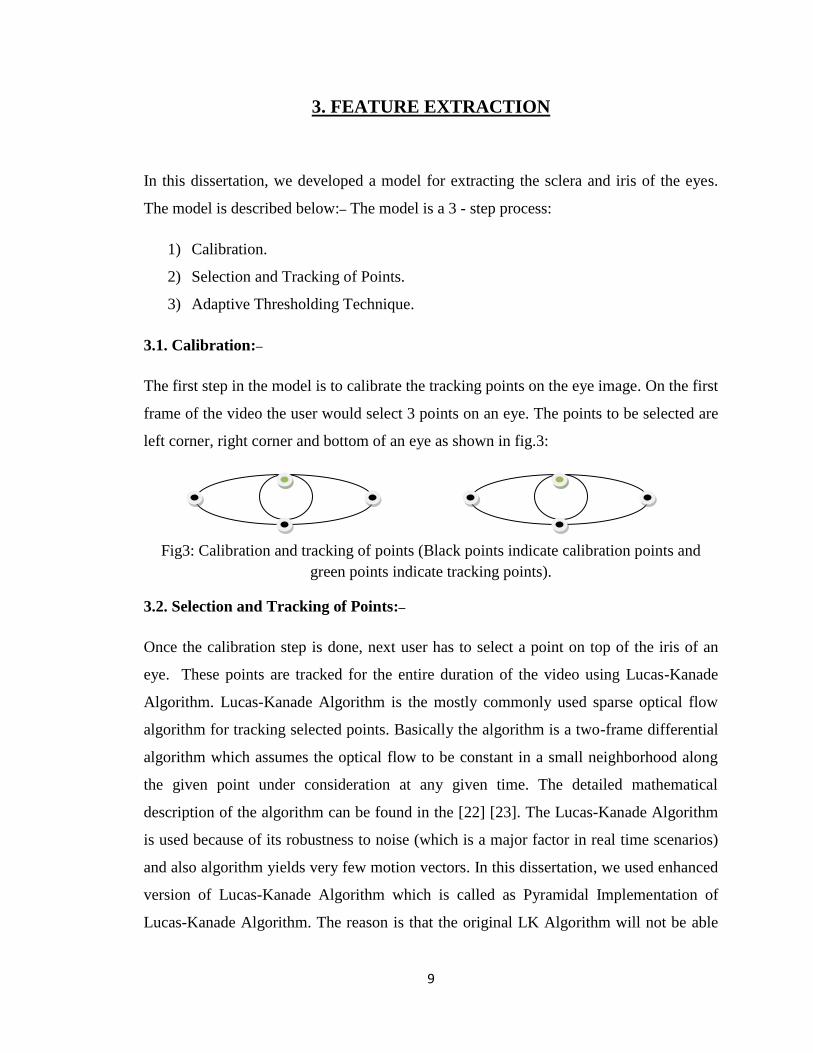
The first step in the model is to calibrate the tracking points on the eye image. On the first
frame of the video the user would select 3 points on an eye. The points to be selected are
left corner, right corner and bottom of an eye as shown in fig.3:
Fig3: Calibration and tracking of points (Black points indicate calibration points and
green points indicate tracking points).
3.2. Selection and Tracking of Points:–
Once the calibration step is done, next user has to select a point on top of the iris of an
eye. These points are tracked for the entire duration of the video using Lucas-Kanade
Algorithm. Lucas-Kanade Algorithm is the mostly commonly used sparse optical flow
algorithm for tracking selected points. Basically the algorithm is a two-frame differential
algorithm which assumes the optical flow to be constant in a small neighborhood along
the given point under consideration at any given time. The detailed mathematical
description of the algorithm can be found in the [22] [23]. The Lucas-Kanade Algorithm
is used because of its robustness to noise (which is a major factor in real time scenarios)
and also algorithm yields very few motion vectors. In this dissertation, we used enhanced
version of Lucas-Kanade Algorithm which is called as Pyramidal Implementation of
Lucas-Kanade Algorithm. The reason is that the original LK Algorithm will not be able

10
to track the points if there is significant motion. In the enhanced version, the image is
divided into layers and points are tracked along each layer [21]. In this dissertation we
used 7 pyramidal layers and a window size of about 21x21 to cope with significant pupil
movement. A rectangular mask is then embedded on the eye using calibrated and tracking
points.
3.3. Thresholding in different color spaces:–
Several other techniques can also be used for extracting regions or features such as
thresholding in different color spaces, by using flood fill algorithms, by using template
matching or by drawing contours. In flood fill algorithm, the user has to select the region
of interest and all the neighborhood regions which are similar according to predefined
criteria are considered as a single region. The predefined criterion can be color of the
pixels or any other statistical property. In Template Matching, the given region of interest
is matched with the trained images. For drawing contours we will first extract edges and
then connect them according to a predefined criterion. The problem with these kinds of
techniques is that the defined criterion is image dependent that is, the criterions has to be
changed for every image and also are affected by noise and illumination. For example, as
shown in fig4: suppose we need to extract the sclera region which is filled with red color
as shown in fig. and will look at the pixel values in different color spaces.
Fig4. Test Image for performing thresholding in different color spaces.
3.3.1. RGB:–
In the fig. 5, the red colored dots indicates sclera region pixel values in the corresponding
channels i.e. for example, in the first figure; red channel is taken along X-Axis and green
channel along Y-axis. Hence we can have a threshold value around 200 along Red-Green
channel for extracting sclera region i.e. pixels whose value greater than 200 along Red-
Green channel are considered to be sclera pixels. However, as we could see from fig.5,
we may lose some data if we choose threshold value to be 200. In the following figures,
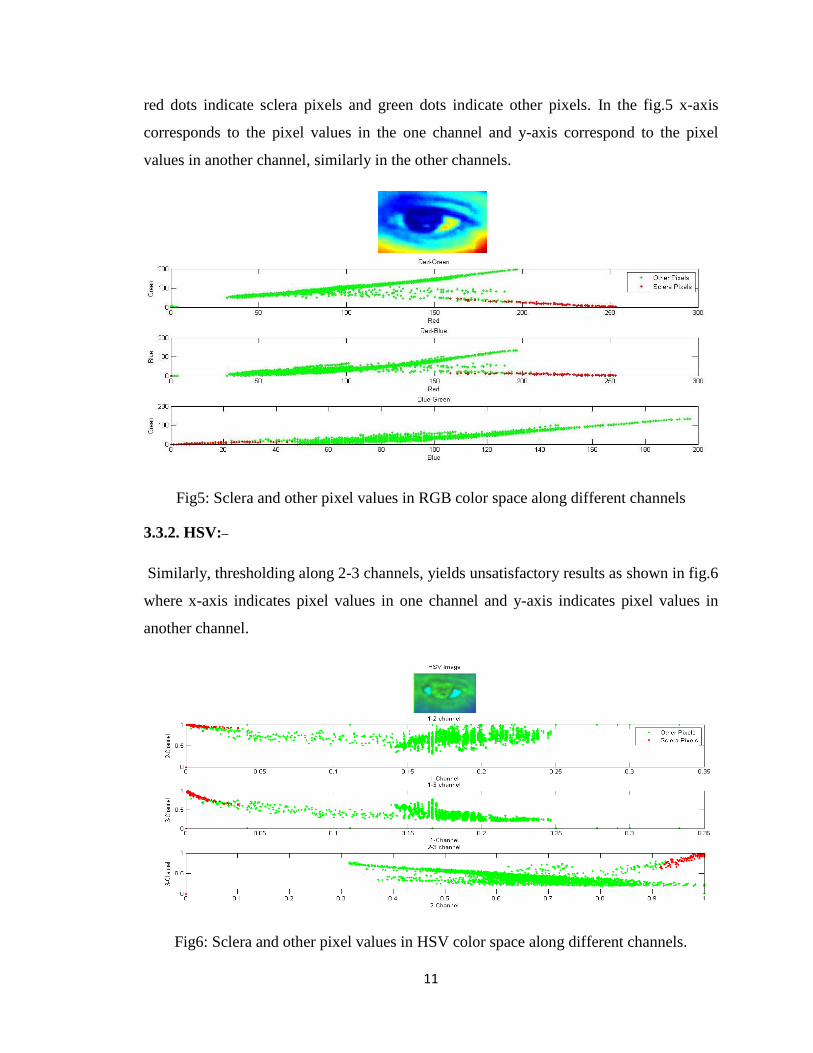
11
red dots indicate sclera pixels and green dots indicate other pixels. In the fig.5 x-axis
corresponds to the pixel values in the one channel and y-axis correspond to the pixel
values in another channel, similarly in the other channels.
Fig5: Sclera and other pixel values in RGB color space along different channels
3.3.2. HSV:–
Similarly, thresholding along 2-3 channels, yields unsatisfactory results as shown in fig.6
where x-axis indicates pixel values in one channel and y-axis indicates pixel values in
another channel.
Fig6: Sclera and other pixel values in HSV color space along different channels.

12
3.3.3. Lab:–
As indicated in fig.7, x-axis indicates pixel values in one channel and y-axis indicates
pixel values in another channel. Thresholding can only be performed along 2-3 channels
even that do not give desired results.
Fig7: Sclera and other pixel values in Lab color space along different channels
3.3.4. YUV:–
As indicated in the fig8 x-axis indicates pixel values in one channel and y-axis pixel
values in another channel. We cannot threshold along any channels as none of them give
desired results.
Fig8: Sclera and other pixel values in YUV color space along different channels

13
3.3.5. YCbCr:–
Similarly, in fig.9 x-axis indicates pixel values in one channel and y-axis pixel values in
another channel. We cannot use this color space for extracting sclera and iris region using
thresholding technique as shown in fig.9.
Fig9: Sclera and other pixel values in YCbCr color space along different channels
3.3.6. NTSC:–
In fig.10 x-axis indicates pixel values in one channel and y-axis pixel values in another
channel. We can threshold along 2-3 channels which may provide partial results as shown
in fig.10:
Fig10: Sclera and other pixel values in NTSC color space along different channels

14
3.4. Adaptive Thresholding Technique:–
After embedding a rectangular mask on an eye using calibration and tracking points, we
then apply adaptive thresholding technique along the region of interests. Before
discussing the proposed adaptive thresholding technique, firstly we need to discuss about
the thresholding techniques and why do we need adaptive thresholding technique, and
why adaptive thresholding is preferred over other techniques like thresholding in
different color spaces, flood filling, template matching and Haar cascade eye detection
for extracting sclera and iris regions of an eye.
The basic idea of a thresholding is to scan the original image pixel by pixel and testing
each pixel value against a specific threshold value i.e. let be an original image
and be any specific threshold, if > then that pixel is classified as a
background pixel otherwise as an object pixel as shown in fig.11:
=
Fig11. Histogram of a gray-level image with dark objects on light background
As shown in above figure if we set the threshold value to, say 100, we can then divide the
image into two groups. However, we don‟t always get an image where we could

15
threshold image easily into two groups using a single threshold value. For example,
consider the histogram of a gray scale image as shown in fig.12:
Fig12: Histogram of gray-level distribution of an image
For the above case more than one threshold is needed to classify the image‟s components.
It is called multi-level thresholding. However, in real world scenarios, the images are
affected by noise and illumination which may affect the results if global thresholding is
used. Hence in order to overcome these problems adaptive thresholding technique is
used.
In an Adaptive Thresholding Technique, the threshold value is not fixed i.e. the value
changes depending on the local property of the image at any given point. The general
definition of a threshold is given as
= [ , ];
In the above equation is the gray-level of a point and is some
local property of this point. When depends only on the i.e. the gray level of
image then it becomes a simple global threshold. In order to take the effect of noise or
illumination into consideration, the calculation of property is important and is

16
usually based on environment of the point at hand. Therefore, global thresholding uses a
fixed threshold for all pixels in the image and therefore works well only if the gray-level
distribution histogram contains distinctively separated peaks and background. Hence we
used a combination of local and adaptive thresholding to extract sclera and iris regions of
eyes.
In this method we will examine the intensity values of local neighborhood of each pixel.
We will then compare the intensity of a pixel with the mean (or any statistic choice of
measurement like median or mode) of the pixels in the neighborhood of the pixel under
consideration at any given point of time. This method faces two problems: Firstly, the
choice of statistic which may vary from one image to another image and is largely
dependent on the nature of the image. Commonly used statistics for comparison are
mean, median, average between the minimal and maximal gray-level in the
neighborhood. The second problem is the choice of neighborhood size. The larger the
size of neighborhood the poorer is the result because it is more affected by illumination
gradient. And also the larger the environment, the more it is expensive to perform the
needed computation. However, if the environment is too small then there is a risk of
being exposed to insufficient data which may lead to poor results when noise is
introduced. For example, consider an image with noise in the form of sporadic points of
extreme intensity like “salt and pepper”. In this case, if we choose a large environment
then there is a good chance of moderating the influence of this noise at the cost of less
resistance to illumination effects. If we choose environment to be small and the statistic
being used is average, then pixels in a noisy environment will be influenced by the noise.
In our dissertation, we kept the neighborhood size to be 3x3 and choice of statistic to be
mean, which worked well as illustrated in results section.
Hence by applying adaptive thresholding technique combined with local threshold we
would determine the pixel to be a sclera pixel and is set to white color if it is greater than
the local mean of the neighboring pixels, or an iris pixel set to black color if it is less
than the local mean of neighboring pixels.
As mentioned earlier the sclera and iris regions can be extracted by applying thresholding
algorithm in any one of the above mentioned color spaces. However, from the above

17
mentioned results we can deduce that by applying thresholding algorithm along different
color spaces will result in non-sclera areas being treated as sclera areas, since we can
observe that in all the color spaces the sclera pixel values overlap with other pixel values
and also the thresholding algorithm depends on image properties i.e.it is affected by noise
and also by illumination. Other techniques such as template matching and contour
methods also depend on image properties such as illumination, shape and size of eye,
color etc.,
Hence for the above mentioned reasons we are using adaptive thresholding combined
with local thresholding for extracting sclera and iris regions.

18
4. Gaze Estimation
The Gaze Pattern can be estimated by using different parameters and by determining the
statistical relationship between those parameters. In this dissertation, we have estimated
the gaze by using 2 methods: 1) using the sclera area and distance between tracking point
and bottom of an eye. 2) Using the Sclera and Iris areas.
4.1. Using sclera area and distance between tracking points along iris axis:–
In this method, eye is divided into two equal halves along the vertical axis at the center of
an eye and sclera area is calculated by counting all the white pixels (which are obtained
after applying adaptive thresholding technique) on either side of an eye. Let
„LELScleraArea‟ be the sclera area from the center to the left side of left eye,
„LERScleraArea‟ be the sclera area from the center to the right side of left eye. Similarly
„RELScleraArea‟ be the sclera area from center to the left side of the right eye and
„RERScleraArea‟ be the sclera area from center to the right side of the right eye. The
sclera areas of both the eyes as shown in fig.13:
Fig13: Sclera areas of each eye
Fig.14 shows the variation of sclera area of each half of both eyes while looking at 9
different locations on the screen namely top left, top center, top right, center left, center,
center right, bottom left, bottom center and bottom right. Fig.15 shows variation of
distance between tracking points along iris axis while looking at different locations on
screen.
LELScleraArea LERScleraArea
Distance between tracking
point and bottom of eye.

19
Fig14: Variation of sclera areas with respect to different points on the screen
0 100 200 300 400 500 600 700 800100
150
200
250
300
350
400
450
500Sclera Area
Number of Frames
Valu
es of S
cle
ra A
reas
LELScleraArea
LERScleraArea
RELScleraArea
RERScleraArea

20
Fig15: Variation of distance between tracking points along iris axis with respect to
different points on screen
4.2. Using sclera and iris areas:–
In this method, eye is divided into two equal halves along vertical and horizontal axis.
The sclera area is calculated in the same way as in first method. The iris area is calculated
by counting all black pixels above the axis line which connects both the corners of an eye
as shown in fig16:
Fig16: Sclera and iris areas
Left and Right Sclera Area
Top Iris Area
Bottom Iris Area

21
The Variation of Iris Area with respect to different points on the screen is shown in
fig.17:
Fig17: Variation of iris areas with respect to different points on the screen
0 100 200 300 400 500 600 700 8000
50
100
150
200
250
300
350
400Iris Area
Number of Frames
Iris
A
rea V
alu
es
LETIrisArea
LEBIrisArea
RETIrisArea
REBIrisArea

22
5. EXPERIMENTAL RESULTS
5.1. Procedure:–
The experimental setup is shown in fig18. below:
Fig18: Experimental set-up.

23
As shown in fig.18, set-up consists of a webcam placed at the bottom of the screen (of a
laptop), a chin rest which restricts the movement of head. Experiment is conducted in a
closed room which consists of 6 lights located on the ceiling. Out of the 6 lights, 3 are
located closer to the camera and rest of them is located farther. The middle light of first
row and the 1st and 3
rd light of second row (farther one‟s) are turned ON and rest of them
are turned OFF. We also used a USB 10 LED laptop light (USB 10-LED Light for
Notebook Laptop PC by HDE company) placed at a height of 23cms or 9 inches above
the camera. We used a chin rest to keep head static and the distance between two
horizontal stands is 18cm or 7 inches. Distance between the camera and the chin rest is
28cms or 11 inches. Height of the camera above the table is 16cms or 6.5 inches.
Distance between light and chin rest is 1.2feet or 34cms and monitor size is 14 inches.
First the user is asked to look at the camera and snapshot of the user is taken for
calibration. From the calibration image we can get the coordinates of eye corners. Then a
test video consisting of 270 frames or 9 sec duration is shown to the user. The test video
frame size is 640 480. The test video consists of a randomly moving object and user is
asked to follow the object. We will select a point on top of the iris of each eye and these
points are tracked for entire video sequence.
5.2. Results:–
Assuming the screen is divided into 9 parts as shown below:
P1 P2 P3
P4 P5 P6
P7 P8 P9
where P1, P2, P3 correspond to top half of the screen, P4, P5, P6 correspond to center
and P7, P8, P9 correspond to the bottom of the screen. The experiment was conducted on
9 people.

24
5.2.1 Advantages and Disadvantages of both methods:–
Both methods have their own benefits and in this section we will be discussing them. In
both the methods the procedure for calculating sclera area is same i.e., gaze estimation
along horizontal axis is same. However, both methods estimate gaze differently along
vertical axis. In the first method, distance between the tracking point and bottom of eye
along iris axis is taken as the criteria for estimating gaze. The advantage of this technique
is that it is computationally faster as we just have to compare the distances between the
tracking and calibration points along iris. However, this technique is user-dependent i.e.,
we cannot have a constant distance between the tracking point and bottom of the eye as
some of the users may have smaller eyes and some may have larger eyes. In the second
technique, gaze is estimated using iris area along vertical axis. The advantage of this
technique is that it is much more efficient since iris area is not user dependent but
computationally expensive. However, both the methods fail to estimate gaze accurately
while looking at the center of the screen, i.e., switching from top to center or from bottom
to center, since there is not much deviation either in the iris position or in the distance
between eye lids. The gaze estimation results using both methods are as shown in fig.19:

25
Fig19: Experimental results for gaze estimation.
In fig.19 X- axis indicates the points on the screen and Y-axis indicates number of people
for which the gaze has been estimated correctly. As you can see from fig.19 that method
2 gives better efficiency while looking at top and bottom of the screen, however its
efficiency decreases while looking at the center.
1 2 3 4 5 6 7 8 90
1
2
3
4
5
6
7
8
9Gaze Estimation Results
Points On the Screen
Num
ber of P
ersons for w
hic
h gaze w
as estim
ated correctly
Method 1
Method 2

26
The fig.20 shows extraction of sclera and Iris using adaptive thresholding technique.
(i) (ii)
(iii) (iv)
(v)
Fig20: Experimental results of sclera and iris extraction using adaptive thresholding. (i)
Input image (ii) Output where white part indicates that sclera region and black part
indicates iris region using adaptive thresholding (iii) Color map of (ii). (iv) Showing
sclera regions (v) Showing Iris regions.

27
Fig21: Results using global and adaptive thresholding
As we can see in fig.21, that global threshold has to be changed for images under
different conditions which is not the case with adaptive thresholding since it considers
only a small region say 3x3 window at any given point of time.

28
6. CONCLUSION
6.1. Contributions and Limitations:
Gaze is an important tool in many applications such as human-computer interaction, in
medical field (helpful in studying neurological, vision and communication disorders) and
also to gain some insight about how people view synthesized images and animations with
the dual purpose of optimizing perceived quality and developing more efficient
algorithms. One of the major drawbacks of gaze estimation is the cost involving
equipments such as high performance cameras followed by the burden of wearing
different equipment‟s by user. In this report, we proposed a new gaze estimation
procedure which makes use of a simple web-cam which reduces the cost considerably.
Contrast to the present gaze estimation techniques, the procedure is an intrusive one i.e.,
the user needs to select a point on each eye, usually selecting a point on top of an eye and
the selected point will be tracked for the entire duration of video. Because of the time
frame we limited our experiments to static head, however, sclera and pupil of an eye have
been extracted with good accuracy and gaze is estimated with a good success rate while
looking at the corners of screen and moderate success rate while looking at other areas of
screen. Even though the gaze estimation is not entirely accurate, but it is right step in the
direction of reducing the cost of gaze estimation and also making it simpler to use. In
future, the onus will be on increasing the accuracy by using a better mathematical
approach for determining gaze by considering the geometric properties of eye, making it
more robust to different lighting conditions, and also estimate gaze with head moment.

29
REFERENCES
1) Eye Tracking Methodology Book by Duchowski, Andrew
2) Dan Witzner Hansen and Qiang Ji. In the Eye of Beholder: A Survey of Models
for Eyes and Gaze. IEEE Transactions on Pattern Analysis and Machine
Intelligence. Vol. 32, No.3, March 2010.
3) Kevin A. Pelphrey, Noah J. Sasson, J. Steven Reznick, Gregory Paul, Barbara D.
Goldman, and Joseph Piven. Visual Scanning of Faces. Journal of Autism and
Developmental Disorders, Vol. 32, No.4, August 2002.
4) Michael L. Spezio, Ralph Adolphs, Robert S.E. Hurley, Joseph Piven. Analysis of
Face Gaze in Autism using “Bubbles”. Neuropsychologia, Vol. 45, Issue 1, pages
144-151, 2007.
5) Ami Klin, Warren Jones, Robert Schultz, Fred Volkmar, Donald Cohen. Visual
Fixation Patterns during Viewing of Naturalistic Social Situations as Predictors of
Social Competence in individuals with Autism. Arch Gen Psychiatry. Vol.59.
pages 809-816. 2002.
6) Andrew T. Duchowski. Eye Tracking Methodology Book. Second Edition. Pages
51 - 54. 2007.
7) Eui Chul Lee, You Jin Ko, Kang Ryoung Park. Gaze Tracking Based on Active
Appearance Model and Multiple Support Vector Regression on Mobile Devices.
Optical Engineering, Vol. 48, July 2009.
8) Scott D. and Findlay J.M. Visual Search, eye movements and display units,
Human factors report, University of Durham, 1993.
9) Hallett, K.Boff, L.Kaufmann & J.Thomas, Handbook of Perception and Human
Performances I, pages 10.25-10.28, 1986.
10) Margrit Betke, William J. Mullally and John J. Magee. Active Detection of Eye
Sclera‟s in Real Time. In IEEE Workshop on Human Modeling, Analysis and
Synthesis, 2000.

30
11) Mohammad Hossein Khosravi and Reza Safabakhsh. Human Eye Sclera
Detection and Tracking using a modified time-adaptive self organizing map.
Journal of Pattern Recognition Society, Vol.41, Pages 2561-2593. January 2008.
12) Erik Murphy-Chutorian, Mohan Manubhai Trivedi. Head Pose Estimation in
Computer Vision: A Survey. IEEE Transactions on Pattern Analysis and Machine
Intelligence, Vol.34, No.4, April 2009.
13) Kathryn Morgan, Computer Gaze Estimation using Image Segmentation, Feature
Extraction and Neural Networks. Utah State University.
14) Zhu J, Yang J. Sub pixel eye gaze tracking. Proceedings of the 5th IEEE
international conference on automatic face and gesture recognition. 2002.
15) M. Betke, W. Mullally, and J. Magee. Active detection of eye sclera‟s in real
time. In Proceedings of the IEEE CVPR Workshop on Human Modeling,
Analysis and Synthesis June 2000.
16) Mohammad Hossein Khosravi, Reza Safabakhsh. Human eye sclera detection
and tracking using a modified time-adaptive self-organizing map, Pattern
Recognition, v.41 n.8, p.2571-2593, August, 2008.
17) R. Stiefelhagen, J. Yang, and A. Waibel. Tracking Eyes and Monitoring Eye
Gaze. In Proceedings Workshop on Perceptual UI, Banff, Canada 1997.
18) Jochen Heinzmann, Alexander Zelinsky, 3-D Facial Pose and Gaze Point
Estimation Using a Robust Real-Time Tracking Paradigm. Pages 142-147, 1998.
19) J-G Wang, E.Sung, Gaze determination via images of irises, Image and Vision
Computing, vol.19, pages 891-911.2001.
20) J-G Wang, E.Sung, R.Venkateshwarlu. Estimating the eye gaze from single eye.
Computer vision and Image understanding vol.98, pages 83-103, 2005.
21) H. Yamazoe, A. Utsumi, T. Yonezawa, and S. Abe. Remote Gaze Estimation
with a Single Camera Based on Facial-Feature Tracking without Special
Calibration Actions, Proceedings 2008 Eye Tracking Research and Applications,
pages 140-145, 2008.
22) Pyramidal Implementation of the Lucas-Kanade feature tracker description of
algorithm, Jean-Yves Bouguet.

31
23) Gary Bradski, Adrain kaehler. Learning OpenCV: Computer vision with the
OpenCV library.

32
Vita
Date and Place of Birth: 06/06/1985, Hyderabad, India.
Educational Institutions: Bachelor of Technology in Electronics and Communications
Engineering at Jawaharlal Nehru Technological University, Hyderabad, India. Master
of Science in Electrical Engineering at University of Kentucky, Lexington, Kentucky,
USA.
Professional Positions: Member of Technical Staff at CloudGrapes Inc, California.
Image Processing Engineer at Logovision Inc, Colorado.
Prashanth Rao Periketi.